
CellTiter Glo Luminescent Cell Viability Assay Protocol
Promega Corporation ·2800 Woods Hollow Road ·Madison, WI 53711-5399 USA Toll F ree in USA 800-356-9526·Phone 608-274-4330 ·F ax 608-277-2516 ·1.Description (1)2.Product Components and Storage Conditions (4)3.Performing the CellTiter-Glo ®Assay (5)A.Reagent Preparation (5)B.Protocol for the Cell Viability Assay (6)C.Protocol for Generating an ATP Standard Curve (optional) (7)4.Appendix (7)A.Overview of the CellTiter-Glo ®Assay..............................................................7B.Additional Considerations..................................................................................8C.References............................................................................................................11D.Related Products. (12)1.DescriptionThe CellTiter-Glo ®Luminescent Cell Viability Assay (a–e)is a homogeneous method to determine the number of viable cells in culture based on quantitation of the ATP present, which signals the presence of metabolically active cells. The CellTiter-Glo ®Assay is designed for use with multiwell-plate formats, making it ideal for automated high-throughput screening (HTS) and cell proliferation and cytotoxicity assays. The homogeneous assay procedure (Figure 1) involves adding a single reagent (CellTiter-Glo ®Reagent) directly to cells cultured in serum-supplemented medium. Cell washing, removal of medium or multiple pipetting steps are not required.The homogeneous “add-mix-measure” format results in cell lysis and generation of a luminescent signal proportional to the amount of ATP present (Figure 2).The amount of ATP is directly proportional to the number of cells present in culture in agreement with previous reports (1). The CellTiter-Glo ®Assay relies on the properties of a proprietary thermostable luciferase (Ultra-Glo™ Recombinant Luciferase), which generates a stable “glow-type” luminescent signal and improves performance across a wide range of assay conditions. The luciferase reaction for this assay is shown in Figure 3. The half-life of the luminescent signal resulting from this reaction is greater than five hours (Figure 4). This extended half-life eliminates the need for reagent injectors and provides flexibility for continuous or batch-mode processing of multiple plates. The unique homogeneous format reduces pipetting errors that may be introduced during the multiple steps required by other ATP-measurement methods.CellTiter-Glo ®Luminescent Cell Viability AssayAll technical literature is available on the Internet at: /protocols/ Please visit the web site to verify that you are using the most current version of this Technical Bulletin. Please contact Promega Technical Services if you have questions on useofthissystem.E-mail:********************Figure 1. Flow diagram showing preparation and use of CellTiter-Glo ®Reagent.Promega Corporation ·2800 Woods Hollow Road ·Madison, WI 53711-5399 USA Toll F ree in USA 800-356-9526·Phone 608-274-4330 ·F ax 608-277-2516 ·3170M A 12_0ACellTiter-Glo CellTiter-Glo MixerLuminometer®System Advantages•Homogeneous:“Add-mix-measure” format reduces the number of plate-handling steps to fewer than that required for similar ATP assays.•Fast:Data can be recorded 10 minutes after adding reagent.•Sensitive:Measures cells at numbers below the detection limits of standard colorimetric and fluorometric assays.•Flexible:Can be used with various multiwell formats. Data can be recorded by luminometer or CCD camera or imaging device.•Robust:Luminescent signal is very stable, with a half-life >5 hours,depending on cell type and culture medium used.•Able to Multiplex:Can be used with reporter gene assays or other cell-based assays from Promega (2,3).Figure 3. The luciferase reaction.Mono-oxygenation of luciferin is catalyzed byluciferase in the presence of Mg 2+, ATP and molecular oxygen.Promega Corporation ·2800 Woods Hollow Road ·Madison, WI 53711-5399 USA Toll F ree in USA 800-356-9526·Phone 608-274-4330 ·F ax 608-277-2516 ·3171M A 12_0A L u m i n e s c e n c e (R L U )Cells per Well10,00060,00020,00030,00040,00050,0000R² = 0.9990.5 × 1061.0 × 1061.5 × 1062.0 × 1062.5 × 1063.0 × 1063.5 × 1064.0 × 106r² = 0.99020,00010,00030,00040,00050,000r² = 0.9900100200300400HO SN S N O S N S N OCOOH +ATP+O 2Ultra-Glo™ Recombinant Luciferase +AMP+PP i +CO 2+LightBeetle Luciferin OxyluciferinMg 2+0Figure 2. Cell number correlates with luminescent output.A direct relationship exists between luminescence measured with the CellTiter-Glo ®Assay and the number of cells in culture over three orders of magnitude. Serial twofold dilutions of HEK293cells were made in a 96-well plate in DMEM with 10% FBS, and assays wereperformed as described in Section 3.B. Luminescence was recorded 10minutes after reagent addition using a GloMax ®-Multi+ Detection System. Values represent the mean ± S.D. of four replicates for each cell number. The luminescent signal from 50HEK293 cells is greater than three times the background signal from serum-supplemented medium without cells. There is a linear relationship (r 2= 0.99)between the luminescent signal and the number of cells from 0to 50,000 cells per well.Figure 4. Extended luminescent half-life allows high-throughput batchprocessing.Signal stability is shown for three common cell lines. HepG2 and BHK-21cells were grown and assayed in MEM containing 10% FBS, while CHO-K1 cells were grown and assayed in DME/F-12 containing 10% FBS. CHO-K1, BHK-21 and HepG2 cells, at 25,000 cells per well, were added to a 96-well plate. After an equal volume of CellTiter-Glo ®Reagent was added, plates were shaken and luminescence monitored over time with the plates held at 22°C. The half-lives of the luminescent signals for the CHO-K1, BHK-21 and HepG2 cells were approximately 5.4, 5.2 and5.8hours, respectively.2.Product Components and Storage ConditionsProduct Size Cat.#CellTiter-Glo ®Luminescent Cell Viability Assay 10ml G7570Substrate is sufficient for 100 assays at 100µl/assay in 96-well plates or 400 assays at 25µl/assay in 384-well plates. Includes:• 1 × 10mlCellTiter-Glo ®Buffer • 1 vial CellTiter-Glo ®Substrate (lyophilized)Product Size Cat.#CellTiter-Glo ®Luminescent Cell Viability Assay 10 × 10ml G7571Each vial of substrate is sufficient for 100 assays at 100µl/assay in 96-well plates or 400 assays at 25µl/assay in 384-well plates (1,000 to 4,000 total assays). Includes:•10 × 10mlCellTiter-Glo ®Buffer •10 vials CellTiter-Glo ®Substrate (lyophilized)Promega Corporation ·2800 Woods Hollow Road ·Madison, WI 53711-5399 USA Toll F ree in USA 800-356-9526·Phone 608-274-4330 ·F ax 608-277-2516 ·R e l a t i v e L u m i n e s c e n c e (%)Time (minutes)CHO-K101020304050607080901003173M A 12_0AProduct Size Cat.# CellTiter-Glo®Luminescent Cell Viability Assay100ml G7572 Substrate is sufficient for 1,000 assays at 100µl/assay in 96-well plates or 4,000assays at 25µl/assay in 384-well plates. Includes:•1 × 100ml CellTiter-Glo®Buffer• 1 vial CellTiter-Glo®Substrate (lyophilized)Product Size Cat.# CellTiter-Glo®Luminescent Cell Viability Assay10 × 100ml G7573Each vial of substrate is sufficient for 1,000 assays at 100µl/assay in 96-well plates or4,000 assays at 25µl/assay in 384-well plates (10,000to 40,000 total assays). Includes:•10 × 100ml CellTiter-Glo®Buffer•10 vials CellTiter-Glo®Substrate (lyophilized)Storage Conditions:For long-term storage, store the lyophilized CellTiter-Glo®Substrate and CellTiter-Glo®Buffer at –20°C. For frequent use, the CellTiter-Glo®Buffer can be stored at 4°C or room temperature for 48hours without loss of activity. See product label for expiration date information. ReconstitutedCellTiter-Glo®Reagent (Buffer plus Substrate) can be stored at room temperaturefor up to 8hours with <10% loss of activity, at 4°C for 48hours with ~5% lossof activity, at 4°C for 4days with ~20% loss of activity or at –20°C for 21weekswith ~3% loss of activity. The reagent is stable for up to ten freeze-thaw cycles,with less than 10% loss of activity.3.Performing the CellTiter-Glo®AssayMaterials to Be Supplied by the User•opaque-walled multiwell plates adequate for cell culture•multichannel pipette or automated pipetting station for reagent delivery•device (plate shaker) for mixing multiwell plates•luminometer, CCD camera or imaging device capable of reading multiwell plates •optional:ATP for use in generating a standard curve (Section 3.C)3.A.Reagent Preparation1.Thaw the CellTiter-Glo®Buffer, and equilibrate to room temperature priorto use. For convenience the CellTiter-Glo®Buffer may be thawed andstored at room temperature for up to 48hours prior to use.2.Equilibrate the lyophilized CellTiter-Glo®Substrate to room temperatureprior to use.Promega Corporation·2800 Woods Hollow Road ·Madison, WI 53711-5399 USA Toll F ree in USA 800-356-9526·Phone 608-274-4330 ·F ax 608-277-2516 ·3.A.Reagent Preparation (continued)3.Transfer the appropriate volume (10ml for Cat.# G7570 and G7571, or 100mlfor Cat.# G7572 and G7573) of CellTiter-Glo ®Buffer into the amber bottlecontaining CellTiter-Glo ®Substrate to reconstitute the lyophilizedenzyme/substrate mixture. This forms the CellTiter-Glo ®Reagent.4.Mix by gently vortexing, swirling or inverting the contents to obtain ahomogeneous solution. The CellTiter-Glo ®Substrate should go intosolution easily in less than 1minute.3.B.Protocol for the Cell Viability AssayWe recommend that you perform a titration of your particular cells todetermine the optimal number and ensure that you are working within thelinear range of the CellTiter-Glo ®Assay. Figure 2 provides an example of sucha titration of HEK293 cells using 0 to 50,000 cells per well in a 96-well format.1.Prepare opaque-walled multiwell plates with mammalian cells in culturemedium, 100µl per well for 96-well plates or 25µl per well for 384-wellplates.Multiwell plates must be compatible with the luminometer used.2.Prepare control wells containing medium without cells to obtain a value forbackground luminescence.3.Add the test compound to experimental wells, and incubate according toculture protocol.4.Equilibrate the plate and its contents at room temperature forapproximately 30 minutes.5.Add a volume of CellTiter-Glo ®Reagent equal to the volume of cell culturemedium present in each well (e.g., add 100µl of reagent to 100µl of mediumcontaining cells for a 96-well plate, or add 25µl of reagent to 25µl ofmedium containing cells for a 384-well plate).6.Mix contents for 2 minutes on an orbital shaker to induce cell lysis.7.Allow the plate to incubate at room temperature for 10 minutes to stabilizeluminescent signal.Note:Uneven luminescent signal within standard plates can be caused bytemperature gradients, uneven seeding of cells or edge effects in multiwellplates.8.Record luminescence.Note:Instrument settings depend on the manufacturer. An integration timeof 0.25–1 second per well should serve as a guideline.Promega Corporation ·2800 Woods Hollow Road ·Madison, WI 53711-5399 USA Toll F ree in USA 800-356-9526·Phone 608-274-4330 ·F ax 608-277-2516 ·3.C.Protocol for Generating an ATP Standard Curve (optional)It is a good practice to generate a standard curve using the same plate onwhich samples are assayed. We recommend ATP disodium salt (Cat.# P1132,Sigma Cat.# A7699 or GE Healthcare Cat.# 27-1006). The ATP standard curveshould be generated immediately prior to adding the CellTiter-Glo®Reagentbecause endogenous ATPase enzymes found in sera may reduce ATP levels.1.Prepare 1µM ATP in culture medium (100µl of 1µM ATP solution contains10–10moles ATP).2.Prepare serial tenfold dilutions of ATP in culture medium (1µM to 10nM;100µl contains 10–10to 10–12moles of ATP).3.Prepare a multiwell plate with varying concentrations of ATP standard in100µl medium (25µl for a 384-well plate).4.Add a volume of CellTiter-Glo®Reagent equal to the volume of ATPstandard present in each well.5.Mix contents for 2 minutes on an orbital shaker.6.Allow the plate to incubate at room temperature for 10 minutes to stabilizethe luminescent signal.7.Record luminescence.4.Appendix4.A.Overview of the CellTiter-Glo®AssayThe assay system uses the properties of a proprietary thermostable luciferase toenable reaction conditions that generate a stable “glow-type” luminescentsignal while simultaneously inhibiting endogenous enzymes released duringcell lysis (e.g., ATPases). Release of ATPases will interfere with accurate ATPmeasurement. Historically, firefly luciferase purified from Photinus pyralis(LucPpy) has been used in reagents for ATP assays (1,4–7). However, it hasonly moderate stability in vitro and is sensitive to its chemical environment,including factors such as pH and detergents, limiting its usefulness fordeveloping a robust homogeneous ATP assay. Promega has successfullydeveloped a stable form of luciferase based on the gene from another firefly,Photuris pennsylvanica(LucPpe2), using an approach to select characteristics thatimprove performance in ATP assays. The unique characteristics of this mutant(LucPpe2m) enabled design of a homogeneous single-reagent-addition approachto perform ATP assays with cultured cells. Properties of the CellTiter-Glo®Reagent overcome the problems caused by factors, such as ATPases, thatinterfere with ATP measurement in cell extracts. The reagent is physicallyrobust and provides a sensitive and stable luminescent output.Promega Corporation·2800 Woods Hollow Road ·Madison, WI 53711-5399 USA Toll F ree in USA 800-356-9526·Phone 608-274-4330 ·F ax 608-277-2516 ·4.A.Overview of the CellTiter-Glo®Assay (continued)Sensitivity and Linearity:The ATP-based detection of cells is more sensitivethan other methods (8–10). In experiments performed by Promega scientists,the luminescent signal from 50HEK293 cells is greater than three standarddeviations above the background signal from serum-supplemented mediumwithout cells. There is a linear relationship (r2= 0.99) between the luminescentsignal and the number of cells from 0 to 50,000 cells per well in the 96-wellformat. The luminescence values in Figure 2 were recorded after 10minutes ofincubation at room temperature to stabilize the luminescent signal as describedin Section3.B. Incubation of the same 96-well plate used in the experimentshown in Figure 2 for 360minutes at room temperature had little effect on therelationship between luminescent signal and number of cells (r2= 0.99).Speed:The homogeneous procedure to measure ATP using the CellTiter-Glo®Assay is quicker than other ATP assay methods that require multiple steps toextract ATP and measure luminescence. The CellTiter-Glo®Assay also is fasterthan other commonly used methods to measure the number of viable cells(such as MTT, alamarBlue®or Calcein-AM) that require prolonged incubationsteps to enable the cells’ metabolic machinery to convert indicator moleculesinto a detectable signal.4.B.Additional ConsiderationsTemperature:The intensity and decay rate of the luminescent signal from theCellTiter-Glo®Assay depends on the luciferase reaction rate. Environmentalfactors that affect the luciferase reaction rate will change the intensity andstability of the luminescent signal. Temperature is one factor that affects therate of this enzymatic assay and thus the light output. For consistent results,equilibrate assay plates to a constant temperature before performing the assay.Transferring eukaryotic cells from 37°C to room temperature has little effect onATP content (5). We have demonstrated that removing cultured cells from a37°C incubator and allowing them to equilibrate to 22°C for 1–2 hours hadlittle effect on ATP content. For batch-mode processing of multiple assayplates, take precautions to ensure complete temperature equilibration. Platesremoved from a 37°C incubator and placed in tall stacks at room temperaturewill require longer equilibration than plates arranged in a single layer.Insufficient equilibration may result in a temperature gradient effect betweenwells in the center and at the edge of the plates. The temperature gradientpattern also may depend on the position of the plate in the stack.Promega Corporation·2800 Woods Hollow Road ·Madison, WI 53711-5399 USA Toll F ree in USA 800-356-9526·Phone 608-274-4330 ·F ax 608-277-2516 ·Chemicals:The chemical environment of the luciferase reaction affects theenzymatic rate and thus luminescence intensity. Differences in luminescenceintensity have been observed using different types of culture media and sera.The presence of phenol red in culture medium should have little impact onluminescence output. Assaying 0.1µM ATP in RPMI medium without phenolred resulted in ~5% increase in luminescence output (in relative light units[RLU]) compared to assays in RPMI containing the standard concentration ofphenol red, whereas assays in RPMI medium containing twice the normalconcentration of phenol red showed a ~2% decrease in luminescence.Solvents for the various test compounds may interfere with the luciferasereaction and thus the light output from the assay. Interference with theluciferase reaction can be detected by assaying a parallel set of control wellscontaining medium without cells. Dimethylsulfoxide (DMSO), commonly usedas a vehicle to solubilize organic chemicals, has been tested at finalconcentrations of up to 2% in the assay and only minimally affects light output.Plate Recommendations:We recommend using standard opaque-walledmultiwell plates suitable for luminescence measurements. Opaque-walledplates with clear bottoms to allow microscopic visualization of cells also maybe used; however, these plates will have diminished signal intensity andgreater cross talk between wells. Opaque white tape may be used to decreaseluminescence loss and cross talk.Cellular ATP Content:Different cell types have different amounts of ATP,and values reported for the ATP level in cells vary considerably (1,4,11–13).Factors that affect the ATP content of cells may affect the relationship betweencell number and luminescence. Anchorage-dependent cells that undergocontact inhibition at high densities may show a change in ATP content per cellat high densities, resulting in a nonlinear relationship between cell numberand luminescence. Factors that affect the cytoplasmic volume or physiology ofcells also will affect ATP content. For example, oxygen depletion is one factorknown to cause a rapid decrease in ATP (1).Promega Corporation·2800 Woods Hollow Road ·Madison, WI 53711-5399 USA Toll F ree in USA 800-356-9526·Phone 608-274-4330 ·F ax 608-277-2516 ·4.B.Additional Considerations (continued)Mixing:Optimal assay performance is achieved when the CellTiter-Glo®Reagent is mixed completely with the cultured cells. Suspension cell lines (e.g., Jurkat cells) generally require less mixing to achieve lysis and extract ATP than adherent cells (e.g., L929 cells). Tests were done to evaluate the effect ofshaking the plate after adding the CellTiter-Glo® Reagent. Suspension cellscultured in multiwell plates showed only minor differences in light outputwhether or not the plates were shaken after adding the CellTiter-Glo®Reagent.Adherent cells are more difficult to lyse and show a substantial differencebetween shaken and nonshaken plates.Several additional parameters related to reagent mixing include the force ofdelivery of CellTiter-Glo®Reagent, sample volume and dimensions of the well.All of these factors may affect assay performance. The degree of reagent mixing required may be affected by the method used to add the CellTiter-Glo®Reagent to the assay plates. Automated pipetting devices using a greater or lesser force of fluid delivery may affect the degree of subsequent mixing required.Complete reagent mixing in 96-well plates should be achieved using orbitalplate shaking devices built into many luminometers and the recommended2-minute shaking time. Special electromagnetic shaking devices that use aradius smaller than the well diameter may be required to efficiently mixcontents of 384-well plates. The depth of medium and geometry of themultiwell plates may have an effect on mixing efficiency. We recommend that you take these factors into consideration when performing the assay andempirically determine whether a mixing step is necessary for the individualapplication.LuminometersFor highly sensitive luminometric assays, the luminometer model and settings greatly affect the quality of data obtained. Luminometers from differentmanufacturers will vary in sensitivities and dynamic ranges. We recommend the GloMax®products because these instruments do not require gainadjustments to achieve optimal sensitivity and dynamic range. Additionally, GloMax®instruments are preloaded with Promega protocols for ease of use.If you are not using a GloMax®luminometer, consult the operating manual for your luminometer to determine the optimal settings. The limits should beverified on each instrument before analysis of experimental samples. The assay should be linear in some portion of the detection range of the instrument used.For an individual luminometer there may be different gain settings. Werecommend that you optimize the gain settings.4.C.References1.Crouch, S.P. et al.(1993) The use of ATP bioluminescence as a measure of cellproliferation and cytotoxicity. J. Immunol. Methods160, 81–8.2.Farfan, A.et al.(2004) Multiplexing homogeneous cell-based assays. Cell Notes10, 2–5.3.Riss, T., Moravec, R. and Niles, A. (2005) Selecting cell-based assays for drugdiscovery screening. Cell Notes13, 16–21.4.Kangas, L., Grönroos, M. and Nieminen, A.L. (1984) Bioluminescence of cellular ATP:A new method for evaluating cytotoxic agents in vitro. Med. Biol.62, 338–43.5.Lundin, A. et al.(1986) Estimation of biomass in growing cell lines by adenosinetriphosphate assay.Methods Enzymol. 133, 27–42.6.Sevin, B.U. et al.(1988) Application of an ATP-bioluminescence assay in human tumorchemosensitivity testing. Gynecol. Oncol.31, 191–204.7.Gerhardt, R.T.et al.(1991) Characterization of in vitro chemosensitivity ofperioperative human ovarian malignancies by adenosine triphosphatechemosensitivity assay. Am. J. Obstet. Gynecol. 165, 245–55.8.Petty, R.D. et al.(1995) Comparison of MTT and ATP-based assays for themeasurement of viable cell number. J. Biolumin. Chemilumin.10, 29–34.9.Cree, I.A. et al.(1995) Methotrexate chemosensitivity by ATP luminescence in humanleukemia cell lines and in breast cancer primary cultures: Comparison of the TCA-100assay with a clonogenic assay. AntiCancer Drugs6, 398–404.10.Maehara, Y. et al.(1987) The ATP assay is more sensitive than the succinatedehydrogenase inhibition test for predicting cell viability. Eur. J. Cancer Clin. Oncol.23, 273–6.11.Stanley, P.E. (1986) Extraction of adenosine triphosphate from microbial and somaticcells. Methods Enzymol.133, 14–22.12.Beckers, B. et al.(1986) Application of intracellular ATP determination in lymphocytesfor HLA-typing. J. Biolumin. Chemilumin.1, 47–51.13.Andreotti, P.E. et al.(1995) Chemosensitivity testing of human tumors using amicroplate adenosine triphosphate luminescence assay: Clinical correlation forcisplatin resistance of ovarian carcinoma. Cancer Res. 55, 5276–82.4.D.Related ProductsCell Proliferation ProductsProduct Size Cat.# ApoLive-Glo™ Multiplex Assay10ml G6410 ApoTox-Glo™ Triplex Assay10ml G6320 CellTiter-Fluor™ Cell Viability Assay (fluorescent)10ml G6080 CellTiter-Blue®Cell Viability Assay (resazurin)20ml G8080 CellTiter 96®AQ ueous One SolutionCell Proliferation Assay (MTS, colorimetric)200 assays G3582 CellTiter 96®AQ ueous Non-RadioactiveCell Proliferation Assay (MTS, colorimetric)1,000 assays G5421 CellTiter 96®AQ ueous MTS Reagent Powder1g G1111 CellTiter 96®Non-RadioactiveCell Proliferation Assay (MTT, colorimetric)1,000 assays G4000 Additional sizes available.Cytotoxicity AssaysProduct Size Cat.# CytoTox-Glo™ Cytotoxicity Assay (luminescent)*10ml G9290Mitochondrial ToxGlo™ Assay*10ml G8000 MultiTox-Glo Multiplex Cytotoxicity Assay(luminescent, fluorescent)*10ml G9270 MultiTox-Fluor Multiplex Cytotoxicity Assay(fluorescent)*10ml G9200 CytoTox-Fluor™ Cytotoxicity Assay (fluorescent)*10ml G9260 CytoTox-ONE™ Homogeneous MembraneIntegrity Assay (LDH, fluorometric)*200–800 assays G7890 CytoTox-ONE™ Homogeneous MembraneIntegrity Assay, HTP1,000–4,000 assays G7892 CytoTox 96® Non-Radioactive Cytotoxicity Assay1,000 assays G1780 (LDH, colorimetric)*GSH-Glo™ Glutathione Assay10ml V691150ml V6912 GSH/GSSG-Glo™ Assay10ml V661150ml V6612 *Additional sizes available.LuminometersProduct Size Cat.# GloMax®-Multi+ Detection System with Instinct™ Software:Base Instrument with Shaking 1 each E8032 GloMax®-Multi+ Detection System with Instinct™ Software:Base Instrument with Heating and Shaking 1 each E9032 GloMax®-Multi+ Luminescence Module 1 each E8041Apoptosis ProductsProduct Size Cat.# Caspase-Glo®2 Assay*10ml G0940 Caspase-Glo®6 Assay*10ml G0970 Caspase-Glo®3/7 Assay* 2.5ml G8090 Caspase-Glo®8 Assay* 2.5ml G8200 Caspase-Glo®9 Assay* 2.5ml G8210Apo-ONE®Homogeneous Caspase-3/7 Assay1ml G7792 DeadEnd™ Fluorometric TUNEL System60 reactions G3250 DeadEnd™ Colorimetric TUNEL System20 reactions G7360Anti-ACTIVE®Caspase-3 pAb50µl G7481Anti-PARP p85 Fragment pAb50µl G7341Anti-pS473Akt pAb40µl G7441 Caspase Inhibitor Z-VAD-FMK, 20mM50µl G7231125µl G7232*Additional sizes available.(a)U.S. Pat. Nos. 6,602,677 and 7,241,584, European Pat. No. 1131441, Japanese Pat. Nos. 4537573 and 4520084 and other patents pending(b)U.S. Pat. No. 7,741,067, Japanese Pat. No. 4485470 and other patents pending.(c)U.S. Pat. No. 7,700,310, European Pat. No. 1546374 and other patents pending.(d)U.S. Pat. Nos 7,083,911, 7,452,663 and 7,732,128, European Pat. No. 1383914 and Japanese Pat. Nos. 4125600 and 4275715.(e)The method of recombinant expression of Coleoptera luciferase is covered by U.S. Pat. Nos. 5,583,024, 5,674,713 and 5,700,673.© 2001–2012 Promega Corporation. All Rights Reserved.Anti-ACTIVE, Apo-ONE, Caspase-Glo, CellTiter 96, CellTiter-Blue, CellTiter-Glo, CytoTox 96 and GloMax are registered trademarks of Promega Corporation. ApoTox-Glo, ApoLive-Glo, CellTiter-Fluor, CytoTox-Fluor, CytoTox-Glo, CytoTox-ONE, DeadEnd, GSH-Glo, GSH/GSSG-Glo, Instinct, Mitochondrial ToxGlo and Ultra-Glo are trademarks of Promega Corporation. alamarBlue is a registered trademark of Trek Diagnostic Ssystems, Inc.Products may be covered by pending or issued patents or may have certain limitations. Please visit our Web site for more information.All prices and specifications are subject to change without prior notice.Product claims are subject to change. Please contact Promega Technical Services or access the Promega online catalog for the most up-to-date information on Promega products.。
promega celltiter-fluor cell viability assay protocol

Promega Corporation ·2800 Woods Hollow Road ·Madison, W I 53711-5399 USA Toll Free in USA 800-356-9526·Phone 608-274-4330 ·Fax 608-277-2516 ·1.Description ..........................................................................................................12.Product Components and Storage Conditions ............................................43.Reagent Preparation and Storage ...................................................................54.Protocols for the CellTiter-Fluor™ Cell Viability Assay ..........................5A.Determining Assay Sensitivity, Method 1........................................................6B.Determining Assay Sensitivity, Method 2........................................................7C.Example Viability Assay Protocol.....................................................................8D.Example Multiplex Assay Protocol (with luminescent caspase assay).......9E.Recommended Controls (10)5.General Considerations ..................................................................................106.References .........................................................................................................117.Related Products ..............................................................................................121.DescriptionThe CellTiter-Fluor™ Cell Viability Assay (a)is a nonlytic, single-reagent-addition fluorescence assay that measures the relative number of live cells in a culture population after experimental manipulation (Figures 1 and 2). The CellTiter-Fluor™ Cell Viability Assay measures a conserved and constitutive protease activity within live cells and therefore serves as a marker of cell viability (1). Results obtained using the CellTiter-Fluor™ Cell Viability Assay correlate well with other established methods of determining cell viability(Figure 3). The live-cell protease activity is restricted to intact viable cells and is measured using a fluorogenic, cell-permeant, peptide substrate (glycyl-phenylalanyl-aminofluorocoumarin; GF-AFC). The substrate enters intact cells where it is cleaved by the live-cell protease activity to generate a fluorescent signal proportional to the number of living cells (Figure 4). This live-cell protease becomes inactive upon loss of cell membrane integrity and leakage into the surrounding culture medium.The CellTiter-Fluor™ Cell Viability Assay also can be used in a single-well,sequential, multiplex format with other downstream chemistries to normalize data by cell number. Data from the assay can serve as an internal control andCellTiter-Fluor™ Cell Viability AssayAll technical literature is available on the Internet at: /protocols/ Please visit the web site to verify that you are using the most current version of this Technical Bulletin. Please contact Promega Technical Services if you have questions on useof this system. E-mail: techserv@allow identification of errors resulting from cell clumping or compoundcytotoxicity. The CellTiter-Fluor™ Cell Viability Assay is compatible with most Promega luminescence assays or spectrally distinct fluorescence assay methods,such as assays measuring caspase activation, reporter gene expression or orthogonal measures of viability. However, some P450-Glo™ multiplexprotocols may require removing culture supernatant to a separate assay well before performing the assay because of isoform-specific competitive inhibition of the cytotochrome P450 enzymes by the coumarin product of the CellTiter-Fluor™ Cell Viability Assay reaction.Figure 1. Schematic diagram of the CellTiter-Fluor™ Cell Viability Assay.Promega Corporation ·2800 Woods Hollow Road ·Madison, W I 53711-5399 USA Toll Free in USA 800-356-9526·Phone 608-274-4330 ·Fax 608-277-2516 ·6868M ACellTiter-Fluor™ ReagentAdd GF-AFC Substrate to Assay Buffer to create the CellTiter-Fluor™ Reagent.MeasureAssay BenefitsMeasure the Relative Number of Live Cells in Culture: Nonlytic, single-reagent-addition, homogeneous, “add-mix-measure” protocol.Get More Data from Every Well: The CellTiter-Fluor™ Cell Viability Assay can be performed in multiplex with most Promega luminescence assays.Normalize Data for Cell Number: Normalizing data for live-cell number makes results more comparable well-to-well, plate-to-plate, day-to-day.Figure 3. The CellTiter-Fluor™ Cell Viability Assay shows strong correlation with established methods for measuring viability. Panel A. The GF-AFC Substrate signal from serial dilutions of live cells plotted against results from the CellTiter-Glo ®Luminescent Cell Viability Assay (Cat.# G7570), which measures cellular ATP.Panel B.The GF-AFC Substrate signal from serial dilutions of live cells plottedagainst results achieved using the CellTiter-Blue ®Cell Viability Assay (Cat.# G8080).Promega Corporation ·2800 Woods Hollow Road ·Madison, W I 53711-5399 USA Toll Free in USA 800-356-9526·Phone 608-274-4330 ·Fax 608-277-2516 ·6867M AA.B.R e s o r u f i n F l u o r e s c e n c e (R F U )2,5002,6002,7002,8002,9003,0003,100GF-AFC Fluorescence (RFU)A T P -B a s e d A s s a y L u m i n e s c e n c e (R L U )0GF-AFC Fluorescence (RFU)LIVE-CELL SUBSTRATE:cell-permeant fluorogenic substrate for the live-cellprotease (Gly-Phe-AFCoumarin)NucleusLive CellLive-cell protease substrate can cross the cell membrane.Active Live-CellProteaseO CF 3GF–N HOGFOCF 3OH 2N6995M AFigure 2. CellTiter-Fluor™ Cell Viability Assay chemistry. The cell-permeant substrate enters the cell, where it is cleaved by the live-cell protease activity to produce the fluorescent AFC. The live-cell protease is labile in membrane-compromised cells and cannot cleave the substrate.Figure 4. The CellTiter-Fluor™ Cell Viability Assay signal derived from viable cells (untreated) is proportional to cell number. Dead cells (treated) do not contribute appreciable signal in the assay.2.Product Components and Storage ConditionsProductSize Cat.#CellTiter-Fluor™ Cell Viability Assay10mlG6080Cat.# G6080 contains sufficient reagents for 100 assays at 100µl/assay in a 96-well plate format or 400 assays at 25µl/assay in a 384-well plate format. Includes:• 1 × 10ml Assay Buffer• 1 × 10µl GF-AFC Substrate (100mM in DMSO)ProductSize Cat.#CellTiter-Fluor™ Cell Viability Assay5 × 10mlG6081Cat.# G6081 contains sufficient reagents for 500 assays at 100µl/assay in a 96-well plate format or 2,000 assays at 25µl/well in a 384-well format. Includes:• 5 × 10ml Assay Buffer• 5 × 10µl GF-AFC Substrate (100mM in DMSO)ProductSize Cat.#CellTiter-Fluor™ Viability Assay2 × 50mlG6082Cat.# G6082 contains sufficient reagents for 1,000 assays at 100µl/assay in a 96-well plate format or 4,000 assays at 25µl/well in a 384-well format. Includes:• 2 × 50ml Assay Buffer• 2 × 50µl GF-AFC Substrate (100mM in DMSO)Storage Conditions: Store the CellTiter-Fluor™ Cell Viability Assaycomponents at –20°C. See product label for expiration date.Promega Corporation ·2800 Woods Hollow Road ·Madison, W I 53711-5399 USA Toll Free in USA 800-356-9526·Phone 608-274-4330 ·Fax 608-277-2516 ·6866M ACells or Cell Equivalents/WellA F C F l u o r e s c e n c e (R F U )3.Reagent Preparation and Storagepletely thaw the CellTiter-Fluor™ Cell Viability Assay components in a37°C water bath. Vortex the GF-AFC substrate to ensure homogeneity, thenbriefly centrifuge for complete substrate volume recovery.2.Transfer the GF-AFC Substrate (10µl for Cat.# G6080 and G6081; 50µl for Cat.#G6082) into the Assay Buffer container (10ml for Cat.# G6080 and G6081; 50mlfor Cat.# G6082) to form a 2X Reagent. Mix by vortexing the contents until thesubstrate is thoroughly dissolved.Note: The solution may initially appear “milky” when the GF-AFC substrate isdelivered to the buffer. This is normal. The substrate will dissolve withvortexing. The CellTiter-Fluor™ Reagent may be scaled to accommodate thevolumes required for downstream multiplexes. To do this, use 1/5 the volumeof buffer when you prepare the reagent (i.e., 10µl of the GF-AFC Substrate in2ml of Assay Buffer). Be sure to label the bottle to indicate that this is a moreconcentrated reagent, suitable for multiplex assays. Add the reagent at 1/5 thevolume of the cell culture.Storage: The CellTiter-Fluor™ Viability Reagent should be used within 24hours if stored at room temperature. Unused GF-AFC Substrate and AssayBuffer can be stored at 4°C for up to 7 days with no appreciable loss ofactivity.4.Protocols for the CellTiter-Fluor™ Cell Viability AssayMaterials to Be Supplied by the User•96-, 384-, or 1536-well opaque-walled tissue culture plates compatible with fluorometer (clear or solid bottom)•multichannel pipettor or liquid-dispensing robot•reagent reservoirs•fluorescence plate reader with filter sets for AFC (380–400nm Ex/505Em)•orbital plate shaker•compound known to cause 100% cytotoxicity or lytic detergent (digitonin, Calbiochem Cat.# 300410 or Sigma-Aldrich Cat.# D141 at 20mg/ml in DMSO).If you have not performed this assay on your cell line previously, werecommend determining assay sensitivity using your cells and one of the two methods described below (Section 4.A or 4.B). If you do not need to determineassay sensitivity for your cells, proceed to Section 4.C.Promega Corporation·2800 Woods Hollow Road ·Madison, W I 53711-5399 USA Toll Free in USA 800-356-9526·Phone 608-274-4330 ·Fax 608-277-2516 ·6.Dilute digitonin to 300µg/ml in water. Using a multichannel pipet,carefully add 10µl of the diluted digitonin to all wells of columns 7–12 to lyse cells (treated samples). Add 10µl of water to all wells of columns 1–6to normalize the volume (untreated cells).7.Add 100µl of the CellTiter-Fluor™ Reagent to all wells, mix briefly by orbital shaking and incubate at 37°C for at least 30 minutes.Note:Longer incubations may improve assay sensitivity and dynamic range. However, do not incubate longer than 3 hours, and be sure to shieldplates from ambient light.Promega Corporation ·2800 Woods Hollow Road ·Madison, W I 53711-5399 USA Toll Free in USA 800-356-9526·Phone 608-274-4330 ·Fax 608-277-2516 ·8.Measure resulting fluorescence with a fluorometer (380–400nm Ex /505nm Em )Note: You may need to adjust instrument gains (applied photomultiplier tube energy).9.Calculate the practical sensitivity for your cell type by making a signal-to-noise calculation for each dilution of cells (10,000 cells/well; 5,000cells/well; 2,500 cells/well, etc.).Viability S:N =(Average Untreated – Average Treated)Note: The practical level of assay sensitivity for the assay is a signal-to-noise ratio of greater than 3 standard deviations (derived from reference 1).4.B.Determining Assay Sensitivity, Method 21.Harvest adherent cells (by trypsinization, etc.), wash with fresh medium (to remove residual trypsin) and resuspend in fresh medium.Note:For cells growing in suspension, proceed to Step2.2.Determine the number of viable cells by trypan blue exclusion using a hemacytometer, then adjust the cells by dilution to 100,000 viable cells/ml in at least 20ml of fresh medium.Note:Concentrate the cells by centrifuging and removing medium if the pool of cells is less than 100,000 cells/ml.3.Divide the volume of diluted cells into separate tubes. Subject one tube to "moderate" sonication (empirically determined by postsonicationmorphological examination) to rupture cell membrane integrity and to simulate a 100% dead population. The second tube of untreated cells will serve as the maximum viable population.4.Create a spectrum of viability by blending sonicated and untreatedpopulations in 1.5ml microcentrifuge tubes as described in Table 2.Promega Corporation ·2800 Woods Hollow Road ·Madison, W I 53711-5399 USA Toll Free in USA 800-356-9526·Phone 608-274-4330 ·Fax 608-277-2516 ·4.B.Determining Assay Sensitivity, Method 2 (continued)5.After mixing each blend by gently vortexing, pipet 100µl of each blend into8 replicate wells of a 96-well plate. Add the 100% viable cells to column 1,95% viable to column 2, etc. Add cell culture medium only to column 10 toserve as a no-cell control.6.Add CellTiter-Fluor™ Reagent in an equal volume (100µl per well) to allwells, mix briefly by orbital shaking, then incubate for at least 30 minutesat 37°C.Note:Longer incubations may improve assay sensitivity and dynamicrange. However, do not incubate longer than 3 hours, and be sure to shieldplates from ambient light.7.Measure resulting fluorescence with a fluorometer (380–400nm Ex/505nm Em).Note: You may need to adjust instrument gains (applied photomultipliertube energy).8.Calculate the practical sensitivity for your cell type by making a signal-to-noise calculation for each blend of cell viability (X = 95, 90%, etc.).Viability S:N = (Average 100% – Average X%)Standard Deviation of 0% (viable cells)Note: The practical level of assay sensitivity for the assay is a signal-to-noise ratio of greater than 3 standard deviations (derived from reference 1).4.C.Example Viability Assay Protocol1.Set up 96-well assay plates containing cells in culture medium at desireddensity.2.Add test compounds and vehicle controls to appropriate wells so that thefinal volume is 100µl in each well (25µl for a 384-well plate).3.Culture cells for the desired test exposure period.4.Add CellTiter-Fluor™ Reagent in an equal volume (100µl per well) to allwells, mix briefly by orbital shaking, then incubate for at least 30 minutesat 37°C.Note:Longer incubations may improve assay sensitivity and dynamicrange. However, do not incubate more than 3 hours, and be sure to shieldplates from ambient light.5.Measure resulting fluorescence using a fluorometer (380–400nm Ex/505nm Em).Note:You may need to adjust instrument gains (applied photomultipliertube energy).Promega Corporation·2800 Woods Hollow Road ·Madison, W I 53711-5399 USA Toll Free in USA 800-356-9526·Phone 608-274-4330 ·Fax 608-277-2516 ·4.D.Example Multiplex Assay Protocol (with luminescent caspase assay)1.Set up 96-well assay plates containing cells in culture medium at the desired density.2.Add test compounds and vehicle controls to appropriate wells so that the final volume is 100µl in each well (25µl for a 384-well plate).3.Culture cells for the desired test exposure period.Note:Caspase activation is a transient event dictated by compound potency and cell cycle susceptibility. Time course experiments are often useful for defining peak caspase activity and cytotoxicity.4.Add 20µl of CellTiter-Fluor™ Reagent (prepared as 10µl substrate in 2ml Assay Buffer) to all wells, and mix briefly by orbital shaking. Incubate for at least 30 minutes at 37°C.Note: Longer incubations may improve assay sensitivity and dynamic range. However, do not incubate longer than 3 hours, and be sure to shield plates from ambient light.5.Measure resulting fluorescence using a fluorometer (380–400nm Ex /505nm Em ).Note: You may need to adjust instrument gains (applied photomultiplier tube energy).6.Add an equal volume of Caspase-Glo ®3/7 Reagent prepared as described in Technical Bulletin #TB323 to wells (100–120µl per well), incubate for 30minutes, then measure luminescence using a luminometer.Figure 5. Multiplex of CellTiter-Fluor™ Assay and Caspase-Glo ®3/7 Assay.The CellTiter-Fluor™ Reagent was added to wells and viability measured after incubation for 30 minutes at 37°C. Caspase-Glo ®3/7 Reagent was added andluminescence measured after a 30-minute incubation (10,000 cells/well).Promega Corporation ·2800 Woods Hollow Road ·Madison, W I 53711-5399 USA Toll Free in USA 800-356-9526·Phone 608-274-4330 ·Fax 608-277-2516 ·6865M A® 3/7 Assaylog 10[paclitaxel] ML u m i n e s c e n c e (R L U )F l u o r e s c e n c e (R F U )4.E.Recommended ControlsNo-Cell Control: Set up triplicate wells without cells to serve as a control to determine background fluorescence.Untreated Cells Control:Set up triplicate wells with untreated cells to serve as a vehicle control. Add the same solvent used to deliver the test compounds to the vehicle control wells.Optional Test Compound Control: Set up triplicate wells without cells but containing the vehicle and test compound to test for possible interference with the assay chemistry.Positive Control for Viability:Set up triplicate wells containing cells treated with a compound known to be toxic to the cells used in your model system.5.General ConsiderationsOptical Filters and Instrumentation:Fluorogenic dyes exhibit distinct absorption (excitation) and emission profiles when a light energy source is applied. Most fluorometers or multimode instruments contain optical band-pass filters that restrict the wavelengths of light used to excite a fluorophore and the wavelengths passing through to the detector. Note that deviation from the optimal filter set recommendations (Figure 6) may adversely affect assay sensitivity and performance.Figure 6. Optimal excitation and emission spectra for AFC.Background Fluorescence and Inherent Serum Activity:Tissue culturemedium that is supplemented with animal serum may contain detectable levels of the protease marker used to measure live-cells. This protease activity may vary among different lots of serum. To correct for variability, determine background fluorescence using samples containing medium plus serum without cells.Temperature: The generation of fluorescent product is proportional to the live-cell protease activity. The activity of this protease is influenced by temperature.Promega Corporation ·2800 Woods Hollow Road ·Madison, W I 53711-5399 USA Toll Free in USA 800-356-9526·Phone 608-274-4330 ·Fax 608-277-2516 ·6864M AWavelength (nm)F l u o r e s c e n c e (R F U )For best results, we recommend incubating at a constant controlled temperatureto ensure uniformity across the plate. After adding reagent and briefly mixing,we suggest one of two options:1.At 37°C in a water-jacketed incubation module (Me’Cour, etc.).Note:Incubation at 37°C in a CO2culture cabinet may lead to edge-effectsresulting from thermal gradients.2.At room temperature with or without orbital shaking.Note: Assays performed at room temperature may require more than30minutes of incubation for optimal sensitivity. However, do not incubatelonger than 3 hours.Assay Controls: In addition to a no-cell control to establish background fluorescence, we recommend including a maximum viability (untreated cells)and maximum cytotoxicity control in the experimental design. The maximum viability control is established by adding vehicle only (used to deliver the test compound to test wells). In most cases, this consists of a buffer system ormedium and the equivalent amount of solvent added with the test compound.The maximum cytotoxicity control can be determined using a compound thatcauses 100% cytotoxicity or a lytic compound added to compromise viability (digitonin). See Section 4.A.Viability Marker Half-Life:The activity of the protease marker found has nohalf-life in viable cells. Viable cells will process the substrate to liberate the AFC fluorophore. However, when cells lose membrane integrity, the protease activity declines very quickly. Therefore enzymatic instability of the live-cell proteaseoutside of viable cells establishes GF-AFC as a good marker for cell viability.Light Sensitivity: Although the GF-AFC Substrate demonstrates good general photostability, the liberated AFC fluorophore (after contact with protease) can degrade with prolonged exposure to ambient light sources. We recommend shielding the plates from ambient light at all times.Cell Culture Medium:The GF-AFC Substrate is introduced into the test wellusing an optimized buffer system that mitigates differences in pH fromtreatment. In addition, the buffer system supports protease activity in a host of different culture media with varying osmolarity. With the exception of media formulations with either very high serum content or phenol red indicator, no substantial performance differences will be observed among media.6.References1.Niles, A.L. et al. (2007) A homogeneous assay to measure live and dead cells in thesame sample by detecting different protease markers. Anal. Biochem.366, 197–206.2.Zhang, J-H. et al.(1999) A simple statistical parameter for use in evaluation andvalidation of high-throughput screening assays. J. Biomol. Screen. 4, 67–73.Promega Corporation·2800 Woods Hollow Road ·Madison, W I 53711-5399 USA Toll Free in USA 800-356-9526·Phone 608-274-4330 ·Fax 608-277-2516 ·7.Related ProductsCell Viability and CytotoxicityAssays ProductSize Cat.#MultiTox-Fluor Multiplex Cytotoxicity Assay 10ml G9200MultiTox-Glo Multiplex Cytotoxicity Assay 10ml G9270CytoTox-Glo™ Cytotoxicity Assay 10ml G9290CytoTox-Fluor™ Cytotoxicity Assay10ml G9260CellTiter-Glo ®Luminescent Cell Viability Assay 10mlG7570CytoTox-ONE™ Homogeneous Membrane Integrity Assay1,000–4,000 assaysG7891CellTiter-Blue ®Cell Viability Assay20mlG8080Additional Sizes Available.Apoptosis AssaysProductSize Cat.#Caspase-Glo ®2 Assay 10ml G0940Caspase-Glo ®6 Assay 10ml G0970Caspase-Glo ®3/7 Assay 10ml G8091Caspase-Glo ®8 Assay 10ml G8201Caspase-Glo ®9 Assay10ml G8211Apo-ONE ®Homogeneous Caspase 3/7 Assay10ml G7790Additional Sizes Available.Reporter Gene AssaysProductSize Cat.#Bright-Glo™ Luciferase Assay System 10ml E2610Steady-Glo ®Luciferase Assay System10ml E2510Additional Sizes Available.Promega Corporation ·2800 Woods Hollow Road ·Madison, W I 53711-5399 USA Toll Free in USA 800-356-9526·Phone 608-274-4330 ·Fax 608-277-2516 ·(a)Patent Pending.© 2007–2012 Promega Corporation. All Rights Reserved.Apo-ONE, Caspase-Glo, CellTiter-Blue, CellTiter-Glo and Steady Glo are registered trademarks of Promega Corporation.BrightGlo, CellTiter-Fluor, CytoTox-Fluor, CytoTox-Glo, CytoTox-ONE and P450-Glo and are trademarks of Promega Corporation.Products may be covered by pending or issued patents or may have certain limitations. Please visit our Web site for more information.All prices and specifications are subject to change without prior notice.Product claims are subject to change. Please contact Promega Technical Services or access the Promega online catalog for the most up-to-date information on Promega products.。
碧云天CellTiter-Lumi

CellTiter-Lumi™ Plus II 发光法细胞活力检测试剂盒产品编号 产品名称包装 C0057S CellTiter-Lumi™ Plus II 发光法细胞活力检测试剂盒 100次 C0057M CellTiter-Lumi™ Plus II 发光法细胞活力检测试剂盒 500次 C0057L CellTiter-Lumi™ Plus II 发光法细胞活力检测试剂盒 2500次 C0057XLCellTiter-Lumi™ Plus II 发光法细胞活力检测试剂盒10000次产品简介:碧云天生产的CellTiter-Lumi™ Plus II 发光法细胞活力检测试剂盒(CellTiter-Lumi™ Plus II Luminescent Cell Viability Assay Kit),简称CTL Plus II 发光法细胞活力检测试剂盒或CTL Plus II ,是一种通过化学发光法测定细胞内ATP 含量从而用于超高灵敏度、超宽线性范围定量检测活细胞数目的试剂盒。
本试剂盒的性能达到甚至在有些方面优于国外同类产品。
本产品是CellTiter-Lumi™ Plus 发光法细胞活力检测试剂盒(简称CTL Plus ,产品编号为C0068)的不同包装版本,两者的检测效果完全一致。
CTL Plus 为即用型液体,优点是无需配制即可以直接使用,缺点是长期保存需要置于-80ºC ,如果在-20ºC 保存时间较长后检测效果会逐渐下降;本产品,即CTL Plus II ,为CTL Plus 的冻干粉版本,使用前需要使用提供的缓冲液充分溶解底物冻干粉后才能使用,优点是在-20ºC 保存特别稳定。
本产品线性范围宽,96孔板中在12个至10万个细胞范围内有良好线性关系。
不同细胞的检测数量上限会有显著不同。
如果检测的细胞数量不超过3万,也可使用性价比更高但线性范围略窄的CellTiter-Lumi™发光法细胞活力检测试剂盒(C0065)。
3D Cell Analysis说明书

欢迎关注Promega官方微信目录1. 什么是3D细胞培养 (3)2. 3D细胞培养的应用 (4)3. 3D细胞培养的分类 (5)4. 3D培养细胞的检测 (6)1)细胞健康检测 (7)• 细胞活性检测 (8)• 细胞凋亡检测 (10)• 细胞毒性检测 (12)2)代谢检测 (14)• 二核苷酸检测系统 (15)• 能量代谢检测系统 (16)• 氧化应激检测系统 (17)5. 检测仪器 (19)33D 细胞培养是能在细胞培养过程中为细胞提供一个更加接近体内生存条件的微环境的细胞培养技术。
■ 什么是3D 细胞培养?很长一段时间以来,科学家们一直依靠平板培养的2D 细胞来研究细胞和疾病的机制。
2D 细胞模型对于细胞培养和处理当然简单且经济。
然而,我们可以看到在过去的十年里,3D 细胞培养越来越受欢迎,因为它们在生理上更为相关,更能代表体内组织。
仔细思考,我们体内没有一种细胞以独立于其他细胞或组织的形式进行单层生长。
相反,大多数细胞自然存在于复杂的三维结构中,包括细胞外基质中的不同细胞类型。
众多的细胞-细胞和细胞-基质相互作用都对它们的行为有着深刻的影响。
此外,2D 单分子膜可以均匀地获得营养和氧气,而肿瘤等细胞团则不是这样。
3D 肿瘤球体更能代表体内肿瘤,与外层相比,内部细胞获得营养和氧气的机会更少,形成自然梯度。
类器官、球状体和3D 细胞模型研究在包括疾病建模和再生医学在内的许多应用中表现出了巨大的潜力。
相对于2D 模型,类器官和球状体等3D 细胞模型使我们有机会在生理学相关背景下更好地理解生物学的复杂性。
经过验证的实验方案和教育资源增强了我们对于培养和分析类器官和球状体的信心,引领3D 模型取得成功。
Quiescence2, nutrientsand assay reagentsDifferences in Cellular Responses“compound is non -toxic”“compound is toxic”4■ 为何要使用3D 培养细胞模型?监测3D 培养物的生物学变化处理iPS 细胞肿瘤活检建好的普通细胞系分化的iPS 细胞CRISPR 转染支持培养敲除一个蛋白(siRNA )用蛋白处理表达一个蛋白用miRNA 处理小分子抑制剂物理学变化(如缺氧)可能在治疗前和/或后发生微球体培养3D Culture真皮成纤维细胞细胞工程细胞健康变化代谢变化表达变化基因组分析细胞模型越来越多地被用来了解疾病机制和药物研发治疗。
PAI-039_Tiplaxtinin_CAS号393105-53-8说明书_AbMole中国
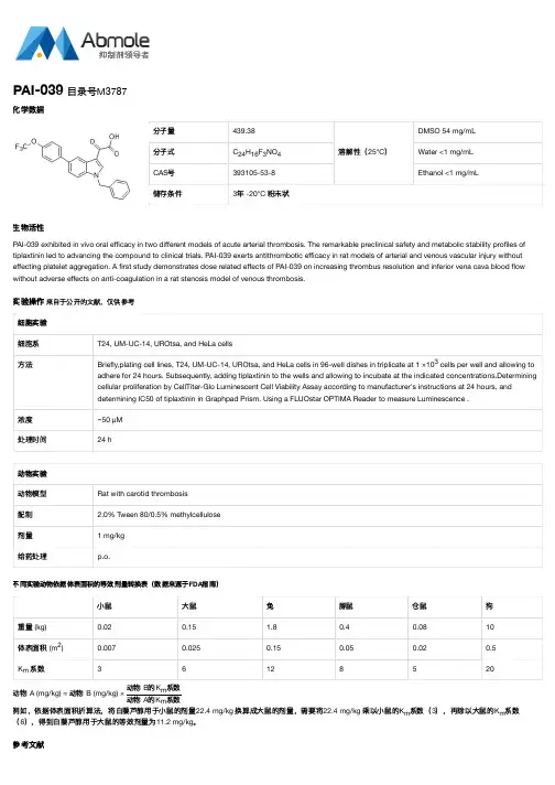
实验操作 来自于公开的文献,仅供参考
细胞实验 细胞系 方法
浓度 处理时间
T24, UM-UC-14, UROtsa, and HeLa cells
Briefly,plating cell lines, T24, UM-UC-14, UROtsa, and HeLa cells in 96-well dishes in triplicate at 1 ×10³ cells per well and allowing to adhere for 24 hours. Subsequently, adding tiplaxtinin to the wells and allowing to incubate at the indicated concentrations.Determining cellular proliferation by CellTiter-Glo Luminescent Cell Viability Assay according to manufacturer's instructions at 24 hours, and determining IC50 of tiplaxtinin in Graphpad Prism. Using a FLUOstar OPTIMA Reader to measure Luminescence .
0.02
0.15
1.8
0.4
0.08
10
体表面积 (m2)
0.007
0.025
0.15
0.05
0.02
0.5
Km 系数
3
6
12
8
5
20
动例物如,A依(m据g/体kg表) =面动积物折算B (法m,g/k将g白) ×藜动动芦物物醇BA用的的于KK小mm系系鼠数数的剂量22.4 (6),得到白藜芦醇用于大鼠的等效剂量为11.2 。 mg/kg
用于CRISPR文库筛选的N2a-Cas9细胞系的构建
中国预防兽医学报Chinese Journal of Preventive Veterinary Medicine第42卷第12期2020年12月V ol.42No.12Dec.2020doi :10.3969/j.issn.1008-0589.202002012用于CRISPR 文库筛选的N2a-Cas9细胞系的构建张纪文,王露露,李芳,刘杏,赵东明*,步志高*(中国农业科学院哈尔滨兽医研究所兽医生物技术国家重点实验室,黑龙江哈尔滨150069)摘要:为了进行CRISPR 文库筛选,本研究采用慢病毒转导的方法利用N2a 细胞构建表达Cas9蛋白的N2a-Cas9单克隆细胞系。
实验将LentiCas9-Blast 、pSPA X2和pMD 2.G 3种质粒共转染人胚胎肾细胞(HEK293T )获取高滴度的重组慢病毒,收集重组慢病毒并感染N2a 细胞,经杀稻瘟菌素(Blasticidin )筛选,通过有限稀释法获得含有Cas9基因的多个单克隆细胞株。
Western blot 结果显示候选细胞系高水平表达Cas9蛋白;利用慢病毒表达报告基因载体系统检测显示候选细胞系的Cas9蛋白具有很高的切割活力;利用CellTiter-Glo 试剂检测结果显示,Cas9基因在候选细胞系内高表达但不影响细胞增殖活性。
本研究利用慢病毒转导系统构建了稳定表达Cas9蛋白的N2a-Cas9单克隆细胞系,为基于CRISPR/Cas9全基因组高通量筛选技术探究神经细胞内关键宿主基因调控狂犬病病毒嗜神经性提供研究基础。
关键词:基因编辑;慢病毒;高通量筛选;鼠神经瘤母细胞中图分类号:S852.65文献标识码:A文章编号:1008-0589(2020)12-1292-04Construction of N2a-Cas9cell line for genome-wide CRISPR screeningZHANG Ji-wen,WANG Lu-lu,LI Fang,LIU Xing,ZHAO Dong-ming *,BU Zhi-gao *(State Key Laboratory of Veterinary Biotechnology,Harbin Veterinary Research Institute,Chinese Academy of Agricultural Sciences,Harbin 150069,China)Abstract :In this study,mouse neuroblastoma (N2a)cells were used to construct N2a-Cas9cell line which stably expresses Cas9protein by using lentivirus transfection technology.This constrcted cells line laid the foundation for CRISPR library screening.First,three plasmids including LentiCas9-Blast,pSPA X2,and pMD 2.G were co-transfected into human renal epithelial cells (HEK293T)to obtain recombinant lentivirus with high titers.N2a cells were infected with the recombinant lentivirus,and then screened by Blasticidin.Finally,several monoclonal cell lines expressing Cas9protein were isolated by limiting dilution.The expression of Cas9expression in N2a-Cas9cell line was determined by western blot,and the high cleavage activity of Cas9was also examined by using lentivirus-based reporter vector system.By using CellTiter-Glo ®reagent,the cell proliferation activity was well detectable and not decreased in N2a-Cas9cell lines,where Cas9gene was expressed in a high level.Therefore,N2a-Cas9cell line was successfully constructed using a lentiviral transfection system,which can provide a basis for genome-wide CRISPR screening to identify crucial host factors regulating Rabies virus neuro-tropism.Key words :gene editing;lentivirus;genome-wide CRISPR screening;murine neuroblastoma收稿日期:2020-02-11基金项目:兽医生物技术国家重点实验室自主课题(SKLVBP201801)作者简介:张纪文(1993-),男,河南驻马店人,硕士研究生,主要从事兽医微生物及其分子生物学研究.*通信作者:E-mail :****************;*********************Corresponding author张纪文,等.用于CRISPR文库筛选的N2a-Cas9细胞系的构建第12期CRISPR-Cas9(Clustered regularly interspaced short palindromic repeat sequences/CRISPR-associated protein 9)是存在于细菌或古生细菌中的一种抵御外源DNA 侵入的适应性免疫反应系统[1]。
psicheck2质粒说明书

Promega Corporation ·2800 Woods Hollow Road ·Madison, WI 53711-5399 USA·Fax 608-277-2516 ·1.Description ..........................................................................................................12.Product Components and Storage Conditions ............................................23.General Considerations (3)A.siCHECK™ Vector Features...............................................................................3B.How the siCHECK™ Vectors Work..................................................................4C.Sample Experiments Using the siCHECK™ Vectors.. (6)4.siCHECK™ Vector Maps .................................................................................95.siCHECK™ Vector Restriction Enzyme Tables . (11)A.Restriction Enzyme Sites for the psiCHECK™-1 Vector..............................11B.Restriction Enzyme Sites for the psiCHECK™-2 Vector (13)6.siCHECK™ Vector Backbones and Components .....................................157.References .........................................................................................................168.Related Products ..............................................................................................181.DescriptionThe psiCHECK™-1 Vector (a–d)(Cat.# C8011) and psiCHECK™-2 Vector (a–f)(Cat.# C8021) are designed to provide a quantitative and rapid approach for optimization of RNA interference (RNAi). The vectors enable the monitoring of changes in expression of a target gene fused to the reporter gene. In both vectors, Renilla luciferase is used as a primary reporter gene, and the gene of interest can be cloned into the multiple cloning region located downstream of the Renilla luciferase translational stop codon. Initiation of the RNAi process toward a gene of interest results in cleavage and subsequent degradation of fusion mRNA. Measurement of decreased Renilla luciferase activity is a convenient indicator of RNAi effect (1).RNAi is a phenomenon by which double-stranded RNA complementary to a target mRNA can specifically inactivate gene function by stimulating the degradation of the target mRNA (2–4). Because of the ability to inactivate genes, RNAi has emerged as a powerful tool for analyzing gene function.siCHECK™ VectorsAll technical literature is available on the Internet at: /tbs Please visit the web site to verify that you are using the most current version of this Technical Bulletin. Please contact Promega Technical Services if you have questions on useof this system. E-mail: techserv@In mammalian systems, including cultured mammalian cells, chemicallysynthesized double-stranded short interfering RNA molecules (<30 nucleotides;siRNA) or endogenously expressed short hairpin RNA molecules (shRNA) result in dsRNA duplexes <30 base pairs in length that induce RNAi (5–10). RNAiduplexes >30bp induce the interferon response and nonspecific degradation ofmRNA and cannot be used as tools for specific gene silencing (11,12).Interestingly, a significant percentage of the siRNA or shRNA designed for aspecific gene are not effective (5,13–16). On average only 1 in 5 of thesiRNA/shRNAs selected for targeting a specific region show efficient genesilencing (16,17). Possible causes for the failure of a particular siRNA/shRNAmay be instability of an siRNA probe in vivo, inability to interact withcomponents of the RNAi machinery or the inaccessibility of the target mRNAdue to local secondary structural constraints. Analysis of nucleotide sequences,melting temperatures and secondary structures have not revealed any obviousdifference between effective and ineffective siRNA/shRNA (18).At present, one of the most serious limitations for the RNAi technology is thelack of a rapid, reliable, quantitative target-site screening method. Variousalgorithm programs exist that aid in the design of potential siRNA targets.However, an experimental method is needed to screen these siRNAs. Currentscreening technologies include such semi-quantitative, time-consuming methods as fluorescence change for GFP-target fusions, Western blot analysis, monitoringphenotypic changes or RT-PCR. In addition, the current screening technologiesare not easily modified for the rapid, simultaneous screening of multiplesiRNA/shRNA.2.Product Components and Storage ConditionsProduct Size Cat.# psiCHECK™-1 Vector20μg C8011 psiCHECK™-2 Vector20μg C8021 Storage Conditions:Store the psiCHECK™-1 and psiCHECK™-2 Vectors at–20°C.Promega Corporation·2800 Woods Hollow Road ·Madison, WI 53711-5399 USA·Promega Corporation ·2800 Woods Hollow Road ·Madison, WI 53711-5399 USA·Fax 608-277-2516 ·3.General Considerations3.A. siCHECK™ Vector FeaturesCurrent methods to monitor changes in gene expression as the result of RNAi are either semi-quantitative, time-consuming or not applicable to high-throughput screening. The siCHECK™ Vectors are easier to use than currently available methods, allow optimal quantitative target site selection and can be adapted for use in high-throughput methodologies.There are two siCHECK™ Vectors, the psiCHECK™-1 Vector and thepsiCHECK™-2 Vector. Both vectors contain as the primary reporter gene the synthetic version of Renilla luciferase, hRluc , which is used to monitor changes in expression as the result of RNAi induction. This synthetic gene is engineered for more efficient expression in mammalian cells and for reduced anomalous transcription.To aid in fusion of the target gene to the synthetic Renilla luciferase reporter gene, a region of restriction sites (i.e., the multiple cloning region) has been added 3´ to the Renilla translational stop. The restriction sites present in the multiple cloning region can be used to create genetic fusions between the gene of interest and the Renilla reporter gene. Because no fusion protein isexpressed, there is no need to be concerned about whether you have cloned into a proper translational reading frame.The multiple cloning region of the psiCHECK™-1 Vector contains unique restriction sites SgfI, XhoI, SmaI, EcoRI, PmeI and NotI. Due to the presence of the firefly expression cassette, the psiCHECK™-2 Vector contains fewer unique restriction sites. The restriction sites in the psiCHECK™-2 Vector multiple cloning region are SgfI, XhoI, PmeI and NotI.The promoter used for Renilla luciferase expression in the siCHECK™ Vectors is the SV40 promoter. Experimental results (data not shown) demonstrate that the SV40 promoter results in the best balance between Renilla luciferaseexpression and the detection of RNAi activity when used with siRNA or vectors expressing shRNA.The difference between the two siCHECK™ Vectors is that the psiCHECK™-2Vector possesses a secondary firefly reporter expression cassette. The firefly expression cassette consists of an HSV-TK promoter, a synthetic firefly luciferase gene and an SV40 late poly(A) signal. To reduce the potential for recombination events, the Renilla luciferase reporter gene in the psiCHECK™-2Vector uses a synthetic poly(A). This firefly reporter cassette has beenspecifically designed to be an intraplasmid transfection normalization reporter;thus when using the psiCHECK™-2 Vector, the Renilla luciferase signal can be normalized to the firefly luciferase signal.If no transfection normalization is required or one would prefer to have the transfection normalization reporter on a second plasmid, the psiCHECK™-1Vector is the vector of choice.3.A. siCHECK™ Vector Features (continued)The psiCHECK™-1 Vector is recommended for use in monitoring RNAieffects in live cells. The changes in Renilla luciferase activity are measuredwith EnduRen™ Live Cell Substrate (Cat.# E6481), which allows continuousmonitoring of intracellular Renilla luminescence (19; Figure 2). EnduRen™ LiveCell Substrate is for use only with Renilla luciferase.Promega offers several reagents that can be used in conjunction with thesiCHECK™ Vectors to monitor Renilla and/or firefly luciferase signals. For thepsiCHECK™-1 Vector, which only contains the Renilla luciferase reporter gene,the Renilla Luciferase Assay System (Cat.# E2810, E2820) can be used. ThepsiCHECK™-2 Vector, which contains Renilla and firefly luciferase reportergenes, requires the use of either the Dual-Luciferase®Reporter Assay System(Cat.# E1910) or the Dual-Glo™ Luciferase Assay System (Cat.# E2920) togenerate the firefly and Renilla luciferase signals.3.B. How the siCHECK™ Vectors WorkFigure 1 provides a basic description of how the siCHECK™ Vectors work.Using the unique restriction sites, the gene of interest is cloned into themultiple cloning region located 3´ to the synthetic Renilla luciferase gene andits translational stop codon. After cloning, the vector is transfected into themammalian cell line of choice, and a fusion of the Renilla luciferase gene andthe gene of interest is transcribed. Vectors expressing potential shRNA orsiRNA can be cotransfected simultaneously or sequentially, depending onyour experimental design. If a specific shRNA/siRNA binds to the targetmRNA and initiates the RNAi process, the fused Renilla luciferase:gene ofinterest mRNA will be cleaved and subsequently degraded, decreasing theRenilla luciferase signal.Promega Corporation·2800 Woods Hollow Road ·Madison, WI 53711-5399 USA·Promega Corporation ·2800 Woods Hollow Road ·Madison, WI 53711-5399 USA·Fax 608-277-2516 ·translation stopcleavage of mRNAlight mRNA5´3´5´3´4339M A 10_3A5´3´hRlucgene of interesthRluc Figure 1. Mechanism of action of the siCHECK™ Vectors.3.C. Sample Experiments Using the siCHECK™ VectorsTo demonstrate the utility of the siCHECK™ Vectors, two experiments are detailed in this Technical Bulletin. In the first experiment, human p53 cDNA was subcloned into the psiCHECK™-1 and the psiCHECK™-2 Vectors using the SgfI and NotI restriction sites located in the multiple cloning region of both vectors. Note the SgfI and NotI restriction sites are located 3´ to the Renilla luciferase translational stop codon. As shown in Figure 2, the psiCHECK™-1Vector containing the human p53 cDNA was cotransfected into HEK-293T cells with the psiLentGene™ Basic Vector expressing either a Renilla luciferase (hRluc ) or p53 shRNA. The negative control was the psiLentGene™ Basic Vector with a nonspecific 19bp insert. (A BLAST search using this 19bp sequence and a threshold >90% revealed no homology to any knownmammalian gene or to the synthetic Renilla luciferase gene.) This nonspecificsequence was used for all RNAi experiments in this Technical Bulletin.Promega Corporation ·2800 Woods Hollow Road ·Madison, WI 53711-5399 USA·4398M A 11_3AR e n i l l a L u m i n e s c e n c e (R L U )Time Post-Transfection (hours)Negative Control Renilla p53Figure 2. Inhibition of Renilla luciferase expression by targeting either the Renilla luciferase or p53 gene.The human p53 cDNA was subcloned into the psiCHECK™-1Vector using the SgfI and NotI restriction sites located 3´ to the Renilla luciferase translational stop codon. To begin the transfection assay, HEK-293T cells were plated in a 96-well plate at 3,000 cells/well. After an overnight incubation, the cells were treated with a transfection mixture consisting of 35μl of serum-free medium,0.3μl of TransFast™ Transfection Reagent (Cat.# E2431), 0.02μg of psiCHECK™-1:p53vector and 0.08μg of psiLentGene™ Basic Vector per well. For this experiment, the psiLentGene™ Vector expressed shRNAs directed against human p53, Renilla luciferase or the nonspecific 19bp sequence, which serves as a negative control,(Section 3.C). After a one-hour incubation, 100μl of serum-containing medium was added to the wells. At 21 hours post-transfection, EnduRen™ Live Cell Substrate (Cat.# E6481) was added to a final concentration of 60μM, and Renilla luciferase activity was monitored. Renilla luciferase activities were normalized to the number of viable cells using the CellTiter-Glo ®Luminescent Cell Viability Assay (Cat.#G7573; 20).At 21 hours post-transfection, nonlytic EnduRen™ Live Cell Substrate wasadded to the wells; luminescence was monitored for the next 27 hours until48 hours post-transfection. The data in Figure 2 show that the psiLentGene™Basic Vector expressing either Renilla luciferase or p53 shRNA inhibits theexpression of the Renilla luciferase reporter gene from the psiCHECK™-1:p53vector. Interestingly, using either Renilla luciferase or p53 shRNA results invirtually identical inhibition of Renilla luciferase expression.In a second experiment, the human p53 cDNA used in Figure 2 was subclonedinto the psiCHECK™-2 Vector using the SgfI and NotI restriction sites. Fivepotential p53 shRNAs designed to bind to five different target sites were clonedinto the psiLentGene™ Basic Vector; the resulting vectors were named Site 1through Site 5. The control is a psiLentGene™ Vector containing the nonspecific19bp sequence. The psiCHECK™-2 Vector containing the p53 cDNA wascotransfected with the psiLentGene™ Vector expressing either a p53 shRNA(Figure 3, Sites 1–5) or the nonspecific shRNA into HEK-293T cells as describedin Figure 3. Forty-eight hours after transfection, the medium was removed andcells were lysed in Passive Lysis Buffer (Cat.# E1941). The firefly and Renillaluciferase signals were generated using the Dual-Luciferase®Reporter 1000Assay System (21).Figure 3, Panel A, displays the Renilla luciferase signal, while Figure 3, Panel B,shows the Renilla luciferase signal normalized (corrected for transfectionefficiency to the firefly luciferase signal). The data in Figure 3, Panel A, isdifficult to interpret due to transfection variations. The Renilla luciferasepositive control, which should demonstrate inhibition of reporter expression, isnot statistically different (i.e., overlapping error bars) from the negative control(no effect on reporter expression was detected). The inability to distinguishbetween the positive and negative controls renders any conclusion regardingthe effectiveness of potential shRNAs suspect.However, when the Renilla luciferase signals are normalized (see Figure 3,Panel B) to the internal firefly luciferase transfection control, the datainterpretation is different, as the Renilla luciferase positive control isstatistically different from the negative control. In addition, the normalizeddata allow the ability to distinguish the effectiveness of the various target siteshRNAs.Promega Corporation·2800 Woods Hollow Road ·Madison, WI 53711-5399 USA·Fax 608-277-2516 ·Promega Corporation ·2800 Woods Hollow Road ·Madison, WI 53711-5399 USA·Figure 3. Target site selection using the psiCHECK™-2 Vector. HEK-293T cells were seeded into a 96-well plate at a density of 3,000 cells/well. Human p53 cDNA was subcloned into the psiCHECK™-2 Vector using the SgfI and NotI restriction sites. After an overnight incubation, the cells were treated with a transfection mixture consisting of 35μl of serum-free medium, 0.3μl of TransFast™ Transfection Reagent (Cat.# E2431), 0.02μg of psiCHECK™-2 Vector:p53 and 0.08μg of psiLentGene™Basic Vector per well. The psiLentGene™ Basic Vector expressed one of five different shRNAs directed against human p53, Renilla luciferase or a nonspecific 19bp sequence (Section 3.C) as a negative control. After a one-hour incubation, 100μl of serum-containing medium was added to the wells. Forty-eight hours post-transfection Renilla and firefly luciferase activities were measured using the Dual-Luciferase ®Reporter 1000 Assay System (Cat.# E1980; 21). Panel A displays the raw Renilla luciferase data, while in Panel B , the Renilla luciferase data has beennormalized to firefly luciferase data. The data represent the mean of 12 wells plus or minus the standard deviation. Note that in other experiments the ability of different shRNAs to inhibit gene expression might vary more dramatically.4399M A 11_3AA.B.S i t e1S i t e2S i t e3S i t e 4 S i t e5 R e ni l l a P o s it i ve C o n t r o l Ne g a t i v e C o n t r o l R e n i l l a L u m i n e s c e n c e (R L U )S i t e 1S i t e 2S i t e 3 S i t e 4 S i t e 5 R e n i l l a Po s i t i v e C o nt r o l N e g a t i v e C o n t r o lN o r m a l i z e d R e n i l l a L u m i n e s c e n c e (R L U )4.siCHECK™ Vector MapspsiCHECK™-1 Vector sequence reference points: SV40 early enhancer/promoter 7–425Chimeric intron489–621T7 RNA polymerase promoter666–684Synthetic Renilla luciferase gene (hRluc )694–1629Multiple cloning region 1636–1680Synthetic poly(A)1688–1736β-lactamase (Amp r ) coding region1874–2734Promega Corporation ·2800 Woods Hollow Road ·Madison, WI 53711-5399 USA·Fax 608-277-2516 ·4343M A 10_3ABglII 1Figure 4. psiCHECK™-1 Vector map. –^– denotes the intron.Synthetic poly(A) signal4342M A 10_3ACCCGGGAATTCGTTTAAACCTAGAGCGGCCGCTGGCCGC AATAAAATA . . . 3′5′ . . . GAGCAGTAA TTCTAGGCGATCGCTCGAG XhoISmaINotIEcoRIPmeISgfIhRlucFigure 5. psiCHECK™-1 Vector multiple cloning region.psiCHECK™-2 Vector sequence reference points:SV40 early enhancer/promoter 7–425Chimeric intron489–621T7 RNA polymerase promoter666–684Synthetic Renilla luciferase gene (hRluc )694–1629Multiple cloning region 1636–1680Synthetic poly(A)1688–1736HSV-TK promoter1744–2496Synthetic firefly luciferase gene (hluc +)2532–4184SV40 late poly(A)4219–4440β-lactamase (Amp r ) coding region4587–5447Figure 6. psiCHECK™-2 Vector map.–^– denotes the intron.4345M A 10_3ABglII 1Synthetic poly(A) signal4344M A 10_3ACCCGGGAATTCGTTTAAACCTAGAGCGGCCGCTGGCCGC AATAAAATA . . . 3′5′ . . . GAGCAGTAA TTCTAGGCGATCGCTCGAGXhoINotIPmeISgfIhRlucFigure 7. psiCHECK™-2 Vector multiple cloning region.5.siCHECK™ Vector Restriction Enzyme Tables5.A.Restriction Enzyme Sites for the psiCHECK™-1 VectorThe following restriction enzyme tables were constructed using DNASTAR ®sequence analysis software. Please note that we have not verified this information by restriction digestion with each enzyme listed. The location given specifies the 3´-end of the cut DNA (the base to the left of the cut site).For more information on the cut sites of these enzymes, or to report adiscrepancy, please contact your local Promega Branch or Distributor. In the U.S., contact Promega Technical Services at 800-356-9526. Vector sequences are available from the GenBank ®database (GenBank ®/EMBL accession number AY535006) and online at:/vectors/Enzyme # of Sites Location AatII 11391Acc65I 154AcyI 21388, 2121AflII 2452, 649Alw44I 21989, 3235AlwNI 13140AspHI 41091, 1993, 2078,3239AvaI 3715, 1643, 1649AvaII 22297, 2519AvrII 1404BamHI 11738BanI 354, 575, 2708BanII 3759, 899, 1650BbsI 1560BbuI 2152, 224BclI 2734, 1187BglI 3357, 694, 2543BglII 11BsaI 3514, 1234, 2595BsaOI51640, 1677, 2143,2292, 3215BsaBI 11453BsaHI 21388, 2121BspHI 21821, 2829BspMI 1476BssSI 21992, 3376Bst98I 2452, 649BstZI 11280Cfr10I 12576Enzyme # of Sites LocationDraI 41663, 2083, 2775,2794DraII 11539DraIII 1882DrdI 2441, 3447DsaI 415, 311, 692, 899EaeI 31674, 1681, 2268EagI 11674EarI 21193, 1862EclHKI 12661Eco52I 11674Eco81I 11280EcoRI 11654EcoRV 11179FspI 28, 2438HaeII 13309HgaI 41570, 2129, 2859,3437HindIII 1420Hsp92I 21388, 2121KpnI 158MspA1I 580, 1679, 2025,2966, 3211NciI 51650, 1651, 2125, 2476, 3172NcoI 315, 311, 692NheI 1684NotI 11674NruI 11355NsiI3154, 226, 913Table 1. Restriction Enzymes That Cut the psiCHECK™-1 Vector Between 1 and 5 Times.Promega Corporation ·2800 Woods Hollow Road ·Madison, WI 53711-5399 USAFax 608-277-2516 ·5.A.Restriction Enzyme Sites for the psiCHECK™-1 Vector (continued)Table 2. Restriction Enzymes That Do Not Cut the psiCHECK™-1 Vector.A ccB7I AccI AccIII AflIII AgeI ApaI AscI BalI BbeI BbrPI BlpI Bpu1102IBsaAI BsaMI BsmI Bsp120I BsrGI BssHII Bst1107I BstEII BstXI ClaI CspI Csp45IEco47III Eco72I EcoICRI EcoNI EheI FseI HincII HindII HpaI I-PpoI KasI MluINaeI NarI NdeI NgoMIV PacI PflMI PinAI PmlI PpuMI PshAI Psp5II RsrIISacI SacII SalI SgrAI SnaBI SpeI SplI SrfI Sse8387I SwaI XbaI XcmITable 3. Restriction Enzymes That Cut the psiCHECK™-1 Vector 6 or More Times. AciI AluI Alw26I BbvI BsaJI Bsp1286I BsrI BsrSI Bst71IBstOI BstUI CfoI DdeI DpnI DpnII Fnu4HI FokI HaeIIIHhaI HinfI HpaII HphI Hsp92II MaeI MaeII MaeIII MboIMboII MnlI MseI MspI NdeII NlaIII NlaIV PleI RsaISau3AI Sau96I ScrFI SfaNI TaqI Tru9I XhoIINote:The enzymes listed in boldface type are available from Promega.Enzyme # of Sites LocationNspI 2152, 224PaeR7I 11643PmeI 11663Ppu10I 3150, 222, 909PspAI 11649PstI 1462PvuI 21640, 2292PvuII 180ScaI 2662, 2180SfiI 1357SgfI 11640SinI 22297, 2519Enzyme # of Sites Location SmaI 11651SphI 2152, 224SspI 11856StuI 1403StyI 515, 311, 404, 692,701TfiI 2426, 805Tth111I 11390VspI 12486XhoI 11643XmaI 11649XmnI21228, 2061Table 1. Restriction Enzymes That Cut the psiCHECK™-1 Vector Between 1 and 5 Times (continued).5.B.Restriction Enzyme Sites for the psiCHECK™-2 VectorThe following restriction enzyme tables were constructed using DNASTAR ®sequence analysis software. Please note that we have not verified this information by restriction digestion with each enzyme listed. The location given specifies the 3´-end of the cut DNA (the base to the left of the cut site).For more information on the cut sites of these enzymes, or to report adiscrepancy, please contact your local Promega Branch or Distributor. In the U.S., contact Promega Technical Services at 800-356-9526. Vector sequences are available from the GenBank ®database (GenBank ®/EMBL accession number AY535007) and online at:/vectors/Promega Corporation ·2800 Woods Hollow Road ·Madison, WI 53711-5399 USAFax 608-277-2516 ·Enzyme # of Sites Location AatII 11391AccI 22079, 3132Acc65I 154AflII 4452, 649 , 1773,1897AflIII 12450Alw44I 24702, 5948AlwNI 22094, 5853ApaI 12562AvrII 2404, 2059BalI 31865, 3513, 4038BamHI 14451BanII 5759, 899, 1650,2050, 2562BbeI 42030, 2815, 3481,3613BbsI 2560, 1743BbuI 2152, 224BclI 5734, 1187, 3112,3853, 4147BglII 11BsaI 4514, 1234, 2123,5308BsaAI 22083, 3734BsaBI 41453, 2979, 4146,4450BsaMI 32504, 4270, 4363BsmI 32504, 4270, 4363Bsp120I 12558BspHI 33115, 4534, 5542BspMI2476, 3463Enzyme # of Sites Location BsrGI 13022BssHII 11978BssSI 33459, 4705, 6089Bst1107I 12080Bst98I 4452, 649, 1773, 1897BstXI 13650BstZI 31674, 4202, 4206Bsu36I 3 1280, 3145, 3745ClaI 14444Csp45I 12390DraI 51663, 4410, 4796,5488, 5507DraIII 1882DrdI 2441, 6160EagI 31674, 4202, 4206EarI 51193, 1874, 2616,2727, 4575EclHKI 15374Eco47III 13519Eco52I 31674, 4202, 4206Eco81I 31280, 3145, 3745EcoNI 32721, 3144, 4149EcoRI 21654, 2386EcoRV 11179EheI 42028, 2813, 3479,3611FseI 23943, 4208FspI 38, 3354, 5151HincII 14349HindII14349Table 4. Restriction Enzymes That Cut the psiCHECK™-2 Vector Between 1 and 5 Times.5.B.Restriction Enzyme Sites for the psiCHECK™-2 Vector (continued)Table 5. Restriction Enzymes That Do Not Cut the psiCHECK™-2 Vector.AccB7I AccIII AgeI AscI BbrPI BlpIBpu1102I BstEII CspI Eco72I EcoICRI I-PpoINdeI PacI PflMI PinAI PmlI PshAIRsrII SacI SalI SgrAI SnaBI SpeISplI SrfI Sse8387I SwaI XcmIEnzyme # of Sites Location HindIII 2420, 2497HpaI 14349KasI 42026, 2811, 3477,3609KpnI 158MluI 12450NaeI 33941, 3962, 4206NarI 42027, 2812, 3478,3610NcoI 515, 311, 692, 2067, 2530NgoMIV 33939, 3960, 4204NheI 1684NotI 11674NruI 11355NsiI 3154, 226, 913NspI 5152, 224, 2336, 3023, 3278PaeR7I 11643PmeI 11663Ppu10I3150, 222, 909Enzyme # of Sites LocationPpuMI 12056Psp5II 12056PspAI 21649, 2019PvuI 21640, 5005PvuII 380, 2268, 2606SacII 12036ScaI 3662, 2697 ,4893SfiI 1357SgfI 11640SmaI 21651, 2021SphI 2152, 224SspI 14569StuI 1403TfiI 2426, 805Tth111I 11390VspI 15199XbaI 14189XhoI 11643XmaI 21649, 2019XmnI 21228, 4774Table 4. Restriction Enzymes That Cut the psiCHECK™-2 Vector Between 1 and 5 Times (continued).6.siCHECK™ Vector Backbones and ComponentsThe vector backbones of the psiCHECK™-1 and psiCHECK™-2 Vectors are based on the phRL-SV40 Vector (Cat.# E6261). Both the psiCHECK™-1 Vector and psiCHECK™-2 Vector contain the synthetic Renilla luciferase reporter gene.The psiCHECK™-2 Vector also contains a synthetic firefly luciferase gene.These synthetic luciferase genes have been codon optimized for more efficient mammalian expression and have been designed with a greatly reduced number of consensus transcription factor binding sites for reduced risk of anomalous transcriptional behavior.SV40 Early Enhancer/PromoterThe psiCHECK™-1 Vector and psiCHECK™-2 Vector contain the SV40 early enhancer/promoter region, which provides strong, constitutive expression of Renilla luciferase in a variety of cell types.Chimeric IntronDownstream of the SV40 enhancer/promoter region is a chimeric introncomposed of the 5´-donor site from the first intron of the human β-globin and the branch and 3´-acceptor site from the intron that is between the leader and the body of an immunoglobin gene heavy chain variable region (22). The sequences of the donor and acceptor sites, along with the branch point site,have been changed to match the consensus sequence for splicing (23).Transfection studies have demonstrated that the presence of an intron flankingthe cDNA insert frequently increases the level of gene expression (24–27).Promega Corporation ·2800 Woods Hollow Road ·Madison, WI 53711-5399 USAFax 608-277-2516 ·Table 6. Restriction Enzymes That Cut the psiCHECK™-2 Vector 6 or More Times. AciI AcyI AluI Alw26I AspHI AvaI AvaII BanI BbvI BglI BsaOI BsaHI BsaJIBsp1286I BsrI Bsr SI Bst71I BstOI BstUI CfoI Cfr10I DdeI DpnI DpnII DraII DsaIEaeI Fnu4HI FokI HaeII HaeIII HgaI HhaI HinfI HpaII HphI Hsp92I Hsp92II MaeIMaeII MaeIII MboI MboII MnlI MseI MspI MspA1I NciI NdeII NlaIII NlaIV PleIPstI RsaI Sau3AI Sau96I ScrFI SfaNI SinI StyI TaqI Tru9I XhoIINote:The enzymes listed in boldface type are available from Promega.T7 PromoterA T7 RNA polymerase promoter is located downstream of the chimeric intron and immediately precedes the synthetic Renilla luciferase reporter gene. This promoter can be used to synthesize RNA transcripts in vitro using T7 RNA Polymerase (Cat.# P2075). Note that the T7 promoter has been verified by sequence only; there has been no functional testing of the T7 promoter. Polyadenylation Signals (SV40 Late and Synthetic)Polyadenylation signals are coupled to the termination of transcription by RNA polymerase II and signal the addition of approximately 200–250 adenosine residues to the 3´-end of the RNA transcript (28). Polyadenylation has been shown to enhance RNA stability and translation (29,30). The late SV40 polyadenylation signal is extremely efficient and has been shown to increase the steady-state level of RNA to approximately fivefold more than that of the early SV40 polyadenylation signal (31). The synthetic poly(A) was cloned from our pCI-neo Vector (Cat.# E1841). The synthetic poly(A) signal is based on the highly efficient polyadenylation signal of the rabbit β-globin gene (32).7.References1.Kumar, R., Conklin, D.S. and Mittal, V. (2003) High-throughput selection of effectiveRNAi probes for gene silencing. Genome Res.13, 2333–40.2.Bass, B.L. (2000) Double-stranded RNA as a template for gene silencing. Cell101, 235–8.3.Zamore, P.D. (2001) RNA interference: Listening to the sound of silence. Nature Struct.Biol.8, 746–50.4.Sharp, P.A. (2001) RNA interference—2001. Genes Dev.15, 485–90.5.Gil, J. and Esteban, M. (2000) Induction of apoptosis by the dsRNA-dependentprotein kinase (PKR): Mechanism of action. Apoptosis5, 107–14.6.Marcus, P.I. and Sekellick, M.J. (1985) Interferon induction by viruses. XIII. Detectionand assay of interferon induction-suppressing particles. Virology142, 411–5.7.Elbashir, S.M. et al.(2001) Duplexes of 21-nucleotide RNAs mediate RNA interferencein cultured mammalian cells. Nature411, 494–8.8.Brummelkamp, T.R., Bernards, R. and Agami, R. (2002) A system for stableexpression of short interfering RNAs in mammalian cell. Science296, 550–3.9.Elbashir, S.M. et al. (2002) Analysis of gene function in somatic mammalian cellsusing small interfering RNAs. Methods26, 199–213.10.Paddison, P.J. et al. (2002) Short hairpin RNAs (shRNAs) induce sequence-specificsilencing in mammalian cells. Genes Dev.16, 948–58.11.Paul, C.P. et al.(2002) Effective expression of small interfering RNA in human cells.Nature Biotechnol. 20, 505–8.。
Proteasome-Glo检测试剂盒说明书
Promega Corporation ·2800 Woods Hollow Road ·Madison, WI 53711-5399 USA·Fax 608-277-2516 ·1.Description ..........................................................................................................12.Product Components and Storage Conditions ............................................73.Reagent Preparation ........................................................................................104.Assay for Detection of Proteasome Activity . (11)A.Assay Conditions................................................................................................11B.Standard Assay (96-well, 100µl Final Reaction Volume).. (12)5.General Considerations ..................................................................................136.References ........................................................................................................147.Related Products .............................................................................................151.DescriptionThe Proteasome-Glo™ 3-Substrate System (a,b,c)consists of three homogeneous bioluminescent assays that measure the three proteolytic activities associated with the proteasome (each of these three assays is also available separately).The proteasome is a multicatalytic complex in the nucleus and cytosol of all eukaryotic cells that is responsible for proteolysis of ubiquitin-tagged proteins.The catalytic core of the complex, the 20S proteasome, is a barrel-shaped assembly of 28 protein subunits that possesses three different proteolytic activities designated as chymotrypsin-like, trypsin-like, and caspase-like (also termed post-glutamyl peptide hydrolase; 1,2). The catalytic sites are located on the inner surface of the central β-rings of the cylindrical particle, and access to them is controlled by narrow gated channels in the outer α-rings of thecomplex. The association of the 20S particle with a 19S regulatory complex at one or both ends of the barrel forms the 26S proteasome and confers an open-channel conformation, resulting in much higher rates of peptide hydrolysis (3,4). The 26S proteasome degrades polyubiquitinated proteins in an ATP-dependent manner. The 19S regulatory unit binds and removes the ubiquitin chains from tagged proteins, and ATPases within the regulatory complex appear to unfold protein substrates and translocate the unfolded polypeptides into the 20S core (2–5). The 20S catalytic core and the 19S regulatory complex are highly conserved from yeast to mammals (1). The 26S proteasome complex processes aberrant and misfolded proteins as well as proteins regulating cell cycle, growth and apoptosis and is essential for cellular function.Proteasome-Glo™ Assay SystemsAll technical literature is available on the Internet at: /tbs/ Please visit the web site to verify that you are using the most current version of this Technical Bulletin. Please contact Promega Technical Services if you have questions on useof this system. E-mail: techserv@The role of the proteasome in degrading several important regulatory proteinshas led to the identification of the proteasome as a therapeutic target for cancertreatment. Proteasome inhibitors can induce apoptosis, and interestingly,transformed cells display greater susceptibility to proteasome inhibition thannonmalignant cells (5). The enhanced proliferative rate of malignant cells maycause accumulation of damaged proteins at a higher rate, which in turn mayincrease dependency on proteasomal degradation (6). The first-generationproteasome inhibitor, bortezomib (PS-341), is now an approved drug for thetreatment of refractory multiple myeloma, and second-generation inhibitors arecurrently being developed (7).The Proteasome-Glo™ 3-Substrate System provides three separate assays thatdiffer in their ability to detect different protease activities based on theirsubstrate components. The luminogenic substrates provided for thechymotrypsin-like, trypsin-like, and caspase-like activities are Suc-LLVY-aminoluciferin, Z-LRR-aminoluciferin, and Z-nLPnLD-aminoluciferin,respectively. Each substrate is added to a buffer system optimized forproteasome activity and luciferase activity to make a Proteasome-Glo™Reagent for a particular catalytic activity. The individual Proteasome-Glo™Reagent is added to test samples in an “add-mix-measure” format, resulting inproteasome-induced cleavage of the particular substrate. Substrate cleavagegenerates a “glow-type” luminescent signal produced by the luciferase reaction(Figure 1). In this homogeneous coupled-enzyme format, the signal isproportional to the amount of proteasome activity (Figure 2).The Proteasome-Glo™ Reagents rely on the properties of a proprietarythermostable luciferase (Ultra-Glo™ Recombinant Luciferase(b)) that isformulated to generate a “glow-type” luminescent signal and providesexcellent performance across a wide range of assay conditions. The proteasomeand luciferase enzyme activities reach a steady-state such that the luminescentsignal peaks rapidly and is maintained for several hours with minimal loss ofsignal (Figure 3). The Proteasome-Glo™ Assays provide rapid, sensitive, andaccurate assays for the three proteolytic activities of the proteasome (Figures 4and 5). The homogeneous Proteasome-Glo™ Assays are designed for use withmultiwell-plate formats, making them ideal for automated high-throughputscreening of proteasome activity and inhibition (Figure 6).Promega Corporation·2800 Woods Hollow Road ·Madison, WI 53711-5399 USA·nLPnLD sequence are recognized by the 20S proteasome. Following cleavage bythe 20S proteasome, the substrate for luciferase (aminoluciferin) is released, allowing the luciferase reaction to produce light.Figure 2. Luminescence is proportional to proteasome concentration. Titrations of 20S proteasome were performed in 96-well plates using the Proteasome-Glo™ 3-Substrate System. Human 20S proteasome was serially diluted in 10mMHEPES (pH 7.6). Thirty minutes after adding the individual Proteasome-Glo™Reagents separately, luminescence was recorded as relative light units (RLU)on a GloMax ®96 Microplate Luminometer. The results were linear over 4 logs of 20S proteasome concentration for all three assays.Each point represents the average of four wells. The background (blank without 20S) was subtracted from each. Values for r 2 and slope were calculated after transforming the data to a log 10-log 10plot.Promega Corporation ·2800 Woods Hollow Road ·Madison, WI 53711-5399 USA·Fax 608-277-2516 ·5855M ANS HSN Suc-LLVY – N Z-LRR – oror or orZ-nLPnLD –Suc-LLVY + Z-LRR Z-nLPnLDAminoluciferin , O 2COOHN S S N H 2N COOH 5862M A1101001,00010,000100,00020S (µg/ml)Suc-LLVY-aminoluciferiny = 1.0394x + 5.4385r 2 = 0.993Z-LRR-aminoluciferin y = 0.9922x + 4.7521 r 2 = 0.9980.0000.000.00.01L u m i n e s c e n c e (R L U , b a c k g r o u n d -s u b t r a c t e d )Figure 3. Signal stability of the Proteasome-Glo™ Assay Systems. Human purified 20S proteasome (1µg/ml) was assayed in 96-well plates using the individual Proteasome-Glo™ Assays. Luminescence was monitored at various time points over 3 hours on a GloMax ®96 Microplate Luminometer. Panel A.The signals peak rapidly and then are very stable for all three assays as shown on a log scale. Panel B.The stable signals generated with 20S proteasome are shown for thechymotrypsin-like (Suc-LLVY-Glo™ ) and trypsin-like (Z-LRR-Glo™) Substrates on a linear scale .Promega Corporation ·2800 Woods Hollow Road ·Madison, WI 53711-5399 USA·5863M ATime (minutes)Suc-LLVY-Glo™ Substrate + 20S Z-LRR-Glo™ Substrate + 20S Z-nLPnLD-Glo™ Substrate + 20S Suc-LLVY-Glo™ Substrate, no 20S Z-LRR-Glo™ Substrate, no 20S Z-nLPnLD-Glo™ Substrate, no 20S50,000200,000250,000300,000Suc-LLVY-Glo™ Substrate + 20S Z-LRR-Glo™ Substrate + 20SL u m i n e s c e n c e (R L U )L u m i n e s c e n c e (R L U )B.Time (minutes)1001,00010,000100,0001,000,000Figure 4. Sensitivity of the Proteasome-Glo™ Assays compared to fluorescent assays. Human purified 20S proteasome was titrated and assayed in 96-well plates using the Proteasome-Glo™ 3-Substrate System or comparable fluorogenic substrates. Luminescence and fluorescence were monitored at 30 minutes on a GloMax ®96 Microplate Luminometer or a Labsystems Fluoroskan Ascent plate reader, respectively. The results are plotted as signal-to-noise ratios. The limit of detection is defined as the amount of 20S proteasome giving a signal-to-noise ratio >3 (dashed lines). Panel A.The Proteasome-Glo™ Chymotrypsin-Like Assay (Suc-LLVY-Glo™ Substrate) was compared to a Suc-LLVY-AMC substrate. The fluorescent assay was performed with or without 0.02% SDS. Panel B.TheProteasome-Glo™ Trypsin-Like Assay (Z-LRR-Glo™ Substrate) was compared to the fluorogenic substrate, Boc-LRR-AMC, and the Proteasome-Glo™ Caspase-Like Assay (Z-nLPnLD-Glo™ Substrate) was compared to the fluorogenic substrate, Ac-nLPnLD-AMC. The signal-to-noise ratios are greater, and the limits of detection are significantly lower for all of the luminescent proteasome assays compared to the fluorescent proteasome assays.Promega Corporation ·2800 Woods Hollow Road ·Madison, WI 53711-5399 USA·Fax 608-277-2516 ·5864M A0.11101001,00010,000100,00020S (µg/ml)Suc-LLVY-Glo™ Substrate Suc-LLVY-AMCSuc-LLVY-AMC + SDS1101001,00010,000100,000Z-LRR-Glo™ Substrate Z-nLPnLD-Glo™ SubstrateBoc-LRR-AMC Ac-nLPnLD-AMCS i g n a l -t o -N o i s e R a t i o0.0000.000.00.01S i g n a l -t o -N o i s e R a t i o20S (µg/ml)0.00000.0000.000.00.1.1B.Figure 5. Determination of IC 50values. The inhibitor concentration that results in 50% inhibition (IC 50) was determined for the proteasome irreversible inhibitor,clasto-lactacystin β-lactone (8), using the Proteasome-Glo™ Chymotrypsin-Like Assay. The inhibitor was resuspended in DMSO, serially diluted in 10mM HEPES (pH 7.6) and combined with 1µg/ml 26S proteasome (Biomol) in 96-well plates.Reagent containing the Suc-LLVY-Glo™ Substrate was added after one hour, and luminescence was recorded 10 minutes after reagent addition. GraphPad Prism ®software was used to calculate the IC 50, which was 30nM.Assay AdvantagesBroad Dynamic Range:The assays are linear over 4 logs of proteasome concentrations and can detect 20S proteasome at concentrations as low as 0.5ng/ml (1pM) (Figures 2 and 4).Fast:Maximum sensitivity is reached in 10–30 minutes after adding reagent (Figure 3), because the assays are not dependent on accumulation of cleaved product for sensitivity.Greater Sensitivity:The coupled-enzyme format and the speed of theProteasome-Glo™ Assay results in low background and excellent signal-to-noise ratios. The assays are significantly more sensitive than fluorescence-based proteasome assays (Figure 4).Accurate: The broad linear range and excellent sensitivity readily translate to accurate kinetic analysis of inhibitors (Figure 5).Simplified Method: The “add-mix-read” protocols make the assays amenable to automation (Figure 6).Amenable to Batch Processing:The coupled-enzyme, homogeneous format results in a stable, glow-type signal, allowing flexibility in read time once thereagent is added (Figure 3).Promega Corporation ·2800 Woods Hollow Road ·Madison, WI 53711-5399 USA·5865M A20,00040,00060,00080,000100,000120,000140,000160,000Clasto-lactacystin β-lactone (µM)L u m i n e s c e n c e (R L U )2.Product Components and Storage ConditionsProduct Size Cat.# Proteasome-Glo™ 3-Substrate System10ml G8531 Includes 3 individual kits. Each kit contains sufficient reagent for 100 assays at100µl/assay or 200 assays at 50µl/assay in 96-well plates or 400 assays at 25µl/assay in384-well plates. Includes:• 1 kit Proteasome-Glo™ Chymotrypsin-Like Assay (Cat.# G8621)• 1 kit Proteasome-Glo™ Trypsin-Like Assay (Cat.# G8631)• 1 kit Proteasome-Glo™ Caspase-Like Assay (Cat.# G8641)Product Size Cat.# Proteasome-Glo™ 3-Substrate System50ml G8532 Includes 3 individual kits. Each kit contains sufficient reagent for 500 assays at100µl/assay or 1,000 assays at 50µl/assay in 96-well plates or 2,000 assays at25µl/assay in 384-well plates. Includes:• 1 kit Proteasome-Glo™ Chymotrypsin-Like Assay (Cat.# G8622)• 1 kit Proteasome-Glo™ Trypsin-Like Assay (Cat.# G8632)• 1 kit Proteasome-Glo™ Caspase-Like Assay (Cat.# G8642)Items Available SeparatelyProduct Size Cat.#Proteasome-Glo™ Chymotrypsin-Like Assay10ml G8621 Includes:•10ml Proteasome-Glo™ Buffer• 1 bottle Luciferin Detection Reagent•50µl Suc-LLVY-Glo™ SubstrateProduct Size Cat.# Proteasome-Glo™ Chymotrypsin-Like Assay50ml G8622 Includes:•50ml Proteasome-Glo™ Buffer• 1 bottle Luciferin Detection Reagent•250µl Suc-LLVY-Glo™ SubstratePromega Corporation·2800 Woods Hollow Road ·Madison, WI 53711-5399 USA·Fax 608-277-2516 ·2.Product Components and Storage Conditions (continued)Product Size Cat.# Proteasome-Glo™ Trypsin-Like Assay10ml G8631 Includes:•10ml Proteasome-Glo™ Buffer• 1 bottle Luciferin Detection Reagent•100µl Z-LRR-Glo™ SubstrateProduct Size Cat.# Proteasome-Glo™ Trypsin-Like Assay50ml G8632 Includes:•50ml Proteasome-Glo™ Buffer• 1 bottle Luciferin Detection Reagent•500µl Z-LRR-Glo™ SubstrateProduct Size Cat.# Proteasome-Glo™ Caspase-Like Assay10ml G8641 Includes:•10ml Proteasome-Glo™ Buffer• 1 bottle Luciferin Detection Reagent•50µl Z-nLPnLD-Glo™ SubstrateProduct Size Cat.# Proteasome-Glo™ Caspase-Like Assay50ml G8642 Includes:•50ml Proteasome-Glo™ Buffer• 1 bottle Luciferin Detection Reagent•250µl Z-nLPnLD-Glo™ SubstrateStorage Conditions: Store the Proteasome-Glo™ Assays at –20°C protectedfrom light. The Proteasome-Glo™ Buffer may be thawed and stored at 4°C for 2 months with no loss in signal. The Proteasome-Glo™ Substrates may berefrozen and stored at –20°C with minimal loss of signal. Proteasome-Glo™Reagent (combined Proteasome-Glo™ Substrate, Proteasome-Glo™ Buffer, andLuciferin Detection Reagent) can be stored at 4°C or –20°C for 1 month withminimal loss of activity.Promega Corporation·2800 Woods Hollow Road ·Madison, WI 53711-5399 USA·Figure 6. Flow diagram showing preparation and use of theProteasome-Glo™ Reagent.Promega Corporation ·2800 Woods Hollow Road ·Madison, WI 53711-5399 USA·Fax 608-277-2516 ·5856M AProteasome-Glo™BufferProteasome-Glo™ReagentLuciferin Detection ReagentAdd the substrate andincubate at room temperature for60 minutes.Label the reagentto indicate the substrate used.Add equal volume of Proteasome-Glo™Reagent to samples.Mix. Incubate 10minutes to 3 hours.Measureluminescence.3.Reagent PreparationDirections are given for performing the Proteasome-Glo™ Assays in a total volume of 100µl using 96-well plates and a luminometer. However, the assays can be easily adapted to different volumes if a 1:1 ratio of Proteasome Glo™Reagent volume to sample volume is preserved (e.g., 25µl of sample + 25µl Proteasome-Glo™ Reagent in a 384-well format).Materials to be Supplied by the User•white-walled multiwell plates (black plates may be used, but RLU will be reduced)•multichannel pipette or automated pipetting station•plate shaker•luminometer capable of reading multiwell plates•20S proteasome enzyme (e.g., Biomol Cat.# PW8720 or Boston Biochem Cat.# E-360)•10mM HEPES Buffer (pH 7.6, for proteasome dilution)Proteasome-Glo™ Reagent Preparation1.Thaw the Proteasome-Glo™ Buffer and equilibrate both buffer and thelyophilized Luciferin Detection Reagent to room temperature before use.2.Reconstitute the Luciferin Detection Reagent in the amber bottle by adding theappropriate volume of Proteasome-Glo™ Buffer (10ml for Cat.# G8621, G8631, G8641; 50ml for Cat.# G8622, G8632, G8642). The Luciferin Detection Reagent should go into solution easily in less than one minute.3.Thaw the appropriate substrate and equilibrate to room temperature beforeuse. For the Chymotrypsin-Like Assay, use the Suc-LLVY-Glo™ Substrate; for the Trypsin-Like Assay, use the Z-LRR-Glo™ Substrate; and for the Caspase-Like Assay, use the Z-nLPnLD-Glo™ Substrate. A slight precipitate may beobserved. Mix well by vortexing briefly.4.Prepare the Proteasome-Glo™ Reagent by adding the Proteasome-Glo™Substrate to the resuspended Luciferin Detection Reagent as per Table 1.Label the reagent bottle to identify the substrate used.5.Allow the Proteasome-Glo™ Reagent to sit at room temperature for 60minutes before use. This allows the removal of any contaminating freeaminoluciferin. Although free aminoluciferin is not detected by HPLC, it ispresent in trace amounts (Figure 7).Note: The Protoeasome-Glo™ Reagent (combined Proteasome-Glo™Substrate, Proteasome-Glo™ Buffer and Luciferin Detection Reagent) can be stored at 4°C or –20°C for 1 month with minimal loss of activity.Figure 7. Time course of free aminoluciferin removal from the Proteasome-Glo™Reagents. The proteasome substrates (Suc-LLVY-Glo™, Z-LRR-Glo™, andZ-nLPnLD-Glo™ Substrates) were added to the individual bottles of reconstituted Luciferin Detection Reagent, and a time course of luminescence loss was recorded.Trace amounts of free aminoluciferin are present in the substrate and are removed by incubation with the reconstituted Luciferin Detection Reagent. To achieve maximal assay sensitivity with minimal background luminescence, the prepared Proteasome-Glo™ Reagent should be incubated for 60 minutes at room temperature before use.4.Assay for Detection of Proteasome Activity4.A.Assay ConditionsPrepare the following reactions to detect proteasome activity (or inhibition of activity) in purified enzyme preparations:•Blank:Proteasome-Glo™ Reagent + vehicle control for test compound orinhibitor, if used.Promega Corporation ·2800 Woods Hollow Road ·Madison, WI 53711-5399 USA·Fax 608-277-2516 ·5866M A1001,00010,000100,0001,000,00010,000,000Time (minutes)L u m i n e s c e n c e (R L U )•Positive Control: Proteasome-Glo™ Reagent + vehicle control + purified proteasome enzyme (20S or 26S).•Assay:Proteasome-Glo™ Reagent + test compound + purified proteasome enzyme (20S or 26S).The blank is used as a measure of any background luminescence associatedwith the test compound vehicle and Proteasome-Glo™ Reagent and should be subtracted from experimental values. The positive control is used to determine the maximum luminescence obtainable with the purified enzyme system.Vehicle refers to the solvent used to dissolve the inhibitor or test compoundused in the study.Notes:1.Prepare the Proteasome-Glo™ Reagent as described in Section 3, and mixthoroughly before starting the assay.2.The final concentration of proteasome should be within the linear range ofthe assay (Figure 2).3.The recommended proteasome dilution buffer is 10mM HEPES (pH 7.6).e identical enzyme concentrations for the assay and positive controlreactions.5.For gentle mixing you may use a plate shaker.6.The maximal luminescent signal will be reached in 10–30 minutes and willhave a half-life of several hours (Figure 3).7.Do not use SDS as an activating agent for the assay.Although a lowpercentage of SDS is frequently used to monitor the chymotrypsin-likeactivity of the proteasome, it is not necessary for activation in thisluminescent assay and is detrimental to the luciferase.4.B.Standard Assay (96-well, 100µl Final Reaction Volume)1.Add 50µl of Proteasome-Glo™ Reagent to each well of a white 96-wellplate containing 50µl of blank, control or test sample. If reusing tips, becareful not to touch pipette tips to the wells containing samples to avoidcross-contamination.2.Gently mix contents of wells using a plate shaker at 300–500rpm for30 seconds. Incubate at room temperature for 10 minutes to 3 hoursdepending upon convenience of reading time (Figure 3, Panel A). Maximalsignal is reached typically within 10–30 minutes using purified 20Sproteasome (Figure 3, Panel B). At this time, sensitivity is optimal.Temperature fluctuations will affect the luminescent readings; if the roomtemperature fluctuates too much, a constant-temperature incubator maybe desired.3.Record luminescence with a plate-reading luminometer.5.General ConsiderationsSensitivityThe bioluminescent Proteasome-Glo™ Assays are more sensitive thancomparable fluorescent assays for several reasons. Biological samples cancontain naturally fluorescent compounds that contribute to background; the luminescent assay eliminates background contributions from such compounds.The luminescent substrates are not substrates for luciferase until they arecleaved; hence, there is insignificant inherent background. Fluorescencesubstrates generally depend on a shift in the excitation/emission wavelengthsafter cleavage by the protease; consequently, there may be some overlap in the emission spectra of the substrate before and after cleavage, creating substantial inherent background. Pre-incubation of the luminescent proteasome substrateswith the Luciferin Detection Reagent insures that any contaminating free aminoluciferin is consumed before beginning the assay (Figure 7). Any contaminating free fluorophore remains in a fluorescent assay, contributing to background. The low background also results in a broad linear range for the luminescent assay (4 logs of proteasome concentration; Figures 2 and 4). Theassay sensitivity allows the researcher to use less proteasome if screening for inhibitors. We recommend defining the linear range for the particularproteasome preparation.These coupled-enzyme assays are not dependent on accumulation of cleaved product because the light output is a result of luciferase consuming the aminoluciferin substrate as it is produced by the protease. Maximum sensitivityis achieved as soon as the proteasome and luciferase activities reach a steady-state. Typically this occurs in 10–30 minutes; therefore the assay is extremely sensitive in a short time frame.Note:Due to the sensitivity of the Proteasome-Glo™ Assays, contaminationwith other luciferin-containing reagents can result in high background luminescence. Be sure that shared luminometers are cleaned thoroughly before performing this assay. Avoid workspaces and pipets that are used withluciferin-containing solutions, including luminescence-based cell viability, apoptosis or reporter gene assays.Temperature and Signal StabilityEnvironmental factors that affect the rate of the luciferase reaction will alsoaffect the intensity of the light output and the stability of the luminescentsignal. Temperature can affect the rate of this enzymatic assay and thus thelight output. For consistent results, equilibrate assay plates to a constant temperature before performing the assay. For batch-mode processing ofmultiple plates, positive and negative controls should be included for eachplate. Additionally, precautions should be taken to ensure completetemperature equilibration.Promega Corporation·2800 Woods Hollow Road ·Madison, WI 53711-5399 USA·Fax 608-277-2516 ·After rapidly reaching peak luminescence, the signal is relatively stable for several hours (Figure 3). Ultimate signal stability may vary depending on the proteasome preparation used.ChemicalsThe chemical environment of the luciferase reaction will affect the enzymatic rate and thus luminescence intensity. Solvents used for various chemical compounds may interfere with the luciferase reaction and thus the light output from the assay. Dimethylsulfoxide (DMSO), commonly used as a vehicle to solubilize organic chemicals, has been tested at final concentrations up to 1% in the assay and found to have a minimal effect on light output.DetergentsThe Ultra-Glo™ Recombinant Luciferase is generally tolerant of a wide variety of detergents. An exception is SDS, which will destroy the activity. Although a low percentage of SDS is frequently used to assay the chymotrypsin-like activity of the proteasome, it is not necessary for activation in this luminescent assay and is detrimental to the luciferase.6.References1.Rechsteiner, M. and Hill, C.P. (2005) Mobilizing the proteolytic machine: Cellbiological roles of proteasome activators and inhibitors. Trends Cell Biol.15, 27–33.2.Kisselev, A.F., Kaganovich, D. and Goldberg, A.L. (2002) Binding of hydrophobicpeptides to several non-catalytic sites promotes peptide hydrolysis by all active sitesof 20S proteasomes. J. Biol Chem.277, 22260–70.3.Kisselev, A.F.et al.(2003) The caspase-like sites of proteasomes, their substratespecificity, new inhibitors and substrates, and allosteric interactions with the trypsin-like sites.J. Biol. Chem.278, 35869–77.4.Ciechanover, A. (2005) Intracellular protein degradation: From a vague idea thru thelysosome and the ubiquitin-proteasome system and onto human diseases and drugtargeting. Cell Death Diff.12, 1178–90.5.Voorhees, P. M. et al.(2003) The proteasome as a target for cancer therapy. Clin.Cancer Res.9, 6316–25.6.Chauhan, D., Hideshima, T. and Anderson, K.C. (2005) Proteasome inhibition inmultiple myeloma: Therapeutic implication. A nn. Rev. Pharmacol. Toxicol.45, 465–73.7.Chauhan, D. et al.(2005) A novel orally active proteasome inhibitor inducesapopotisis in multiple myeloma cells with mechanisms distinct from Bortezomib.Cancer Cell8, 407–19.8.Dick, L.R. et al. (1996) Mechanistic studies on the inactivation of the proteasome bylactacystin: A central role for clasto-lactacystin-β-lactone.J. Biol. Chem. 271, 7273–6.7.Related ProductsProtease AssaysProduct Size Cat.# Proteasome-Glo™ Cell-Based Assay10ml1G8660 Calpain-Glo™ Protease Assay10ml1G8501 DPPIV-Glo™ Protease Assay10ml1G8350For Laboratory Use.1Additional Sizes Available.Apoptosis AssaysProduct Size Cat.# Caspase-Glo®8 Assay*100ml1G8202 Caspase-Glo®9 Assay*100ml1G8212 Caspase-Glo®3/7 Assay*100ml1G8092Apo-ONE®Homogeneous Caspase-3/7 Assay100ml1G7791 DeadEnd™ Colorimetric TUNEL System40 reactions1G7130 DeadEnd™ Fluorometric TUNEL System60 reactions G3250*For Laboratory Use. 1Additional Sizes Available.Cell Viability and Cytotoxicity AssaysProduct Size Cat.#CellTiter-Glo®Luminescent Cell Viability Assay (ATP)10ml1G7570 CellTiter-Blue®Cell Viability Assay (Resazurin)20ml1G8080 CellTiter 96®AQ ueous One SolutionCell Proliferation Assay (MTS)*200 assays1G3582 CellTiter 96®AQ ueous Non-RadioactiveCell Proliferation Assay (MTS)*1,000 assays1G5421 CellTiter 96®Non-Radioactive CellProliferation Assay (MTT)*1,000 assays1G4000 MultiTox-Fluor Multiplex Cytotoxicity Assay*10ml1G9200 CytoTox-Fluor™ Cytotoxicity Assay*10ml1G9260 CytoTox 96®Non-Radioactive Cytotoxicity Assay (LDH)*1,000 assays G1780 CytoTox-ONE™ Homogeneous MembraneIntegrity Assay (LDH)1,000–4,000 assays1G7891*For Laboratory Use. 1Additional Sizes Available.Promega Corporation·2800 Woods Hollow Road ·Madison, WI 53711-5399 USA·Fax 608-277-2516 ·。
普洛麦格G9711 G9712 G9713 CTB288 中文说明书
G9711, G9712 and G97132021版 CTB288原英文技术手册TB288中文说明书适用产品目录号:G7570、G7571、G7572和G7573CellTiter-Glo® LuminescentCell Viability Assay普洛麦格(北京)生物技术有限公司Promega (Beijing) Biotech Co., Ltd 地址:北京市东城区北三环东路36号环球贸易中心B座907-909电话:************网址:技术支持电话:400 810 8133(手机拨打)技术支持邮箱:*************************CTB2882021制作1所有技术文献的英文原版均可在/ protocols 获得。
请访问该网址以确定您使用的说明书是否为最新版本。
如果您在使用该试剂盒时有任何问题,请与Promega 北京技术服务部联系。
电子邮箱:*************************1. 产品描述 (2)2. 产品组分和储存条件 (5)3. 进行CellTiter-Glo®检测 (6)3. A. 试剂制备 (6)3. B. Cell Viability Assay操作步骤 (7)3. C. 建立ATP标准曲线的操作步骤(可选) (7)4. 附录 (8)4. A. CellTiter-Glo® Assay概述 (8)4. B. 其他注意事项 (9)4. C. 参考文献 (11)4. D. 相关产品 (12)5. 内容变更总结 (14)CellTiter-Glo® Luminescent Cell Viability Assay普洛麦格(北京)生物技术有限公司Promega (Beijing) Biotech Co., Ltd 地址:北京市东城区北三环东路36号环球贸易中心B座907-909电话:************网址:技术支持电话:400 810 8133(手机拨打)技术支持邮箱:*************************CTB2882021制作21. 产品描述CellTiter-Glo® Luminescent Cell Viability Assay(a-d)是通过定量存在的ATP(它是新陈代谢活跃细胞存在的信号),测定培养物中活细胞数量的均质方法。
7 遗传报告基因和转染系统
Assay System
1,000 assays E1980
1
9,897
可单独购买的产品
Passive Lysis 5X Buffer
30ml E1941
1
508
说明:Dual-Luciferase® Reporter (DLRTM) Assay System(a-f)(DualLuciferase®双萤光素酶报告基因检测系统)为双报告基因检测提供有 效的手段。 在DLRTM检测中,萤火虫(Photinus pyralis)萤光素酶和 海肾(Renilla reniformis)萤光素酶的活性可在单个样品中依次检测。 首先检测萤火虫萤光素酶,将萤光素酶检测试剂II(Luciferase Assay Reagent II, LAR II)加入样本中,产生的光信号至少持续1分钟。定 量萤火虫萤光强度后,再在同一个样品中加入Stop & Glo®试剂,将 上述反应终止,同时启动海肾萤光素酶反应。使用带有试剂自动注 射器的发光检测仪,可在4秒内完成两个检测。在DLRTM检测系统 中,两个报告基因产生的线性检测的灵敏度均可达 <10-18attomole, 在实验宿主细胞内均无内源活性。而且,DLRTM检测的一体化模式既 可快速定量检测转染细胞,也可用于快速定量检测无细胞转录/翻译 反应体系中的两个报告基因。
若任一是 双报告基因系统
若均否 单报告基因系统
检测限是关键的要求吗,即使会损 害通量和易用性?
是
否
• 为获得更高的通量加入一种试剂对
您的工作重要吗?
是
• 您的使用体积是否受限,比如必须
加入更少体积的试剂?
否
• 您有在萤光模式下能够进行滤光检
测的仪器吗?
BacTiter-GloTM 微生物细胞活性检测操作手册英文版
Promega Corporation ·2800 Woods Hollow Road ·Madison, WI 53711-5399 USA.Fax 608-277-2516 I.Description (1)II.Product Components and Storage Conditions (5)III.Protocol for Performing the BacTiter-Glo™ Assay (6)A.Reagent Preparation (6)B.Protocol for Measuring ATP from Bacteria (7)C.Protocol for Generating an ATP Standard Curve (optional) (7)IV.Appendix (8)A.Overview of the BacTiter-Glo™ Assay.............................................................8B.Additional Considerations..................................................................................9C.Examples of BacTiter-Glo™ Assay Applications..........................................11D.References............................................................................................................14E.Related Products.................................................................................................15I.DescriptionThe BacTiter-Glo™ Microbial Cell Viability Assay (a,b)is a homogeneous method for determining the number of viable bacterial cells in culture based on quantitation of the ATP present. ATP is an indicator of metabolically active cells.The BacTiter-Glo™ Assay is designed for either single-tube or multiwell-plate formats for high-throughput screening (HTS). The homogeneous assay procedure involves adding a single reagent (BacTiter-Glo™ Reagent) directly to bacterial cells in medium and measuring luminescence (Figure 1). Washing cells, removing culture medium and performing multiple pipetting steps are not required. The formulation of the reagent supports bacterial cell lysis and generation of a luminescent signal in a homogeneous “add, mix, measure” format. The lumine-scent signal is proportional to the amount of ATP present, which is directly proportional to the number of cells in culture (Figure 2). The BacTiter-Glo™Reagent relies on the properties of a proprietary thermostable luciferase (Ultra-Glo™ Recombinant Luciferase) and a proprietary formulation for extracting ATP from bacteria. The BacTiter-Glo™ Assay generates a “glow-type” luminescent signal, produced by the luciferase reaction shown in Figure 3, which has a signal half-life generally over 30 minutes depending on the bacterium and medium. The assay has been shown to detect a variety of bacteria, yeast and fungi (Table 1). The homogeneous format reduces pipetting errors that may be introduced during the multiple steps required by other methods of ATP measurement.BacTiter-Glo™ Microbial Cell Viability AssayAll technical literature is available on the Internet at: /tbs/ Please visit the web site to verify that you are using the most current version of this Technical Bulletin. Please contact Promega Technical Services if you have questions on useof this system. E-mail: techserv@.Advantages•Simplify your Assay:The add, mix, measure format reduces the number of handling steps to fewer than that required for similar ATP assays, with noinjectors required.•Get Results Quickly: Data can be recorded 5 minutes after adding andmixing reagent, and sensitivity allows you to detect growth sooner.•Increase your Sensitivity:Measures ATP from as few as 10 bacterial cells.•Choose your Format:Can be used with various multiwell or single-useformats. Data can be recorded by luminometer or CCD camera.•Achieve Robust Signal:Luminescent signal is stable, with a 30-minutehalf-life.Promega Corporation ·2800 Woods Hollow Road ·Madison, WI 53711-5399 USA· 4609M ABacTiter-Glo™SubstrateBacTiter-Glo™ReagentLuminometerBacTiter-Glo™BufferFigure 1. Diagram of the BacTiter-Glo™ Microbial Cell Viability Assay protocol. The assay is suitable for single-tube or multiwell-plate formats shown here.Promega Corporation ·2800 Woods Hollow Road ·Madison, WI 53711-5399 USA·Fax 608-277-2516 · 4608M A 101010101010S i g n a l :N o i s e Cells/Well110101010101010108Figure 2. Bacterial cell numbers correlate with luminescent signal.Four bacterial strains [Escherichia coli (ATCC25922), Staphylococcus aureus (ATCC25923),Pseudomonas aeruginosa (ATCC27853) and Bacillus cereus (ATCC10987)] were grown in Mueller Hinton II (MH II) Broth (BD Cat.# 297963; see Section IV.B for growthmedium recommendations) at 37°C overnight. The overnight culture was diluted 50-fold in fresh MH II Broth and then incubated for several hours to reach log phase.Samples of the culture were serially diluted using MH II Broth in a 96-well plate.The assay was performed according to the protocol described in Section III.The reconsituted BacTiter-Glo™ Reagent was equilibrated for 1.5 hours at roomtemperature to achieve better sensitivity (see Reagent Background in Section IV.B).Luminescence was recorded on a GloMax ®96 Microplate Luminometer (Cat.#E6501). Signals represent the mean of three replicates for each measurement.Bacterial cell numbers were determined by plate counting of colony forming units on Luria-Bertani agar plates. The signal-to-noise ratio was calculated: S:N = [mean of signal – mean of background]/standard deviation of background. There is a linear correlation between luminescent signal and the number of cells over five orders of magnitude. The limits of detection drawn from this experiment for E. coli ,S. aureus ,P. aeruginosa and B. cereus are approximately 40, 150, 70 and 10 cells, respectively.Promega Corporation ·2800 Woods Hollow Road ·Madison, WI 53711-5399 USA· HO SN S N O SN SN O COOHFireflyLuciferaseBeetle Luciferin + ATP + O 2Oxyluciferin + AMP + PP i + CO 2 + LightMg 2+5768M A ––4607M A Time (minutes)R e l a t i v e L u m i n e s c e n c e (%)Figure 3. The luciferase reaction. Mono-oxygenation of luciferin is catalyzed byluciferase in the presence of Mg 2+, ATP and molecular oxygen.Figure 4. BacTiter-Glo™ Reagent generates a glow-type luminescent signal.E. coli cells were grown and assayed as described in Figure 2. Three media weretested: Luria-Bertani Broth, Mueller Hinton II (MH II) Broth (BD Cat.# 297963),and Trypticase Soy Broth (TSB, BD Cat.# 299113). Approximately 106E. coli cellswere used for the assay. The stability of the luminescence signal was monitoredover time. Luminescence was recorded on a GloMax ®96 Microplate Luminometer (Cat.# E6501). The half-lives of the luminescence signals in MH II, LB and TSB were 26, 28 and 68 minutes, respectively.II.Product Components and Storage ConditionsCat.#Product Size BacTiter-Glo™ Microbial Cell Viability Assay 10ml G8230For Laboratory Use. Substrate is sufficient for 100 assays at 100µl/assay in 96-well platesor 400 assays at 25µl/assay in 384-well plates. Includes:•10ml BacTiter-Glo™ Buffer• 1 vial BacTiter-Glo™ Substrate (lyophilized)•1ProtocolCat.#Product Size BacTiter-Glo™ Microbial Cell Viability Assay 10 × 10ml G8231For Laboratory Use. Each vial of substrate is sufficient for 100 assays at 100µl/assay in 96-well plates or 400 assays at 25µl/assay in 384-well plates. Includes:•10 × 10ml BacTiter-Glo™ Buffer•10 vials BacTiter-Glo™ Substrate (lyophilized)•1ProtocolCat.#Product Size BacTiter-Glo™ Microbial Cell Viability Assay 100ml G8232For Laboratory Use. Substrate is sufficient for 1,000 assays at 100µl/assay in 96-wellplates or 4,000 assays at 25µl/assay in 384-well plates. Includes:•100ml BacTiter-Glo™ Buffer• 1 vial BacTiter-Glo™ Substrate (lyophilized)•1ProtocolCat.#Product Size BacTiter-Glo™ Microbial Cell Viability Assay 10 × 100ml G8233For Laboratory Use. Each vial of substrate is sufficient for 1,000 assays at 100µl/assay in96-well plates or 4,000 assays at 25µl/assay in 384-well plates (10,000 to 40,000 totalassays). Includes:•10 × 100ml BacTiter-Glo™ Buffer•10 vials BacTiter-Glo™ Substrate (lyophilized)• 1 ProtocolStorage Conditions: For long-term storage, the lyophilized BacTiter-Glo™Substrate and BacTiter-Glo™ Buffer should be stored at –20°C. For frequentuse, the BacTiter-Glo™ Buffer can be stored at 4°C or at room temperature for48 hours without loss of activity. For optimal performance, reconstitutedBacTiter-Glo™ Reagent (buffer plus substrate) should be used within eighthours when the reagent is kept at room temperature. The reconstitutedBacTiter-Glo™ Reagent can be stored at 4°C for four days, at –20°C for oneweek or at –70°C for one month with less than 20% loss of activity.Promega Corporation·2800 Woods Hollow Road ·Madison, WI 53711-5399 USA·Fax 608-277-2516 ·III.Protocol for Performing the BacTiter-Glo™ AssayMaterials to Be Supplied by the User•opaque-walled multiwell plates•multichannel pipette or automated pipetting station for delivering reagent•plate shaker or other device for mixing contents of multiwell plates•luminometer (e.g., GloMax ®96 Microplate Luminometer [Cat.# E6501] orGloMax ®20/20 Luminometer [Cat.# E5311]), or CCD cameracapable of reading multiwell plates•optional: ATP for generating a standard curveCaution: Skin contains ATP. Because this assay is so sensitive, we recommendwearing gloves to avoid contamination.III.A.Reagent Preparation1.Thaw the BacTiter-Glo™ Buffer and equilibrate to room temperature beforeuse. For convenience the BacTiter-Glo™ Buffer may be thawed and storedat room temperature for up to 48 hours before use.2.Equilibrate the lyophilized BacTiter-Glo™ Substrate to room temperature.3.Transfer the appropriate volume (10ml for Cat.# G8230, G8231 or 100ml forCat.# G8232, G8233) of BacTiter-Glo™ Buffer into the amber bottlecontaining BacTiter-Glo™ Substrate to reconstitute the lyophilizedenzyme/substrate mixture. This forms the BacTiter-Glo™ Reagent.4.Mix by gently vortexing, swirling or by inverting the bottle to obtain ahomogeneous solution. The BacTiter-Glo™ Substrate should go intosolution easily, in less than one minute.5.Equilibrate Reagent at room temperature for at least 15 minutes before use.To achieve maximum sensitivity, additional equilibration time may berequired. See “Reagent Background” in Section IV.B for more information.Promega Corporation ·2800 Woods Hollow Road ·Madison, WI 53711-5399 USA·III.B.Protocol for Measuring ATP From BacteriaNote: All steps are performed at room temperature (22–25°C).1.Prepare an opaque-walled multiwell plate with microbial cells in culturemedium (e.g., 100µl for each well of a 96-well plate or 25µl for each well ofa 384-well plate).2.Prepare control wells containing medium without cells to obtain a value forbackground luminescence.3.Equilibrate the plate and its contents to room temperature.4.Add a volume of BacTiter-Glo™ Reagent equal to the volume of cellculture medium present in each well (e.g., add 100µl of reagent to 100µl ofmedium containing cells for the 96-well plate format or 25µl of reagent forthe 384-well plate format).5.Mix contents briefly on an orbital shaker and incubate for five minutes.6.Record luminescence.Note: Instrument settings depend on the manufacturer. An integration timeof 0.25–1 second per well should serve as a guideline.III.C.Protocol for Generating an ATP Standard Curve (optional)Note: All steps are performed at room temperature (22–25°C).1.Prepare 1µM ATP in culture medium (100µl of 1µM ATP solution contains10–10moles ATP).2.Prepare 10-fold serial dilutions of ATP in culture medium (1µM to 10pM;100µl volumes would contain 10–10to 10–15moles of ATP).3.Prepare a multiwell plate with varying concentrations of standard ATPsolution in 100µl medium.4.Add a volume of BacTiter-Glo™ Reagent equal to the volume of ATPstandard present in each well (1:1 ratio).5.Mix contents briefly on an orbital shaker and incubate for one minute.Since there is no lysis required to release ATP, longer incubations are notrequired.6.Record luminescence.Promega Corporation·2800 Woods Hollow Road ·Madison, WI 53711-5399 USA·Fax 608-277-2516 ·IV.AppendixIV.A.Overview of the BacTiter-Glo™ AssayThe BacTiter-Glo™ Assay System utilizes a proprietary thermostable luciferase(Ultra-Glo™ Recombinant Luciferase) to enable extraction of ATP frombacterial cells and to support a stable “glow-type” luminescent signal.Historically, firefly luciferase purified from Photinus pyralis has been used inreagents for ATP assays (1–3). However, this enzyme has only moderatestability in vitro and is sensitive to factors such as pH and detergents, limitingits usefulness in a robust homogeneous ATP assay. Promega has successfullydeveloped a stable form of luciferase (Ultra-Glo™ Recombinant Luciferase)based on the gene from another firefly, Photuris pennsylvanica , using anapproach to select for characteristics that improve performance in ATP assays(4). In addition, we developed a proprietary formulation to achieve rapid andmore efficient extraction of ATP from a variety of microbial cells (Table 1). Thecombination of these two essential elements in the BacTiter-Glo™ Reagentenabled design of a homogeneous single-reagent system for performing ATPassays on cultured cells. The reagent is physically robust and provides asensitive and stable luminescent output.Table 1. BacTiter-Glo™ Reagent Works with a Variety of MicrobialOrganisms.*For Candida albicans a 15-minute incubation time is required for optimal signal,and the limit of dete ction is around 5 × 10³cells.Promega Corporation ·2800 Woods Hollow Road ·Madison, WI 53711-5399 USA·IV.B.Additional ConsiderationsTemperature: The intensity and rate of decay of the luminescent signal fromthe BacTiter-Glo™ Assay depend on the rate of the luciferase reaction.Environmental factors that affect the rate of the luciferase reaction will resultin a change in the intensity of light output and the stability of the luminescentsignal. Temperature is one factor that affects the rate of this enzymatic assayand thus the light output. For consistent results, equilibrate assay plates toroom temperature before performing the assay. Insufficient equilibration mayresult in a temperature gradient effect between the wells in the center and onthe edge of the plates.Growth Medium: Growth medium is another factor that could contribute tothe background luminescence and affect the luciferase reaction in terms ofsignal level and signal stability (Figure 4). We have used MH II Broth (cation-adjusted Mueller Hinton Broth; Becton, Dickinson and Company Cat.# 297963)for all our experiments unless otherwise stated. It supports growth of mostcommonly encountered aerobic and facultative anaerobic bacteria and isselected for use in food testing and antimicrobial susceptibility testing by theFood and Drug Administration and the National Committee for ClinicalLaboratory Standards (NCCLS) (5,6). MH Medium has low luminescencebackground and good batch-to-batch reproducibility.Chemicals: The chemical environment of the luciferase reaction will affect theenzymatic rate and thus luminescence intensity. Solvents used for the variouschemical compounds tested for their antimicrobial activities may interfere withthe luciferase reaction and thus the light output from the assay. Interferencewith the luciferase reaction can be determined by assaying a parallel set ofcontrol wells containing medium without cells. Dimethylsulfoxide (DMSO),commonly used as a vehicle to solubilize organic chemicals, has been tested atfinal concentrations up to 2% in the assay and has little effect on light output(<5% loss of activity).Plate and Tube Recommendations:The BacTiter-Glo™ Assay is suitable formultiwell-plate or single-tube formats. Standard opaque-walled multiwellplates suitable for luminescence measurements are recommended for use.Opaque-walled plates with clear bottoms allowing microscopic visualizationof cells also may be used; however, these plates will have diminished signalintensity and greater cross-talk between wells. Opaque white tape may beused to reduce luminescence loss and cross-talk. For single-tube assays, thestandard tube accompanying the luminometer used should be suitable.Cellular ATP Content: Different bacteria have different amounts of ATP percell, and values reported for the ATP level in cells vary considerably (7,8).Factors that affect the ATP content of cells such as growth phase, medium, andpresence of metabolic inhibitors, may affect the relationship between cellnumber and luminescence (7).Promega Corporation·2800 Woods Hollow Road ·Madison, WI 53711-5399 USA·Fax 608-277-2516 ·Mixing: Optimum assay performance is achieved when the BacTiter-Glo™Reagent is completely mixed with the sample of cultured cells. For all the bacteria we tested, maximum luminescence signals were observed after efficiently mixing and incubating for 1–5 minutes. However, complete extraction of ATP from certain bacteria, yeast or fungi may take longer. Automated pipetting devices using a greater or lesser force of fluid delivery may affect the degree of subsequent mixing required. Ensure complete reagent mixing in 96-well plates by using orbital plate shaking devices built into many luminometers. We recommend considering these factors when performing the assay and determining whether a mixing step and/or longer incubation is necessary.Reagent Background: Despite the rigorous ATP-free manufacturing process, a trace amount of ATP is still present in the BacTiter-Glo™ Substrate and Buffer. In addition, ATP could be introduced by the user during the reconstitution step. When the BacTiter-Glo™ Substrate and Buffer are mixed together to reconstitute BacTiter-Glo™ Reagent, a background luminescence signal is generated that decreases over time as the ATP is being consumed. This process is referred to as “burn-off.” Complete burn-off to the lowest achievable background could take up to two hours. However, this is only necessary when the maximum sensitivity is required (e.g., detection of very low numbers of microorganisms).IV.C.Examples of BacTiter-Glo™ Assay ApplicationsThe BacTiter-Glo™ Assay provides a simple and robust way to quantify bacteria with superb sensitivity and dynamic range. Some examples of its applications are shown below.Screening for Antimicrobial CompoundsWe used the BacTiter-Glo™ Assay to screen one rack of Library ofPharmacologically Active Compounds from Sigma (LOPAC, #8, enzyme inhibitors, total of 80 compounds) for antimicrobial activity againstStaphylococcus aureus . The results are shown in Figure 5. All positive controls of standard antibiotics (boxed points) and three LOPAC compounds (circled points) exhibited signficant anti-S. aureus activity. The three LOPAC hits were D6, emodin; D11, sanguinarine chloride; and H7, minocycline. The anti-S. aureus activities of these compounds have been reported in theliterature (9–11).Promega Corporation ·2800 Woods Hollow Road ·Madison, WI 53711-5399 USA·Fax 608-277-2516 ·4615M A10101010 L u m i n e s c e n c e (R L U )Sample Number20406080100Figure 5. Screening for antimicrobial compounds using the BacTiter-Glo™ Assay.S. aureus ATCC 25923 strain was grown in Mueller Hinton II (MH II) Broth (BD Cat.#297963; see Section IV.B for growth medium recommendations) at 37°C overnight. The overnight culture was diluted 100-fold in fresh MH II Broth and used as inoculum for the antimicrobial screen. Working stocks (50X) of LOPAC compounds and standard antibiotics were prepared in DMSO. Each well of the 96-well multiwell platecontained 245µl of the inoculum and 5µl of the 50X working stock. The multiwell plate was incubated at 37°C for 5 hours. One hundred microliters of the culture was taken from each well, and the BacTiter-Glo™ Assay was performed according to the protocol described in Section III. Luminescence was measured using a GloMax ®96Microplate Luminometer (Cat.# E6501). The samples and concentrations are: Wells 1–4and 93–96, negative control of 2% DMSO; wells 5–8 and 89–92, positive controls of 32µg/ml standard antibiotics tetracycline, ampicillin, gentamicin, chloramphenicol,oxacillin, kanamycin, piperacillin, and erythromycin; wells 9–88, LOPAC compounds at 10µM.Evaluating Antimicrobial Compound ActivityWe examined the dosage effects of oxacillin on S. aureus using theBacTiter-Glo™ Assay. The results are shown in Figure 6. Oxacillin showedanti-S. aureus activity in a dosage-dependent fashion. The reported and observed minimal inhibitory concentration (MIC) values for oxacillin on S. aureus ATCC 25923 in cation-adjusted MH II Broth are 0.125–0.5µg/ml (6), corresponding to approximately IC 75–IC 90values on the dosage curve determined using the BacTiter-Glo™ Assay.4614M A% R L U v s . N o -D r u g C o n t r o l0.51.01.52.0Oxacillin (µg/ml)Figure 6. Evaluating antimicrobial compounds using the BacTiter-Glo™ Assay. S. aureus ATCC 25923 strain and oxacillin were prepared as described in Figure 5and incubated at 37°C; the assay was performed after 19 hours of incubation asrecommended for MIC determination by NCCLS (6). The relative percentage of RLU compared to the no-oxacillin control is shown. Luminescence was recorded on a GloMax ®96 Microplate Luminometer (Cat.# E6501).Examining Bacterial Growth with Extended Sensitivity and Range We examined the growth of E. coli using either the BacTiter-Glo™ Assay or optical density (O.D.) measurement. The results are shown in Figure 7. The extended sensitivity and range of the BacTiter-Glo™ Assay allows users to monitor E. coli growth immediately after inoculation. When measuring growth by O.D., the first significant measurement (0.025) did not occur until 5 hours after inoculation. The growth curve determined by the BacTiter-Glo™ Assay has a dynamic range over six orders of magnitude compared to the growth curve determined by O.D. measurement, which only has a range of about two orders of magnitude. The increased dynamic range allows researchers to moreeasily monitor slow-growing bacteria.Promega Corporation ·2800 Woods Hollow Road ·Madison, WI 53711-5399 USA·Fax 608-277-2516 ·4616M A10102103104105106107108109L u m i n e s c e n c e (R L U )Time (hours)Figure 7. Evaluating bacterial growth using the BacTiter-Glo™ Assay. E. coli ATCC 25922 strain was grown in Mueller Hinton II (MH II) Broth (B.D. Cat.# 297963; see Section IV.B for growth medium recommendations) at 37°C overnight. The overnight culture was diluted 1:106in 50ml of fresh MH II Broth and incubated at 37°C with shaking at 250rpm. Samples were taken at various time points, and the BacTiter-Glo™Assay was performed according to the protocol described in Section III. Luminescence was recorded on a GloMax ®96 Microplate Luminometer. Optical density was measured at 600nm (O.D.600) using a Beckman DU650 spectrophotometer. Diluted samples were used when readings of RLU and O.D. exceeded 108and 1, respectively.IV.D.References1.DeLuca, M.A. and McElroy, W.D. (1978) Purification and properties of fireflyluciferase. Methods. Enzymol. 57, 3–15.2.McElroy, W.D. and DeLuca, M.A. (1983) Firefly and bacterial luminescence: Basicscience and applications.J. Applied Biochem. 5, 197–209.3.Lundin, A. and Thore, A. (1975) Analytical information obtained by evaluation of thetime course of firefly bioluminescence in the assay of ATP. Anal. Biochem.66, 47–63.4.Hall, M.P.et al. (1998) Stabilization of firefly luciferase using directed evolution. In:Bioluminescence and Chemiluminescence, Perspectives for the 21st Century.Roda, A.,Pazzagli, M., Kricka, L.J. and Stanley, P.E. (eds) New York: John Wiley & Sons, 392–5.5.Association of Official Analytical Chemists. (1995) Bacteriological Analytical Manual,8th ed. AOAC International, Gaithersburg, MD.6.National Committee for Clinical Laboratory Standards (2000) Methods for DilutionAntimicrobial Susceptibility Tests for Bacteria that Grow Aerobically; approved standard-fifth edition M7-A5. National Committee for Clinical Laboratory Standards, Wayne,PA.7.Stanley, P.E. (1986) Extraction of adenosine triphosphate from microbial and somaticcells. Methods. Enzymol.133, 14–22.8.Hattori, N. et al. (2003) Enhanced microbial biomass assay using mutant luciferaseresistant to benzalkonium chloride. Anal. Biochem.319, 287–95.9.Hatano, T.et al. (1999) Phenolic constituents of Cassia seeds and antibacterial effect ofsome napthalenes and anthraquinones on methicillin-resistant Staphylococcus aureus.Chem. Pharm. Bull.47, 1121–7.10.Godowski, K.C. et al. (1995) Whole mouth microbiota effects following subgingivaldelivery of sanguinarium.J. Periodontol.66, 870–7.11.Raad, I. et al. (2003) In vitro and ex vivo activities of minocycline and EDTA againstmicroorganisms embedded in biofilm on catheter surfaces. Antimicrob. AgentsChemother.47, 3580–5.IV.E.Related ProductsLuminometersProduct Size Cat.# GloMax®96 Microplate Luminometer 1 each E6501 GloMax®20/20 Luminometer 1 each E5311 Disposable Polypropylene Test Tubes1,000 tubes E4221Cell Viability AssaysProduct Size Cat.# CellTiter-Glo®Luminescent Cell Viability Assay*10ml G7570 (luminescent, ATP)10 × 10ml G7571100ml G757210 × 100ml G7573 CellTiter-Blue®Cell Viability Assay20ml G8080 (colorimetric, resazurin)100ml G808110 × 100ml G8082 CellTiter 96®AQ ueous One Solution CellProliferation Assay*200 assays G3582 (colorimetric, MTS)1,000 assays G35805,000 assays G3581CytoTox-ONE™ Homogeneous MembraneIntegrity Assay200–800 assays G7890 (Fluorometric LDH)1,000–4,000 assays G7891 CytoTox-ONE™ Homogeneous MembraneIntegrity Assay, HTP1,000–4,000 assays G7892 MultiTox-Fluor Multiplex Cytotoxicity Assay*10ml G9200 (Fluorometric, measure live and dead cells) 5 × 10ml G92012 × 50ml G9202 CytoTox-Fluor™ Cytotoxicity Assay*10ml G92605 × 10ml G92612 × 50ml G9262*For Laboratory Use.Promega Corporation·2800 Woods Hollow Road ·Madison, WI 53711-5399 USA·Fax 608-277-2516 ·Apoptosis AssaysProduct Size Cat.# Caspase-Glo® 3/7 Assay* 2.5ml G809010ml G8091100ml G809210 × 10ml G8093Caspase-Glo®8 Assay* 2.5ml G820010ml G8201100ml G8202 Caspase-Glo®9 Assay* 2.5ml G821010ml G8211100ml G8212 Apo-ONE®Homogeneous Caspase-3/7 Assay1ml G779210ml G7790100ml G7791 Proteasome AssaysProduct Size Cat.# Proteasome-Glo™ Cell-Based Assay*10ml G86605 × 10ml G86612 × 50ml G8662Proteasome-Glo™ 3-Substrate System*10ml G853150ml G8532 *For Laboratory Use.(a)U.S. Pat. Nos. 6,602,677 and 7,241,584, Australian Pat. No. 754312 and European Pat. No. 1131441 have been issued to Promega Corporation for thermostable luciferases and methods of production. Other patents are pending.(b)The method of recombinant expression of Coleoptera luciferase is covered by U.S. Pat. Nos. 5,583,024, 5,674,713 and5,700,673.© 2004–2007 Promega Corporation. All Rights Reserved.Apo-ONE, Caspase-Glo, CellTiter 96, CellTiter-Blue, CellTiter-Glo and GloMax are registered trademarks of Promega Corporation. BacTiter-Glo, CytoTox-Fluor, CytoTox-ONE, Proteasome-Glo and Ultra-Glo are trademarks of Promega Corporation.Products may be covered by pending or issued patents or may have certain limitations. Please visit our Web site for more information.All prices and specifications are subject to change without prior notice.Product claims are subject to change. Please contact Promega Technical Services or access the Promega online catalog for the most up-to-date information on Promega products.。