Traumatic Brain Injury and Aging
创伤性脑损伤台湾

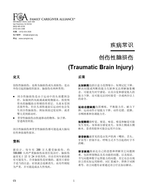
690 Market Street, Suite 600San Francisco, CA 94104(800) 445-8106(415) 434-3388传真:(415) 434-3508电子邮件:info@网址:疾病常识创伤性脑损伤(Traumatic Brain Injury)定义创伤性脑损伤,也称为脑损伤或头部损伤,是由外伤引起的脑组织损害。
脑损伤有两种类型:闭合性脑损伤是由于运动中的头部遭到急停,如撞到挡风玻璃或被钝物撞击,致使颅骨内的脑髓拍击坚硬的骨质层。
头部未受到直接外伤,但在头部快速前后运动时也会发生闭合性脑损伤。
例如颈部过度屈伸,或者婴儿受到摇动时。
贯穿性脑损伤由快速移动的物体,如子弹,穿透颅骨所致。
闭合性脑损伤和贯穿性脑损伤都可能造成大脑局灶性和弥漫性损害。
资料据估计,每年有 200 万人遭受脑损伤。
约500,000 人因严重脑损伤而需住院治疗。
脑损伤最常见于 15 至 24 岁的男性,但在任何年龄段都有可能发生。
许多脑损伤是轻微的,通常只要给予适当的注意,症状就会逐渐消失。
而有些则较为严重,并可能造成永久性残疾。
后果认知缺陷包括注意力范围缩小、短期记忆下降、解决问题或判断的能力欠缺和无法理解抽象概念。
可能丧失时空感觉,以及自觉和感觉他人的能力下降。
还可能无法同时接受一步或两步以上的命令。
运动功能缺陷包括瘫痪、平衡能力差、耐力下降、运动动作计划能力下降、动作迟缓、震颤、吞咽困难和协调能力差。
知觉障碍指听觉、视觉、味觉、嗅觉和触觉可能发生变化,身体部分感觉丧失、身体左侧或右侧麻木。
患者的肢体可能会运用不自如。
语音缺陷最常见的是由发声肌肉(嘴唇、舌头、牙齿等)控制不良、呼吸方式不当引起的吐字不清晰。
语言缺陷指表达自己的思想和理解其它问题困难。
包括辨别物品及其功能的问题,以及阅读、书写问题和数字运算能力的问题。
其它还会出现语言的实际运用障碍、词汇量减少、替换字词困难等。
语言问题有必要通过语言疗法加以解决。
抗氧化活性药物筛选模型的建立

抗氧化活性药物筛选模型的建立刘衍鹏;刘天强;黄冠军;张小维;肖丹【摘要】The screening model for antioxidant samples were established by using hydrogen peroxide to induce oxidative damage in grass carp liver cells in vitro. Results showed that the optimal concentration of H2O2 was 10 mmol/L and the reaction time with liver cells was two hours. The antioxidant effect of vitamin C and another two drugs at different concentrations were tested by the model. All of the 3 drugs were proved to possess some antioxidant ability.%以草鱼(Ctenopharyngodon idellus)肝细胞为研究对象,以过氧化氢刺激肝细胞,建立抗氧化活性药物体外筛选模型.结果显示:过氧化氢的最佳使用浓度为10 mmol/L,对草鱼肝细胞的作用时间为2h.使用建立的模型测定了维生素C和两种药物在不同浓度下的抗氧化作用,结果表明维生素C和两种药物均具有一定的抗氧化能力.【期刊名称】《淡水渔业》【年(卷),期】2011(041)005【总页数】4页(P52-55)【关键词】过氧化氢;草鱼(Ctenopharyngodon idellus);肝细胞;抗氧化;超氧化歧化酶【作者】刘衍鹏;刘天强;黄冠军;张小维;肖丹【作者单位】通威股份有限公司,成都610041;通威股份有限公司,成都610041;通威股份有限公司,成都610041;通威股份有限公司,成都610041;通威股份有限公司,成都610041【正文语种】中文【中图分类】S948在渔业生产、运输等过程中,水质、饲养、环境等因素经常导致鱼类氧化应激损伤。
病理学脑萎缩英文

病理学脑萎缩英文Pathological Brain AtrophyThe human brain is a remarkable and complex organ, responsible for a vast array of cognitive, emotional, and physical functions. However, the brain is not immune to the ravages of disease and aging, and one of the most devastating conditions that can affect it is pathological brain atrophy.Brain atrophy is a term that describes the progressive loss of brain tissue, leading to a decrease in the overall volume and weight of the organ. This process can occur due to a variety of underlying causes, ranging from genetic disorders to acquired conditions such as Alzheimer's disease, Parkinson's disease, and traumatic brain injury.One of the most well-known forms of pathological brain atrophy is Alzheimer's disease, a neurodegenerative disorder that is characterized by the progressive loss of brain cells and the accumulation of abnormal proteins, such as amyloid-beta and tau. As the disease progresses, the brain begins to shrink, and the affected individual experiences a gradual decline in cognitive function, including memory loss, difficulty with problem-solving, and changesin personality and behavior.Another common form of pathological brain atrophy is Parkinson's disease, a neurological disorder that primarily affects the motor system. In Parkinson's disease, the loss of dopamine-producing neurons in the brain's substantia nigra region leads to the characteristic tremors, rigidity, and slowness of movement that are hallmarks of the condition. Over time, the brain's overall volume may decrease as the disease progresses.Traumatic brain injury (TBI) is another major cause of pathological brain atrophy. TBI can occur as a result of a sudden impact or rapid acceleration and deceleration of the head, such as during a car accident or a fall. The initial injury can cause immediate damage to brain tissue, and in the aftermath, the brain may continue to shrink and lose volume due to the ongoing inflammatory and degenerative processes.In addition to these well-known conditions, pathological brain atrophy can also be caused by a variety of other factors, including vascular disorders, such as stroke, and genetic disorders, such as Huntington's disease and frontotemporal dementia.Regardless of the underlying cause, the consequences of pathological brain atrophy can be devastating. As the brain losesvolume and tissue, the affected individual may experience a range of cognitive, emotional, and physical impairments, including memory loss, difficulty with language and communication, changes in personality and behavior, and even physical disabilities.One of the most challenging aspects of pathological brain atrophy is the fact that it is often a progressive and irreversible condition. While some treatments and interventions may help to slow the rate of decline or manage the associated symptoms, there is currently no cure for most forms of pathological brain atrophy.Despite these challenges, researchers and clinicians continue to work tirelessly to better understand the underlying mechanisms of pathological brain atrophy and to develop new and more effective treatments. Through advances in neuroimaging, genetics, and molecular biology, scientists are gaining a deeper understanding of the complex processes that lead to brain atrophy, and this knowledge is paving the way for the development of new therapeutic approaches.In the meantime, individuals affected by pathological brain atrophy and their loved ones must navigate the difficult and often overwhelming challenges that come with this condition. This may involve managing symptoms, adapting to changes in cognitive and physical abilities, and seeking support from healthcare professionals,caregivers, and support groups.Despite the many challenges, there is also hope. With continued research, innovation, and a commitment to improving the lives of those affected by pathological brain atrophy, we can work towards a future where these devastating conditions are better understood, more effectively treated, and ultimately, prevented.。
tbi常用造模方法

tbi常用造模方法
TBI(Traumatic Brain Injury,脑外伤)常用的造模方法包括:
1. 自由落体撞击法:该方法通过使用一定质量的金属物体从一定高度自由落体,撞击大鼠头顶部特定区域,以制作TBI模型。
2. 液体冲击法:在开窗部位暴露完整硬脑膜后,连接液体冲击装置,瞬时液体脉冲冲击导致侧壁液体冲击脑损伤。
3. 脑组织标本留取:取脑后用4%多聚甲醛溶液固定,蔗糖溶液脱水后,经OCT包埋做冰冻切片,切片厚度通常为10um。
此外,有些方法在TBI模型中采用的药物和操作可能因实验需求和条件而有所不同。
tbi在医学上的应用

tbi在医学上的应用脑损伤(Traumatic Brain Injury,TBI)是一种常见的医学状况,它是由于头部受到外力的剧烈撞击、震动或穿透性伤害引起的。
TBI在全球范围内广泛存在,并经常导致严重的病理变化和功能障碍。
在医学上,TBI的应用主要涉及其诊断和治疗方面。
首先,TBI的诊断对于提供适当的治疗和护理至关重要。
在医学上,医生通常通过临床评估、病史询问、神经系统检查和神经影像学等多种方法来确诊TBI。
临床评估包括对患者的意识状态、记忆、注意力、协调性和感觉等方面进行评估。
病史询问可以了解头部损伤的原因、持续时间和相关症状。
神经系统检查用于评估神经系统的功能状态,如肌力、感觉和反射等。
而神经影像学(如CT扫描和MRI)可以提供详细的脑部结构信息,帮助医生确定损伤的程度和范围。
在TBI的治疗方面,医学上采取了多种方法来帮助患者康复。
首先,早期治疗是TBI患者康复的重要环节之一。
早期治疗主要包括维持患者的呼吸道通畅、监测生命体征、控制颅内压力、预防脑水肿和颅内感染等。
另外,药物治疗也在TBI中发挥重要作用。
常用的药物包括镇静剂、抗惊厥药物、抗血栓药物和兴奋剂等。
这些药物能够减轻患者的不适症状,控制颅内压力和预防并发症。
除了药物治疗,物理疗法在医学上也被广泛应用于TBI的康复中。
物理疗法包括物理治疗、康复训练、职业治疗和言语治疗等。
物理治疗可通过运动、按摩和物理器械等方法来减轻疼痛、增强肌肉力量和改善关节灵活度。
康复训练则通过日常生活技能训练、平衡训练和步态训练等来提高患者的日常功能。
职业治疗则主要关注患者在工作和生活中的适应能力。
言语治疗可帮助恢复受损的语言和认知功能。
另外,TBI的疾病预防也是医学上的关注焦点之一。
在预防方面,医生通常会提供相关的教育和建议,包括遵循交通安全规则、佩戴头盔和安全带等防护装备、避免高风险活动、保持良好的生活习惯等。
此外,医学研究也致力于寻找更有效的预防措施,如开发创新的头盔材料、改进车辆安全装置和推广交通法规等。
小鼠脑外伤后脑血流的动态变化及其与行为学恢复的相关性

·408·创伤性脑损伤 (traumatic brain injury,TBI ) 是世界范围内最主要的致死和致残原因[1]。
在中国,每10万人中有13人因脑损伤而死亡[2]。
TBI显著增加其他神经系统疾病风险,包括慢性创伤性脑病、阿尔茨海默病和抑郁等[3],从而带来巨大社会压力和经济损失[2]。
脑血管损伤是TBI重要的病理生理过程,脑血管结构破坏、功能障碍参与了TBI的发病、发展及损伤修复过程,并与继发性损伤密切相关。
血管功能障碍常表现为脑出血、脑梗死、脑水肿等[4]。
脑血流是评估血管功能的重要指标之一,大多数TBI 患者在受伤后可以观察到局部或整体的脑血流减· 论著 ·小鼠脑外伤后脑血流的动态变化及其与行为学恢复的相关性李坤航1,张旭东1,赵丹2,梁宇2,钟诗雨1,赵伟东2,包义君1 (中国医科大学 1. 附属第四医院神经外科,沈阳 110032; 2. 生命科学学院发育细胞生物学教研室,沈阳 110122) 摘要 目的 明确小鼠创伤性脑损伤 (TBI ) 后脑血流的动态变化,及其与运动功能受损和修复的相关性。
方法 构建小鼠中度TBI 模型。
通过激光散斑成像技术监测活体脑血流;通过转棒、悬绳和壁架实验分析小鼠运动协调能力,并评估脑血流与运动功能损伤和恢复的相关性。
结果 脑血流在TBI 急性期显著减少,TBI 后6 h、1 d 和3 d 分别减少了69.8%、62.9%和67.8%,TBI 后3 d 开始恢复,至TBI 后14 d 恢复到正常水平。
TBI 导致小鼠运动协调功能障碍,TBI 后3 d 运动功能损伤最严重,TBI 后28 d 基本恢复到正常水平;TBI 后脑血流恢复和运动功能恢复呈显著正相关。
结论 TBI 导致小鼠脑损伤部位脑血流下降和运动功能障碍,此后脑血流和运动功能逐渐恢复,二者的恢复有相关性,且脑血流的恢复早于运动功能的恢复。
提示TBI 后通过改善脑血流能够促进运动功能的恢复。
艾本多夫—科隆量表和格拉斯哥昏迷评分对创伤后意识障碍患者预后评估的比较
艾本多夫—科隆量表和格拉斯哥昏迷评分对创伤后意识障碍患者预后评估的比较关键词:艾本多夫-科隆量表;格拉斯哥昏迷评分;创伤后意识障碍;预后评估1. 引言创伤后意识障碍(Traumatic brain injury, TBI)是头部受创后引起的一系列神经系统疾病,主要包括脑震荡、脑挫裂伤、脑血管意外等多种类型。
其疾病程度和预后严峻程度直接相关,因此对于TBI患者的预后评估分外关键。
艾本多夫-科隆量表(Extended Glasgow Outcome Scale,EGOS)和格拉斯哥昏迷评分(Glasgow Coma Scale,GCS)是常用的用于评估TBI患者疾病严峻程度的工具。
EGOS主要通过衡量TBI患者的长期功能恢复和生活质量来评估疾病严峻程度;GCS则是通过观察患者的意识状态、眼部反应和运动反应等指标来评估患者的疾病严峻程度。
本文旨在探讨这两种工具在TBI患者预后评估中的比较,以期为临床医师在评估TBI患者预后方面提供参考。
2. EGOS评估内容及评分标准EGOS共评估了六个不同的类别来描述患者的长期恢复(Table 1),包括死亡、重度伤残、中度伤残、轻度伤残、复原并需要援助,以及复原且未需要援助。
Table 1 EGOS评估内容和评分标准类别评分死亡 1重度伤残 2中度伤残 3轻度伤残 4复原并需要援助 5复原且未需要援助 63. EGOS评估的优缺点EGOS的优点在于其能够全面评估TBI患者的长期功能恢复和生活质量,同时在评估过程中思量到了患者对自身恢复的主观感受。
此外,EGOS在对于不同类型的TBI患者进行预后评估时能够针对性地进行评分。
然而,EGOS评分的主观性较强,因为其需要思量到患者自身的感受和生活环境等多种因素,容易导致评分不准确。
4. GCS评估内容及评分标准GCS主要关注TBI患者的意识状态、眼部反应和运动反应等指标。
详尽来说,GCS通过以下三个指标进行评定(Table 2):Table 2 GCS评估内容和评分标准指标评分眼部反应(Eye response) 1-4运动反应(Motor response) 1-6言语反应(Verbal response) 1-55. GCS评估的优缺点GCS评估的优点在于其评分标准比较周密、准确,能够准确地反映TBI患者的疾病严峻程度。
脑外伤护理查房英文版本
CDC 2006
Anatomy and physiology review
Skull Anatomy
The skull is a rounded layer of bone designed to protect the brain from penetrating injuries
The base of the skull is rough, with many bony protuberances These ridges can result in injury to the temporal and frontal lobes of the brain during rapid 颅底 acceleration
Case report 1
Name: Tim Sex: male Age: 17 Date: 2011-10-12 6pm
Medical history: While riding a bicycle without a helmet
on a busy street and was struck by a car
Increased Intracranial Pressure
剧烈头痛
喷射性呕吐 视神经 乳头水肿
Etiology and Pathophysiology of Intracranial Hypertension
颅腔占位性 病变 脑组织增加
脑血流量增加
脑脊液增加
Cerebral Herniation
Nursing care of patient with TBI
Zhan Yu xin Neurosurgery Department 0109-kitty@
创伤性脑损伤中“脑-肠轴”调节机制的研究进展
科
脑-肠轴;创伤性颅脑损伤;肠道微生物群
中图分类号 R741;R741.02;R651.1 文献标识码 A
DOI 10.16780/ki.sjssgncj.20221136
武汉 430015
本文引用格式:胡博玄, 刘子华, 赵小云, 刘红朝. 创伤性脑损伤中“脑-肠轴”调节机制的研究进展[J]. 神经损伤
@
of TBI, there exists a brain-gut axis with bidirectional regulation affecting the progression and prognosis of TBI,
but the functional and mechanistic aspects of the brain-gut axis after TBI have not yet been fully clarified. This review will summarize and analyze the progress in research on the bidirectional regulation mechanism of the
释放出的氧自由基也同样进一步加重黏膜损伤[10-12]。此外,过量
被下丘脑神经元直接感受到[23]。
的儿茶酚胺还可导致胃肠道运动障碍,包括胃轻瘫和食物不耐
受[11]。免疫反应也是造成肠道损伤的因素之一。TBI 激活免疫
3 肠道变化对 TBI 的影响
系统,导致炎症介质如核因子κB、肿瘤坏死因子-α与白细胞介
2.3 TBI 后肠道微生物群的变化
是中枢神经系统功能的基础[25]。
[10]
肠道微生物群是近来许多医学学科研究的热点,指的是定
创伤性脑损伤的评估和康复
创伤性脑损伤的评估和康复一、引言在现代社会,创伤性脑损伤(Traumatic Brain Injury, TBI)已经成为一种常见的健康问题。
它是指由于头部遭受外力撞击或突然剧烈晃动而导致脑组织受损的情况。
TBI不仅对个体产生了严重的生理和心理影响,还对家庭和社会造成了巨大负担。
因此,对TBI进行及时、准确的评估并实施科学有效的康复方案至关重要。
二、TBI的评估方法2.1 临床评估临床评估是最常用和广泛接受的TBI评估方法之一。
医生通过观察病史、神经系统检查和认知功能测量等手段来确定患者脑损伤程度和功能障碍情况。
此外,还可采用诸如格拉斯哥昏迷指数(Glasgow Coma Scale, GCS)等工具来衡量患者意识水平。
2.2 影像学评估影像学技术在TBI的评估中起着重要作用。
常见的影像学检查方法包括计算机断层扫描(Computed Tomography, CT)和磁共振成像(Magnetic Resonance Imaging, MRI)。
这些检查可以帮助医生确定脑内出血、损伤范围及其他结构异常,以便制定相应的治疗和康复计划。
2.3 神经心理学评估神经心理学评估可用于评估TBI患者认知、情绪和行为功能的状况。
例如,通过使用标准化测试工具如韦氏智力量表、威斯康星卡片分类测验等,可以全面了解患者在注意力、记忆、执行功能等方面的表现。
这将有助于制定个体化的康复方案。
三、TBI的康复方案3.1 早期干预在TBI发生后的早期阶段,及时干预是至关重要的。
早期干预包括急救处理、手术治疗和药物管理等措施。
在此阶段内,专业医务人员需要集中精力处理并控制颅内感染风险,并促进脑组织再生修复。
针对不同严重程度的TBI患者,需要制定个体化的早期干预计划。
3.2 身体康复身体康复是TBI患者整体康复的重要组成部分。
它包括物理疗法、作业治疗和运动疗法等多种方法。
通过这些方法,可以恢复受损的运动功能、平衡能力和日常生活技能,提高患者生活质量。
- 1、下载文档前请自行甄别文档内容的完整性,平台不提供额外的编辑、内容补充、找答案等附加服务。
- 2、"仅部分预览"的文档,不可在线预览部分如存在完整性等问题,可反馈申请退款(可完整预览的文档不适用该条件!)。
- 3、如文档侵犯您的权益,请联系客服反馈,我们会尽快为您处理(人工客服工作时间:9:00-18:30)。
A variety of additional factors such as age and age-associated systemic alterations can alsoaffect both survival and recovery from severe injury. These include changes in hormonal anddrug metabolism, nutritional status, immune function, and increased frailty, among otherfactors. Given this added layer of complexity, it is not obvious that the injury itself or putativetreatments for it will behave in the same way in older subjects as they do in young adults. Thispopulation might therefore require not only special research consideration, but also a differenttreatment paradigm, which could potentially involve combination treatments designedspecifically to address an age-altered physiology.TBI is a Multiorgan Systemic Inflammatory Disorder Acute phase events with the greatest import for survival after TBI appear to be those related to inflammatory processes, especially the production of inflammatory cytokines, which is a well-recognized aspect of the physiological response to trauma.8 Levels of inflammatory cytokines are the most consistent prognostic markers of outcomes in patients with systemic inflammatory response syndrome, sepsis, multiorgan dysfunction syndrome, and multiorgan failure,9 all of which are commonly the proximate cause of death after CNS trauma 7,10,11 and generally result from an imbalance between pro- and anti-inflammatory responses to severe injury.10 We therefore support the view that TBI should be considered a systemic and not just a focal problem, especially in the context of inflammation and short-term survival.Systemic effects are also evident in response to oxidative stress. Shohami et al.12 reported that TBI induces a cascade of highly reactive oxygen species that can damage brain tissue and other organs throughout the body. Using sophisticated methods to measure the ability of tissue to scavenge free radicals as a measure of oxidative stress, the investigators found that closed head injury in male rats led to widespread changes in brain, liver, lung, skin, kidney, and intestine within 24 h of the insult. More recently Moinard et al.13 found significantly altered metabolism and mitochondrial activity in the liver triggered by the TBI-induced inflammatory cascade in the brain. Histological examination of liver tissue showed that the TBI caused immune cellinfiltration, edema, fibrosis, and necrosis that helped to block normal liver metabolism andhomeostasis.Inflammatory markers are not just causally related to mortality after TBI, but also appear tobe a good index of the extent of injury 9,14 and reliable independent predictors of injuryoutcome.15–18 Although the levels of the cytokines tumor necrosis factor-α (TNF α) andinterleukin-1β (IL-1β) increase after severe injury, it is the level of IL-6 that is considered themost accurate prognostically, because it is the chief regulator of acute hepatic response 19 andcorrelates with systemic inflammation and outcome.9 There is also evidence that earlyincreased levels of IL-6 can be a marker of high risk of complication and organ failure.20Although it has been argued that systemic inflammation is so complex that it defies traditionalreductionist definitions and may not be useful in predicting system behaviors,21 modulatingthe production of inflammatory cytokines during the acute phase could be of benefit in reducingmortality and improving the prognosis for better functional outcomes. For example, there isrecent evidence of sex differences in patients with multiple trauma, with premenopausalwomen showing significantly lower plasma cytokines and less multiorgan failure and sepsis,compared with age-matched men.22 It is possible that these sex differences in response to injuryare attributable to higher circulating levels of progesterone (P 4) in female subjects,23,24 anintriguing hypothesis, given the success of P 4 in treating TBI.Progesterone and Brain Injury TreatmentA number of recent publications have demonstrated effectiveness of P 4 treatment inexperimental models of TBI.25–29 It has been consistently shown that post-injuryadministration of P 4 can attenuate the cytological, morphological, and functional deficitsNIH-PA Author ManuscriptNIH-PA Author ManuscriptNIH-PA Author Manuscriptcaused by traumatic injury.27,30–35 Unlike other sex steroids, P 4 is synthe-sized byoligodendrocytes in the brain, where it then acts on membrane-bound and classical nuclearsteroid receptors.36 Progesterone can also affect water channel proteins (aquaporins) tomodulate both vasogenic and cytotoxic edema,37,38 reduce glutamate toxicity, mediate toxiccalcium influx by its antagonist effects on sigma receptors, upregulate GABA A receptors toreduce excitotoxic damage, and reduce apoptosis and necrosis.29,36 It is likely that thepleiotropic and systemic nature of P 4 is responsible for its effectiveness in the treatment ofTBI. As one recent review suggests, “given the multifactorial nature of the secondary injuryprocess, it is unlikely that targeting a single factor will result in significant improvement inoutcome. Conversely, targeting several injury factors may be the most likely therapeuticapproach to improve outcome ”4 (p. 29, emphasis added). Progesterone is just such amultifactorial agent.The preclinical findings on neuroprotective effects of P 4 have been confirmed in humans in arecently completed phase IIa single-center clinical trial for P 4 in the treatment of moderate tosevere adult TBI (N = 100).39 Mortality among patients given P 4 intravenously for 3 days afterthe injury was less than half that of controls provided standard-of-practice care but no hormone(13.6% vs 30.4%). Functional outcomes at 30 days were significantly better for moderatelyinjured patients given P 4 than for the placebo group, and a data safety monitoring boardappointed by the U.S. National Institutes of Health found no serious adverse events attributableto P 4 treatment. This was the first successful clinical trial for the treatment of TBI in more than30 years of research.A second independent, randomized double-blind study from China examined P 4 in 159 patientswith severe TBI given an intramuscular course of P 4 treatment over 5 days. Similar beneficialoutcomes on morbidity and mortality were observed at both 30 days and 6 months after injury,again without any serious adverse events caused by the treatment.40A phase III, 17-center, double-blind, randomized clinical trial for TBI will start enrolling about1000 patients in January 2010 (NCT00822900; ).One specific reason for increased survival in P 4-treated TBI patients may be the ability of P 4to substantially reduce systemic acute-phase inflammation and so prevent multiorgandysfunction syndrome and multiorgan failure. In support of this idea, Chen et al.41 found thatcontusion injury to the cerebral cortex in rats induced expression of IL-1β and TNF α in theintestinal mucosa, followed by apoptosis in the intestinal mucosa. A 5-day course of treatmentwith P 4 reduced both the cytokines and cell death in the intestine, demonstrating the systemicbenefits of P 4 administration.Aging and Brain InjuryAlthough the incidence of TBI has decreased in most age groups because of improved primaryprevention (such as seat belt and safety helmet use), it has increased by 21% in people overthe age of 65.42 Age is itself an independent predictor of mortality due to TBI, which in thegeriatric population is twice that of younger age groups.43 The age distribution ofhospitalizations due to TBI has the most pronounced peak late in the eighth decade,44 and thehighest mortality rates from TBI, ranging from 60.9% to 86.8% depending on the study, occurin people 75 to 80 years old.45 Given the magnitude of the problem, it is important to addressthe effectiveness of any treatment specifically in aged subjects.Old animals are fatter, more sedentary and irritable, and (like most old people) have sensory,cognitive, and motor deficits that can exacerbate injury-induced impairments and impedefunctional recovery. Old rats and old people also metabolize drugs differently than young ones,so doses and agents that work in younger subjects might not work in older animals, or couldNIH-PA Author Manuscript NIH-PA Author Manuscript NIH-PA Author Manuscripteven aggravate the injury. A number of systemic physiological changes associated with theaging process could also alter drug effectiveness, such as altered metabolism and endogenoushormonal milieu,46–50 impaired immune function,51–53 increased levels of systemicinflammation,54 and reduced physiological reserve or frailty.55,56 Additional changes withinthe CNS, including reduced plasticity 57 and more subtle chemical, structural, morphological,and network alterations,58,59 can also significantly affect the ability of an older animal or personto survive and recover from severe injury.CNS effects of agingAlthough it is established that there is no overt loss of neurons in the brain with normal aging,a number of more subtle structural, chemical, and metabolic changes occur,58,59 both at thelevel of individual neurons and in medium-scale neuronal networks, that can significantlyaffect the ability of the CNS to adapt to internal and environmental changes. Other changes inthe CNS in response to aging are similar to the changes that occur in other cells, and includeincreased oxidative stress, altered protein accumulation, nucleic acid damage, and dysfunctionof energy homeostasis.59 The cellular changes during normal aging make neurons increasinglysusceptible to excitotoxic damage 59 through the impairment of ion pumps,60 dysregulation ofCa 2+ homeostasis,59 and decreased mitochondrial function.61 Given that all these processesare involved in the evolution of the injury after traumatic insult, the process of aging itselfcould significantly increase vulnerability and impair the potential for recovery from TBI inaged individuals.There is evidence that the brains of aged animals exhibit gene expression profiles characteristicof microglial activation and neuroinflammation.62–65 Microglia themselves also appear torespond to stimulation with an amplified inflammatory response in aged animals.66 Thisneuroinflammatory priming can significantly affect the development of neurologicaldysfunction. Under normal conditions these inflammatory changes are temporary, and themicroglia return to a dormant state as soon as the immune challenge is resolved. Aging,however, creates a brain environment in which microglial sensitivity to immune activation isincreased and microglial activation does not resolve, which can lead to the pathogenesis ofneurological disease.66 This situation is likely to exacerbate secondary injury in older subjectsafter TBI, in that trauma can lead not only to a severe systemic immune response,67 but alsoto immune factor release from the damaged cells within the brain itself.68Exaggerated neuroinflammation can also interfere with neuroplasticity during the recoveryphase, and inflammatory cytokines such as TNF α, IL-1β, and IL-6 appear to directly interferewith long-term potentiation,69–71 memory consolidation,72 neurite outgrowth,73 andhippocampal neurogenesis.74 These data suggest that continued presence of reactive microgliain the aged brain creates an environment permissive to a prolonged and amplifiedneuroinflammatory response that can lead to subsequent complications, especially after TBI.66Systemic effects of aging and immunosenescenceIn addition to alterations in the CNS, age-related changes in the immune system are alsocommon and have been implicated in virtually all age-associated disease processes.53 Agingis associated with a general activation of the inflammatory response, which, because of thechronic antigenic stress on innate immunity experienced over a lifetime, becomes the basis forthe onset of inflammatory diseases 51 and an increasing inability to mount an appropriateimmune response to antigenic stimulation.53 With aging there is also a decrease in theproduction of anti-inflammatory hormones,75 as well as a general tendency toward productionof elevated amounts of proinflammatory cytokines by peripheral blood mononuclear cells.54NIH-PA Author Manuscript NIH-PA Author Manuscript NIH-PA Author ManuscriptThe fulminating inflammatory state associated with increasing age has been dubbedinflammaging .54 This condition is systemic but also has specific effects within the CNS.66Given the importance of inflammatory cytokines such as TNF α, IL-1β, and IL-6 in bothbehavioral modulation and the evolution of traumatic injury, it is likely that an increasedneuroinflammatory cytokine response in the elderly patient can disrupt neuronal synapticplasticity, establishing a CNS environment predisposed to long-lasting complications, as wellas a reduced ability to recover from trauma.66 In fact, the aged brain appears to exist in a chronicstate of inflammation that is associated with increased immune reactivity and continuous low-level production of central inflammatory cytokines.68Endocrine effects of agingIn addition to structural and immune decline, human aging is associated with decreased activityin a number of systemic hormones, including thyroid hormone,47 sex steroids,46,49,76 growthhormone,48 insulin-like growth factor type 1 (IGF-I),48 and 25-hydroxyvitamin D 3. 77,50,78Especially intriguing is the relationship between endocrine decline and the change inimmunological function. Some research suggests that proinflammatory cytokines maydownregulate the physiological responses to a variety of hormones, including insulin, growthhormone, IGF-I, thyroid hormone, and estrogens.79TNF α, IL-1, and IL-6 can specifically modulate the release of growth hormone, growthhormone releasing hormone, and somatostatin,80 and are known to reduce concentrations ofIGF-I 81 and increase the concentrations of glucocorticoids 82 in the serum of human patients.Lower serum IGF-I and dehydroepiandrosterone sulfate levels were found in frail (comparedwith nonfrail) elderly individuals,56 and an inverse correlation between serum IL-6 and IGF-I was noted only in frail individuals.56 Elevated levels of inflammatory cytokines 83–86 and thedevelopment of frailty in older adults 87,88 have also been associated with vitamin D deficiency(D-deficiency).These data can be taken to suggest that the immune system and its age-associated dysfunctionmay be intimately related to the functioning of the neuroendocrine system, and that thisrelationship may further contribute to the development of systemic insufficiency andvulnerability to injury.Aging and vitamin D deficiencyOf all the endocrine and nutritional disruptions that affect the elderly population, D-deficiencyhas recently received considerable attention, partly because an estimated 1 billion peopleworldwide exhibit D-deficiency or insufficiency.89 In the northeastern United States, studiesshow a prevalence from 32% in healthy adults 90 to approximately 50% for adolescents andpreadolescents.91–93 The most dramatic statistics for D-deficiency, however, come fromstudies in the elderly population, which show that 40 to 100% of community-dwellingAmerican and European older men and women are D-deficient. Even higher averages are seenin the ill and institutionalized.94–102Vitamin D-deficiency has been associated with many systemic disorders, including infectious,inflammatory, and autoimmune conditions,103–109 cardiovascular disease,110,111 hypertensionand atherosclerosis,112 neuromuscular function,113 cancer,114 neurodegenerative diseases,115,116 and neuropsychological and functional outcomes in the elderly population.117 In fact,D-deficiency appears to be correlated with many if not most of the problems associated withadvanced age, especially those with an inflammatory component.103,106 There is also evidencethat the level of serum vitamin D is a key marker of frailty,83,88 and that it is associated withelevated levels of IL-6.118NIH-PA Author Manuscript NIH-PA Author Manuscript NIH-PA Author ManuscriptIt is worth noting here that vitamin D is in fact not a vitamin, but rather a secosteroid hormonewith the same cholesterol backbone as other steroids (including P 4) and with its own class ofnuclear steroid receptors and signaling mechanisms.119,120 In its biologically active form, 1,25-dihydroxyvitamin D 3 (vitamin D hormone, or VDH) is a hormone with sites of actionthroughout the CNS; it can be considered a neuroactive steroid, because it is both synthesizedand has actions in the nervous system. Vitamin D hormone is also a known and potentmodulator of the cell cycle, immune function, and calcium homeostasis. As such, it may be inimportant compound not only as an endogenous hormone, but also as a treatment in its ownright, especially in combination with P 4.Aging, Brain Injury, and Neurosteroids Progesterone in aged rats Given the promising results with progesterone in young adult animals, we sought to determine if it would work as well in aged animals.121 The working hypothesis in our aging studies was that P 4 would be effective in treating TBI in aged rats. We measured levels of inflammatory proteins, cell death, edema, and behavior during the acute phase of injury (24–72 h after TBI)in aged rats (20 months old, approximately equivalent to 60 years in humans) to determine the potential of P 4 in reducing mortality after TBI. Injured animals treated with 8 mg/kg and 16mg/kg P 4 beginning within the first hour after surgery showed decreased expression of cyclooxygenase-2, IL-6, and nuclear factor κB (NF κB) at all time points examined, indicating a reduction in the acute inflammatory process compared with the aged rats given vehicle. The 16 mg/kg P 4 group also showed reduced neuronal apoptosis at all time points, and decreased edema and improved locomotor outcomes. Although the lowest P 4 dosage used in previous studies in younger animals (8 mg/kg) was effective in the aged animals, overall it was not as effective as 16 mg/kg, suggesting potentially altered P 4 kinetics and metabolism in the older animals.We also observed an association between increased levels of P-glycoprotein and reducededema, suggesting that P 4 may reduce cerebral edema both through its antiinflammatory effectson cytokine levels and through direct effects on the integrity of the blood–brain barrier. P-glycoprotein is regulated through the pregnane X receptor (PXR)122,123 and plays a criticalrole in removing toxic products from the cell. Progesterone has been shown to exertneuroprotective effects through the PXR.124 This raises the intriguing possibility that some ofthe post-TBI benefits of P 4 may be effected through this relatively novel signaling mechanism.Consequences of D-deficiency for TBI and P 4 treatmentGiven the effectiveness of P 4 in aged rats after TBI and the prevalence of D-deficiency in theelderly population, we asked whether D-deficiency would affect the outcome of a brain injuryand whether it would interfere with the benefits of P 4 treatment in aged rats. Based on thecurrent literature, we hypothesized that D-deficiency would exacerbate inflammation andreduce or eliminate the benefits of P 4 treatment after TBI in aged animals. Because early onsetof inflammation is taken as a reliable prognostic indicator of mortality in human patients withsignificant trauma, we measured acute inflammatory proteins, cell death, DNA damage, andshort-term behavior as indicators of inflammation and secondary damage in D-deficient agedanimals after TBI.In general, we observed increased inflammation in aged rats with D-deficiency, whether oursubjects were uninjured, injured but untreated, or injured and treated with P 4.125 Our resultsconfirmed previous studies 103,106,126 suggesting that D-deficiency establishes a higherbaseline level of inflammation, in effect priming the system for an increased immune–inflammatory response after brain injury. This elevated acute-phase response correlated withNIH-PA Author ManuscriptNIH-PA Author ManuscriptNIH-PA Author Manuscriptincreased cell death and DNA damage, indicating a more severe secondary injury process.Vitamin D deficiency also affected sickness behaviors, such as movement and grooming, whichstrongly correlated (by general linear models) with the expression of TNF α and IL-6.Our data can be taken to indicate that short-term alterations in locomotor behavior could serveas a rough behavioral indicator of brain inflammation in the acute phase after injury. Ourmodels also indicated that both TNF α and IL-6 were significantly increased by D-deficiencyand that both cytokines showed deficiency–treatment interaction effects. We take this to meanthat the increased expression of these inflammatory cytokines may play an important role inthe cascade of toxic mechanisms underlying the attenuation of P 4 benefits in D-deficiency.Also notable is the fact that, although most of the variability in TNF α was accounted for byinjury, the level of IL-6 was affected primarily by D-deficiency. This fits with otherindependent data connecting IL-6 levels with D-deficiency, frailty, and inflammation 88,118,127,128 and suggests to us that IL-6 may be the key cytokine involved in the detrimental effectsof D-deficiency. The elevation in IL-6 was most evident in our data comparing D-deficientand D-normal animals after TBI and P 4 treatment. Most of the other cytokines were elevatedin D-deficient animals twofold or threefold, but IL-6 was increased nearly fivefold by 72 hafter injury.Management of VDH deficiencyGiven the data from human studies showing that the level of IL-6 is the most accurateprognostic indicator of survival in the acute phase after TBI, our findings could have significantimplications in the clinical setting. We suggest that elderly people be screened for potential D-deficiency and provided with supplementation therapy after TBI, because they could be morefrail and thus more likely to die from their injuries. Powner et al.129 point out that, duringrehabilitation from severe TBI, as many as 40% of patients suffer from endocrine dysfunctionsassociated with sickness behaviors such as chronic fatigue, decreased libido, amenorrhea,increased anxiety, memory failure, depression, and anorexia, among others. Although for someof these symptoms it is speculative that vitamin D could be a helpful treatment, the costs ofvitamin D supplementation are minor and it is easy to give, and the implications for thetreatment of a number of disease conditions could be important for clinical practice.Although it seems reasonable that supplementation with vitamin D in a deficient state wouldbe equivalent to ongoing vitamin D sufficiency (i.e., acute correction of deficiency should beequivalent to no deficiency), there is no a priori reason to make this assumption. At 24 h afterinjury, we observed no differences in inflammatory cytokines between VDH-treated deficientand D-normal animals. By 72 h after injury, while there was still no difference with TNF α andIL-1β was still lower than in D-normal animals, levels of IL-6, NF κB p65, and cell death wereall higher in VDH-treated deficient than in D-normal animals. These findings can be taken tosuggest that, at least with reference to IL-6 levels, acutely correcting D-deficiency is not asgood as maintaining D-normal status. In other words, prevention could be better than acutetreatment.Vitamin D deficiency as a public health issue has received much attention recently in thepopular press, as well as in the medical literature. Now we can add another benefit tomaintaining a normal vitamin D status: it is not just good for the arteries and protective againstcancer, it can also be a primary intervention against TBI—in some ways, the equivalent ofwearing a seatbelt. Although further research needs to be done on this issue, we suggest thatthe findings are likely to apply across the developmental spectrum and not just to the elderlypopulation (where it might be exacerbated because of complications from other health-relatedfactors).NIH-PA Author Manuscript NIH-PA Author Manuscript NIH-PA Author ManuscriptCombining P 4 With Vdh In Treating TBI In The Aged PopulationRationale for combinatorial therapy in treating TBICombination therapy is somewhat new to the area of brain injury, but it is a well-established pharmacological approach to a number of other diseases, such as HIV–AIDS or tuberculosis,although most drug development still focuses on individual drugs targeting one or at most a few specific mechanisms. Two issues are involved: drug combination and target promiscuity,or pleiotropy .Rationales for combining drugs include targeting multiple divergent mechanisms and overcoming single-drug limitations such as receptor kinetics, pharmacology, and signaling pathways.130,131 This idea has recently gained ground in the treatment of TBI.5 Because both P 4 and VDH are pleiotropic and multifunctional, it is difficult to predict specific mechanisms of interaction, but both could be used to target cell death after TBI, for example, as one of the many mechanisms by which a brain injury evolves. On the one hand, P 4 would reduce cell death by preventing release of cytochrome c from the mitochondria, upregulating the antiapoptotic Bcl-2 protein, and downregulating the proapoptotic Bax protein.1,26,132 Vitamin D hormone, on the other hand, would prevent the reactivation of cell cycle machinery,133–135 which is a common step toward apoptosis in terminally differentiated neurons,136,137 and would upregulate nerve growth factor (NGF), providing a strong external prosurvival signal.138,139 The two compounds would thus act in synergy and provide benefits greater than either compound bining P 4 and VDH, pleiotropic drugs that work primarily through intracellular receptors and transcription factors likewise merits serious consideration, as meeting the criteria posited by Vink and Nimmo 4 in their review of multifunctional drugs for head injury. We strongly agree that a combinatorial approach to treatment is not only reasonable but may be essential,given the complexity and heterogeneity of human TBI and the fact that, of the 130 preclinicalmonotherapy drugs that have shown promise in treating brain injury, all have failed when takento clinical trial.5Effectiveness of P 4–VDH combination after TBI in aged ratsWe recently demonstrated effectiveness of a combination of P 4 and VDH in both in vitro andin vivo models. Combining VDH and P 4 was shown to be effective in vitro in increasing thesurvival of primary cortical neurons under glutamate challenge, with the combination showingmore neuroprotection than either compound alone at best dose.140 We applied the sametreatment concept to D-deficient aged rats with TBI and showed that the only treatment thatreduced proteins measured (TNF α, IL-1β, IL-6, NF κB p65, activated caspase-3, and p53) inall cases by 72 h after injury was the combination of P 4 and VDH (5 mg/kg in a single dose)compared with vehicle or either compound given alone. The combination treatment was alsothe only one that dramatically improved behavioral parameters, which our statistical modelsagain showed to be strongly correlated with systemic inflammation and levels of TNF α andIL-6. Should these results be translatable to humans, the clinical implications are obvious: acombination treatment of P 4 and VDH given to elderly patients with TBI should improvesurvival over P 4 given alone to the same population.Briefly summarized, we conclude that 1) in aged rats with brain injury, P 4 is effective inreducing acute inflammation, a key indicator of survival in human patients; 2) D-deficiencyincreases acute-phase inflammation and attenuates the benefits of P 4 treatment in aged ratswith TBI, suggesting that such a deficiency could increase mortality after brain injury in humanpatients; 3) a combination of P 4 and VDH partially reverses the effects of D-deficiency andreduces post-TBI acute inflammation in aged rats, suggesting that, should these results translateNIH-PA Author ManuscriptNIH-PA Author ManuscriptNIH-PA Author Manuscript。
