Fluctuation-induced interactions between dielectrics in general geometries
FRET 维基百科
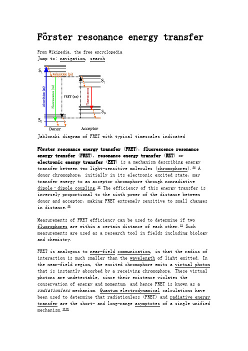
Förster resonance energy transferFrom Wikipedia, the free encyclopediaJump to: navigation, searchJablonski diagram of FRET with typical timescales indicatedFörster resonance energy transfer (FRET), fluorescence resonance energy transfer (FRET), resonance energy transfer (RET) orelectronic energy transfer (EET) is a mechanism describing energy transfer between two light-sensitive molecules (chromophores).[1] A donor chromophore, initially in its electronic excited state, may transfer energy to an acceptor chromophore through nonradiative dipole–dipole coupling.[2] The efficiency of this energy transfer is inversely proportional to the sixth power of the distance between donor and acceptor, making FRET extremely sensitive to small changes in distance.[3]Measurements of FRET efficiency can be used to determine if two fluorophores are within a certain distance of each other.[4] Such measurements are used as a research tool in fields including biology and chemistry.FRET is analogous to near-field communication, in that the radius of interaction is much smaller than the wavelength of light emitted. In the near-field region, the excited chromophore emits a virtual photon that is instantly absorbed by a receiving chromophore. These virtual photons are undetectable, since their existence violates the conservation of energy and momentum, and hence FRET is known as a radiationless mechanism. Quantum electrodynamical calculations have been used to determine that radiationless (FRET) and radiative energy transfer are the short- and long-range asymptotes of a single unified mechanism.[5][6]Contents[hide]∙ 1 Terminology∙ 2 Theoretical basis∙ 3 Experimental co nfirmation of the Förster resonance energy transfer theory∙ 4 Methods to measure FRET efficiencyo 4.1 Sensitized emissiono 4.2 Photobleaching FRETo 4.3 Lifetime measurements∙ 5 Fluorophores used for FRETo 5.1 CFP-YFP pairso 5.2 BRETo 5.3 Homo-FRET∙ 6 Applicationso 6.1 Biology∙7 Other methods∙8 See also∙9 References∙10 External linksTerminology[edit]Förster resonance energy transfer is named after the German scientist Theodor Förster.[7] When both chromophores are fluorescent, the term "fluorescence resonance energy transfer" is often used instead, although the energy is not actually transferred by fluorescence.[8][9]In order to avoid an erroneous interpretation of the phenomenon that is always a nonradiative transfer of energy (even when occurring between two fluorescent chromophores), the name "Förster resonance energy transfer" is preferred to "fluorescence resonance energy transfer;" however, the latter enjoys common usage in scientific literature.[10] It should also be noted that FRET is not restricted to fluorescence. It can occur in connection with phosphorescence as well.[8]Theoretical basis[edit]The FRET efficiency () is the quantum yield of the energy transfer transition, i.e. the fraction of energy transfer event occurring per donor excitation event:[11]where is the rate of energy transfer, the radiative decayrate, and the 's are the rate constants of any other de-excitation pathways.[12]The FRET efficiency depends on many physical parameters that can be grouped as follows:∙The distance between the donor and the acceptor (typically in the range of 1-10 nm)∙The spectral overlap of the donor emission spectrum and the acceptor absorption spectrum.∙The relative orientation of the donor emission dipole moment and the acceptor absorption dipole moment.depends on the donor-to-acceptor separation distance with an inverse 6th power law due to the dipole-dipole coupling mechanism:with being the Förster distance of this pair of donor andacceptor, i.e. the distance at which the energy transfer efficiency is 50%.[12]The Förster distance depends on the overlap integral of the donor emission spectrum with the acceptor absorption spectrum and their mutual molecular orientation as expressed by the following equation.[13][14]where is the fluorescence quantum yield of the donor in the absence of the acceptor, κ2 is the dipole orientation factor, is the refractive index of the medium, is Avogadro's number, and is the spectral overlap integral calculated aswhere is the normalized donor emission spectrum, and is the acceptor molar extinction coefficient.[15] The orientation factor κ is given by,Where denotes the normalized transition dipole moment of therespective fluorophore and denotes the normalized inter-fluorophore displacement. κ2 =2/3 is often assumed. This value is obtained when both dyes are freely rotating and can be considered to beisotropically oriented during the excited state lifetime. If either dye is fixed or not free to rotate, then κ2 =2/3 will not be a valid assumption. In most cases, however, even modest reorientation of the dyes results in enough orientational averaging that κ2 = 2/3 doesnot result in a large error in the estimated energy transfer distance due to the sixth power dependence of R0 on κ2. Even when κ2 is quite different from 2/3 the error can be associated with a shift in R0 and thus determinations of changes in relative distance for a particular system are still valid. Fluorescent proteins do not reorient on a timescale that is faster than their fluorescence lifetime. In this case 0 ≤ κ2≤ 4.[15]The FRET efficiency relates to the quantum yield and the fluorescence lifetime of the donor molecule as follows:[16]where and are the donor fluorescence lifetimes in the presenceand absence of an acceptor, respectively, or aswhere and are the donor fluorescence intensities with and without an acceptor, respectively.Experimental confirmation of the Förster resonance energy transfer theory[edit]The inverse sixth-power distance dependence of Förster resonance energy transfer was experimentally confirmed by Wilchek, Edelhoch and Brand[17][18] using tryptophyl peptides. Stryer, Haugland and Yguerabide[19] also experimentally demonstrated the theoretical dependence ofFörster resonance ene rgy transfer on the overlap integral by using a fused indolosteroid as a donor and a ketone as an acceptor. However, a lot of contradictions of special experiments with the theory was oserved. The reason is that the theory has approximate character and gives overstimated distances of 50-100 Angstrems (Vekshin N.L. Energy Transfer in Macromolecules, SPIE, 1997; Vekshin N.L. Photonics of Biopolymers, Springer, 2002).Methods to measure FRET efficiency[edit]In fluorescence microscopy, fluorescence confocal laser scanning microscopy, as well as in molecular biology, FRET is a useful tool to quantify molecular dynamics in biophysics and biochemistry, such as protein-protein interactions, protein–DNA interactions, and protein conformational changes. For monitoring the complex formation between two molecules, one of them is labeled with a donor and the other with an acceptor. The FRET efficiency is measured and used to identify interactions between the labeled complexes. There are several ways of measuring the FRET efficiency by monitoring changes in the fluorescence emitted by the donor or the acceptor.[20]Sensitized emission[edit]One method of measuring FRET efficiency is to measure the variationin acceptor emission intensity.[14] When the donor and acceptor are in proximity (1–10 nm) due to the interaction of the two molecules, the acceptor emission will increase because of the intermolecularFRET from the donor to the acceptor. For monitoring protein conformational changes, the target protein is labeled with a donor and an acceptor at two loci. When a twist or bend of the protein brings the change in the distance or relative orientation of the donor and acceptor, FRET change is observed. If a molecular interaction or a protein conformational change is dependent on ligand binding, this FRET technique is applicable to fluorescent indicators for the ligand detection.Photobleaching FRET[edit]FRET efficiencies can also be inferred from the photobleaching rates of the donor in the presence and absence of an acceptor.[14] This method can be performed on most fluorescence microscopes; one simply shines the excitation light (of a frequency that will excite the donor but not the acceptor significantly) on specimens with and without the acceptor fluorophore and monitors the donor fluorescence (typically separated from acceptor fluorescence using a bandpass filter) over time. The timescale is that of photobleaching, which is seconds to minutes, with fluorescence in each curve being given bywhere is the photobleaching decay time constant and depends on whether the acceptor is present or not. Since photobleaching consists in the permanent inactivation of excited fluorophores, resonance energy transfer from an excited donor to an acceptor fluorophore prevents the photobleaching of that donor fluorophore, and thus high FRET efficiency leads to a longer photobleaching decay time constant:where and are the photobleaching decay time constants of thedonor in the presence and in the absence of the acceptor, respectively. (Notice that the fraction is the reciprocal of that used for lifetime measurements).This technique was introduced by Jovin in 1989.[21] Its use of anentire curve of points to extract the time constants can give it accuracy advantages over the other methods. Also, the fact that time measurements are over seconds rather than nanoseconds makes it easierthan fluorescence lifetime measurements, and because photobleaching decay rates do not generally depend on donor concentration (unless acceptor saturation is an issue), the careful control of concentrations needed for intensity measurements is not needed. It is, however, important to keep the illumination the same for the with- and without-acceptor measurements, as photobleaching increases markedly with more intense incident light.Lifetime measurements[edit]FRET efficiency can also be determined from the change in the fluorescence lifetime of the donor.[14] The lifetime of the donor will decrease in the presence of the acceptor. Lifetime measurements of FRET are used in Fluorescence-lifetime imaging microscopy.Fluorophores used for FRET[edit]If the linker is intact, excitation at the absorbance wavelength of CFP (414nm) causes emission by YFP (525nm) due to FRET. If the linker is cleaved by a protease, FRET is abolished and emission is at the CFP wavelength (475nm).CFP-YFP pairs[edit]One common pair fluorophores for biological use is a cyan fluorescent protein (CFP) – yellow fluorescent protein (YFP) pair.[22] Both are color variants of green fluorescent protein (GFP). Labeling with organic fluorescent dyes requires purification, chemical modification, and intracellular injection of a host protein. GFP variants can be attached to a host protein by genetic engineering which can be more convenient. Additionally, a fusion of CFP and YFP linked by a protease cleavage sequence can be used as a cleavage assay.[23]BRET[edit]A limitation of FRET is the requirement for external illumination to initiate the fluorescence transfer, which can lead to background noise in the results from direct excitation of the acceptor or to photobleaching. To avoid this drawback, Bioluminescence Resonance Energy Transfer (or BRET) has been developed.[24] This technique uses a bioluminescent luciferase (typically the luciferase from Renilla reniformis) rather than CFP to produce an initial photon emission compatible with YFP.Homo-FRET[edit]In general, "FRET" refers to situations where the donor and acceptor proteins (or "fluorophores") are of two different types. In many biological situations, however, researchers might need to examine the interactions between two, or more, proteins of the same type—or indeed the same protein with itself, for example if the protein folds or forms part of a polymer chain of proteins[25] or for other questions of quantification in biological cells.[26]Obviously, spectral differences will not be the tool used to detect and measure FRET, as both the acceptor and donor protein emit light with the same wavelengths. Yet researchers can detect differences in the polarisation between the light which excites the fluorophores andthe light which is emitted, in a technique called FRET anisotropy imaging; the level of quantified anisotropy (difference in polarisation between the excitation and emission beams) then becomes an indicative guide to how many FRET events have happened.[27]Applications[edit]Biology[edit]FRET has been used to measure distance and detect molecular interactions in a number of systems and has applications in biology and chemistry.[28] FRET can be used to measure distances between domains in a single protein and therefore to provide information about protein conformation.[29] FRET can also detect interaction between proteins.[30] Applied in vivo, FRET has been used to detect the location and interactions of genes and cellular structures including intergrins and membrane proteins.[31] FRET can be used to obtain information about metabolic or signaling pathways.[32] FRET is also used to study lipid rafts in cell membranes.[33]FRET and BRET are also the common tools in the study of biochemical reaction kinetics and molecular motors.The applications of Fluorescence Resonance Energy Transfer (FRET) have expanded tremendously in the last 25 years, and the technique has become a staple technique in many biological and biophysical fields. FRET can be used as spectroscopic ruler in various areas such as structural elucidation of biological molecules and their interactions in vitro assays, in vivo monitoring in cellular research, nucleic acid analysis, signal transduction, light harvesting and metallic nanomaterial etc. Based on the mechanism of FRET a variety of novel chemical sensors and biosensors have been developed.[34]Other methods[edit]A different, but related, mechanism is Dexter Electron Transfer.An alternative method to detecting protein–protein proximity is the bimolecular fluorescence complementation (BiFC) where two halves of a YFP are fused to a protein. When these two halves meet they form a fluorophore after about 60 s – 1 hr.[35]See also[edit]∙Förster coupling∙Surface energy transfer∙Dexter electron transfer∙Time-resolved fluorescence energy transferReferences[edit]1.Jump up ^ Cheng, Ping-Chin (2006). "The Contrast Formation in OpticalMicroscopy". In Pawley, James B. Handbook Of Biological Confocal Microscopy(3rd ed.). New York, NY: Springer. pp. 162–206. doi:10.1007/978-0-387-45524-2_8. ISBN 978-0-387-25921-5.2.Jump up ^ Helms, Volkhard (2008). "Fluorescence Resonance Energy Transfer".Principles of Computational Cell Biology. Weinheim: Wiley-VCH. p. 202.ISBN 978-3-527-31555-0.3.Jump up ^ Harris, Daniel C. (2010). "Applications of Spectrophotometry".Quantitative Chemical Analysis (8th ed.). New York: W. H. Freeman and Co.pp. 419–44. ISBN 978-1-4292-1815-3.4.Jump up ^ Zheng, Jie (2006). "Spectroscopy-Based Quantitative FluorescenceResonance Energy Transfer Analysis". In Stockand, James D.; Shapiro, MarkS. Ion Channels: Methods and Protocols. Methods in Molecular Biology,Volume 337. Totowa, NJ: Humana Press. pp. 65–77. doi:10.1385/1-59745-095-2:65. ISBN 978-1-59745-095-9.5.Jump up ^ Andrews, David L. (1989). "A unified theory of radiative andradiationless molecular energy transfer". Chemical Physics135 (2): 195–201. Bibcode:1989CP....135..195A. doi:10.1016/0301-0104(89)87019-3.6.Jump up ^ Andrews, David L; Bradshaw, David S (2004). "Virtual photons,dipole fields and energy transfer: A quantum electrodynamical approach".European Journal of Physics25 (6): 845. doi:10.1088/0143-0807/25/6/017.7.Jump up ^Förster, Theodor (1948). "Zwischenmolekulare Energiewanderung undFluoreszenz" [Intermolecular energy migration and fluorescence]. Annalender Physik (in German) 437: 55–75. Bibcode:1948AnP...437...55F.doi:10.1002/andp.19484370105.8.^ Jump up to: a b Valeur, Bernard; Berberan-Santos, Mario (2012). "ExcitationEnergy Transfer". Molecular Fluorescence: Principles and Applications, 2nded. Weinheim: Wiley-VCH. pp. 213–261. doi:10.1002/9783527650002.ch8.ISBN 9783527328376.9.Jump up ^ FRET microscopy tutorial from Olympus10.Jump up ^Glossary of Terms Used in Photochemistry (3rd ed.). IUPAC. 2007.p. 340.11.Jump up ^ Moens, Pierre. "Fluorescence Resonance Energy Transferspectroscopy". Retrieved July 14, 2012.12.^ Jump up to: a b Schaufele, Fred; Demarco, Ignacio; Day, Richard N. (2005)."FRET Imaging in the Wide-Field Microscope". In Periasamy, Ammasi; Day, Richard. Molecular Imaging: FRET Microscopy and Spectroscopy. Oxford:Oxford University Press. pp. 72–94. doi:10.1016/B978-019517720-6.50013-4.ISBN 978-0-19-517720-6.13.Jump up ^Förster, Th. (1965). "Delocalized Excitation and ExcitationTransfer". In Sinanoglu, Oktay. Modern Quantum Chemistry. IstanbulLectures. Part III: Action of Light and Organic Crystals3. New York and London: Academic Press. pp. 93–137. Retrieved 2011-06-22.14.^ Jump up to: a b c d Clegg, Robert (2009). "Förster resonance energytransfer—FRET: what is it, why do it, and how it's done". In Gadella,Theodorus W. J. FRET and FLIM Techniques. Laboratory Techniques inBiochemistry and Molecular Biology, Volume 33. Elsevier. pp. 1–57.doi:10.1016/S0075-7535(08)00001-6. ISBN 978-0-08-054958-3.15.^ Jump up to: a b Demchenko, Alexander P. (2008). "Fluorescence DetectionTechniques". Introduction to Fluorescence Sensing. Dordrecht: Springer.pp. 65–118. doi:10.1007/978-1-4020-9003-5_3. ISBN 978-1-4020-9002-8. 16.Jump up ^ Majoul, Irina; Jia, Yiwei; Duden, Rainer (2006). "PracticalFluorescence Resonance Energy Transfer or Molecular Nanobioscopy of Living Cells". In Pawley, James B. Handbook Of Biological Confocal Microscopy (3rd ed.). New York, NY: Springer. pp. 788–808. doi:10.1007/978-0-387-45524-2_45. ISBN 978-0-387-25921-5.17.Jump up ^ Template:ISRAEL JOURNAL OF CHEMISTRY. Vol. 1. No. Sa. 196318.Jump up ^ {Edelhoch, H., Brand, L., Wilchek, M. (1967). "Fluorescencestudies with tryptophyl peptides". Biochemistry 6 (2): 547–559.doi:10.1021/bi00854a024. PMID 6047638}19.Jump up ^ Lakowicz, Joseph R., ed. (1991). Principles. New York: PlenumPress. p. 172. ISBN 978-0-306-43875-2.20.Jump up ^ "Fluorescence Resonance Energy Transfer Protocol". WellcomeTrust. Retrieved 24 June 2012.[dead link]21.Jump up ^Szöllősi, János; Alexander, Denis R. (2007). "The Application ofFluorescence Resonance Energy Transfer to the Investigation ofPhosphatases". In Klumpp, Susanne; Krieglstein, Josef. ProteinPhosphatases. Methods in Enzymology, Volume 366. Amsterdam: Elsevier.pp. 203–24. doi:10.1016/S0076-6879(03)66017-9. ISBN 978-0-12-182269-9. 22.Jump up ^ Periasamy, Ammasi (July 2001). "Fluorescence resonance energytransfer microscopy: a mini review". Journal of Biomedical Optics6 (3): 287–291. Bibcode:2001JBO.....6..287P. doi:10.1117/1.1383063.PMID 11516318.23.Jump up ^ Nguyen, AW; Daugherty, PS (March 2005). "Evolutionaryoptimization of fluorescent proteins for intracellular FRET.". Naturebiotechnology23 (3): 355–60. doi:10.1038/nbt1066. PMID 15696158.24.Jump up ^ Bevan, Nicola; Rees, Stephen (2006). "Pharmaceutical Applicationsof GFP and RCFP". In Chalfie, Martin; Kain, Steven R. Green FluorescentProtein: Properties, Applications and Protocols. Methods of Biochemical Analysis, Volume 47 (2nd ed.). Hoboken, NJ: John Wiley & Sons. pp. 361–90. doi:10.1002/0471739499.ch16. ISBN 978-0-471-73682-0.25.Jump up ^ Gautier, I.; Tramier, M.; Durieux, C.; Coppey, J.; Pansu, R.B.;Nicolas, J.-C.; Kemnitz, K.; Coppey-Moisan, M. (2001). "Homo-FRETMicroscopy in Living Cells to Measure Monomer-Dimer Transition of GFP-Tagged Proteins". Biophysical Journal80 (6): 3000–8.Bibcode:2001BpJ....80.3000G. doi:10.1016/S0006-3495(01)76265-0.PMC 1301483. PMID 11371472.26.Jump up ^ Bader, Arjen N.; Hofman, Erik G.; Voortman, Jarno; Van Bergen EnHenegouwen, Paul M.P.; Gerritsen, Hans C. (2009). "Homo-FRET ImagingEnables Quantification of Protein Cluster Sizes with SubcellularResolution". Biophysical Journal97 (9): 2613–22.Bibcode:2009BpJ....97.2613B. doi:10.1016/j.bpj.2009.07.059. PMC 2770629.PMID 19883605.27.Jump up ^ Gradinaru, Claudiu C.; Marushchak, Denys O.; Samim, Masood;Krull, Ulrich J. (2010). "Fluorescence anisotropy: From single molecules to live cells". The Analyst135 (3): 452–9. Bibcode:2010Ana...135..452G.doi:10.1039/b920242k. PMID 20174695.28.Jump up ^ Lakowicz, Joseph R. (1999). Principles of fluorescencespectroscopy (2nd ed.). New York, NY: Kluwer Acad./Plenum Publ. pp. 374–443. ISBN 978-0-306-46093-7.29.Jump up ^ Truong, Kevin; Ikura, Mitsuhiko (2001). "The use of FRET imagingmicroscopy to detect protein–protein interactions and proteinconformational changes in vivo". Current Opinion in Structural Biology11(5): 573–8. doi:10.1016/S0959-440X(00)00249-9. PMID 11785758.30.Jump up ^ Pollok, B; Heim, R (1999). "Using GFP in FRET-basedapplications". Trends in Cell Biology9 (2): 57–60. doi:10.1016/S0962-8924(98)01434-2. PMID 10087619.31.Jump up ^ Sekar, R. B.; Periasamy, A (2003). "Fluorescence resonance energytransfer (FRET) microscopy imaging of live cell protein localizations". The Journal of Cell Biology160 (5): 629–33. doi:10.1083/jcb.200210140.PMC 2173363. PMID 12615908.32.Jump up ^ Ni, Qiang; Zhang, Jin (2010). "Dynamic Visualization of CellularSignaling". In Endo, Isao; Nagamune, Teruyuki. Nano/Micro Biotechnology.Advances in Biochemical Engineering/Biotechnology, Volume 119. Springer.pp. 79–97. Bibcode:2010nmb..book...79N. doi:10.1007/10_2008_48.ISBN 978-3-642-14946-7. PMID 19499207.33.Jump up ^ Silvius, John R.; Nabi, Ivan Robert (2006). "Fluorescence-quenching and resonance energy transfer studies of lipid microdomains in model and biological membranes (Review)". Molecular Membrane Biology23(1): 5–16. doi:10.1080/09687860500473002. PMID 16611577.34.Jump up ^ S. A., Hussain et al. (2015). "Fluorescence Resonance EnergyTransfer (FRET) sensor" (PDF). J. Spectrosc. Dyn.5 (7): 1–16.35.Jump up ^ Hu, Chang-Deng; Chinenov, Yurii; Kerppola, Tom K. (2002)."Visualization of Interactions among bZIP and Rel Family Proteins in Living Cells Using Bimolecular Fluorescence Complementation". Molecular Cell9(4): 789–98. doi:10.1016/S1097-2765(02)00496-3. PMID 11983170。
泛素调节蛋白质降解途径

意义
了解了泛素为媒介的蛋白质裂解作用和 过程,使得科学家对细胞如何控制及分裂蛋 白质的研究有可能深入到分子层级。而当蛋 白质裂解作用发生异常时,人体就会产生不 适甚至疾病,如子宫颈癌症和囊肿纤维症等, 因此,从分子层面角度去了解泛素调节的蛋 白质降解的化学过程和机理,及对生命过程 进一步的探索,具有十分重要的应用意义。
第二阶段: 靶蛋白在26 s蛋白酶体的作用下,由泛素介导的蛋白水解过程。 经泛素活化的底物蛋白被展平后,通过两个狭孔,进入26 s蛋白酶体的催化 中心,蛋白降解在20 s蛋白酶体内部发生。进入26 s蛋白酶体的底物蛋白质 被多次切割,最后形成3~22个氨基酸残基的小肽。 整个流程分为以下六个步骤: 1、E1类酶激活泛素,该过程需要ATP(三磷酸腺苷)提供一定能量; 2、泛素转移至E2类酶; 3、E3类酶具有特异性,可以识别出需破坏的目标蛋白质,与目标蛋白质 接近的E2-泛素复合体将泛素转移至目标蛋白质; 4、E3类酶释放出被泛素标记的蛋白质; 5、被标记的蛋白质分子尾端形成一小段泛素分子链; 6、泛素分子链在蛋白酶体的端口被识别并脱离蛋白质,目标蛋白质进入 蛋白酶复合体的桶状通道最终降解为缩氨酸并由另一端口释放出去。
泛素—蛋白酶体途径( upp ) 一系列相关的酶
泛素活化酶(E1)是催化泛素与底物结合所需的第一个酶。 细胞内仅有单一的泛素活化酶基因。利用不同的转录起始点, 它可产生E1a 和E1b两种泛素活化酶,它们的生物功能可能有所 不同。 泛素偶连酶(E2)是泛素与蛋白底物结合所需的第二个酶。 细胞内有多种泛素偶连酶基因,大多数泛素偶连酶有一个14~ 16 KD的核心,含有活性所必需的半胱氨酸残基。在不同的泛素 偶连酶间有约35%的同源性,这一区域可能参与泛素偶连酶和蛋 白底物的结合。 泛素-蛋白连接酶是泛素与底物蛋白结合所需的第三个酶。 泛素-蛋白连接酶在决定泛素介导的底物降解方面有特殊的作 用。不同类型的泛素-蛋白连接酶间缺乏序列同源性,而且分子 量差异较大。
depletion interactions 解释 -回复

depletion interactions 解释-回复什么是depletion interactions?Depletion interactions,或者称为排除相互作用,是一种物理现象,描述了溶液中大分子聚集体的形成。
这种相互作用是由于在溶液中存在的聚集体或颗粒能够“排除”周围的小分子而产生的。
在溶液中,大分子聚集体(如高分子聚合物或胶体颗粒)会引起周围小分子的局部浓度降低。
这种浓度降低导致小分子聚集体之间的排斥力增加,从而产生depletion interactions。
这种排斥力对于聚合体的形状和结构有重要影响,并且在胶束形成、蛋白质折叠和细胞凝集等生物学过程中都起着关键作用。
Depletion interactions的存在可以通过一种简单的实验来证明。
可以将大量的聚合物分散在溶液中,然后加入一种亲水性小分子,如甘油。
由于聚合物的存在,甘油分子会聚集在聚合物周围形成一个较为浓缩的层。
这个浓缩层导致了聚合物之间的排斥力增加,从而使聚合物分子彼此聚集在一起形成聚集体。
这个实验表明,depletion interactions是由于小分子的浓度降低导致的。
那么,depletion interactions是如何产生的呢?首先,要了解depletion interactions的机制,需要了解depletion力的来源。
depletion力起源于小分子在大分子排列周围的热运动引起的熵效应。
在一个高分子溶液中,小分子趋向于从高浓度区域向低浓度区域扩散,这是因为大分子的存在限制了小分子在体积方向上的自由扩散。
当大量的小分子聚集在大分子周围时,会形成一个浓缩层,而这个层的存在会阻碍其他小分子的扩散,从而导致小分子的熵降低。
这种熵降低是由于小分子在大分子聚集体周围的“排斥区”内的限制聚合引起的。
其次,depletion interactions的强度取决于多种因素。
一是大分子和小分子之间的体积比。
当大分子的体积较大时,浓缩层的厚度也会增加,从而增加depletion interactions的力量。
细胞器之间相互作用在非酒精性脂肪性肝病发生发展中的作用
2 DOI:10.3969/j.issn.1001-5256.2023.01.028细胞器之间相互作用在非酒精性脂肪性肝病发生发展中的作用刘天会首都医科大学附属北京友谊医院肝病中心,北京100050通信作者:刘天会,liu_tianhui@163.com(ORCID:0000-0001-6789-3016)摘要:细胞器除了具有各自特定的功能外,还可与其他细胞器相互作用完成重要的生理功能。
细胞器之间相互作用的异常与疾病的发生发展密切相关。
近年来,细胞器之间相互作用在非酒精性脂肪性肝病(NAFLD)发生发展中的作用受到关注,特别是线粒体、脂滴与其他细胞器之间的相互作用。
关键词:非酒精性脂肪性肝病;细胞器;线粒体;脂肪滴基金项目:国家自然科学基金面上项目(82070618)RoleoforganelleinteractioninthedevelopmentandprogressionofnonalcoholicfattyliverdiseaseLIUTianhui.(LiverResearchCenter,BeijingFriendshipHospital,CapitalMedicalUniversity,Beijing100050,China)Correspondingauthor:LIUTianhui,liu_tianhui@163.com(ORCID:0000-0001-6789-3016)Abstract:Inadditiontoitsownspecificfunctions,anorganellecanalsointeractwithotherorganellestocompleteimportantphysiologicalfunctions.Thedisordersoforganelleinteractionsarecloselyassociatedthedevelopmentandprogressionofvariousdiseases.Inrecentyears,theroleoforganelleinteractionshasattractedmoreattentionintheprogressionofnonalcoholicfattyliverdisease,especiallytheinteractionsbetweenmitochondria,lipiddroplets,andotherorganelles.Keywords:Non-alcoholicFattyLiverDisease;Organelles;Mitochondria;LipidDropletsResearchfunding:NationalNaturalScienceFoundationofChina(82070618) 细胞器可以通过膜接触位点与其他细胞器相互作用,完成物质与信息的交换,形成互作网络[1]。
Plant-Microbe Interactions in the Rhizosphere
Plant-Microbe Interactions in theRhizospherePlant-microbe interactions in the rhizosphere are a crucial aspect of the soil ecosystem, playing a significant role in plant growth, nutrient uptake, andoverall soil health. The rhizosphere is the narrow region of soil that is directly influenced by the roots of plants, where a complex network of interactions occurs between the plant, soil, and various microorganisms. These interactions can beboth beneficial and detrimental, depending on the specific microorganisms involved and the environmental conditions. Understanding the dynamics of plant-microbe interactions in the rhizosphere is essential for developing sustainableagricultural practices and improving crop productivity. One of the most important aspects of plant-microbe interactions in the rhizosphere is the exchange of nutrients between the plant and the microorganisms. Plants release a variety of compounds, such as sugars, amino acids, and organic acids, into the rhizosphere through their roots. These compounds serve as an energy source for the diverse microbial community in the soil, including bacteria, fungi, and archaea. In return, the microorganisms help the plant acquire essential nutrients, such as nitrogen, phosphorus, and iron, by solubilizing and mineralizing soil nutrients, making them more available for plant uptake. This mutualistic relationship between plants and microorganisms is crucial for the overall health and productivity of the plant.In addition to nutrient exchange, plant-microbe interactions in the rhizosphere also play a vital role in plant defense against pathogens. Certain microorganismsin the rhizosphere, known as plant growth-promoting rhizobacteria (PGPR), have been shown to stimulate plant growth and enhance resistance to diseases. These beneficial microorganisms can directly inhibit the growth of plant pathogens by producing antimicrobial compounds or competing for space and resources in the rhizosphere. Furthermore, PGPR can also induce systemic resistance in plants, activating their defense mechanisms against a wide range of pathogens. Understanding the mechanisms by which PGPR confer disease resistance to plants can have significant implications for reducing the reliance on chemical pesticides in agriculture. However, not all plant-microbe interactions in the rhizosphere arebeneficial. Some microorganisms can have detrimental effects on plant health, causing diseases and reducing crop yields. For example, soil-borne pathogens, such as Fusarium and Phytophthora species, can infect plant roots and cause root rot, leading to stunted growth and wilting of the plant. These pathogenic microorganisms can outcompete beneficial microbes in the rhizosphere, disrupting the delicate balance of the soil ecosystem. Understanding the factors that contribute to the proliferation of pathogenic microorganisms in the rhizosphere is essential for developing effective strategies to manage plant diseases and maintain soil health. Moreover, the composition and diversity of the microbial community in the rhizosphere are influenced by various factors, including soil type, plant species, and environmental conditions. Different plants release different types and amounts of root exudates, which can selectively promote the growth of specific groups of microorganisms in the rhizosphere. Furthermore, the physical and chemical properties of the soil, such as pH, moisture, and organic matter content, can also have a significant impact on the structure and function of the rhizosphere microbial community. Understanding the complex interplay between these factors and their effects on plant-microbe interactions is crucial for optimizing soil management practices and promoting sustainable agriculture. In conclusion, plant-microbe interactions in the rhizosphere are a dynamic and intricate network of relationships that profoundly impact plant growth, nutrient cycling, and soil health. The exchange of nutrients, the promotion of plant defense mechanisms, and the influence of environmental factors all contribute to the complexity of these interactions. By gaining a deeper understanding of the mechanisms underlying plant-microbe interactions in the rhizosphere, we can develop innovative strategies to enhance crop productivity, reduce the reliance on chemical inputs, and promote sustainable agricultural practices. Ultimately, this knowledge can contribute to the development of a more resilient and environmentally friendly agricultural system, benefiting both farmers and the broader ecosystem.。
微透析高效液相色谱法研究金属酶与小分子的相互作用
2003年第60卷第1期,95~99化学学报ACT A CHIMICA SINICAV ol.61,2003N o.1,95~99微透析高效液相色谱法研究金属酶与小分子的相互作用郭 明a ,b 孔 亮a 厉 欣a 邹汉法Ξ,a Ξ(a 中国科学院大连化学物理研究所国家色谱研究中心 大连116011)(b 大连大学化学与化工系 大连116622)摘要 将微透析(microdialysis )高效液相色谱法(high performance liquid chromatography ,HP LC )联用技术应用于定量研究超氧化物歧化酶(superoxide dismutase )与组氨酸和组氨酸-金属离子配合物的相互作用.实验结果表明:Cu 2Zn 2S OD 酶不仅与组氨酸,而且还可以与组氨酸2C o (Ⅱ)和组氨酸2Ni (Ⅱ)相互作用,Cu 2Zn 2S OD 酶中的部分Cu (Ⅱ)和Zn (Ⅱ)被诱导和置换出来,并相应地影响了酶的活性;组氨酸-金属离子配合物的作用比组氨酸强,酶活性降低也更明显;酶中结合的外来金属离子的量比从酶上被诱导和顶替下来的金属离子的量要少,说明在溶液中有脱辅基酶的存在.关键词 微透析,配合物,超氧化物歧化酶,相互作用I nteraction of Metalloenzyme with Small Molecules Studiedby Microdialysis/HPLCG UO ,Ming a ,b K ONG,Liang a LI ,X in a Z OU ,Han 2Fa Ξ,a(a National Chromatography R.&A.Center ,Dalian Institue o f Chemical Physics ,Chinese Academy o f Sciences ,Dalian 116011)(b Department o f Chemistry and Chemical Technology ,Dalian Univer sity ,Dalian 116622)Abstract In this paper ,a method of microdialysis coupled with high performance liquid chromatography (HP LC )was established to study the quantitative interaction of superoxide dismutase (Cu 2Zn 2S OD )with histidine and histidine 2metal ion com plexes.It was observed that not only histidine ,but als o histidine 2C o (Ⅱ)and histidine 2Ni (Ⅱ)could induce and substitute s ome Cu (Ⅱ)and Zn (Ⅱ)from Cu 2Zn 2S OD ,eventually in fluence the activity of this metalloenzyme.Histidine 2metal ion com plexes have stronger ability than histidine to displace the bound metal ion from S OD.In addition ,the am ount of foreign metal ion binding to the S OD was less than that of metal ion lost from S OD.It obviously means that there was apoenzyme in the s olution.K eyw ords microdialysis ,com plex ,superoxide dismutase ,interaction 超氧化物歧化酶(superoxide dismutase ,Cu 2Zn 2SOD )广泛地存在于需氧和耐氧的生物体组织内,在生物体内起着消除超氧离子的作用,是生物体防御氧化损伤的一种十分重要的金属酶[1].Cu 2Zn 2SOD 酶分子量约为32000,每个Cu 2Zn 2SOD酶分子内含有2个亚基,每个亚基含有一个由Cu (Ⅱ)和Zn (Ⅱ)以及氨基酸构成的活性中心[2,3].在活性中心内Cu (Ⅱ)与四个组氨酸(His )和一个水分子形成五配位,Zn (Ⅱ)与三个组氨酸和一个天冬氨酸(Asp )结合,Cu (Ⅱ)与Zn (Ⅱ)之间通过共同的His 61连接[3].由于在生物体内存在大量的氨基酸,而金属离子大部分也以氨基酸配合物的形式存在于有机体内[4],因此研究金属酶与氨基酸以及氨基酸的金属离子配合物的相互作用具有重要的生理学意义.胡皆汉等[5]发现Cu 2Zn 2SOD 酶活性中心金属离子在溶液中能与外部的氨基酸发生相互作用,部分SOD 酶中的Cu (Ⅱ)和Zn (Ⅱ)可以被诱导出来,并相应地影响了SOD 酶的催化活性.郑学仿等[6]证明氨基酸2C o (Ⅱ)可以将Cu 2Zn 2SOD 酶中的部分Cu (Ⅱ)和Zn (Ⅱ)诱导、置换出来,C o (Ⅱ)进入酶中Cu (Ⅱ)和Zn (Ⅱ)的结合位点,形成C o (Ⅱ)的SOD 酶衍生物.这些实验都是利用谱学方法进行研究,可以用来进行结合位点的定性研究.但是,这种谱学方法受到探针种类少和很难进行精确定量分析的限制,难以将作用的程度与酶的生物学特性———酶活性相关联.ΞE 2mail :z ouh fa @Received July 15,2002;revised September 18,2002;accepted October 15,2002.微透析(microdialysis)与高效液相色谱(high performance liquid chromatography)相结合曾被用于研究药物及金属离子等与蛋白质的相互作用[7~13],证明该方法可以用于定量研究生物大分子与小分子的相互作用.本文利用该方法研究了Cu2Zn2SOD酶与组氨酸(histidine,His)的金属离子配合物的相互作用,探讨了作用的程度对酶活性的影响.1 实验部分1.1 主要试剂及仪器牛血红细胞铜锌超氧化物歧化酶(bovine erythrocyte superoxide dismutase,简称Cu2Zn2SOD,Cu(Ⅱ)Zn(Ⅱ)2SOD或SOD)由河北大学生物工程研究所提供,酶活力为4000U/mg 酶蛋白;邻苯三酚(1,2,32benzenetriol)购自Sigma公司;组氨酸购自上海化学试剂商店;磺化四苯基卟啉(Meso2tetra(42 sulfophenyl)2porphyrin,TPPS4)购自日本和光纯药工业株式会社(浓度1.5×10⁻⁴m ol/L);其它试剂见文献[11].实验用水为M illipore2Q超纯水.实验所用微透析系统见文献[11];微透析所用探针为C M A/M icrodialysis,购自C M A/M icrodialysis公司(C M A,USA),微透析探针膜长4mm,内径015mm,膜截止分子量为20000.1.2 实验方法1.2.1 组氨酸-金属离子2S OD溶液的制备称取一定量的SOD酶,加10m L水溶解,装入透析袋,在15℃超纯水中透析除盐2d,用光度法测定其浓度,然后用含011m ol/L NaCl的011m ol/L NaH2PO42Na2HPO4(pH= 7143)作缓冲溶液配成10μm ol/L的SOD溶液.每次实验取10μm ol/L的SOD酶溶液3m L,按与SOD酶的不同比例加入小于10μL体积的组氨酸或组氨酸-金属离子溶液,在4℃下放置168h(7d).将其转移至4m L样品瓶中,在37℃下微透析取样,微透析取样方法见文献[10].微透析取样后的SOD酶溶液放入透析袋中,在15℃超纯水中透析除盐2d,按1.2.4方法测定作用后酶的活性.1.2.2 微透析取样及HP LC分析微透析液微透析取样方法见文献[11].本实验采用金属离子与TPPS4柱前衍生,HP LC法检测微透析灌流液中游离金属离子的含量.检测方法见文献[11].以前的研究工作表明微透析取样对相互作用结合平衡的影响可以忽略不计[7~11].本实验由于采用HP LC柱前衍生法检测,衍生试剂TPPS4的特征谱带(S oret带,即400~500 nm波长范围)摩尔吸光系数高达105L・m ol-1・cm-1以上,且HP LC可以有效地分离各个化合物,因此,检测方法的重现性和灵敏度大大增强.1.2.3 回收率微透析回收率是准确定量测定相互作用的一个重要参数,它是组分在透析液中的浓度与原溶液中的浓度比值.在本文实验条件下测定各金属离子的回收率Cu(Ⅱ)为65174%(RSD310%,n=6),Zn(Ⅱ)为70145%(RSD of 312%,n=6),Ni(Ⅱ)为67168%(RSD112%,n=6),C o(Ⅱ)为66121%(RSD416%,n=6).1.2.4 酶活性测定Cu2Zn2SOD酶的活性分析采用邻苯三酚自氧化法[14].在25℃下测定酶的活性,以抑制邻苯三酚自氧化速率的50%为一个活力单位.本实验以原SOD酶的活性为100%,测定作用后酶的相对活性.1.2.5 Cu2Zn2S OD酶中铜和锌含量测定称取20mg的Cu2Zn2SOD酶,加入10m L水溶解,放入透析袋中,在超纯水中透析除盐2d,测定其浓度,取5m L酶溶液,加入20m L浓硝酸预消化过夜.第二天在电炉上加热消化,当溶液接近蒸发完全时逐滴滴入浓硝酸,再滴加双氧水至溶液变澄清,将双氧水蒸发完毕,加水溶解,用011m ol・L-1氢氧化钠溶液调pH至6,定容稀释到25m L.按文献[10]方法测定铜和锌含量,再计算出每个SOD酶分子含金属铜和锌的含量,单位为金属离子与SOD酶分子的摩尔比.每个SOD酶分子的分子量按32000计算.按以上方法进行测定,得到的结果为每个Cu2Zn2SOD原酶分子中含铜和锌的个数分别为11932(±01024,n=6)和11892(±01043,n=6). 1.2.6 S OD酶与配合物相互作用的计算方法用HP LC方法测定出灌流液中金属离子铜和锌的含量,按照回收率公式计算出作用后酶溶液中被顶替下来的游离金属离子铜和锌的量,由此可以得到作用后SOD酶中剩余的铜和锌的含量.外加金属离子与SOD酶结合量的计算方法与此相同.实验时以未加其它试剂的SOD原酶溶液为空白值.2 结果与讨论2.1 组氨酸对微透析/HP LC分析的影响在水溶液中,组氨酸可以与金属离子络合,形成不同摩尔配比的配合物,金属离子与组氨酸主要以[M(His)]2+和[M(His)2]2+两种络合方式存在[13].本实验主要研究Cu2Zn2SOD酶与组氨酸及组氨酸-金属离子的相互作用,由于在溶液中存在游离的组氨酸,因此需考察组氨酸对微透析/HP LC检测金属离子的影响.HP LC测定不同组氨酸浓度下透析液中金属离子的含量,实验结果见图1.六次实验结果的峰面积相对平均偏差为:Cu(Ⅱ)316%;Zn(Ⅱ)418%,;Ni(Ⅱ)210%;C o(Ⅱ) 314%.由此可以表明组氨酸对金属离子含量的测定结果影响不大.2.2 Cu2Z n2SOD酶与组氨酸的作用按上述的实验方法研究Cu2Zn2SOD酶与组氨酸的相互作用,微透析/HP LC分析酶中活性中心离子Cu(Ⅱ)和Zn(Ⅱ)的含量变化,测定作用后SOD酶的活性.实验结果见图2和表1.图中横坐标“m"为外加组氨酸与SOD酶的摩尔比;左边的纵坐标表示作用后每一个SOD酶分子中含Cu(Ⅱ)或Zn(Ⅱ)的个数;右边的纵坐标表示作用后SOD酶的相对69 化学学报V ol.61,2003图1 组氨酸对微透析/HP LC 测定金属离子的影响Figure 1 In fluence of histidine on measurement of metal ions by microdialysis/HP LC●—HPLC peak area of Zn (Ⅱ)in dialysate ;■—HP LC peak area ofCu (Ⅱ)in dialysate ;▲—HP LC peak area of C o (Ⅱ)in dialysate ;∀—HP LC peak area of Ni (Ⅱ)indialysate 图2 不同摩尔配比(m )组氨酸作用下S OD 酶中金属含量及酶活性的变化Figure 2 Variation of metal content and enzyme relative activity of S OD with addition of histidine [in m olar ratio (m )of His to S OD]○—relativeactivity of S OD ;■—Zn (Ⅱ)content in S OD ;●—Cu (Ⅱ)content in S OD活性,以原SOD 酶测得活性作归一化处理.得到的数据列在表1中.由图2和表1可见,随着组氨酸2Cu 2Zn 2SOD 酶溶液中组氨酸量的增加,透析液中游离的Cu (Ⅱ)和Zn (Ⅱ)的浓度逐渐增大,也就是Cu 2Zn 2SOD 酶中Cu (Ⅱ)和Zn (Ⅱ)的含量逐渐减少.作用后SOD 酶中Zn (Ⅱ)的含量降低了912%.酶中Cu (Ⅱ)的含量降低了1510%.酶的活性则降低了1317%.由于组氨酸与Cu 2Zn 2SOD 酶混合溶液中没有外加Cu (Ⅱ)和Zn (Ⅱ),微透析在溶液中测定出的游离Cu (Ⅱ)和Zn (Ⅱ)必定来自于Cu 2Zn 2SOD 酶,也就是说在组氨酸的作用下,SOD 酶中部分Cu (Ⅱ)和Zn (Ⅱ)进入到溶液中.这种作用被称作诱导作用[5].组氨酸不仅可以与溶液中游离的金属离子形成组氨酸配合物,同样也可以与金属酶中的活性中心金属离子形成配位平衡,将活性中心的金属离子诱导出来,形成具有空位的SOD 酶,即脱辅基酶(apoenzyme ).另外,从实验结果可以看出酶中Cu (Ⅱ)含量的降低与酶活性降低程度基本一致.这是由于Cu 2Zn 2SOD 中Cu (Ⅱ)是酶的活性中心离子,只有当Cu (Ⅱ)结合在SOD 酶的活性中心位点时,SOD 酶才有活性,如果将酶的活性中心中的Cu (Ⅱ)去掉,形成脱辅基酶,则该酶没有任何催化活性[3].在Cu 2Zn 2SOD 中,Zn (Ⅱ)不是酶的活性中心离子,Zn (Ⅱ)的含量变化并不会影响SOD 酶的活性[3].因此作用后酶的活性变化与酶中Cu (Ⅱ)含量的变化是一致的.由于组氨酸诱导作用的影响大,SOD 酶中诱导出来的Cu (Ⅱ)和Zn (Ⅱ)的量较多,微透析/HP LC 分析检测更加容易和准确.因此用微透析/HP LC 法定量研究这种相互作用更具有优越性.2.3 Cu 2Z n 2SOD 酶与组氨酸-金属离子络合物的作用按上述同样的方法分别研究Cu 2Zn 2SOD 酶与组氨酸2C o (Ⅱ)和组氨酸2Ni (Ⅱ)的相互作用,这里组氨酸与金属离子的比例是1∶1的关系.分析的结果见图3、图4和表1.图中坐标表示的意义见2.2,图中右边的另一个纵坐标表示SOD 酶结合外来金属离子的含量,也以每一个酶分子含金属表1 组氨酸或组氨酸-金属离子作用后酶中金属离子含量及酶活性的变化aT able 1 Data of loss of metal ions ,binding am ount of foreign metal ions and relative activity of S OD in phosphate bu ffer with addition of His or His 2metal酶中金属离子和酶活性变化与酶作用的化合物组氨酸组氨酸2C o (Ⅱ)组氨酸2Ni (Ⅱ)Cu (Ⅱ)损失量摩尔/摩尔S OD 酶分子0.289(±0.015)0.293(±0.032)0.427(±0.132)百分数/%15.015.222.1Zn (Ⅱ)损失量摩尔/摩尔S OD 酶分子0.174(±0.054)0.657(±0.112)0.524(±0.086)百分数/%9.234.727.6结合外来金属离子量摩尔/摩尔S OD 酶分子0.661(±0.151)0.643(±0.135)S OD 酶活性降低量/%(RS D )13.7(0.5)21.9(1.2%)34.2(2.1%)aCu (Ⅱ)和Zn (Ⅱ)损失量及结合金属量下面括号内数值为标准偏差,相对酶活性下面括号内是相对标准偏差(The value in parentheses under loss of Cu (Ⅱ),Zn (Ⅱ)and binding metal ion is S D.The value in parentheses under relative activity is RS D ).79N o.1郭 明等:微透析高效液相色谱法研究金属酶与小分子的相互作用离子的个数来表示.从图3、图4和表1可以看出:在组氨酸-金属离子作用下,SOD 酶中的Cu (Ⅱ)和Zn (Ⅱ)的含量逐渐减少,而结合外来金属离子的量逐渐增加,同时酶活性逐渐降低.图3 不同摩尔配比(m )组氨酸2C o (Ⅱ)作用下S OD 酶中金属含量及酶活性.的变化Figure 3 Variation of metal content and enzyme relative activity of S OD with addition of His 2C o (Ⅱ)[in m olar ratio (m )of His 2C o(Ⅱ)to S OD]○—relativeactivity of S OD ;■—Zn (Ⅱ)content in S OD ;●—Cu (Ⅱ)content in S OD ;□—C o (Ⅱ)content in S OD 图4 不同摩尔配比(m )组氨酸2Ni (Ⅱ)作用下S OD 酶中金属含量及酶活性的变化Figure 4 Variation of metal content and enzyme relative activity of S OD with addition of His 2C o (Ⅱ)[in m olar ratio (m )of His 2C o (Ⅱ)to S OD]○—relativeactivity of S OD ;■—Zn (Ⅱ)content in S OD ;●—Cu (Ⅱ)content in S OD ;□—Ni (Ⅱ)content in S OD.从表1可以看出:组氨酸2Ni (Ⅱ)诱导出来的Cu (Ⅱ)的量高于组氨酸.原因是组氨酸与金属离子形成的配合物与Cu 2Zn 2SOD 酶活性中心的结构相似,组氨酸2Ni (Ⅱ)可以很快与活性中心离子达到交换平衡.组氨酸2C o (Ⅱ)和组氨酸2Ni (Ⅱ)诱导出来的Zn (Ⅱ)的量也高于组氨酸,由于C o (Ⅱ)与Zn (Ⅱ)的离子半径相近,配位构型也相同[16],C o (Ⅱ)比较容易进入SOD 酶中的Zn (Ⅱ)位点,而Ni (Ⅱ)与Zn (Ⅱ)的最适配位构型是不一致的,因此,组氨酸2C o (Ⅱ)置换SOD 酶中Zn (Ⅱ)的能力要比组氨酸2Ni (Ⅱ)强.在组氨酸2C o (Ⅱ)和组氨酸2Ni (Ⅱ)的作用下,SOD 酶结合的金属离子量比从SOD 酶中诱导和置换出来的Cu (Ⅱ)和Zn (Ⅱ)总和要低,说明酶中有空的结合位点出现,也就是有脱辅基酶形成,这主要是由于C o (Ⅱ)或Ni (Ⅱ)与Cu (Ⅱ)或Zn (Ⅱ)的最适配位结构不完全相同,不能完全顶替由于组氨酸诱导作用形成的空位.组氨酸2C o (Ⅱ)与SOD 作用后酶中Cu (Ⅱ)的含量减少了1512%,而酶活性却降低了2119%.二者不相等的原因是由于一部分C o (Ⅱ)占据了酶中的Zn (Ⅱ)位点,使酶活性降低.在SOD 酶中Zn (Ⅱ)的含量变化并不会影响酶的活性,但是如果在Zn (Ⅱ)的位置上结合了其它的金属离子,由于结合离子的性质与Zn (Ⅱ)不同,会对酶的结构有微小的影响,从而导致酶活性发生改变[17].由于C o (Ⅱ)取代Zn (Ⅱ)形成的SOD 衍生酶Cu (Ⅱ)C o (Ⅱ)SOD 的活性是原酶活性的90%[17],而原酶中活性中心的Cu (Ⅱ)换上其它金属离子,生成的衍生酶都没有活性[3],因此,作用后酶活性降低要比酶中Cu (Ⅱ)含量的减少更明显.组氨酸2Ni (Ⅱ)与SOD 作用后酶的活性降低了3412%,同样比活性中心Cu (Ⅱ)的减少量2211%要低,原因也是Ni (Ⅱ)取代Zn (Ⅱ)形成的SOD 酶衍生酶活性较低,Cu (Ⅱ)Ni (Ⅱ)SOD 衍生酶的活性只有原酶活性的30%[18].3 结论从以上实验可以看出:利用微透析结合高效液相色谱不仅可以定性研究金属酶与小分子的相互作用,而且可以定量讨论作用的程度以及对酶活性的影响.实验结果表明:Cu 2Zn 2SOD 酶不仅可以与组氨酸发生相互作用,还可以与组氨酸2C o (Ⅱ)和组氨酸2Ni (Ⅱ)发生相互作用.诱导和置换出Cu 2Zn 2SOD 酶中的部分Cu (Ⅱ)和Zn (Ⅱ),形成脱辅基酶和金属衍生酶,引起酶活性的下降.组氨酸-金属离子的作用要比单独组氨酸的作用更强烈,并且在溶液中有脱辅基酶存在.R eferences1Vallee ,B.L.;Wacker ,W. E. C.;Bartholomay ,A. F.Ann.Internal Med.1959,50,1077.2Fee ,J.A.Trends Biochem.Sci.1984,7,84.3Sawyer ,D.T.;Valentine ,J.S.Acc.Chem.Res.1982,15,200.4Jones ,P.W.;T aylor ,D.J.;Williams ,M. D.R.J.Inorg.Biochem.2000,81,1.5Hu ,J.2H.;Su ,Z.2Y.Sci.China ,Ser.B 1993,23,793(in Chinese ).89 化学学报V ol.61,2003(胡皆汉,舒占永,中国科学(B ),1993,23,793.)6Zheng ,X.2F.;Hu ,J.2H.;Cheng ,G.2B.;Xu ,Y.2T.;Zhao ,Y.2K.;Liu ,M.H.Spectroscopy and Spectral Analysis 1999,19,798(in Chinese ).(郑学仿,胡皆汉,程国宝,许永廷,赵永魁,刘美华,光谱学与光谱分析,1999,19,798.)7Wang ,H.2L.;Z ou ,H.2F.;Feng , A.S.;Zhang ,Y.2K.Anal.Chim.Acta 1997,342,159.8Wang ,H.2L.;Z ou ,H.2F.;Zhang ,Y.2K.Anal.Chem.1998,70,373.9Wang ,H.2L.;Z ou ,H.2F.;Zhang ,Y.2K.Rev.Anal.Chem.1999,18,383.10G uo ,M.;Z ou ,H.2F.;Wang ,H.2L.;K ong ,L.;Ni ,J.Y.Anal.Chim.Acta 2001,443,91.11G uo ,M.;K ong ,L.;Mao ,X.2Q.;Li ,X.;Z ou ,H.2F.Sci.China ,Ser.B 2002,45,151.12Zhang ,S.;Huang , F.;Cao ,X.2N.;Y ang ,P.2Y.;Zhang ,W.;Jin ,L.2T.Analyst 2002,127,485.13Chung ,L.2S.;Hu ,T.2T.J.Chromatogr.,B 2002,769,351.14Marklund ,S.;Marklund ,G.Eur.J.Biochem.1974,47,469.15Cars on ,R.H.;Brown ,T.L.Inorg.Chem.1966,5,268.16Wang ,K.;Xu ,H.2B.;T ang ,R.2H.;Luo ,X.2M.TraceElement in Life Science ,Chinese Measurement Publish C ompany ,1992,pp.117~119(in Chinese ).(王夔,徐碧辉,唐任寰,罗贤懋,生命科学中的微量元素,中国计量出版社,1992,pp.117~119.)17Beem,K.M.;Rich ,W. E.;Rajag opalan ,K.V.J.Biol.Chem.1974,249,7298.18Ming ,L.J.;Valentine ,J.S.J.Am.Chem.Soc.1990,11,6374.(A0207159 ZH AO ,X.J.;DONG,L.J.)99N o.1郭 明等:微透析高效液相色谱法研究金属酶与小分子的相互作用。
假单胞菌TCd-1 对不同镉耐性水稻品种镉吸收及根际土壤酶活性与镉形态的影响
DOI: 10.12357/cjea.20210854柳玲林, 汪敦飞, 黄明田, 肖清铁, 游武, 钱鑫, 郑新宇, 林瑞余. 假单胞菌TCd-1对不同镉耐性水稻品种镉吸收及根际土壤酶活性与镉形态的影响[J]. 中国生态农业学报 (中英文), 2022, 30(8): 1362−1371LIU L L, WANG D F, HUANG M T, XIAO Q T, YOU W, QIAN X, ZHENG X Y, LIN R Y. Effects of Pseudomonas TCd-1 inocu-lation on Cd uptake, rhizosphere soils enzyme activities and Cd bioavailability in rice (Oryza sativa ) varieties with different Cd toler-ance[J]. Chinese Journal of Eco-Agriculture, 2022, 30(8): 1362−1371假单胞菌TCd-1对不同镉耐性水稻品种镉吸收及根际土壤酶活性与镉形态的影响*柳玲林1, 汪敦飞1, 黄明田1, 肖清铁1,2, 游 武1, 钱 鑫1, 郑新宇1,2, 林瑞余1,2**(1. 福建农林大学生命科学学院/福建省农业生态过程与安全监控重点实验室 福州 350002; 2. 福建农林大学作物生态与分子生理学福建省高校重点实验室 福州 350002)摘 要: 为探究假单胞菌TCd-1降低水稻镉吸收的根际生态机制, 以高镉耐性水稻品种‘特优671’和低镉耐性水稻品种‘百香139’为材料, 通过盆栽土培试验, 研究了接种TCd-1菌株对10 mg∙kg −1镉处理水稻镉吸收、根际土壤镉形态及酶活性的影响。
结果表明: 接种菌株后高、低镉耐性水稻品种各部位的镉含量显著降低(P <0.05), 镉富集系数分别降低35.14%和47.79%, 转移系数无显著变化; 根际土壤可交换态镉含量分别显著降低15.89%和23.81%(P <0.05) , 铁锰氧化结合态镉含量显著提高39.58%和28.81% (P <0.05), 有机态镉含量显著提高36.11%和25.00%(P <0.05); 低镉耐性水稻品种根际土壤酸性磷酸酶、脲酶、蔗糖酶、纤维素酶和过氧化氢酶活性依次提高26.74%、12.07%、62.50%、81.17%和5.13%, 多酚氧化酶活性降低12.40%, 高镉耐性水稻的酸性磷酸酶、脲酶、蔗糖酶、纤维素酶和多酚氧化酶活性依次降低7.19%、9.39%、25.53%、16.20%和11.44%, 过氧化氢酶活性提高5.13%。
证实2种蛋白相互作用的高分文献
证实2种蛋白相互作用的高分文献在生物学研究中,蛋白质相互作用的研究对于揭示细胞内分子的功能和调控机制至关重要。
下面将介绍两种蛋白质相互作用的高分文献。
1. 文献标题:Structural basis for the recognition and ubiquitination of a single nucleosome residue by Rad6-Bre1发表日期:2024年2月13日主要内容:该文献描述了蛋白质Rad6-Bre1与核小体结构中一个特定残基的相互作用。
通过利用X射线晶体学技术,研究人员解析了Rad6-Bre1与核小体残基的结合模式,并确定了该相互作用的结构基础。
通过表征Rad6-Bre1与该残基的相互作用,研究人员发现该相互作用在细胞染色质修饰和胚胎发育中起到关键作用。
此外,研究人员还揭示了这种相互作用中的部分结构变异对于细胞分化和疾病发展的潜在影响。
2. 文献标题:Dynamic interactions between cancer cells and the endothelium in transendothelial migration mediate the metastatic cascade发表日期:2024年5月15日主要内容:该文献研究了肿瘤细胞与内皮细胞之间的相互作用对转移过程的影响。
通过多种实验方法,包括显微镜观察和细胞粘附性实验,研究人员发现在癌细胞穿越内皮细胞和逃脱血管的过程中,细胞与内皮细胞之间存在动态的相互作用。
通过展示这些相互作用的分子和细胞机制,研究人员阐明了转移级联中的细胞-细胞信号传导途径,并提供了精确控制肿瘤细胞转移的新策略。
这两篇文献都具有较高的分数和重要性,揭示了蛋白相互作用在生物学中的重要性和相关的分子机制。
这些研究为我们了解细胞内分子交互的功能和调控提供了重要的基础。
双膦配体修饰铑催化乙酸乙烯酯氢甲酰化反应(英文)
Ch n du61 0 4 ih n e g 0 6 ,Sc ua ,Chna i
A s atR oim— ty e yrfr lino i l ct e t t s f i op i gnsw s td d A h曲 rg sl t i Байду номын сангаасbt c: hdu c a zdh doomy t f n e t wi eue p shn l ad a s i . i ei e cvt r al ao v ya a hh o dh ei u e o ei y
wo l eu eul o du tilvny c tt yd o om y ain. u db s f ri sra i l eaeh r f r lto f n a
K e o d :viyl c tt ; y r o m y ain; e oslci iy iho p n ia d; - c t x p o na yw r s n eae h d ofr lto rgi ee tvt ;dp s hielg n 2 a e o y r pa l a
双膦 配体 修饰铑 催化 乙酸 乙烯 酯氢 甲酰化 反应
梁 浩然,张 林,郑 学丽, 海 燕,袁茂林 ,李瑞祥,陈 华 付
四) 大 学 化 学 学 院绿 色 化 学 与技 术 教 育部 重 点 实验 室,四) 成 都 60 6 J I J l 10 4
碧云天生物技术DEPC水说明书
碧云天生物技术/Beyotime Biotechnology 订货热线:400-1683301或800-8283301 订货e-mail :******************技术咨询:*****************网址:碧云天网站 微信公众号DEPC 水(DNase 、RNase free)产品编号 产品名称包装 R0021DEPC 水(DNase 、RNase free)100ml产品简介:碧云天生产的DEPC 水,即DEPC-treated Water ,是用DEPC(diethypyrocarbonate ,焦碳酸二乙酯)处理过并经高温高压消毒的Milli-Q 纯水。
经检测不含RNase 、DNase 和proteinase 。
DEPC 水可以用于RNA 沉淀的溶解,含有RNA 的各种反应体系如反转录、siRNA 的退火等,以及其它各种要求无RNase 、DNase 和proteinase 的反应体系。
DEPC 水不同于DEPC 。
DEPC 水是使用DEPC 处理过的水,基本上不含DEPC ,99.99%以上是水。
如需购买用于去除RNA 酶的DEPC ,可以选购碧云天的DEPC(ST036)。
包装清单:产品编号 产品名称包装 R0021 DEPC 水(DNase 、RNase free)100ml —说明书1份保存条件:室温保存,一年有效。
注意事项:如果每次的使用量很小,可以适当分装后再使用。
手上通常有RNase ,必须戴一次性手套操作,以防RNase 污染。
本产品仅限于专业人员的科学研究用,不得用于临床诊断或治疗,不得用于食品或药品,不得存放于普通住宅内。
为了您的安全和健康,请穿实验服并戴一次性手套操作。
相关产品:产品编号 产品名称包装 R0021 DEPC 水(DNase 、RNase free) 100ml R0022 DEPC 水(DNase 、RNase free)500ml ST036DEPC10g使用本产品的文献:1. Xu F, Yang T, Chen Y. Quantification of microRNA by DNA–Peptide Probe and Liquid Chromatography–Tandem MassSpectrometry-Based Quasi-Targeted Proteomics. Anal Chem. 2016 Jan 5;88(1):754-63. 2. Zheng Y, Liang W, Yuan Y, Xiong C, Xie S, Wang H, Chai Y, Yuan R. Wavelength-resolved simultaneous photoelectrochemical bifunctional sensor on single interface: A newly invitro approach for multiplexed DNA monitoring in cancer cells. Biosens Bioelectron. 2016 Jul 15;81:423-30. 3. Zheng X, Pang X, Yang P, Wan X, Wei Y, Guo Q, Zhang Q, Jiang X. A hybrid siRNA delivery complex for enhanced brain penetration and precise amyloid plaque targeting inAlzheimer's disease mice. Acta Biomater. 2017 Feb;49:388-401. 4. Liu L, Xu Q, Hao S, Chen Y. A Quasi-direct LC-MS/MS-based Targeted Proteomics Approach for miRNA Quantification via a Covalently Immobilized DNA-peptide Probe. SCI Rep-UK. 2017 Jul 18;7(1):5669. 5. Xu F, Zhou W, Cao J, Xu Q, Jiang D, Chen Y. A Combination of DNA-peptide Probes and Liquid Chromatography-Tandem MassSpectrometry (LC-MS/MS):A Quasi-Targeted Proteomics Approach for Multiplexed MicroRNA Quantification. Theranostics. 2017 Jul 8;7(11):2849-2862. 6. Zheng X, Pang X, Yang P, Wan X, Wei Y, Guo Q, Zhang Q, Jiang X. A hybrid siRNA delivery complex for enhanced brainpenetration and precise amyloid plaquetargeting in Alzheimer's disease mice. Acta Biomater. 2017 Feb;49:388-401. 7. Guo S, Meng XW, Yang XS, Liu XF, Ou-Yang CH, Liu C. Curcumin administration suppresses collagen synthesis inthe hearts ofrats with experimentaldiabetes. Acta Pharmacol Sin. 2018 Feb;39(2):195-204. 8. Han B,Zhang Y,Zhang Y,Bai Y,Chen X,Huang R,Wu F,Leng S,Chao J,Zhang JH,Hu G,Yao H. Novel insight into circular RNAHECTD1 in astrocyte activation via autophagy by targeting MIR142-TIPARP: implications for cerebral ischemic stroke. Autophagy. 2018;14(7):1164-1184.9.Xu L,Yu QW,Fang SQ,Zheng YK,Qi JC. MiR-650 inhibits the progression of glioma by targeting FAM83F. Eur Rev MedPharmaco. 2018 Dec;22(23):8391-8398.10.Yu C,Zhang X,Sun X,Long C,Sun F,Liu J,Li X,Lee RJ,Liu N,Li Y,Teng L. Ketoprofen and MicroRNA-124 Co-loaded poly (lactic-co-glycolic acid) microspheres inhibit progression of Adjuvant-induced arthritis in rats. Int J Pharmacol. 2018 Dec 1;552(1-2):148-153.11.Guo S,Meng XW,Yang XS,Liu XF,Ou-Yang CH,Liu C. Curcumin administration suppresses collagen synthesis in the hearts of ratswith experimental diabetes. Acta Pharmacol Sin. 2018 Feb;39(2):195-204.12.Xie Y,Hu JZ,Shi ZY. MiR-181d promotes steroid-induced osteonecrosis of the femoral head by targeting SMAD3 to inhibitosteogenic differentiation of hBMSCs. Eur Rev Med Pharmaco. 2018 Jul;22(13):4053-4062.13.Kang Q,Zou H,Zhou L,Liu LX,Cai JB,Xie N,Li WH,Zhang C,Shi WH,Wang LM,Zhang WH,Zhu H,Wang SF,Zhang XW. Role ofthe overexpression of TRAF4 in predicting the prognosis of intrahepatic cholangiocarcinoma. Int J Oncol. 2018 Jul;53(1):286-296.14.Wang P,Chen Y,Wang L,Wu Y,Wang L,Wu Y,Gong Z. The intervention mechanism of folic acid for benzo(a)pyrene toxic effects invitro and in vivo. Eur J Cancer Prev . 2018 Jul 16.15.Cai Y,Dong ZY,Wang JY. MiR-520b inhibited metastasis and proliferation of non-small cell lung cancer by targeting CHAF1A. EurRev Med Pharmaco. 2018 Nov;22(22):7742-7749.16.Pan SC,Cui HH,Qiu CG. HOTAIR promotes myocardial fibrosis through regulating URI1 expression via Wnt pathway. Eur RevMed Pharmaco. 2018 Oct;22(20):6983-6990.17.Zhang YQ,Chen Y,Ding YM,Yu TH. Protective effect of cyclosporine on inflammatory injury of renal tubular epithelial cells. EurRev Med Pharmaco. 2018 Oct;22(19):6551-6559.Version 2020.02.242 / 2 R0021 DEPC水(DNase、RNase free) 400-1683301/800-8283301 碧云天/Beyotime。
- 1、下载文档前请自行甄别文档内容的完整性,平台不提供额外的编辑、内容补充、找答案等附加服务。
- 2、"仅部分预览"的文档,不可在线预览部分如存在完整性等问题,可反馈申请退款(可完整预览的文档不适用该条件!)。
- 3、如文档侵犯您的权益,请联系客服反馈,我们会尽快为您处理(人工客服工作时间:9:00-18:30)。
a r X i v :0704.2171v 2 [c o n d -m a t .s t a t -m e c h ] 5 O c t 2007Fluctuation-induced interactions between dielectrics in generalgeometriesS.Pasquali, A.C.MaggsLaboratoire de Physico-Chime Th´e orique,Gulliver CNRS-ESPCI 7083,10rue Vauquelin,75231Paris Cedex 05,France.Abstract We study thermal Casimir and quantum non-retarded Lifshitz interactions between dielectrics in general geometries.We map the calculation of the classical partition function onto a determinant which we discretize and evaluate with the help of Cholesky factorization.The quantum partition function is treated by path integral quantization of a set of interacting dipoles and reduces to a product of determinants.We compare the approximations of pairwise additivity and proximity force with our numerical methods.We propose a “factorization approximation”which gives rather good numerical results in the geometries that we study.PACS numbers:05.10.-a 41.20.CvI.INTRODUCTIONMuch is known aboutfluctuation-induced interactions between bodies.One distinguishes two parts to the interaction[1,2,3]:Thefirst is quantum in nature,and corresponds to a multi-center generalization of London dispersion forces.The second,the so called thermal Casimir effect,has as its origin the thermal excitation of polarization modes in dielectrics,giving rise to temperature dependent forces.Together they form the multi-body generalization of the long-ranged part of the van der Waals interaction.An alternative,but equivalent vision comes from associating these forces to the energy and entropy offluctuating electrodynamicfields.A number of sophisticated theoretical techniques have been applied to the calculation of this interaction,but only the simplest of geometries are analytically tractable to exact solution,for instance planar surfaces[4],spheres or cylinders.More complicated physical situations are generally studied with perturbation theory,or in exceptional cases with non-perturbative methods adapted to individual geometries[5]. Such methods,that keep into account the totality of the interaction,are usually developed on the basis of Lifshitz theory for dielectrics.Analytic methods are typically based on an approximation around one of the few solvable problems.These range from the simple proximity force approximation,to a semi-classical interactions approximation[6],to the multipole expansion[7],to the more recent and sophisticated ray optics approximation[8]. The main problem with these methods is they are limited when the system under study departs too far from the unperturbed system.Given the limitations of all analytic methods, which are in practice only capable of studying very regular geometries,numerical approaches have been developed.These methods have been limited both by storage space required and by long computational times,making it possible to study only two-dimensional systems, or three-dimensional systems translationally invariant in one direction[9,10].It is only very recently that more powerful methods have been developed based on the numerical calculation of the Maxwell stress tensor[11].The numerical approach we present in this paper is complementary to this last method,but it is based on somewhat different theoretical grounds.We will explain in the discussion how the two methods compare in detail.We note,as an aside,that so called“atomistic modeling”of materials,as performed with most molecular dynamics codes,with the aim of understanding micro-mechanical response, conformations of macromolecules or interfaces,often uses assumptions such as pairwise ad-ditivity of interactions[12]parameterized with phenomenological Lennard-Jones potentials. These methods neglect retardation,screening of the classical interaction by ions and multi-body effects.It is of clear interest to develop methods which will enable one to have a better quantitative understanding of such effects in both soft and hard condensed matter physics.More sophisticated(and even more costly)quantum simulation methods based on local density functionals exsist,but are known to miss long-ranged dispersion interactions completely[13].In this paper we want to focus on important geometries which are clearly beyond per-turbative study.One simple example is the case of a tip near a structured surface,where the curvature of the surface makes most approximation methods ineffective,unless the di-electric contrast is very weak[14].This paper has as its principle aim the generalization of a recent paper[15]which used direct diagonalization of a large matrix in order to calcu-late the free energy offluctuating dielectrics in the classical(thermal Casimir)limit.The methods in[15]were rather limited,only very small three dimensional systems could be studied.Quantum effects were neglected entirely.In this paper we use more sophisticated factorization techniques which allow us to treatfiner discretizations of physical systems.We will,in addition,show how to introduce quantum mechanics into our formalism and use it to study the full,non-retarded interaction between two dielectric bodies.In order to com-pare with our numerical method,we analyze the performance of two simple approximation methods:pairwise additivity,and the proximity force approximation.We also introduce an approximation based on the factorization of the geometric and material properties that works surprisingly well for the geometries that we study.We begin by formulating the theory of classicalfluctuating dielectrics.In this we prefer a formulation of the classical interaction in terms of the true microscopicfluctuatingfield, the polarization,rather than the electricfield and electric displacement.We then show how to efficiently factorize the resulting quadratic forms and apply our formalism to calculate the free energy of interaction between a tip and an indentation.We then generalize our microscopic energy functional in order to consider the quantumfluctuations of a dielectric and evaluate the non-retarded interaction between a tip and a structured surface.II.CLASSICAL FLUCTUATING DIELECTRICSWe begin by evaluating the classical thermal interaction,since most of the technical difficulties of discretization and factorization are already present,before showing how to treat the quantum case.We start with the energy of a heterogeneous dielectric system[16] written in terms of the polarization density p.We note that formulation in terms of the polarizationfield is also much more convenient for the generalization to scale dependent dielectric effects[17],which are particularly important in water based systems.The energy of a linear dielectric has two contributions[16],firstly a long-ranged Coulomb energy for the induced charged densityρi=−∇·p and secondly a local contribution which depends on the local electric susceptibilityχ0(r).U p= ∇·p(r)∇·p(r′)2χ0(r)d3r.(1) We note that the dielectric constantǫ(r)=1+χ0(r);we use units whereǫ0=1.The partition function of thefluctuating dipolesZ= D p exp(−βU p),(2) can be simplified by re-writing the Coulomb potential,1/4πr as an integral over a potential φ.We thenfind the effective energyU p,φ= (∇φ)22χ0(r) d3r.(3) One now performs the Gaussian integral over p,tofind the partition function expressed as an integral overφ.To do this it is useful to integrate by parts replacingφ∇·p with−p·∇φ; boundary terms vanish in periodic systems or those in whichfields decay at infinity.In order to perform numerical calculations,we derive a discretized theory.We discretize by placing scalar quantities such asφon V=L3nodes of a cubic lattice.We choose a length scale such that the lattice spacing is unity.The lattice spacing should be much larger than the atomic scale,so that a formulation in terms of continuum dielectric properties is possible;the lattice should however be sufficientlyfine to resolve features of physical interest, such as points or rough surfaces.Vectorfields,such as∇φ,are associated with the3V links,ǫandχ0are also associated with the links.WefindZ(ǫ)= lχ1/20,l Dφexp −β lǫlwhere l is a link;the discretization of the derivative∂l evaluates the difference between variables on the corresponding nodes.The integral in eq.(4)is over all modes excluding the uniform mode,φ=const which has eigenvalue zero[15].We write the quadratic form appearing in eq.(4)asφMφ/2with a symmetric matrix M.The interesting,long ranged, part of the free energy of interaction comes fromk B TF=determinant without forming intermediate dense matrices,saving computer memory and accelerating calculations.We evaluate the determinant byfirstly modifying the matrix in order to render it positive definite,so that we are not troubled by the zero eigenvalue.We do this by adding V to a single arbitrary diagonal element of the matrix;this can be shown to be equivalent to neglecting the zero mode of the determinant[15].With the matrix,M now positive definite we write it as a product of Cholesky factors M=R T R where R is an upper triangular matrix.Because R is triangular the determinant is given by the product of the diagonal elements.From the factor R wefind det M=(det R)2.This factorization has a number of remarkable properties that make it far more powerful than diagonalization.A good choice for the ordering that is used for evaluating the Cholesky factors renders the method particularly interesting:Nested dissection numbers the nodes in a non-consecutive manner by recursively cutting a graph into equal pieces[18].For this ordering of our matrix the Cholesky factor(in three dimensions)can be generated with a storage of S∼V4/3,and with arithmetic effortτe∼V2[19].We used the software package Taucs[20]to perform the Cholesky factoring.Wefind that a system of V=643can be factored on a3.2GHz64-bit Xeon workstation in approximately300seconds,using3GB of memory.IV.INTERACTIONS BETWEEN A TIP AND INDENTATIONWe now consider the interactions between a tip and a surface indentation in a system in which the thermal contribution is dominant.Experimentally this can occur either with tips in water which are made with materials which have similar optical properties(so that the high frequency contributions to the Lifshitz energy cancel out),or in systems that are sufficiently separated such that the quantum interactions have died out requiring separations larger than c/2k B T∼5µm at room temperature[3,21].We evaluated the free energy of interaction for a system composed of a three dimensional rounded tip close to an indentation in a surface discretized to a lattice of dimension L=803, Figure1.We evaluated the free energies with two vertical displacements of l=6and l=20.The physically interesting case for optically matched systems has a contrast ratio of approximately rǫ=ǫ1/ǫ2=50between the two media.However in order to study the evolution of the interactions with contrast we worked with a minimum ratio of rǫ=1.05toa maximum of rǫ=100.FIG.1:Section of the tip-indentation system taken on a plane going through the center of the tip. The system is discretized to a lattice with L=80.The tip and the indentation are taken to be half ellipsoids with vertical semi-axes a=18and horizontal semi-axes b=15and b=19respectively. In thefigure l=6.In the absence of a general method to compute dispersion forces in arbitrary geometries, two approximations are commonly used.Thefirst is the proximity force approximation according to which the interaction between surfaces of any geometry is found by assuming that the surfaces are locallyflat.One then sums an effective,local free energy of the formAU(h,A)=FIG.2:Minimal distance map for l=6(top)and l=20(bottom).Black corresponds to the minimal distance,while white corresponds to the maximal distance.As the tip moves up the geometry of the closest approach changes from a central disk to a ring from the rim of the indentation.Mesh corresponds to the discretization lattice.thefinal free energy.The maps for the two chosen gaps are shown in Figure2.Wefind that the free energies computed with the proximity force approximation can deviate to up to40%from the full numerical evaluation.In particular the proximity force approximation performs the worse for high dielectric contrast(rǫ=50)and when the two surfaces are close together.Results for this system are shown in Figure3.When the l=20proximity force approximation performs better with deviations from the full evaluation of8%for large contrast.The second method,widely used in computer modeling,is an additivity assumption according to which the overall interaction is found by summing pairwise interactions of ele-mentary components.A typical example of such approximation performs an integration of the long-ranged tail of Lennard-Jones1/r6potentials between infinitesimal constituents of the interacting macroscopic objects[22].More generally,if one assumes a pairwise interac-tion between elements of a dielectric of the form V(r)=αv(r)whereαcharacterizes theFIG.3:Ratio between the free energies computed via proximity force approximation and our numerical results as a function of the dielectric contrast between the two materials for l=6.strength of the potential,then the long-ranged part of the interaction energy between two macroscopic bodies can be written asU(R,α)=αG(R)(7)where the coordinates R are the relative positions of the bodies and the function G encodes all the geometric information.We notice that both eq.(6)and eq.(7)display a factorization property(they are expressed as a product of material and of geometric terms)so that if we for instance consider the ratioR12=U(R1,α)/U(R2,α)(8)for two different geometries wefind a result independent of the material property.Since this factorization property is true in two very different limiting approximations it seems interesting to study the degree to which it remains valid over a wide range of dielectric contrasts in a geometry which is far from planar.Surprisingly,Figure4,wefind that the ratio of free energies computed at different dis-tances exhibits only a moderate variation with the dielectric contrast.The robustness of this result has been tested by changing the system size and the accuracy of the tip/indentation discretization.Our study suggests a way of inferring free energy values at high dielectricFIG.4:Test of the factorization property,eq.(8).The ratio of interaction energies for two separations of the tip-indentation system of Figure1is plotted as a function of rǫ.The ratio varies by just13%over the whole range of dielectric contrasts,despite the great variation in geometry displayed in Figure2.contrast once the results at low contrast are known.It is sufficient to determine the inter-action at lowǫto obtain the whole set of measurements for the desired system.Our results show that for classical,thermal interaction,the factorization method we propose,works better than proximity force approximation when large surface deformations are present and for high dielectric contrasts,both instances where proximity force approximation performs poorly.This approach can be combined with analytic approximations that compute free energies for arbitrary geometries in a small dielectric contrast expansion[14].V.NON-RETARDED QUANTUM INTERACTIONSThe quantum interaction between two materials is classified as non-retarded when the interaction is instantaneous,or retarded when one must take into account thefinite propa-gation speed of electromagnetic radiation.In thefirst case the decay of interactions between two atoms is1/r6;in the second the interaction falls as1/r7.The characteristic crossover distance between these two forms is determined by the wavelength of typical spectral fea-tures that dominate dielectric response.This is often a feature in the ultraviolet,leading toa cross-over length of tens of nanometers for most materials.We now generalize our treatment of the interaction to the short distance,non-retarded regime.The systems for which this effect is dominant are limited to the nano-scale,however we will show that this case is particularly simple to treat with the methods developed above. We leave the generalization to retarded interactions,which require the correct treatment of the propagating electromagneticfield,for a future publication.A.QuantizationSince in the energy eq.(1)thefield p represents a microscopic polarization vector we can study its dynamics by adding the kinetic energy.T=ρ(r)˙p2/2(9) whereρ(r)is a mass density.The thermodynamics of a quantum system are particularly simple to treat with the method of path integral quantization[23].The potential and kinetic energies are combined in an effective statistical weight for an ensemble of N identical replicas of the original system; these replicas are coupled in the time/temperature direction by harmonic springs.The exact quantum partition function is then generated in the limit of large N.The effective action at each time slice,m isU m= d3rρ(r)(p m−p m+1)2χ(ω)=12τ2(2−2cosω)with Fourier frequencyω=2πn/N.We again perform the transformation from eq.(1)to eq.(3)by introducing an integral over the potential.After integrating over p the partition function of the quantum system is given by a productZ=N−1n=0Z(ǫ(n))(11)where in the Z(ǫ)of eq.(4),the dielectric function is replaced byχ0ǫ(n)=1+χ0(r)ρ(r).If we introduce the Matsubara frequenciesωn=2nπ/βwefind that for N large eq.(12)simplifies to the single pole approximationχ0ǫ(n)=1+N=240for the evaluation of the free energy landscape described below.For this value F∞/F240=0.987.Higher accuracy and faster convergence(in1/N4)are possible if we work with two values of N and extrapolate to F∞.FIG.5:Convergence of free energy differences with copy number N.We evaluated the free energy difference of two parallel plates separated by l=2and l=10for N varying from80to260. The free energy is plotted as a function of N−2.From values of N above180we extrapolate F∞=2.051×10−3.L=48.We then evaluated the free energy of a system composed of a sharp point over a surface with regular wells as a function of the horizontal position.Given that typical small force microscopy tip sizes are of the order of10nm−50nm forces measured experimentally by this technique will generally fall into the crossover between the retarded and non-retarded regimes.However,there do exist extreme cases of tips of radii of2nm(super sharp silicon tips),where this case could be relevant,even if we are beginning to be close to the atomic scale where a continuum dielectric description is more difficult to justify.We scanned a region of12×12lattice points,covering the area of one well and its surroundings.Results of the free energy landscape are shown in Figure6.The free energy of each point F(x,y)is evaluated for N=240,for which,due to the symmetryǫ(n)=ǫ(N−n), only N/2+1=121determinants are needed.Firstly,the matrices(122×121∼17,500) were built using Matlab,and stored on disk.This took approximately two days on a single workstation.Then,the free energy evaluation was performed on a cluster of nine processors,and took approximately2days to complete,with the evaluation of the interaction at each position taking about3hours.The energy landscape we obtained reflects the underlying well profile,but with smoothed features.FIG.6:Quantum free energy landscape for a system composed of a sharp,conical,tip near a surface with regular wells,side and depth l=6,separated by6lattice spaces in both x and y direction.Box size L=48.A portion of the system of dimensions12×12is scanned and the result reproduced periodically.Top surface:the free energy landscape at one lattice space above the top of the wells.Lower surface indicates the positions of the wells.VI.DISCUSSIONQuitefine and useful discretization offluctuating dielectrics can be studied using methods based on Cholesky factorization of a sparse determinant.One can study the full multibody dispersion interaction at nanometric length scales,a scale suitable for the study of macro-molecules and nano-particles,in non-trivial three dimensional geometries.Conventional methods for interpreting such systems use extensive modeling with forcefields that contain many simplifying assumptions which can now be checked(in the nearfield regime)against explicit numerical results.We now compare our approach with recent numerical work[11].The authors work withthe full discretized Maxwell equations which enables them to consider the full retarded interaction between bodies;they calculate the stress tensor rather than the free energy that we chose to evaluate.The method requires the calculation of Green functions which are then integrated over a closed surface surrounding the body of interest.The authors propose several different methods of solving for the Green functions,including fast multipole and multigrid methods,which they argue can solve the problem in with memory S∼V and time τe∼V2−1/d.They work with modest system sizes of20×40in simplified2+1dimensional geometries,suitable for studing grooved surfaces.In practice they used a conjugate method which requires a number of iterations which increase with the systems size as V1/d[26],so that their actual implementation scales like our own asτe∼V2.It could have been useful to compare real computing times,and not only theoretically derived asymptotic behaviors,but unfortunately the authors do not report these data.Any consideration of the choice of algorithm must take into account the prefactors in these laws;fast multipole methods have been abandoned in applications such as molecular dynamics due to the enormous prefactors in the(apparently favorable) asymptotic scaling.For system sizes comparable to those used in this paper onefinds that the number of floating point operations needed to perform the Cholesky factorization is comparable to N flop=6.5V2[27].Multigrid methods while being asymptotically fast can require hundreds of iterations in order to converge[28].It thus seems possible that they perform less well than the methods of the present paper for moderate system sizes.It would be most interesting to study the crossover point in the efficiency of the various methods.When we restrict ourselves to systems in2+1dimensions the Cholesky factorization we use also improves in performance.Storage requirement in this case is only S∼N log(N), and computation time isτe∼N4/3.The interaction of grooved surfaces is studied by Fourier transforming in the uniform direction before performing the factorization[15].In such a system evaluation of the interaction of a system discretized to a lattice of dimensions 5003can be performed in30minutes on a3.2GHz64-bit Xenon workstation.The main physical problems that are still not possible to study with this method have length scales in the range20nm−10µm where the retarded interaction dominates.This requires a different discretization strategy based on the full Maxwell equations and will be considered in an future paper[29].We wish to thank F.Nitti for many useful discussions.Workfinanced in part by the Volkswagenstiftung.[1]V.Parsegian and B.Ninham,Biophysical Journal10,664(1970).[2] B.Ninham and V.Parsegian,Biophysical Journal10,646(1970).[3] B.W.Ninham and J.Daicic,Phys.Rev.A57,1870(1998).[4] B.W.Ninham and V.A.Parsegian,The Journal of Chemical Physics53,3398(1970).[5]R.Buscher and T.Emig,Physical Review A(Atomic,Molecular,and Optical Physics)69,062101(pages18)(2004).[6]M.Schaden and L.Spruch,Phys.Rev.A58,935(1998).[7]R.Balian and B.Duplantier,Ann.Phys.112,165(1978).[8]R.L.Jaffe and A.Scardicchio,Physical Review Letters92,070402(2004).[9]R.Buesher and T.Emig,Physical Review Letters94,133901(2005).[10]H.Gies and K.Klingm¨u ller,PRL96,220401(2006).[11] A.Rodriguez,M.Ibanescu, D.Iannuzzi,J.Joannopoulos,and S.Johnson,URL/pdf/0705.3661.[12]W.Hofer,A.Foster,and A.Shluger,Reviews of Modern Physics75,1287(2003).[13]M.Ardhammar,P.Lincoln,and B.Nordn,PNAS99,15313(2002).[14]R.Golestanian,Physical Review Letters95,230601(2005).[15]S.Pasquali,F.Nitti,and A.Maggs,submitted,cond-mat/0703229,(2007).[16]R.A.Marcus,J.Chem.Phys.24,966(1956).[17] A.C.Maggs and R.Everaers,Physical Review Letters96,230603(2006).[18] A.George,SIAM Journal Numer.Anal.10,345(1973).[19]I.S.Duff,A.Erisman,and J.Reid,SIAM Journal Numer.Anal.13,686(1976).[20] D.Irony,G.Shklarski,and S.Toledo,Future Generation Computer Systems20,425(2004),URL http://www.tau.ac.il/∼stoledo/taucs.[21] B.Cappella and G.Dietler,Surface Science Reports34,1(1999).[22] C.Argento and R.French,Journal of Applied Physics80,6081(1996).[23] D.M.Ceperley,Rev.Mod.Phys.67,279(1995).[24]J.N.Israelashvili,Intermolacular and surface forces(Academic Press,1992).[25]L.Brualla,K.Sakkos,J.Boronat,and J.Casulleras,Journal of Chemical Physics121,636(2004).[26]G.H.Golub and C.F.V.Loan,Matrix Computations(The Johns Hopkins University Press,Baltimore,MD,USA,1989),2nd ed.[27]P.R.Amestoy,I.S.Duff,J.-Y.L’Excellent,and X.S.Li,ACM Trans.Math.Softw.27,388(2001),ISSN0098-3500.[28] A.J.Roberts,Aust N.Z.Ind.Appl.Math.J.43,E1(2001).[29]S.Pasquali,F.Nitti,and A.Maggs,In preparation(2007).。