olecranon fractur 鹰嘴骨折
尺骨鹰嘴骨折AO治疗原则

Postoper ative treatmen t
Hidden trouble and complica tion
Clinical efficacy
Introduction
The olecranon is close to the skin and is prone to fracture under direct violence. Ulnar olecranon fracture caused by hyperextension and torsion is the most common type of elbow injury.
Thank for your listening!
Note:
The involution of radial head and humeral head must be carefully evaluated to identify whether there is displacement or instability. Simple transverse or oblique fractures are not necessarily stable because they can be associated with dislocation of the elbow or forearm.
II. Most patients have functional recovery of elbow joint mobility.
III. Despite the mild limitation of elbow extension, there is no obvious dysfunction.
3、Preoperative plan
尺骨鹰嘴骨折fractureofolecranon
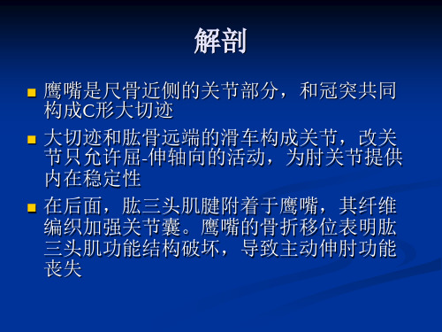
鹰嘴是尺骨近侧的关节部分,和冠突共同 构成C形大切迹
大切迹和肱骨远端的滑车构成关节,改关 节只允许屈-伸轴向的活动,为肘关节提供 内在稳定性
在后面,肱三头肌腱附着于鹰嘴,其纤维 编织加强关节囊。鹰嘴的骨折移位表明肱 三头肌功能结构破坏,导致主动伸肘功能 丧失
鹰嘴的骨化中心在10岁时出现,大约16岁融 合。在成人可以一直存在骺板;常为双侧 存在并具有家族遗传性
治疗
鹰嘴骨折的治疗目标应包括以下几个客观 标准:关节恢复,伸肌运动力量的保留, 稳定,避免可能发生的并发症引起的僵硬 和运动受限。
无移位骨折(nondisplaced fractures)
无移位骨折 由于尺骨鹰嘴有肱三头肌腱的附着, 骨折后很少不发生移位。对于没有条件进行手术 治疗和骨折移位较小的患者,可进行屈肘45-90度 长臂石膏后托固定2-3周,不能在完全伸肘位固定, 5-7天内行X线片检查,以保证骨折不发生再移位。 固定6-8周骨折也不能获得完全愈合,但固定3周 即可获得充分的稳定。此时可去除外固定,在保 护下进行功能锻炼,直至骨折在X线片上表现为 完全愈合之前,避免屈肘超过90度。因为对无移 位的尺骨鹰嘴骨折进行内固定治疗可使肘关节早 期进行功能锻炼,以改善临床效果,对于有条件 患者也可考虑手术固定治疗。
斜形-近端骨折:骨折线向远端延伸致冠突,影响肘关节稳定性。 骨折-脱位:通常由严重创伤所致。
colton分类
Colton把鹰嘴骨折分为两大类:无移位骨折 (Ⅰ型)和移位骨折(Ⅱ型)。 Ⅰ型无移 位骨折定义为分离小于2mm,肘关节屈曲 到90度时移位无增加,患者可以克服重力伸 展肘关节。 Colton把Ⅱ型骨折进一步分为: ⅡA型,撕脱性骨折;ⅡB型,斜形和横行 骨折;ⅡC型,粉碎性;和ⅡD型,骨折脱 位型。
手术_3.3.3 加压外固定治疗尺骨鹰嘴骨折

【编号】3.3.3【手术名称】加压外固定治疗尺骨鹰嘴骨折【英文名称】external fixation of olecranon fracture of ulna【别名】骨外固定加压治疗尺骨鹰嘴骨折【ICD编码】78.1301【概述】尺骨鹰嘴位于肘后皮下,为肱三头肌的止点,其扩张的腱膜附着在鹰嘴后方。
鹰嘴骨折虽多由直接暴力引起,但肱三头肌收缩的间接力量引起的撕脱亦为鹰嘴骨折的机制之一。
这种骨折要求准确复位和早期功能活动,以免后遗关节面不平整,关节粘连和僵硬。
尺骨鹰嘴骨折的影像学表现见下图(图3.3.3-1,3.3.3-2)。
【适应证】1.伴有严重软组织伤的开放性骨折。
2.火器性骨折由于软组织失活变化很大和需多次清创,骨外固定便于观察处理伤口和多次搬运病人。
3.重度烧伤伴有骨折,外固定器治疗既可为骨折提供牢稳固定,也便于观察治疗创面,防止植皮区受压和关节瘢痕挛缩。
4.伴有多发伤的开放性骨折或多发骨折,骨外固定可迅速将骨折制动,可减轻疼痛和减少进一步失血的危险,有利于抗休克和争取时间对威胁病人生命的损伤施行手术。
5.有广泛软组织挤压伤的闭合性骨折,不适应作切开复位内固定,使用经皮穿针外固定可避免夹板、石膏或手术加重软组织损伤。
6.骨折伴有骨缺损者可用牵伸固定法保持伤肢长度,以便日后修复骨缺损。
7.感染性骨折和感染性骨不连,使用外固定器有利于伤口治疗,可同时将骨折牢固固定,能有效地兼顾两者的治疗。
8.骨折伴有神经与血管损伤。
【禁忌证】【术前准备】【麻醉与体位】在臂丛神经阻滞或局部麻醉下进行,肘关节屈曲100°~110°。
【手术步骤】1.穿针 为避免误伤尺神经,近心骨折端须由尺侧向桡侧穿针。
远心骨折段穿针平面宜距骨断面2c m,在尺骨嵴旁1c m处进针。
如骨折为粉碎型,则须经肱三头肌附着部穿针(图3.3.3-3)。
穿针应与尺骨长轴垂直和相互平行。
2.复位与加压固定 穿针后将肘关节置于半伸展位,手法矫正侧方移位后,拧紧固定夹顶端螺钉,旋转螺杆同时伸展肘关节,使骨折的关节面平整对合,加压固定骨折。
解剖型锁定加压接骨板修复尺骨鹰嘴骨折:肘关节功能6个月随访评价

解剖型锁定加压接骨板修复尺骨鹰嘴骨折:肘关节功能6个月随访评价齐岩;田军;姚宇;刘浩;孙磊【摘要】BACKGROUND:Improper treatment of olecranon fractures wil affect elbow function. To reduce the incidence of complications and good recovery of elbow function, it is important to give accurate reduction and rigid internal fixation of olecranon fracture. <br> OBJECTIVE:To investigate the anatomic locking compression plate in the repair of olecranon fracture. <br> METHODS:Anatomical locking compression plate was used for open reduction and fixation in 36 cases of olecranon fracture. The recovery of elbow joint function of patients after operation was evaluated. <br> RESULTS AND CONCLUSION:36 cases of olecranon fracture received anatomical locking compression plate for open reduction and fixation, and were folowed up for more than 6 months. No early complications were found in the incision such as infection or hematoma. They were primarily healed. Broberg and Morrey evaluation showed that the excelent and good rate was 94% (34/36). It is concluded that anatomical locking compression plate for olecranon fracture is simple to be operated. The fixation is firm. The patients can do early exercise postoperatively, less complications and good recovery of elbow joint function.%背景:尺骨鹰嘴骨折治疗不当会影响肘关节功能,为了减少并发症的发生,使肘关节功能得到良好恢复,给予尺骨鹰嘴骨折准确复位和牢固的内固定尤为重要。
上肢骨折

• 多见于青少年
• 发生在尺桡骨任何节段
• 骨折影响前臂旋转功能
病因病机
直 接 暴 力 打 击
桡尺骨干双骨折
传 导 暴 力 所 致
扭 转 暴 力 所 致
桡尺骨干双骨折
桡尺骨干双骨折
桡尺骨干双骨折
桡尺骨干双骨折
诊断要点
• 外伤史
• 局部肿胀、疼痛、前臂功能丧失 • 重叠、成角、旋转和侧方移位 • 骨擦音、异常活动 • X线照片(桡尺骨干全长,包肘腕关节)
3.骨间膜是坚韧致密的纤维
膜,附着于桡、尺骨嵴,
几乎连接桡尺骨的全长,
由桡骨斜向下至尺骨(见 右图)。其松紧度是随着 前臂的旋转而改变。
骨 间 膜 行走方向
下尺桡 关 节 桡、尺骨的连接
桡尺骨干双骨折
4.前臂的中立位:屈肘90°, 拇指向上掌心向内。 前臂处于中立位时尺桡 骨接近平行,骨间膜上 下紧张一致,前臂稳定 性最大。故处理尺桡骨 双骨折时,应将前臂固 定在中立位。
尺骨鹰嘴骨折
间接暴力所致骨折
肌肉牵拉所致尺骨鹰嘴骨折
病因病机(2)
尺骨鹰嘴骨折
肱三头肌
直接暴力 所致骨折
直接暴力
少见
跌倒撞击,暴力直接打击尺骨鹰嘴部, 骨折多为粉碎性骨折,因鹰嘴支持带及 骨膜保持完整,无移位。
尺骨鹰嘴骨折
诊断要点
1.明显受伤病史。
2.症状:鹰嘴部疼痛、局限 性肿胀,鹰嘴两旁的凹陷 消失 主动伸肘功能丧失。
注意复位前先抽出关节内积血再复位。
拇指抵于尺骨鹰嘴
推挤伸肘
尺骨鹰嘴骨折
㈡ 固定:复位后,尺骨 鹰嘴上端抱骨圈固定
肘关节于屈曲0~20°
位3周,再逐渐改为
复位前
尺骨鹰嘴骨折

病因病理(2)
诊断要点
01
02
03
外伤史
疼痛、肿胀、功能障碍(伸肘)X线尺骨鹰嘴骨折尺骨鹰嘴骨折治疗
手法整复(较容易) 问题: 1、固定在非功能位(伸直)对功 能恢复有影响; 2、关节面复位不易达解剖对合(粉 碎性骨折更明显)
肘后三角
尺骨鹰嘴骨折
单击添加副标题
单击此处添加文本具体内容,简明扼要地阐述你的观点
了解尺骨鹰嘴骨折的解剖、病因病理、诊断、整复和夹板固定方法。
尺骨鹰嘴骨折
尺骨鹰嘴骨折
间接暴力 肘关节突然屈曲,肱三头肌强烈收缩而发生尺骨鹰嘴的撕脱性骨折,近端向上移位 病因病理(1)
尺骨鹰嘴骨折
打击尺骨鹰嘴部,骨折多为粉碎性骨折,无移位。
手术治疗(近年趋向) 操作易,可达理想对位,可早进入肘部练功,关节功能恢复快
临床问题?
点击此处添加正文,文字是您思想的提炼,为了演示发布的良好效果,请言简意赅地阐述您的观点。
ONE
靴状畸形(1)
靴状畸形(2)
靴状畸形(3)
??
THANKS FOR WATCHING
鹰嘴钩钢板在治疗尺骨鹰嘴骨折中的可行性研究
鹰嘴钩钢板在治疗尺骨鹰嘴骨折中的可行性研究邹祝艺;徐剑锋;李金生;利东升【期刊名称】《中国医药科学》【年(卷),期】2016(006)004【摘要】Objective To explore the application effect of olecranon hook plate in the treatment of olecranon fracture. MethodsA total of 83 patients with olecranon fracture were selected in our hospital in 2013 January 2015 June, 83 patients were randomly divided to control group (n=43) and observation group (n=40) according to the sequence of admission time. Patients in control group received the tension band fixation with Kirschner wire for the treatment, the observation group received internal fixation for theolecranon hook plate. The operation time, hospitalization time, healing time of fracture, fracture healing time, fracture anatomical reduction rate and elbow function were compared between the two groups.Results The operation time, hospitalization time and clinical healing time of the observation group were significantly shorter than those in the control group(t=7.0660,2.7149, 5.1244,P<0.05).The anatomic reduction rate of the observation group was 80%, significantly higher than the control group 58.1%(x2=4.6034,P<0.05). The Schatzker score of observationgroup'sexcellent rate was 65%, which was significantly higher than 34.8% of the control group(P<0.05). The incidence of adverse reactions in observation group was 5%, and there was no significant difference inadverse reactions with the control group(x2=0.1430, P>0.05). ConclusionThe olecranon fracture treated with olecranon hook plate has good clinical effect, can effectively shorten the operation and hospitalization time, fracture healing time, postoperative elbow function was improved, safe and effective, worthy of clinical application.%目的:探究鹰嘴钩钢板内固定在治疗尺骨鹰嘴骨折中的应用效果。
尺骨鹰嘴解剖型钢板治疗粉碎性尺骨鹰嘴骨折
尺骨鹰嘴解剖型钢板治疗粉碎性尺骨鹰嘴骨折米博斌;刘国辉;杨述华;熊家伟;李强【摘要】Objective To evaluate the therapeutic effect of treatment of comminuted olecranon fractures with anatomical plates. Methods A total of 36 patients with comminuted olecranon fractures were treated by anatomical plates. Results All fractures were anatomical reduced by operation. All cases were followed up for 8 ~ f5months. According to Broberg and Morrey scoring system, 28 patients were rated as excellent, 5 good, and 3 fair. The rate of excellent and good results was 9f. 6%. No cases of infection, neural injury was found. 2 cases had traumatic arthritis, and both cases were relived by phannacotherapy. Conclusions The application of anatomical plates has advantages of effective fixation, early exercise and fewer complications. It is one of the idea choices to treat comminuted olecranon fractures.%目的探讨尺骨鹰嘴解剖型钢板治疗粉碎性尺骨鹰嘴骨折的疗效.方法采用尺骨鹰嘴解剖型钢板治疗36例粉碎性尺骨鹰嘴骨折患者.结果患者骨折均获得解剖复位.36例均获随访,时间8~15个月.按Broberg 和Morrey 评估标准进行功能评定:优28例,良5例,可3例,优良率为91.6%.无伤口感染、神经损伤等并发症.2例发生创伤性关节炎,经药物治疗后症状缓解.结论尺骨鹰嘴解剖型钢板治疗尺骨鹰嘴粉碎性骨折疗效好,能够达到解剖复位,手术固定牢靠.【期刊名称】《临床骨科杂志》【年(卷),期】2012(015)006【总页数】2页(P654-655)【关键词】解剖型钢板;尺骨鹰嘴骨折;骨折,粉碎性【作者】米博斌;刘国辉;杨述华;熊家伟;李强【作者单位】华中科技大学同济医学院附属协和医院骨科,湖北,武汉,430022;华中科技大学同济医学院附属协和医院骨科,湖北,武汉,430022;华中科技大学同济医学院附属协和医院骨科,湖北,武汉,430022;华中科技大学同济医学院附属协和医院骨科,湖北,武汉,430022;华中科技大学同济医学院附属协和医院骨科,湖北,武汉,430022【正文语种】中文【中图分类】R683.41;R687.322008年3月~2011年2月,我院应用尺骨鹰嘴解剖型钢板治疗36例粉碎性尺骨鹰嘴骨折患者,效果满意,现报道如下。
尺骨鹰嘴骨折
的风险。
穿戴防护装备
03
在进行可能发生碰撞或摔倒的活动时,应穿戴适当的防护装备
,如护肘、护膝等。
术后护理
定期换药
术后定期换药,保持伤口清洁干燥,预防感染。
观察伤口情况
密切观察伤口情况,如出现红肿、疼痛、渗出等异常症状,应及 时就医。
遵循医嘱
遵循医生的建议,按时服药、按时复查,以及按照医嘱进行康复 训练。
康复训练与注意事项
早期康复训练
术后早期可进行手指、腕关节等小关 节的活动,以促进血液循环和关节灵 活性。
逐渐增加运动量
根据医生的建议,逐渐增加运动量和 强度,以促进骨折愈合和功能恢复。
注意疼痛和肿胀
在康复训练过程中,注意观察疼痛和 肿胀情况,如出现明显不适或症状加 重,应及时停止训练并就医。
避免二次受伤
后期康复
骨折愈合后,进行全面的功能锻炼和康复训练,包括力量训练、柔 韧性训练和协调性训练等,以恢复关节的正常功能。
04
尺骨鹰嘴骨折的预防与护理
预防措施
保持身体健康
01
保持健康的生活方式,包括均衡饮食、适量运动和规律作息,
以提高骨骼和肌肉的强度和韧性。
避免高风险活动
02
避免参与高风险活动,如极限运动、竞技体育等,以降低骨折
THANK YOU
感谢各位观看
关节镜下复位固定
在关节镜下进行手术,复位骨折并使用钢丝、缝 线等固定器材进行固定。
外固定架
使用外固定架对骨折部位进行固定,操作简便, 适用于严重粉碎性骨折等复杂情况。
康复治疗
早期康复
在骨折愈合初期,进行适当的关节活动和肌肉锻炼,以预防关节 僵硬和肌肉萎缩。
中期康复
钛合金接骨板治疗尺骨鹰嘴粉碎性骨折的临床研究
钛合金接骨板治疗尺骨鹰嘴粉碎性骨折的临床研究摘要】目的探讨尺骨鹰嘴粉碎性骨折的手术治疗和结果。
方法肘后正中纵形切口约10-14cm,显露骨折断端,将骨折复位,接骨板充分塑形尺骨鹰嘴的形状,先用2枚螺钉将接骨板固定于近端尺骨鹰嘴上,再用螺钉固定骨折近端,缝合肱三头肌腱膜,关闭切口。
术后可早期进行功能锻炼。
结果本组51例全部随访6个月-4年,平均14个月,术后X线片显示骨折均已愈合,平均骨性愈合时间7.1月,无切口感染,骨不连等并发症。
按庞桂根疗效评价标准评定[1]:优40例,良6例,可3例,差2例。
优良率:90.2%。
结论钛合金接骨板内固定是治疗粉碎性尺骨鹰嘴骨折的有效手段,具有固定坚强,能早期功能锻炼,肘关节功能良好,并发症少的优点。
【关键词】钛合金接骨板尺骨鹰嘴骨折本院自2006年5月-2011年12月,应用钛合金尺骨鹰嘴接骨板内固定治疗尺骨鹰嘴粉碎性骨折51例,取得满意疗效,现报告如下:1. 资料与方法1.1一般资料:本组51例,男46例,女9例,最小年龄20岁,最大年龄63岁,平均年龄38.16岁,左侧36例,右侧15例。
受伤原因:直接暴力18例,跌伤23例(交通事故5例),坠落伤10例。
按改良Schatzkker分型[2],A型损伤15例,A1型损伤8例,A2型损伤9例,B1型12例,B2型4例,C型损伤2例,D型损伤1例合并肘关节前脱位和桡骨头骨折。
1.2手术方法:采用臂丛麻醉仰卧位,患肢上气囊止血带,置于胸前,常规消毒铺巾,取肘后纵形切口长约10-14cm,依次切开皮肤,皮下组织后向两侧剥离骨膜,暴露骨折断端,清理血肿及碎骨片,生理盐水冲洗,直视下复位骨折,用巾钳钳住以维持复位临时固定,选择长度合适的钛合金接骨板,近折段放置于尺骨鹰嘴后方,远折段放置于尺骨脊内侧面上,用克氏针临时固定,在C臂透视下确定骨折复位好,钢板位置理想,鹰嘴部用松质骨螺钉固定,尺骨干部用皮质骨螺钉固定。
1.3术后处理:常规使用抗生素3-5天,石膏托固定1周,1周后行肘关节功能被动锻炼,抬高患肢以利消肿,术后2周拆线,开始主动功能锻炼,粉碎较重的骨折,结合术中内固定物的稳定情况,决定何时功能锻炼,一般在术后4周复查X线片之后确定相关的康复方法,并在医生指导下进行循序渐进的肘关节功能康复锻炼。
- 1、下载文档前请自行甄别文档内容的完整性,平台不提供额外的编辑、内容补充、找答案等附加服务。
- 2、"仅部分预览"的文档,不可在线预览部分如存在完整性等问题,可反馈申请退款(可完整预览的文档不适用该条件!)。
- 3、如文档侵犯您的权益,请联系客服反馈,我们会尽快为您处理(人工客服工作时间:9:00-18:30)。
Mechanism of injury
Direct
trauma High energy trauma such as car accident
Treatment goals
Orthopaedic objectives Alignment Stability
Functional goals Restore ADL
Methods of treatment
Closed
reduction and splint or cast Indication: nondisplaced stable fracture Open reduction and internal fixation Indication :displaced and comminuted fracture Excision and triceps advancement and reattachment Indication: elderly patient with osteroporotic bone and significantly comminuted fracture
motion flexion extension Pronation supination Normal 150 -5 -0 90 90 functional 90 Lacking20-30 50 50
Functional
goals Expected time of bone healing Expected duration of rehabis of treatment :special aspects
Closed reduction and splint or cast gently perform actively range of motion exercises of elbow in flexion,extension,supination,pronation Active-assistive range of motion exercises to the wrist Open reduction and internal fixation Active –assistive exercises Excision and triceps advancement and reattachment supination and pronation are encouraged
Methods of treatment :special aspects
Closed reduction and splint or cast gently perform actively range of motion exercises of elbow in flexion,extension,supination,pronation Active-assistive range of motion exercises to the wrist Open reduction and internal fixation Active –assistive exercises Excision and triceps advancement and reattachment after six weeks ,remove the cast
Olecranon fractures
lizhiyong
Difinition and classification
Proximal end of ulna Extraarticlar or intraarticlar fracture Displaced or nondisplaced fracture Transverse or oblique comminuted fracture Stable or unstable fracture Corresponding by disruption of extensor mechanism Associated with coronoid fracture and MCL injury
Treatment (two weeks)
1.
2. 3. 4. 5.
6.
7.
Orthopaedic and rehabilitation Physical examination :excessive swelling \loosened cast or splint danger : no pressure in the antecubital fossa Radiography Weight bearing Range of motion shoulder movement \finger movement Muscle strength gentle isometric exercise o f biceps and wrist \isotonic exercise of fingers Functional activities self-care and hygiene
Treatment (six to eight weeks)
Orthopaedic and rehabilitation
Physical examination : 1. check the fracture site tendernes and motion 2. Check the improving elbow range of motion and strength 3. Check for the resolvement of the reflex sympathetic dystrophy 4. Check the status of any existing ulnar nerve deficits Radiography Weight bearing full weight bearing is allowed Range of motion 1. continue active and active –assistive range of motion to the shoulder\elbow\wrist 2. Avoiding Gently passive range of motion at the elbow 3. Dynamic splint may be used for elbow stretch Muscle strength: continue resistive exercise to the wrist and elbow graded weights to improve strength Functional activities start using the affected extremity for all activities o f selfcare
Rehabilitation objectives
Range
of motion Muscle strength including triceps\ biceps \supinators of the forearm\ wrist pronators of the forearm and wrist\ Wrist extensor group\ wrist flexor group
Methods of treatment :special aspects
Closed
reduction and splint or cast Adequacy of cast\skin breakdown \trim the cast edge for full motion of fingers Open reduction and internal fixation Erythema discharge \early movement Excision and triceps advancement and reattachment Check the wound \remove drainage
1. 2. 1. 2.
Treatment (four to six weeks) Orthopaedic and rehabilitation
Physical examination : check the fracture site tenderness\crepitus \motion Check the elbow range of motion danger : reflex sympathetic dystrophy Radiography Weight bearing Range of motion continue active and active –assistive range of motion to the shoulder\elbow\wrist Avoiding passive range of motion at the elbow Muscle strength: advance isometric exercise o f biceps and wrist \isotonic exercise of fingers Functional activities start using the affected extremity for grooming
Methods of treatment :special aspects
Closed reduction and splint or cast Actively perform isometric exercises of elbow extensors within the cast Open reducthon and internal fixation Continue elbow flexion in the patient with stable tension band fixation Excision and triceps advancement and reattachment Avoiding range of motion