(参考)Chitosan-graft-polyethylenimine as a gene carrier n
美国 Gibco 最小必要基质 MEM 产品说明书
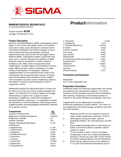
Product Information MINIMUM ESSENTIAL MEDIUM EAGLED-VALINE MODIFICATIONProduct Number M7395Storage Temperature 2-8°CProduct DescriptionMinimum Essential Medium (MEM), developed by Harry Eagle, is one of the most widely used of all synthetic cell culture media. Early attempts to cultivate normal mammalian fibroblasts and certain subtypes of HeLa cells revealed that they had specific nutritional requirements that could not be met by Eagle’s Basal Medium (BME). Subsequent studies using these and other cells in culture indicated that additions to BME could be made to aid growth of a wider variety of fastidious cells. MEM, which incorporates these modifications, includes higher concentrations of amino acids. MEM has been used for cultivation of a wide variety of cells grown in monolayers. Optional supplementation of non-essential amino acids to the formulations that incorporate either Hanks’ or Earle’s salts has broadened the usefulness of this medium. The formulation has been further modified by optional elimination of calcium to permit growth of cells in suspension culture.MINIMUM ESSENTIAL MEDIUM EAGLE, Product No. M 7395 is one of the cell culture media available from Sigma. The selection of a nutrient medium is strongly influenced by 1] type of cell, 2] type of culture [monolayer, suspension, clonal] and 3] degree of chemical definition necessary. It is important to review the literature for recommendations concerning medium, supplementation and physiological parameters required for a specific cell line.Components g/L Calcium Chloride•2H2O0.265 Magnesium Sulfate (anhydrous)0.09767 Potassium Chloride0.4 Sodium Chloride 6.8 Sodium Phosphate Monobasic0.122 (anhydrous)L-Arginine•HCl0.126L-Cystine•2HCl0.0313 L-Glutamine0.292L-Histidine•HCl•H2O0.042L-Isoleucine0.052L-Leucine0.052L-Lysine•HCl0.0725 L-Methionine0.015L-Phenylalanine0.032L-Threonine0.048L-Tryptophan0.01L-Tyrosine•2Na•2H2O0.0519D-Valine0.092 Choline Chloride0.001Folic Acid0.001myo-Inositol0.002 Niacinamide0.001D-Pantothenic Acid (hemicalcium)0.001 Pyridoxal•HCl0.001 Riboflavin0.0001 Thiamine•HCl0.001 Glucose 1.0 Phenol Red•Na0.011Precautions and DisclaimerREAGENTFor In Vitro Diagnostic UsePreparation InstructionsPowdered media are extremely hygroscopic and should be protected from atmospheric moisture. The entire contents of each package should be used immediately after opening. Preparing a concentrated solution of medium is not recommended as precipitates may form. Supplements can be added prior to filtration or introduced aseptically to sterile medium. The nature of the supplement may affect storage conditions and shelf life of the medium.1.Measure out 90% of final required volume ofwater. Water temperature should be 15-20°C.2.While gently stirring the water, add thepowdered medium. Stir until dissolved. Do NOTheat.3.Rinse original package with a small amount ofwater to remove all traces of powder. Add tosolution in step 2.4.To the solution in step 3, add 2.2 g sodiumbicarbonate or 29.3 ml of sodium bicarbonatesolution [7.5%w/v] for each liter of final volumeof medium being prepared. Stir until dissolved.5.While stirring, adjust the pH of the medium to0.1-0.3 pH units below the desired pH since itmay rise during filtration. The use of 1N HCl or1N NaOH is recommended.6.Add additional water to bring the solution tofinal volume.7.Sterilize immediately by filtration using amembrane with a porosity of 0.22 microns.8.Aseptically dispense medium into sterilecontainer.Storage/StabilityStore the dry powdered medium at 2-8°C under dry conditions and liquid medium at 2-8°C in the dark. Deterioration of the powdered medium may be recognized by any or all of the following: [1] color change, [2] granulation/clumping, [3] insolubility. Deterioration of the liquid medium may be recognized by any or all of the following: [1] pH change, [2] precipitate or particulate matter throughout the solution, [3] cloudy appearance [4] color change. The nature of supplements added may affect storage conditions and shelf life of the medium. Product label bears expiration date.ProcedureWater for tissue culture use [W-3500]Sodium Bicarbonate [S-5761] orSodium Bicarbonate Solution, 7.5% [S-8761]1N Hydrochloric Acid [H-9892]1N Sodium Hydroxide [S-2770]Medium additives as requiredProduct ProfileAppearance off-white powder Moisture content 2.0% Solubility clear solution at 1x concentrationpH at room temperature 5.8 ± 0.3 [without sodium bicarbonate]pH at room temperature 7.5 ± 0.3 [with sodium bicarbonate]Osmolality250 mOsm/kg H2O ± 5% [without sodium bicarbonate]Osmolality290 mOsm/kg H2O ± 5% [with sodium bicarbonate]Amino Acid Analysis Analysis has confirmedby HPLC that amino acids are present atconcentrations consistent withthe formula.Key Element Analysis Analysis has confirmed that by ICAP key elements are present atconcentrations consistent withthe formula.BIOLOGICAL PERFORMANCE CHARACTERISTICS Biological performance is assessed using an appropriate cell line(s). Growth studies are carried through 2 subculture generations. Cells are counted and growth is plotted as a logarithmic function of time in culture. Seeding efficiencies, doubling time, and final cell densities are determined. During the testing period cultures are examined microscopically for atypical morphology and evidence of cytotoxicity. Test results are available upon request.References1.Eagle, H. et al (1956) myo-Inositol as anEssential Growth Factor for Normal andMalignant Human Cells in Tissue Culture.J.Biol. Chem. 214, 845-847.2.Eagle, H.(1976) Media for Animal Cell Culture.Tissue Culture Association Manual. 3, 517-520.3.Eagle, H. (1959). Amino Acid Metabolism inMammalian Cell Cultures. Science. 130, 432-437.4.Eagle, H. (1955) Nutrition Needs of MammalianCells in Culture. Science. 122, 501.5.Gilbert, S.F. and Migeon, B.R. (1975) D-valineas a selective agent for normal human androdent epithelial cells in culture. Cell. 5, 11-17.7H027Sigma brand products are sold through Sigma-Aldrich, Inc.Sigma-Aldrich, Inc. warrants that its products conform to the information contained in this and other Sigma-Aldrich publications. Purchaser must determine the suitability of the product(s) for their particular use. Additional terms and conditions may apply. Please see reverse side ofthe invoice or packing slip.。
李瑛,女,博士,教授。四川省化学化工学会理事,四川大学化学学
李瑛,女,博士,教授。
四川省化学化工学会理事,四川大学化学学各位读友大家好,此文档由网络收集而来,欢迎您下载,谢谢工作业绩。
从事有机化学教学与科研工作29年。
主持或者参与“863”计划2项。
国家自然科学基金9项。
教育部博士点基金5项和其他省部级项目8项。
在国内外重要学术刊物上发表论文60余篇。
SCI收录40余篇。
获省部级鉴定成果2项。
授权发明专利2项。
四川大学第七届教学名师。
四川省精品课程《有机化学》负责人。
主编本科生教材《有机化学基础》。
参编“十一五”研究生教材《现代有机合成化学》。
获四川省教学成果二等奖一项。
代表性成果。
1. Xiaopeng Xu, Yulei Wu, Junfeng Fang,* Zuojia Li, ZhenguoWang, Ying Li, Qiang Peng*. Side chain engineering of benzodithiophene-fluorinated quinoxaline low band gap copolymers for high performance polymer solar cells. Chemistry-A European Journal, 2016, 20, Zhenguo Wang, Jie Zhao,Ying Li, Qiang Peng*. Low band-gap copolymers derived from fluorinated isoindigo and dithienosilole: synthesis, properties and photovoltaic applications. Polymer Chemistry, 2016, 5, Zhi Zeng, Ying Li, Jufu Deng, Qin Huang, Qiang Peng*. Synthesis and photovoltaic performance of low band gap copolymers based on diketopyrrolopyrrole and tetrathienoacene with different conjugated bridges. J. Mater. Chem. A, 2016, 2, Kui Feng, Xiaoyan Shen, Ying Li*, Yujiang He, Dong Huang, Qiang Peng*. Ruthenium Containing Supramolecular Polymers with Cyclopentadithiophene-BenzothiazoleConjugated Bridges for Photovoltaic Applications. Polymer Chemistry, 2016, 4, Zuojia Li, Dan Zhou, Lixin Li, Ying Li,* Yujiang He, Jian Liu。
siRNA

Tumor-speci fic delivery of siRNA using supramolecular assembly of hyaluronic acid nanoparticles and 2b RNA-binding protein/siRNA complexesKyung-mi Choi a ,Mihue Jang b ,Jong Hwan Kim b ,c ,Hyung Jun Ahn b ,*aInstitute of Global Environment and Department of Biology,Kyung Hee University,Dongdaemun-Gu,Seoul 130-701,South KoreabCenter for Theragnosis,Biomedical Research Institute,Korea Institute of Science and Technology,Seongbuk-Gu,Seoul 136-791,South Korea cDepartment of Chemistry,Kyung Hee University,Dongdaemun-Gu,Seoul 130-701,South Koreaa r t i c l e i n f oArticle history:Received 17March 2014Accepted 21April 2014Available online xxxKeywords:siRNAsiRNA carrierSupramolecular assemblyHyaluronic acid-choleresterol conjugatea b s t r a c tAnticancer therapeutics delivering exogenous siRNA have been explored to suppress the tumor-associated genes,but several limitations of siRNA delivery such as tumor-targeted delivery,controlled siRNA release at the sites of interest,or instabilities of siRNA in physiological fluids should be prefer-entially addressed for its clinical applications.As an attempt to meet these criteria,we designed a su-pramolecular assembly,which was composed of cholesterol-bearing hyaluronic acid (HA-Chol)conjugates and 2b RNA-binding protein (2b)/siRNA complexes.In contrast to the traditional siRNA polyplexes using electrostatic interactions,HA-Chol nanoparticles,as a results of self-assembly of HA-Chol conjugates,provide the hydrophobic core that acts as the container for 2b protein/siRNA com-plexes,where a high af finity of 2b protein for siRNA could neutralize the negative-charged siRNA.Here,we investigated the potential of HA-Chol/2b/siRNA complexes as the siRNA carriers that provide encapsulation,protection,and targeted delivery of siRNA.The HA-Chol nanoparticles could selectively deliver 2b protein/siRNA complexes to the tumor cells with up-regulated CD44receptors and suppress the expression of target gene.The pH-associated binding properties of siRNA for 2b proteins allowed the controlled release of siRNA in the endocytic compartments,and ultimately the released siRNA could obtain the RNAi acitivities in the cells,whereas the encapsulated 2b proteins still stayed within the HA-Chol nanoparticles.Our delivery systems demonstrate the promising potential of the ef ficient siRNA carriers in the anticancer therapeutic applications.Ó2014Elsevier Ltd.All rights reserved.1.IntroductionRecently,RNA interference (RNAi)has provided a promising tool to suppress the expression of target gene via degradation of mRNA [1,2].RNAi mechanism is fundamentally triggered by small inter-fering RNA (siRNA),which initiates the cleavage and degradation of its complementary mRNA after assembled with RNA-induced silencing complex (RISC),and consequently,the synthesis of the corresponding protein is blocked.However,the inherent properties of naked siRNA such as its anionic property,immune response,large molecular weight,and enzymatic degradation in serum havelimited its applications in vitro or in vivo [3e 5].Also,the naked siRNA does not possess any functional moiety that selectively rea-ches the target tissues,an appropriate carrier system should address these criteria for therapeutic RNAi applications.A large range of delivery vehicles have been exploited for siRNA,including viruses,lipids,proteins,polymers,and gold nanoparticles [6e 9].In particular,the traditional polycationic polymers such as polyethylenimines (PEIs),chitosans,and reducible polyamido-amines (rPAAs)have been explored to achieve the effective siRNA delivery and gene knockdown [10e 12].The amino groups present in the polycationic polymers can complex with anionic siRNA via electrostatic interactions and thus provide a driving force to form the condensed siRNA-complexed nanoparticles (siRNA polyplexes),which protect the bound siRNA against RNases attack as well as neutralize the anionic siRNA for cellular internalization.Since the effective delivery systems should avoid potentially toxic side effects of the vehicles,there have been many efforts to*Corresponding author.Korea Institute of Science and Technology,39-1Hawolgok-dong,Seongbuk-Gu,Seoul 136-791,South Korea.Tel.:þ8229585938;fax:þ8229585909.E-mail address:hjahn@kist.re.kr (H.J.Ahn).Contents lists available at ScienceDirectBiomaterialsjournal homepage:w ww.elsevi/locate/biomaterials/10.1016/j.biomaterials.2014.04.0960142-9612/Ó2014Elsevier Ltd.All rights reserved.Biomaterials xxx (2014)1e 12reduce the inherent cytotoxicities derived from a high charge density or limited degradability of polycationic polymers,of which cytotoxicities lead to necrotic cell death or apoptosis in the un-controlled manner[13].For example,many researchers have modified polycationic polymers or synthesized new polycationic polymers,while keeping a balance between their complexing ability and cell viability[14].Nevertheless,it is still challenging to condense siRNA into the less cytotoxic siRNA polyplexes.Interestingly,many globular proteins have been experimentally reported to complex with the self-assembled nanoparticles such as cholesterol-bearing pullulan through the hydrophobic interactions between these proteins and hydrophobic moieties within the nanoparticles[15,16].In this respect,the self-assembled nano-particles,composed of amphiphilic polymers,have extensively drawn much attention as a protein drug carrier because they permit the encapsulation of hydrophobic therapeutic proteins, thereby protecting them from harsh in vivo conditions[17].In the current study,hyaluronic acids(HA)as a starting building block is a highly anionic mucopolysaccharide,of which the alter-nating disaccharide units are composed of D-glucuronic acid and N-acetyl-D-glucosamine with b(1e4)interglycosidic linkage[18].HA is the biodegradable non-toxic biomaterial,and notably,has been used as the popular delivery agent for cancer treatment,because it specifically binds CD44and RHAMM receptors abundantly pre-sented in many types of tumor cells as well as regulates the angiogenesis during tumor growth[19,20].For example,HA-modified macromolecular prodrugs and nanocarriers showed a specific binding to CD44-overexpressing tumor cells regardless of their negatively-charged surface charge[21,22].Also,thiol-functionalized HA nanogels encapsulating siRNA by an inverse emulsion method showed the cellular internalization and gene knockdown effects in the CD44-overexpressing HCT-116cells[23].For delivering siRNA to tumor cells,we prepared the self-assembled HA nanoparticles(HA-Chol NPs)in an aqueous envi-ronment after hydrophobically modifying HA backbone with cho-lesterols.Although the HA nanoparticles are the attractive tumor-specific carrier for siRNA delivery,their negatively charged prop-erty cannot encapsulate the anionic siRNA directly[24].Instead of encapsulating the naked siRNA,we here proposed an alternative method to incorporate siRNA/2b protein complexes into the HA nanoparticles.The2b proteins,derived from Tomato aspermy virus, are known to bind dsRNA to counter host defense during viral infection[25],and here showed the stable complexing ability with siRNA in the physiological pH condition.Based on the3D structural analysis of siRNA/2b protein complex,2b proteins neutralize the negative-charged phosphate backbone of siRNA when they wraparound the siRNA duplex[26].Here,we demonstrate that the neutralized siRNA,as a result of complexing with2b proteins,can be encapsulated into the HA-Chol NPs to form the HA-Chol/2b/ siRNA complexes in the absence of polycationic polymers(Fig.1).We enzymatically degraded the HA-Chol/2b/siRNA complexes with hyaluronidase to show whether siRNA locates deeply inside the core of HA-Chol nanoparticles or is simply bound to the surface of nanoparticles,because the siRNA stabilities in serum can be explained by inaccessibility of RNases.Also,we investigated the CD44-mediated tumor-targeting abilities of the HA-Chol/2b/siRNA NPs and their cellular uptakes via endocytosis pathways.To better understand the siRNA dissociation in the acidity environments such as endosomes or endolysosomes,we assessed the variation of siRNA binding affinity for2b proteins or HA-Chol NPs in the acidic ing the FITC-labeled2b proteins,we could examine the retention of2b proteins within the nanoparticles after cellular internalization.Ultimately,we investigated whether the HA-Chol/ 2b/RFP-siRNA NPs effectively suppressed the expression of tar-geted RFP gene in tumor pared to the traditional delivery methods utilizing electrostatic interactions,these studies provide an alternative strategy to deliver siRNA to the tumor cells for cancer therapy and control siRNA release,as well as to efficiently encap-sulate siRNA into HA nanoparticles.2.Materials and methods2.1.MaterialsCholesteryl chloroformate,ethylenediamine,N-(3-dimethylaminopropyl)-N0-ethylcarbodiimide(EDC),N-hydroxysulfosuccinimide(sulfo-NHS),bovine testicular hyaluronidase,and pyrene were purchased from Sigma Aldrich(St.Louis,MO).So-dium hyaluronate(HA,M w234kDa)was obtained from Lifecore Biomedical(Chaska, MN).NHS-Cy5.5fluorescence dye was purchased from Amersham Bioscience(Pis-cataway,NJ).Primers and siRNA were available from Bioneer(Daejeon,Korea).2.2.Synthesis of HA-Chol conjugatesFirst,cholesteryl chloroformate was converted to3b-Cholest-5-en-3-yl N-(2-Aminoethyl)carbamate(Chol-Am)that could be reacted with the carboxylic acids of HA,as shown in the Fig.2.In brief,cholesteryl chloroformate(2.25g,5mmol)was dissolved in anhydrous toluene(50mL),and to this solution,ethylenediamine (16.7ml,250mmol)dissolved in anhydrous toluene(150mL)was dropwisely added.After stirred for16h at room temperature,the mixture was washed outwith Fig.1.Schematic illustration of supramolecular assembly for tumor-specific siRNA delivery.The amphiphilic HA-Chol conjugates self-assemble to form HA-Chol nano-particles,which provide the hydrophobic core that can act as a container for hydro-phobic cargo,2b protein/siRNA complexes.The molecular structures explain how2b protein dimer recognizes siRNA duplex.2b proteins and siRNA are colored magenta and yellow,respectively.The molecular structures were prepared by the Pymol pro-gram,and the structures of2b protein,siRNA,and2b/siRNA complex are from the Protein Data Bank(PDB IDs:2ZI0).(For interpretation of the references to color in this figure legend,the reader is referred to the web version of this article.)K.-m.Choi et al./Biomaterials xxx(2014)1e12 2distilled water and subjected to freeze-drying.The resulting powder was then dis-solved in dichloromethane/methanol solution (1/1,v/v,100mL)with stirring.We filtered off the suspension solution with a syringe filter (PTFE,MWCO:1m m,Whatman,Piscataway,NJ)to eliminate biscarbamate,and lyophilized the filtrate to obtain cholesteryl amine conjugate (Chol-Am)as a powder (750mg,1.60mmol,32%).We characterized its chemical structure using 1H NMR (Unity Plus 600,Varian,400MHz).Second,we chemically modi fied the backbone of HA with the hydrophobic Chol-Am in the presence of EDC and sulfo-NHS.Brie fly,we dissolved sodium hyaluronate (100mg,244m mol)in distilled water (10mL)containing EDC (23.4e 93.5mg)and sulfo-NHS (26.4e 106.0mg),and to this solution,we slowly added Chol-Am conju-gate (5.35mg e 21.4mg,12.2m mol e 48.8m mol)dissolved in THF (10mL).After stirred for 6h,the reaction mixture was dialyzed against distilled water/THF (1/1,v/v)solution and distilled water,respectively.We freeze-dried the dialyzed solution and could obtain HA-Cholesterol (HA-Chol)conjugates as a powder.The amount of cholesterol molecules conjugated in the backbone of HA was determined by 1H NMR in CDCl 3.2.3.Preparation and characterization of HA-Chol NPsTo form HA-Chol nanoparticles via a self-assembling process,we dissolved the amphiphilic HA-Chol conjugates in PBS buffer with sonicating for 2min (Digital Soni fier-250,Branson,MO).After filtering the nanoparticles (2mg/mL)with a membrane filter (pore-size;0.45m m),we could measure their sizes and zeta po-tential charges using a Zetasizer Nano ZS (Malvern,UK),which was operated at 633nm and 22Æ0.3 C.The autocorrelator collected the scattered light at an angle of 90 and each of diameter values reported in the current studies was an average of more than three measurements.Each of measurement composed of more than 11data collections.The transmission electron microscopy images of HA-Chol nanoparticles could be obtained in a similar way as described previously [27].Moreover,we measured the critical aggregation concentration (CAC)following a procedure described elsewhere [28],and ultimately,could estimate CAC value of HA-Chol conjugates by plotting the fluorescence intensity ratio (I 338/I 333)versus HA-Chol concentration.2.4.Cy5.5-labeling to HA-Chol NPs and FITC-labeling to 2b proteinsWe added NHS-Cy5.5(14m mol)to an excess of ethylenediamine (700m mol)dissolved in dichloromethane,and stirred for 6h.We could remove the unreacted ethylenediamine with solvent extraction method using distilled water/dichloro-methane (1/1,v/v),and then could prepare amine-bearing Cy5.5.Next,HA-Chol NPs (1mg)dissolved in PBS buffer were reacted with the amine-bearing Cy5.5(1m mol,in 200m L of THF)in the presence of EDC/sulfo-NHS (1m mol/1m mol)for 2h,and then the reaction mixture was subjected to dialysis against distilled water.The content of Cy5.5molecules in the Cy5.5-HA-Chol NPs was 4.5wt.%,as determined using UV/VIS spectrophotometer (Agilent,Santa Clara,CA)at 686nm.To label 2b proteins with FITC fluorescence dye,we chemically conjugated the amine functional groups of 2b proteins with NHS-Fluorescein,which was obtained from Thermo Scienti fic.Brie fly,we dissolved 20m g of NHS-Fluorescein in 1mL of sodium carbonate (pH 9.3)and mixed the solution with 1mg of 2b proteins,which was stored in PBS buffer.After incubating 3h at room temperature,we dialyzed the reaction mixture against PBS buffer using membrane tubes (MWCO ¼3000)to remove the unreacted NHS-Fluorescein.When we calculated the molar concentra-tions of FITC and 2b proteins,the ratio of FITC/protein was between 2and 5.2.5.Biosynthesis and puri fication of 2b proteinFor cloning of expression vectors,we first ampli fied an insert gene corre-sponding to 2b proteins from Tomato aspermy virus by PCR reaction with forward and reverse primers.Based on the restriction enzyme sites NdeI and XhoI,we could ligate the insert gene into plasmid pET-22b(þ)using T4DNA ligase.The insert gene sequences of plasmid pET-22b(þ)/2b were con firmed by DNA sequencing analysis,and then the plasmid was transformed into Escherichia coli strain BL21(DE3)on the ampicillin-containing agar plates.After 10mL of seed culture was inoculated to 1L of LB medium containing ampicillin antibiotics,cells were grown at 37 C.At OD 600of 0.5,we added 0.5m M of IPTC into the cells to induce the protein expression.After overnight growth,we harvested the cells by using centrifugation.The collected cell pellet was resuspended with 30mL of buffer A containing 50m M Tris e HCl (pH 8.0),and then fully sonicated.After centrifugation at 13,000rmp for 1h,we loaded the supernatant fractions into Ni-NTA resin (Qiagen af finity system)for protein puri fi-cation.The resin was fully washed with 100mL of buffer A,and then the puri fied proteins could be eluted by 10mL of buffer B containing 50m M Tris e HCl (pH 8.0)and 300m M imidazole.We con firmed the protein purity using SDS-PAGE analysis and calculated its concentration based on a Bradford method.To concentrate the protein solution,we used Amicon Ultra 3K MWCO centrifugal filters (Millipore).2.6.Supramolecular assembly of HA-Chol NPs,2b protein,and siRNAFirst,we estimated the complexing ratio between 2b protein and siRNA using a gel retardation assay.FITC-siRNA (20m g)dissolved in 20m L of PBS buffer was incubated with varying amounts of 2b protein for 10min.On 20%polyacrylamide gel,we examine the electrophoretic mobilities of each sample under TBE running buffer.We could detect the fluorescent bands corresponding to FITC fluorescence using a 12bit CCD camera (KODAK Image Station 4000MM,Japan).Second,we measured the loading content of siRNA in the self-assembled HA-Chol/2b/siRNA NPs.We dissolved each type of HA-Chol NPs (1.6m g)in 20m L of PBS buffer,and then mixed the nanoparticle solutions with 2b/FITC-siRNA complexes (1.5m g,2:1M ratio).At the indicated incubation time,each sample was centrifuged at 13,000rpm for 30min and the supernatant was collected to separate the unbound 2b/FITC-siRNA from HA-Chol/2b/FITC-siRNA NPs,which were precipitated as a fluorescent pellet.When we measured the amounts of FITC-siRNA in the superna-tant fraction,the amounts of FITC-siRNA assembled into HA-Chol/2b/FITC-siRNA NPs could be estimated.To calculate the loading content of siRNA in HA-Chol/2b/FITC-siRNA NPs,we divided the amount of siRNA assembled intoHA-Chol/2b/Fig.2.Synthetic scheme and 1H NMR spectrum of HA-Cholesterol conjugate.K.-m.Choi et al./Biomaterials xxx (2014)1e 123FITC-siRNA NPs by the weight of HA-Chol conjugates plus2b proteins.In contrast of HA-Chol/2b/FITC-siRNA NPs,there was no precipitation seen when we centrifuged 2b/FITC-siRNA solution.Thefluorescent intensities of FITC-siRNA were determined by a12bit CCD camera in a similar way as described above.2.7.Hyaluronidase digestionWith50U/mL of bovine testicular hyaluronidase dissolved in PBS buffer,we degraded the HA-Chol24/2b/FITC-siRNA NPs(10m g/mL)for the given incubation time ranging from0to48h.After centrifuging the reaction solutions at13,000rpm for30min,we separated the supernatant from the pellet,and then measured the relative amounts of FITC-siRNA in the supernatant using a12bit CCD camera.The fluorescent pellet corresponding to HA-Chol24/2b/FITC-siRNA NPs was resuspended with PBS buffer and itsfluorescent intensity was also quantitatively measured.2.8.siRNA stability test and pH-associated dissociation of siRNA from HA-Chol/2b/ siRNA NPsWe incubated HA-Chol24/2b/FITC-siRNA NPs(corresponding to0.2m g of siRNA) with PBS buffer containing30%of fetal bovine serum(FBS)at37 C.According to the given incubation times,we quenched the enzymatic degradation by adding RNase inhibitor(3units),and then loaded each sample to20%polyacrylamide gel.During the electrophoresis under TBE running buffer,the FITCfluorescence bands corre-sponding to HA-Chol24/2b/siRNA NPs were continuously seen only in the loading wells due to their large molecular weights,whereas the FITCfluorescence bands of free FITC-siRNA showed the fast electrophoretic mobility.Next,to investigate pH-associated dissociation behavior of siRNA from the assembled nanoparticles,we incubated HA-Chol24/2b/FITC-siRNA NPs for30min under the different buffer conditions ranging from pH7.4to pH5.0.Subsequently, each sample was electrophoresised on20%polyacrylamide gel and the resulting fluorescent bands were measured in a similar way as described above.For quanti-tative analysis offluorescent bands,we used GelQuant software.Following the same procedures,we also examined the pH-associated dissociation behavior of siRNA from2b/FITC-siRNA complexes and plotted the amounts of dissociated siRNA along the indicated pH conditions.2.9.Cytotoxicity studies and TNF-a analysisFor in vitro cytotoxicity studies,we carried out MTT assay following a procedure described elsewhere[27].We examined the cytotoxicities of empty HA-Chol NPs and HA-Chol/2b/siRNA NPs in the B16F10/RFP murine melanoma cells,and plotted their cytotoxicities along the concentrations ranging from5m g/mL to100m g/mL.We also studied the cytotoxicities of branched polyethylenimine(PEI,25kDa),which is one of the representative polycationic polymers,and compared their cytotoxicities with those of HA-Chol nanoparticles.For the innate immunity studies,we performed TNF-a analysis following the same procedures as previously described[29].Briefly,to the murine macrophage RAW264.7cells(1Â104cell/mL)prepared on a96-well plate,we treated empty HA-Chol24NPs(10m g/mL),2b/siRNA complexes(50n M),HA-Chol24/2b/siRNA NPs(cor-responding to50n M siRNA),or Lipofectamine2000(LF)/siRNA complexes(corre-sponding to50n M siRNA),and then collected the cell culture supernatants at4h and 24h post-treatment,respectively.Next,we measured the released TNF-a cytokines using TNF-a Platinum ELISA kit(eBioscience,San Diego,CA)according to the manu-facturer’s instructions.As a positive control for TNF-a release,we treated0.1m g/mL of lipopolysaccharide and the resulting TNF-a secretion was also measured.2.10.Cell internalization studies,blocking control experiments,endocytic inhibitor studies,and intracellular localization studiesTo estimate the amounts of CD44on the cells,we incubated200mL of murine B16F10cells melanoma cells with anti-CD44antibody(PE tag,R&D systems)for1h and then washed three times with PBS buffer.In parallel,we treated human foreskin fibroblast(HFF)normal cells,for the negative control set,in the same procedure. After detaching both sets of cells from the plates,we could collect1Â104cells/set. The relative amounts of CD44on both sets of cells were measured byflow cytometer (FC-500flow cytometer,Beckman Coulter,FL).For cellular uptake studies,we treated Cy5.5-HA-Chol24/2b/FITC-siRNA NPs (10m g/mL)to B16F10cells,which were cultured in RPMI-1640medium supple-mented with10%FBS using a5%CO2incubator.At2h post-treatment,we washed the cells three times with PBS buffer.We could measure the Cy5.5and FITCfluo-rescence images by using a DeltaVision Deconvolution microscope(Applied Preci-sion,Issaquah,WA)equipped with60Âoil immersion lens,and quantitatively analyze theirfluorescence intensities with CXP software.DAPI(10m g/mL)was treated to cells for the nuclear staining.As a control,we treated the free FITC-siRNA to the cells and could obtain theirfluorescence microscopic images.To perform the blocking studies,we pretreated free HA polymer(10mg/mL)to B16F10cells for1h prior to treatment with HA-Chol24/2b/FITC-siRNA NPs(10m g/ mL)for2h.In parallel,we treated the cells only with HA-Chol24/2b/FITC-siRNA NPs (10m g/mL)for2h.Next,we washed both cell samples with PBS buffer and obtained theirfluorescence images by using thefluorescence microscope.The relative fluorescence intensities of both cell samples were analyzed according to the same procedure as described above,quantified,and plotted.To examine each effect of four types of chemical endocytic inhibitors,wefirst searched the concentration ranges that would result in less than10%of cytotoxicities in the B16F10cells.Such nontoxic concentration ranges were as follows;chlor-promazine(10e20m g/mL),filipin III(1e5m g/mL),amiloride(50e80m M),and cyto-chalasin D(10e30m g/mL).We pretreated each drug in its non-toxic concentration range to the B16F10cells,which then incubated with HA-Chol24/2b/FITC-siRNA NPs (10m g/mL)for2h.After washing three times with PBS buffer,we visualized FITC fluorescence images by thefluorescence microscope and quantified thefluorescence intensities.We indicated the error bars as meanÆstandard deviation for three replicate measurements and the*represented p<0.05when determined using one-way ANOVA.For labeling of early endosomes,we transfected the B16F10cells with CellLight Early Endosomes-RFP(Rab5a)markers24h prior.In parallel,we stained the B16F10 cells with Lysotracker Red DND-99markers for labeling of late endosomes and endolysosomes.To both sets of stained cells,we treated HA-Chol/2b/FITC-siRNA NPs (10m g/mL)for1h,and then acquired the cellular images by thefluorescence mi-croscope,which was equipped with polychroicfilter providing excitation and emission wavelength for FITC or RFP.CellLight Early Endosomes-RFP and Lysotracker Red DND-99markers were purchased from Invitrogen and Life Technologies.2.11.Nuclear localization of2b proteinTo visualize the nuclear localization of2b proteins,we prepared FITC-labeled2b proteins(FITC-2b proteins)as described above and delivered them directly into B16F10cells by using Xfect Protein Transfect Reagent(Clontech Laboratoreis,Inc., Mountain View,CA).First,we seeded the cells on a6-well plate the day before doing transfection.To prepare the transfection reagent/protein mixture,we diluted15m L of Xfect Protein Transfection Reagent stock solution in85m L of distilled water and mixed them with100m L of FITC-2b protein/siRNA complexes(50mg/L)following the manufacturer’s protocol.While incubating the mixture for30m,we washed the cells with PBS.After removing PBS,we added serum-free medium to each well.For transfection,we added the prepared transfection reagent/protein mixture to the cells and incubated for1h.In parallel,we prepared HA-Chol/FITC-2b protein/siRNA NPs and treated the cells with them in the absence of transfection reagents.We obtained thefluorescence images of both sets of cells by thefluorescence micro-scope.For clarity,we stained nucleus with DAPI and also measured itsfluorescence images.The nuclear localization images of transfection reagents/FITC-2b proteins/ siRNA mixture were compared with those of HA-Chol/FITC-2b protein/siRNA NPs.2.12.Targeted RFP gene knockdown in RFP-expressing B16F10cellsAfter RFP-expressing B16F10(B16F10/RFP)cells were seeded on96-well plates the day before siRNA treatments,the cells were grown in RPMI-1640medium supplemented with10%FBS using a5%CO2incubator.While preparing the mixture of medium and HA-Chol24/2b/siRNA NPs(corresponding to50n M of siRNA),we washed the cells with the medium.For RFP gene knockdown,the cells were treated with the mixture and incubated for2h.The cells were washed with the medium and then cultured further for the additional22h.To examine the RFP gene knockdown, we measured the RFPfluorescence images using thefluorescence microscope.In the same procedure,we treated the cells with HA-Chol24/2b/scrambled siRNA NPs (equivalent to50n M siRNA),or empty HA-Chol24NPs(10m g/mL)and obtained their fluorescence images.RFP targeting siRNA and scrambled(sc)siRNA could be pre-pared as previously described[27].Similarly,the RFP gene silencing abilities of LF/ siRNA complexes,as the traditional gene transfection reagents,were examined.To quantify the RFP gene knockdown using RT-PCR,we completely removed DNA in the extracted total RNAs from the harvested cells following the procedures as previously described[27].After transcribing RFP mRNA to its complementary DNA by using Reverse Transcriptase(Sigma Aldrich),we amplified the reaction mixture using PCR reaction(denaturing steps,92 C for1m;annealing steps,50 C for1m; elongation steps,65 C for1m;30cycles).The amplified DNAs were analyzed on2% agarose gel electrophoresis.Also,we estimated the relative amounts of RFP proteins in the RFP gene-suppressed cells following the Western blotting methods as pre-viously described[27].Finally,we performed aflow cytometry analysis(FC-500flow cytometer)to quantify the RFP gene knockdown in the HA-Chol24/2b/siRNA-treated cells.For ten thousands of cells,we estimated the RFPfluorescence-positive frac-tions and the meanfluorescence intensities.The excitation and emission wavelength for RFP detection was550and600nm,respectively.3.Results and discussion3.1.Synthesis of cholesterol-bearing hyaluronic acidsFor the synthesis of amphiphilic HA-Chol conjugates,chlor-oformate group of cholesteryl chloroformate wasfirst converted to amine group in the presence of excess ethylenediamine,leading to formation of3b-Cholest-5-en-3-yl N-(2-Aminoethyl)carbamateK.-m.Choi et al./Biomaterials xxx(2014)1e12 4(cholesteryl amine)(Fig.2).Depending on a feed ratio of cholesteryl amine to HA polymers in the presence of EDC and sulfo-NHS,the number of cholesterol per 100sugar residues on HA backbones (degree of substitution,DS)was tunable,and the cholesterol-bearing HA conjugates (HA-Chol conjugates)could be obtained.When we examined the chemical structures of HA-Chol conjugates using 1H NMR (CDCl 3),formation of cholesteryl amine could be con firmed by the peaks appearing from ethylene group (d ¼2.82ppm [2H,e CH 2NH 2],d ¼3.22ppm [2H,e NHCH 2e ]).When the amount of cholesteryl amine was calculated based on the integration ratio between the characteristic peak of N-acetyl group in HA (d ¼2.01ppm,[3H,e COCH 3])and that of methine group of cholesterol (d ¼ 5.37ppm [1H,e CH]C]),the DS of cholesterol increased proportionally to the feed ratio of cholesteryl amine,as shown in the Table 1.3.2.Characterization of HA-Chol nanoparticlesThe freshly prepared HA-Chol conjugates were sonicated in aqueous conditions to spontaneously form the nano-sized particles (HA-Chol NPs).When the particle sizes of nanoparticles from HA-Chol conjugates with different DS (HA-Chol3e HA-Chol24)were evaluated by dynamic light scattering (DLS),the average sizes of HA-Chol NPs ranged from 190to 410nm.As the DS increased,the particle size of HA-Chol NPs decreased,and these results indicated the formation of dense nanoparticles was derived from the for-mation of more hydrophobic core.Table 1shows that the x po-tentials of HA-Chol NPs are negative in the range of À64.3to À45.1mV,indicating that the negatively charged HA polymers surrounded the nanoparticle surface.Moreover,the moderately low polydispersity factors (0.22e 0.298)showed that HA-Chol NPs had a narrow size distribution.According to the TEM images,the shape of HA-Chol NPs was uniformly spherical and the sizes of particles in a dehydrated state were moderately smaller than those determined from DLS analysis (Fig.3A).When the sizes of particles were monitored for 6days in the PBS buffer,there was no signi ficant variation in size,and these sta-bilities might be due to the negatively charged surface of HA-Chol NPs (Fig.3B).Moreover,the sizes of particles were rarely affected by their concentrations ranging from 0.5to 5mg/mL and not aggregated by interactions among nanoparticles (data not shown).The Fig.3C shows the critical aggregation concentration (CAC)of HA-Chol NPs estimated by pyrene fluorescence studies.When the intensity ratio (I 338/I 333)of pyrene excitation spectra measured with varying the HA-Chol NPs concentrations,the CAC values of HA-Chol NPs ranged from 0.006to 0.15mg/mL depending on their DS.In particular,as the number of conjugated cholesterol molecule increased,the CAC values of HA-Chol NPs remarkably decreased,and thus these results show that the molecular associations required for self-assembled nanoparticle formation increases pro-portionally to the amounts of conjugated cholesterol molecules.3.3.Supramolecular assembly of HA-Chol NPs,2b protein,and siRNABased on the structural simulation as shown in the Fig.1,two 2b proteins recognize a single siRNA duplex.When a complexing ratio of 2b protein and FITC-siRNA was estimated on the gel shift assay,where the relative amounts of 2b proteins were varied for a fixed amount of FITC-siRNA,a single FITC-siRNA duplex was recognizedTable 1Characteristics of HA-Chol conjugates.The number behind HA-Chol indicates the degree of substitution of cholesterol.Sample a DS b M n c Size (nm)d x (mV)ePDI f HAe 234,000e ee HA-Chol3 2.6241,000410À64.30.282HA-Chol1312.7270,000268À54.70.22HA-Chol2424301,000190À45.10.298a HA-Chol conjugates with different DS values.b Degree of substitution (DS,the number of cholesterol per 100sugar residue).c Number-average molecular weight,estimated from 1H NMR spectrum.d Average size of HA-Chol NPs,measured by dynamic light scattering in PBS.e The zeta potential of HA-Chol NPs in distilled water (1mg/mL).fPolydispersity Index of HA-CholNPs.Fig.3.(A)Size distribution of HA-Chol NPs,prepared from HA-Chol24conjugates (2mg/mL)in PBS buffer,is shown.The inset TEM image shows the particle shape of nanoparticles in distilled water.(B)Stability of HA-Chol nanoparticles,estimated by size change in PBS buffer as a function of time.(C)Determination of critical aggre-gation concentration (CAC).Fluorescence intensity ratios from pyrene excitation spectra were plotted against concentration of HA-Chol conjugates.CAC of HA-Chol3,HA-Chol13,and HA-Chol24conjugates were 0.15,0.025,and 0.006mg/mL,respectively.K.-m.Choi et al./Biomaterials xxx (2014)1e 125。
纳米制剂在胶质瘤诊治中的应用进展
纳米制剂在胶质瘤诊治中的应用进展张安可;毕云科;徐远志;楼美清【期刊名称】《中华神经外科疾病研究杂志》【年(卷),期】2017(016)006【总页数】3页(P570-572)【关键词】胶质母细胞瘤;纳米制剂;早期诊断;治疗进展【作者】张安可;毕云科;徐远志;楼美清【作者单位】上海交通大学附属第一人民医院神经外科,上海200080;上海交通大学附属第一人民医院神经外科,上海200080;上海交通大学附属第一人民医院神经外科,上海200080;上海交通大学附属第一人民医院神经外科,上海200080【正文语种】中文【中图分类】R739胶质母细胞瘤(glioblastoma multiform, GBM)是中枢神经系统最常见的恶性肿瘤,约占所有成人原发性胶质瘤的51%,是小于35岁成人肿瘤致死的第二大病因[1]。
近些年,GBM的诊断和治疗有了较大进展,但患者的复发率、致死率仍很高,预后不容乐观。
经传统的联合治疗后,GBM患者的中位生存期仅14.6个月,而无进展生存期少于24 w[2]。
针对GBM的化疗有诸多弊端,比如:神经毒性,特异性差,肿瘤内药物浓度低和严重的毒副作用等,这些缺点限制了传统化疗药物在临床的广泛应用。
目前,研究人员正着力从脑肿瘤发生发展的分子及生物学机制方面设计新的治疗方案,例如肿瘤细胞如何克服细胞周期;如何逃避程序性死亡;如何诱导血管生成;如何逃避免疫调节;并涉及自杀基因、抑癌基因、细胞因子基因等[3]。
近年来,纳米技术的发展已经扩展到生物医学领域,成为针对GBM诊断和治疗的全新工具。
纳米医学的发展为GBM的早期诊治提供了希望。
由于血脑屏障的存在,98%的有效药物无法透过屏障而达到治疗浓度,但纳米制剂的应用成功的解决了这一难题[4]。
纳米制剂可自我组装,具有体积小,稳定性及生物兼容性好的特点,且与细胞的接触面大。
此外,纳米制剂还具有以抗体或配体为基础的肿瘤特异靶向功能,可包封、递送抗肿瘤药物,是目前对GBM进行研究和诊治的新型工具。
碧云天MTT细胞增殖及细胞毒性检测试剂盒说明书.pdf_1694034687.4777226

MTT 细胞增殖及细胞毒性检测试剂盒产品编号 产品名称包装 C0009S MTT 细胞增殖及细胞毒性检测试剂盒 500次 C0009MMTT 细胞增殖及细胞毒性检测试剂盒2500次产品简介:MTT 细胞增殖及细胞毒性检测试剂盒(MTT Cell Proliferation and Cytotoxicity Assay Kit)是一种非常经典的细胞增殖和细胞毒性检测试剂盒,被广泛应用于细胞增殖和细胞毒性的检测。
MTT 可以被线粒体内的一些脱氢酶还原生成结晶状的深紫色产物formazan (图1A)。
在特定溶剂存在的情况下,可以被完全溶解(图1B)。
然后通过酶标仪可以测定570nm 波长附近的吸光度(图2)。
细胞增殖越多越快,则吸光度越高;细胞毒性越大,则吸光度越低。
图1. MTT 细胞增殖及细胞毒性检测试剂盒实测效果图。
A. HeLa 细胞加入使用本试剂盒配制的MTT 溶液,在细胞培养箱内孵育4小时,显微镜下可见大量结晶状的深紫色产物formazan 生成。
B. 不同数量HeLa 细胞在MTT溶液(MTT solution)加入后4小时的效果图(上图)及深紫色产物formazan 生成后加入Formazan 溶解液(Formazan solvent),充分溶解后的效果图(下图)。
图2. 本试剂盒检测不同数量HeLa 细胞的效果图。
不同的检测条件下,实际读数会因标准品的配制、检测仪器等的不同而存在差异,图中数据仅供参考。
本试剂盒采用了独特的Formazan 溶解液配方,无需去除原有的培养液,可以直接加入Formazan 溶解液溶解formazan 。
从而避免了由于去除培养液时formazan 被部分去除而引起的误差。
本试剂盒本底低,灵敏度高,线性范围宽,使用方便。
碧云天各种细胞增殖和细胞毒性检测试剂盒的比较和选择,请参考/support/cell-proliferation.htm 。
本试剂盒C0009S 包装可以测定500个样品,C0009M 包装可以测定2500个样品。
多甲氧基黄酮及其衍生物用于防治与SIRT6水平低下相关疾病[发明专利]
![多甲氧基黄酮及其衍生物用于防治与SIRT6水平低下相关疾病[发明专利]](https://img.taocdn.com/s3/m/61c8a6b7ad02de80d5d8406c.png)
专利名称:多甲氧基黄酮及其衍生物用于防治与SIRT6水平低下相关疾病
专利类型:发明专利
发明人:金满文,高雯祺,吴建朝,薛刚
申请号:CN201410198360.7
申请日:20140512
公开号:CN104161749A
公开日:
20141126
专利内容由知识产权出版社提供
摘要:本发明公开了一种如通式(Ⅰ)所示的基本结构为多甲氧基黄酮及其衍生物,用于制备预防和/或治疗SIRT6水平低下相关疾病的药品和用法。
其代表药物为:五甲基槲皮素(化学名:
3,5,7,3’,4’-五甲基槲皮素;英文名:Pentamethylquercetin)。
本发明涉及五甲基槲皮素及相关多甲氧基黄酮类物质在SIRT6水平低下的动物模型和细胞模型,通过上调SIRT6水平,防治SIRT6水平低下相关的肥胖、代谢综合征、2型糖尿病、非酒精性脂肪肝病、血管内皮细胞功能失常、癌症。
申请人:华中科技大学
地址:430074 湖北省武汉市洪山区珞喻路1037号
国籍:CN
代理机构:华中科技大学专利中心
更多信息请下载全文后查看。
胺、酰胺和脲的N-亚硝基化

胺、酰胺和脲的N-亚硝基化中国医药工业杂志ChineseJournalofPharmaceuticals2007.38(12)[15][16][17][18][19][20][21]intracellulardeliveryofrecombinantcaspase一3forinducing apoptosis[J].JControlledRelease,2005,108(1):121—131.HwaKS,HoonJJ,ChulCK,eta1.Target—specificgene silencingbysiRNAplasmidDNAcomplexedwithfolate—modifiedpoly(ethylenimine)[J].JControlledRelease,2005,104(1):223—232.HaradaA,TogawaH,KataokaK.eta1.Physicochemical propertiesandnucleaseresistanceofantisense—OngOdeOxynuc1eOtidesentrappedinthecoreofpolyion complexmicellescomposedofpoly(ethyleneglyco1)一poly(L—lysine)blockcopolymers[J].EurJPharmSci,2001,13(1):35—42.AhnCH,ChaeSY,BaeYH,eta1.Synthesisof biodegradablemulti—blockcopolymersofpoly(L—lysine)andpoly(ethyleneglyco1)asanon—viralgenecarrier[J].J ControlledRelease,2004,97(3):567—574.NahJW,YuL,HanSO,eta1.Arterywallbindingpeptide—poly(ethyleneglyco1)一grafted—poly(L-lysine)一basedgenedeliverytoarterywallcells[J].JControlledRelease,2002,78(1—3):273—284.KimJS,MaruyamaA,AkaikeLeta1.TerplexDNAdelivery systemasagenecarrier[J].PharmRes,1998,15(1):l16—121.AffleckDG,YuL,BullDA,eta1.Augmentationof myocardialtransfectionusingTerplexDNA:anovelgene deliverysystem[J].GeneTher,2001,8(5):349—353.LimYB,HanSO,KongHU,eta1.Biodegradablepolyester,889poly[alpha一(4-aminobuty1)一L—glycolicacid],asanon—toxic genecarrier[J].PharmRes,2000,17(7):8l1—816.[22]LynnD,LangerR.Degradablepoly(beta—aminoesters): synthesis,characterization,andself-assemblywithplasmidDNA[J].JAmChemSoc,2000.122:10761—10768.[23]KimHJ,KwonMS,ChoiJS,eta1.Highlyeffectiveandslow--biodegradablenetwork--typecationicgenedelivery polymer:smalllibrary—likeapproachsynthesisand characterization[J].Biomaterials,2006,27(10):2292—2301. [24]PunSH,BellocqNC,LiuA,eta1.Cyclodextrin—modified polyethyleniminepolymersforgenedelivery[J].Bioconjug Chem,2004,15(4):831—840.[25]GaoSY,ChenJN,DongL,eta1.TargetingdeliveryofO1igOnuc1eOtideandplasmidDNAtohepatocytevia galactosylatedchitosanvector[J].EurJPharmBiopharm,2005,60(3):327—334.[26]徐茏,益敏,徐宇虹.纳米树突状多聚物颗粒在基因治疗中的应用[J].中国肿瘤生物治疗杂志,2003,10(1):1—4. [27]OkudaT,SugiyamaA,NiidomeT,eta1.Charactersofdendriticpoly(L—lysine)analogueswiththeterminallysines replacedwithargininesandhistidinesasgenecarriersinvitro[J].Biomaterials,2004,25(3):537—544.[28]李宁,张韵惠,熊晓莉.树状聚合物在药物制剂中的应用[J].中国医药工业杂志,2005,37(4):275—279.[29]JeongJH,KimSW,ParkTG.Biodegradab1etriblock copolymerofPLGA-?PEG--PLGAenhancesgenetransfection efficiency[J].PharmRes,2004,21(1):50—54.$38-83胺,酰胺和脲的Ⅳ_亚硝基化CelariesB等[Synthesis, 2006,(14):2371]仲胺,叔胺,酰胺和脲用SnC14~[JNaNO,进行Ⅳ一亚硝基化,这种方法通过在反应位点上生成亚硝酰氯(NOC1)完成, 反应有选择性,收率高,条件温和的特点.反应在室温下进行,溶剂为氯仿,二氯甲烷,乙醚,乙酸乙酯和乙醇.12例反应中9例收率大于70%.[隋强摘]$38-84氢解脱苄更有效的钯催化剂LiY[nthCommun, 2006,36:925]以四氢呋喃和异丙醇(3:1)为溶剂,Pd/C~IPd(OH)/C(1:1)为催化剂,底物在2kg氢压下反应6~48h,脱去与氮或氧相连的苄基或取代苄基,10例收率70%~95%.优于相同条件下单独用Pd/C或Pd(OH)2/C催化.[李建华摘]$38-85醇用Oxone-NaC!氧化SchulzeA等[SynthCommun, 2006,36:1147]伯醇于水一乙酸乙酯中,加入过硫酸氢钾和催化量NaC1,室温反应1~7h,得对称酯,6例收率79~98%(部分实例含游离酸).同样条件下,苄醇和仲醇分别被氧化成相应的醛(4 例收率30%~85%)和酮(5例收率80%~94%).[陈莉莉摘]。
(生物科技行业)密歇根大学生物系实验室的常用试剂配方

Table of ContentsLB Medium (1)NZ Medium (2)SM Buffer (3)SET Buffer (4)6X Prehyb Soln (5)10 X TBE (6)10 X TAE (7)20 X SSC (8)1% SDS, 0.2 M NaOH (9)14% PEG (8000), 2M NaCl, 10 mM MgSO4 (10)20% SDS (11)1.0 M Tris, pH 8.0, 1.5 M NaCl (12)10mM Tris-HCl, pH 7.5, 10mM MgSO4 (13)10 mM Tris, 50 mM EDTA, pH 7.5 (14)10 mM Tris-HCl, 1 mM EDTA, pH 7.5 (15)3 M Sodium Acetate, pH 4.8 (16)Electrophoresis dye (17)Labelling Stop dye (18)Sequencing gel dye (19)5% Acrylamide (20)6% Acrylamide in TBE, 50% Urea (21)40% Acrylamide (22)LB Medium (1 Liter)10g Bacto-tryptone5g Bacto-yeast extract10g NaClFor forty plates add 1% agar--1g. Autoclave media. When cool, add ampicillin and pour plates. For 1L of media, add 1.8 mL amp.NZ Medium (500 mL)5 g Bacto-tryptone2.5 g Bacto-yeast extract2.5 g NaCl1.25 g MgSO4For 20 plates add 1.2% agar--6g. Autoclave and pour plates at 50o CSM Buffer (1L)5.8 g NaCl1.2 g MgSo450 mL 1M Tris-HCl, pH 7.50.1 g Gelatin (doesn't dissolve)AutoclaveUsed for phage dilution and storage.SET Buffer50 mM Tris-HCl, pH 8.0, 50 mM EDTA, 20% w/v Sucroseto make 200mL:40 g Sucrose10 mL of 1M Tris20 mL of 0.5 M EDTA, disodium saltbring to 200 mL with H206X Prehybridization Solutionto make 500 mL300 mL ddH20150 mL 20X SSC50 mL 50X Denhardt's solution1 mL 0.5 M EDTA (disodium salt)2.5 mL 20% SDS6X refers to the concentration of SSC10X TBE Buffer (for polyacrylamide gels) to make one liter:60.75 g Tris3.7 g EDTA (tetrasodium salt)30 g Boric acid10X TAE Buffer (For agarose gels)to make one liter:48.20 g Tris6.75 g NaAce3.75 g EDTA (disodium salt)Adjust pH to 7.6 with acetic acid. (Approx. 20 mL)20X SSCto make one liter:175.3 g NaCl88.2 g NaCitrateadd water to bring volume to one liter.adjust to pH 7.0 with HCl.1% SDS, 0.2 M NaOHto make 100 mL:93 mL ddH205 mL 20% SDS2 mL 10 M NaOH14% PEG (8000), 2M NaCl, 10 mM MgSO4 to make one liter:140 g PEG117 g NaCl2.46 g MgSO4For use in phage DNA preparation.20% SDSto male 250 mL:50 g of SDS in a beakerAdd stir bar and H20 last.This solution will have to be heated for the SDS to dissolve.1.0 M Tris, pH 8.0, 1.5 M NaClto make one liter:121.1 g Trizma87.6 g NaClin a volume of water less than 1L. Adjust pH with HCl, then bring to 1L with H2010 mM Tris-HCl, pH 7.5, 10 mM MgSO4to make one liter:10 mL 1 M Tris-HCl2.46 g MgSO4for use in phage DNA preparation10 mM Tris, 50 mM EDTA, pH 7.5to make 200 mL:2 mL 1 M Tris20 mL 0.5 M EDTA (tetrasodium salt)178 mL ddH20adjust pH with HCl.10 mM Tris-HCl, 1 mM EDTA, pH 7.5to make 200 mL:2.0 mL 1 M Tris-HCl, pH 7.50.4 mL 0.5 M EDTA197.6 mL ddH203 M Sodium Acetate, pH 4.8to make one liter:408.1 g NaAce (trihydrate; gets cold in soln)about 700 mL H20adjust pH with glacial acetic acid (takes a lot)Measure tru pH by dilution with water; range will be between 4.8 and 5.5.Electrophoresis Dyeto make 4 mL:3 mL 50 mM EDTA, 10 mM Tris-HCl, pH 8.01 mL glycerol20 μL BPB10 μL Xylene cyanolStop dye for labelled probe1 mL 50 mM EDTA, 10 mM Tris, pH 7.5-8.5about 200 μl glyceroladd a few grains of blue dextran (8000)Sequencing gel dyefor approx 1 mL:1 mL formamide10 μL xylene cyanol10 μl BPB3 μL 10 M NaOH5% acrylamideto make 200 mL:20 mL 10X TBE25 mL 40% acrylamide155 mL H206% Acrylamide in TBE, 50% Ureato make 500 mL:50 mL 10X TBE75 mL 40% acrylamide250 g Ureabring to 500 mL with H2O40% Acrylamide (38:2 acrylamide:bis acrylamide) to make 200 mL:76 g acrylamide4 g bis acrylamidebring to 200 mL with H2O。
- 1、下载文档前请自行甄别文档内容的完整性,平台不提供额外的编辑、内容补充、找答案等附加服务。
- 2、"仅部分预览"的文档,不可在线预览部分如存在完整性等问题,可反馈申请退款(可完整预览的文档不适用该条件!)。
- 3、如文档侵犯您的权益,请联系客服反馈,我们会尽快为您处理(人工客服工作时间:9:00-18:30)。
Chitosan-graft-polyethylenimine as a gene carrierHu-Lin Jiang a ,You-Kyoung Kim a ,Rohidas Arote a ,Jae-Woon Nah b,⁎,Myung-Haing Cho c ,Yun-Jaie Choi a ,Toshihiro Akaike d ,Chong-Su Cho a,⁎aSchool of Agricultural Biotechnology,Seoul National University,Seoul 151-921,South KoreabDepartment of Polymer Science and Engineering,Sunchon 540-742,South KoreacLaboratory of Toxicology,College of Veterinary Medicine,Seoul National University,Seoul 151-742,South KoreadDepartment of Biomolecular Engineering,Tokyo Institute of Technology,Yokohama 226-8501,JapanReceived 20July 2006;accepted 24October 2006Available online 28October 2006AbstractChitosans have been proposed as biocompatible alternative cationic polymers that are suitable for non-viral delivery.However,the transfection efficiency of chitosan-DNA nanoparticles is still very low.To improve transfection efficiency,we prepared chitosan-graft-polyethylenimine (CHI-g-PEI)copolymer by an imine reaction between periodate-oxidized chitosan and polyethylenimine (PEI).The molecular weight and composition of the CHI-g-PEI copolymer were characterized,using multi-angle laser scattering (GPC-MALS)and 1H nuclear magnetic resonance (1H NMR),respectively.The copolymer was complexed with plasmid DNA (pDNA)in various copolymer/DNA (N/P)charge ratios,and the complex was characterized.CHI-g-PEI showed good DNA binding ability and high protection of DNA from nuclease attack.Also,with an increase in charge ratio,the sizes of the CHI-g-PEI/DNA complex showed a tendency to decrease,whereas the zeta potential of the complex showed an increase.The CHI-g-PEI copolymer had low cytotoxicity,compared to PEI 25K from cytotoxicity assays.At high N/P ratios,the CHI-g-PEI/DNA complex showed higher transfection efficiency than PEI 25K in HeLa,293T and HepG2cell lines.Our results indicate that the CHI-g-PEI copolymer has potential as a gene carrier in vitro .©2006Elsevier B.V .All rights reserved.Keywords:Non-viral gene delivery;Chitosan;Polyethylenimine;Chitosan-graft-PEI;Cytotoxicity;Transfection efficiency;Gene therapy1.IntroductionGene therapy refers to the transmission of DNA encoding a therapeutic gene of interest into the targeted cells or organs with consequent expression of the transgene.In the past several years,gene therapy has received significant attention due to its potential application in the replacement of dysfunctional genes and treatment of acquired diseases [1,2].In biomedical research,gene delivery is an essential tool for elucidating gene structure,regulation,and function and is the technological basis for gene therapy [3].While most gene therapy protocols presently inclinical trials employ recombinant viral vectors,safety concerns have led to the pursuit of non-viral alternatives.Viral vectors are mostly constructed from adenovirus,retrovirus,adeno-associ-ated virus,herpes simplex virus,and lentivirus.They are all highly efficient in specific circumstances,but the potential risks of undesired immune and toxic side reactions have raised concerns [4,5].Non-viral vectors can be classified into lipoplex-and polyplex-forming carriers [6],and they have several advantages over viral vectors,including safety,lower immuno-genicity,and the ability to transfer larger DNA molecules [7].Therefore,the research and development of non-viral vectors are a very important undertaking.Chitosans,a family of linear binary polysaccharides,com-prised of beta (1–4)linked 2-amino-2-deoxy-β-D -glucose (GlcN;D-unit)and the N -acetylated analogue (GlcNAc;A-unit),have been proposed as biocompatible alternative cationic polymers that are suitable for non-viral gene delivery [8].However,this system has a significant limitation,owing toitsJournal of Controlled Release 117(2007)273–280⁎Corresponding authors.Cho is to be contacted at School of Agricultural Biotechnology,Seoul National University,Seoul 151-921,South Korea.Tel.:+8228804636;fax:+8228752494.Nah,Department of Polymer Science and Engineering,Sunchon 540-742,South Korea.Tel.:+82617503566;fax:+82617503508.E-mail addresses:jwnah@sunchon.ac.kr (J.-W.Nah),chocs@plaza.snu.ac.kr (C.-S.Cho).G E N E D E L I V E R Y0168-3659/$-see front matter ©2006Elsevier B.V .All rights reserved.doi:10.1016/j.jconrel.2006.10.025low transfection efficiency [9].The transfection efficiency may depend on several factors,such as the chemical structure of polycations,cell type,nanoparticle size and composition,and interactions with cells [10].One of the primary causes of poor gene delivery is the inefficient release from endosomes into the cytoplasm [9].To improve transfection efficiency,several derivatives of chitosan have been designed,based on reactions with free amino groups.Park et al.prepared modified galactosylated chitosan (GC)/DNA complexes,and these systems could efficiently transfect liver cells,expressing asialoglycoprotein receptors,which specifically recognize the galactose ligands on chitosan,although the transfection was still low [11–13].Kim et al.also reported chitosan coupled to urocanic acid (UA),which bears an imidazole ring that can play a crucial role in endosomal rupture through a proton sponge mechanism [14].Wong et al.synthesized PEI-graft-chitosan by performing a cationic polymerization of aziridine to water-soluble chitosan [15].The results indicated that PEI-g-chitosan had a lower cytotoxicity and showed higher transfection efficiency than PEI 25K both in vitro and in vivo .Among non-viral vectors,polyethylenimine (PEI)has been shown to effectively condense plasmids into colloidal particles that can effectively transfect a variety of cells both in vitro and in vivo due to its buffering capacity [16].PEI exists as a branched polymer,as well as in linear form.The branched form of PEI shows a theoretical ratio of primary to secondary to tertiary amine groups of 1:2:1.These amines have p K a values spanning the physiological pH range,resulting in buffering capacity [17].On the other hand,many studies have addressed concerns about the toxicity of conventional PEI.The cytotoxi-city of PEI is dependent on its molecular weight.A lower molecular weight PEI has a lower cytotoxicity [18,19].In this study,we designed the chitosan-graft-PEI (CHI-g-PEI)copolymer by an imine reaction between periodate-oxidized chitosan and low molecular weight PEI to reduce cytotoxicity and enhance the transfection efficiency,a design which has not been reported elsewhere.Physicochemical properties of CHI-g-PEI/DNA complex were analyzed.Cy-totoxicity,transfection efficiency,and serum resistance abil-ity of the CHI-g-PEI/DNA complexes were also investigated in vitro .2.Materials and methods 2.1.MaterialsChitosan (molecular weight,100kDa;deacetylation degree,87.7%)was kindly supplied from Jakwang (Ansung,Korea).Branched PEI 25K,potassium periodate,and DNA (sodium salt from calf thymus)were obtained from Sigma-Aldrich (St.Louis,MO,USA).Branched PEI 1800Da was purchased from Wako (Osaka,Japan).A 5.3kb expression vector,pGL3-control (Promega,Madison,USA),contained a luciferase gene,driven by an SV40promoter and enhancer.pEGFP-N2(4.7kb),encoding a green fluorescent protein,driven by an immediate early promoter of CMV ,was purchased from Clontech Laboratories (Palo Alto,CA,USA).The plasmidswere propagated in Escherichia coli ,extracted by the alkali-lysis technique,and purified by a QIAGENR kit (Chatsworth,CA,USA).The purity of the plasmids (consisting of super-coiled and open circular forms)was checked by electrophoresis on a 1%agarose gel,and the concentration of DNA was determined by measuring UV absorbance at 260nm.DNA from calf thymus was used for the measurement of particle size and zeta potential.2.2.Synthesis of copolymerCHI-g-PEI copolymer was synthesized in two steps.In the first step,periodate-oxidized chitosan was prepared by a modified method of V old and Christensen [20].Briefly,chitosan 100K (0.1M)and potassium periodate (0.01M)were each dissolved in sodium acetate buffer (pH 4.5).Prior to mixing the solutions,each solution was degassed with N 2and adjusted to 4°C.After mixing the solutions,the reaction was performed for 48h and stopped by adding ethylene glycol (10%v/v).Then the solution was dialyzed (Spectra/Por®membrane:MWCO=3500)against NaCl (0.2M,pH 4.5)and,thereafter,against deionized water (pH 4.5).In the second step,PEI 1800(20mmol)was reacted with the periodate-oxidized chitosan solution (10mmol)with magnetic stirring for 2days at 4°C.Subsequently,the solution was treated with sodium borohydride (2g NaBH 4/g chitosan),dialyzed against NaCl (0.2M,pH 4.5)(MWCO=12–14K),and thereafter,against deionized water (pH 4.5)at 4°C to remove unreacted PEI.After dialysis,the copolymer was lyophilized.The reaction scheme is shown in Fig.1.2.3.Characterization of copolymerThe composition of the prepared CHI-g-PEI copolymer was estimated by measuring 1H nuclear magnetic resonance (1H NMR)(Avance ™600,Bruker,Germany).The molecular weight of the CHI-g-PEI copolymer was measured,using a gel permeation chromatography column with multi-angle laser scattering (GPC-MALS)at 690nm laser wavelength (Dawn Eos,Wyatt,USA).The column temperature was maintained at 25°C.The flow rate was 0.5ml/min,and the mobile phase was 0.5M ammonium acetate (pH5.5).Fig.1.Proposed reaction scheme for synthesis of CHI-g-PEI.G E N E D E L I V E R Y274H.-L.Jiang et al./Journal of Controlled Release 117(2007)273–2802.4.Cell lines,cell culture and cell viability assaysHeLa(human cervix epithelial carcinoma cells),293T (human kidney cells),and HepG2(human hepatoblastoma cells)were cultured in Dulbecco's modified Eagle medium (DMEM,Gibco BRL,Paris,France),supplemented with10% fetal bovine serum(FBS,HyClone,Logan,Utah),streptomycin at100μg/ml,and penicillin at100U/ml.All cells were incubated at37°C in humidified5%CO2atmosphere.Cells were split,using trypsin/EDTA medium when almost confluent.In vitro cytotoxicity tests were evaluated by Cell Titer96®A queous One Solution Cell Proliferation Assay(Promega).Cells were seeded in96-well plates at an initial density of1×104cells/ well in0.2ml of growth medium and incubated for24h prior to the addition of filtered polymers.Growth media were replaced by fresh,serum-free media,containing various amounts of polymers. After an additional incubation for24h,the media were changed with growth media containing20μl of Cell Titer96®A queous One Solution Reagent.Finally,after further incubation for3h,the absorbance was measured at570nm,using an ELISA plate reader (GLR1000,Genelabs Diagnostics,Singapore)to measure the metabolic activity of the cells.Cell viability(%)was cal-culated according to the following equation:cell viability(%)= (OD570(sample)/OD570(control))×100,where OD570(sample)represents a measurement from a well treated with polymer and OD570(control) from a well treated with PBS buffer only.2.5.Preparation of CHI-g-PEI/DNA complexesAll CHI-g-PEI/DNA complexes were freshly prepared before use.Charge ratio(N/P)of the CHI-g-PEI/DNA complexes was expressed as the ratio of moles of the amine groups of copolymer to moles of phosphates of DNA.For calculation of N/P ratios,330Da was used as an average mass per charge for DNA.Complexes were prepared by adding copolymer solution to equal volumes of calf thymus DNA(Sigma)(for size and zeta potential measurements),to pGL3-control solution(for lucifer-ase assay),or to pEGFP-N2(for in vitro GFP transfection)with gentle vortexing and incubated at room temperature for30min.2.6.Gel retardation assayTo confirm DNA condensation ability of the copolymer, electrophoresis was plex formation was induced at various N/P ratios from0.1to5,and the final volume with the 6×agarose gel loading dye mixture(Biosesang,Korea)was 12μl.The complexes were loaded onto1%agarose gels with EtBr(0.1μg/ml)and run with Tris-acetate(TAE)buffer at100V for40min.DNA retardation was observed by irradiation with UV light and assayed with Cam2com software.2.7.Observation with energy-filtering transmission electron microscopy(EF-TEM)The morphology of CHI-g-PEI/DNA(pGL3)complex was observed using EF-TEM(LIBRA120,Carl Zeiss,Germany).One drop of CHI-g-PEI/DNA complex was placed on a coppergrid and stained with1%uranyl acetate solution for10s.Thegrid was allowed to dry for10min further and was then examined under the electron microscope.2.8.Measurement of particle size and zeta potentialThe size and surface charge of the CHI-g-PEI/DNA complexwere measured,using an electrophoretic light scattering spectrophotometer(ELS8000,Otsuka Electronics,Osaka, Japan),with90and20scattering angles at25°C,respectively.CHI-g-PEI/DNA complexes were prepared in water at N/Pratios of0.1–35.The volume of each sample was2ml, containing a final DNA concentration of40μg/ml.2.9.Protection and release assay of DNAProtection and release of DNA in complexes were carriedout by electrophoresis,according to the modified method ofPark et al.[21].Briefly,1μl of DNase I(2units)or PBS in DNase/Mg2+digestion buffer(50mM Tris–Cl,pH7.6and10mM MgCl2)was added to4μl of polyplex solution or to0.1μg of naked plasmid DNA(N/P ratio:5),and incubated at37°C with shaking at100rpm for30min.For DNase inactivation,all samples were treated with4μl of EDTA(250mM)for10min and mixed with1%sodium dodecyl sulfate(SDS),dissolved in1M NaOH(pH7.2)at a final volume of18μl.The final samples were incubated for2h,and electrophoresis was performed in1%agarose gel with TAE running buffer for1h at50V.2.10.Cell transfection—luciferase activity assayCells were seeded in24-well plates at an initial density of10×104cells/well in1ml of growth medium.After incubationfor18h(to reach70%confluence at the time of transfection),the media were replaced with serum-free or10%serum-containing media with polymer/pGL3-control(1μg)complexesat various N/P ratios and additionally incubated for4h.Thenthe media were exchanged for fresh media,containing serum,and allowed to incubate for24h at37°C.The luciferase assaywas performed according to the manufacturer's protocols. Relative light units(RLUs)were measured with a chemilumin-ometer(Autolumat LB953,EG and G,Berthold,Germany). Protein quantification was determined by the BCA method,andRLUs were normalized to protein concentration in the cell extracts[22].Each transfection experiment was carried out in triplicate,and transfection activity was expressed as relativelight units.2.11.Confocal laser scanning microscopy and flow cytometryThe HeLa cells were plated on collagen-coated glass coverslips in24-well plates(Iwaki Glass Co.,Tokyo,Japan)at5×104cells/well.The cells were incubated for18h.Then the media were replaced with serum-free media,containing polymer/pEGFP-N2(1μg)complex at N/P ratio35.AfterGENEDELIVERY275H.-L.Jiang et al./Journal of Controlled Release117(2007)273–280incubation for 4h,serum-free media were changed with fresh media,containing serum.After 24h incubation,cells were washed in PBS,and the coverslips were mounted on microslides.Cells were directly observed with a confocal laser scanning microscope (MRC-1024,Bio-Rad,UK).For flow cytometry,transfected cells were prepared by the same method as for confocal microscopy.After transfection,cells were washed once with PBS and detached with 0.25%trypsin/EDTA.Transfection efficiency was evaluated by scoring the percentage of cells expressing GFP,using a FACS Calibur System from Becton-Dickinson (San Jose,CA).Fluorescence parameters from 10,000cells were acquired,and transfection was carried out in triplicate.3.Results and discussion3.1.Synthesis and characterization of copolymerWe succeeded in synthesizing CHI-g-PEI copolymer by an imine reaction between periodate-oxidized chitosan and anamine group of PEI (Fig.1).The periodate ion,IO 4−,attacks vicinal diols to cleave the carbon –carbon bond by an oxidation reaction,leading to the formation of a dialdehyde [20].Com-position of synthesized copolymer was analyzed by 1H NMR (Fig.2and Table 1).After oxidation,the proton peak of the 2-carbon of chitosan was decreased (the part shown in dotted line)due to cleavage of the carbon –carbon bond by the periodate ion,leading to the formation of a dialdehyde.In addition to the vicinal diols,other 1,2-dioxygenated groups and 1,2-amino alcohols are also oxidatively cleaved by the periodate.N -acetylation groups in the chitosan,however,are not cleaved [20].The degree of oxidized chitosan determined from integral values from the proton peak of the 2-carbon was about 45mol%(Table 2).The assignment of chemical shifts of PEI was determined by the same method previously reported [21].Asshown in Fig.2,the proton peaks of PEI (–NHCH 2CH 2–)appeared at 3.3–2.5ppm (Table 1),indicating that PEI was grafted to the chitosan chain.The characteristics of synthesized copolymer are shown in Table 2.The molecular weight of CHI-g-PEI (24.99kDa)was similar to the molecular weight of PEI 25K,which was used as a control [23].Wong et al.also synthesized PEI-graft-chitosan using water-soluble chitosan [15].However,in our study,we used commercial chitosan with low water-solubility.After PEI was grafted,CHI-g-PEI was completely water-soluble at physiological pH conditions due to the hydrophilic property of PEI,although chitosan itself is only soluble in acidic conditions [24].3.2.Characterization of CHI-g-PEI/DNA complexesOne prerequisite of a polymeric gene carrier is DNA condensation.The condensation capability of CHI-g-PEI with DNA was evaluated,using agarose gel electrophoresis.Fig.3demonstrates that the migration of DNA was completely retarded when the N/P ratio of CHI-g-PEI/DNA complex was around three.The formation of CHI-g-PEI/DNA complexes was also monitored by observation of the morphology and changes in particle size and zeta potential of the complex as a function of N/P ratio.Fig.4(a)shows a representative morphology of a CHI-g-PEI/DNA complex.The result indicated that the complex had a well-formed spherical shape and compacted structure.Surface properties,such as particle size and surface charge of the complex,are necessary to assure its uptake by cells.In particular,the particle size of a complex is an important factor that influences the access and passage of the complex through the targeting site.As shown in Fig.4(b),all sizes of the complexes were less than 250nm,and the particle sizes of the complex decreased with an increase in the N/P ratio.It is thought that the polymer complexes with DNA throughionicFig.2.Representative 1H NMR spectrum of chitosan-g-PEI in D 2O:δ=3.2–3.1(–CH 2–,2-carbon of chitosan)and 3.3–2.5(–NHCH 2CH 2–,PEI ethylene).Table 1Assigned proton peaks Polymer Assigned peak Chitosan –CH –3.1ppm PEI–NHCH 2CH 2–3.3–2.5ppmTable 2Characteristics of prepared chitosan-g-PEI Initial MW (kDa)Degree of oxidation (mol%)MW of periodate-oxidized chitosan (kDa)MW of chitosan-g-PEI (kDa)Degree of grafted PEI (mol%)1004513.425.07.9Fig. 3.Agarose gel electrophoresis of copolymer/DNA (pGL3-control)complexes at various N/P ratios.G E N E D E L I V E R Y276H.-L.Jiang et al./Journal of Controlled Release 117(2007)273–280interactions,and at high N/P ratios,there are net electrostatic repulsive forces to prevent aggregation among complexes.The relatively homogenous size distributions of complexes,mea-sured by dynamic light scattering,were unimodal [Fig.4(c)].Upon self-assembly of DNA and cationic polymer,the highly negative charge of DNA is neutralized rapidly,and the surface charge of the complex becomes positive at higher N/P ratios.A positive surface charge of polyplexes is necessary for binding to anionic cell surfaces,which consequently facilitates uptake by the cell [19,25].Fig.4(b)shows the zeta potentials of the complexes at various N/P ratios.At N/P ratio 0.1,where thecomplex could not form completely,the zeta potential of the copolymer/DNA complex was negative.With increasing N/P ratios,the zeta potential of the complex rapidly increased to positive values.For effective gene expression,the DNA in the gene vehicle should be protected from degradation by enzymes [21].As shown in Fig.5,in contrast to the control naked plasmid DNA,DNA in the complex was protected from DNase I.These results suggested that more integral DNA could transfer to cells without degradation byenzymes.Fig.4.EF-TEM images of CHI-g-PEI/DNA complex at N/P ratio 35(a),particle sizes and surface charges of copolymer/DNA complexes at various N/P ratios (b)and size distribution of complex prepared at N/P ratio 35(c)(n =3,error bars represent standarddeviation).Fig.5.Protection and release assay of DNA.DNA was released by adding 1%SDS to the copolymer/DNA complex at N/P ratio 5.(1)DNA marker,(2)plasmid DNA (pGL3-control)alone,(3)plasmid DNA treated with DNase I,(4)CHI-g-PEI/DNA complex and (5)CHI-g-PEI/DNA complex treated with DNaseI.Fig.6.Cytotoxicity of copolymer at various concentrations in different cell lines.(a)293T,(b)HeLa and (c)HepG2(n =3,error bars represent standard deviation).G E N E D E L I V E R Y277H.-L.Jiang et al./Journal of Controlled Release 117(2007)273–2803.3.Cytotoxicity of CHI-g-PEITo investigate the cytotoxicity of CHI-g-PEI copolymer,cell viability was determined by the Cell Titer96®AQ ueous One Solution Cell Proliferation Assay at various concentrations of copolymer.Fig.6shows that the copolymer has low cytotoxicity,compared to PEI 25K,in three different cell lines.The cytotoxicity of cationic polymers is probably caused by polymer aggregation on cell surfaces,impairing importantmembrane functions.Also,the cationic polymers may interfere with critical intracellular processes of cells:in particular,the primary amine was reported to disrupt PKC function through disturbance of protein kinase activity [15,26].It has been reported that high molecular weight PEI is significantly more toxic than low molecular weight PEI [18,19,27].Our results also showed that,by increasing the molecular weight of grafted PEI,the cell viability decreased (figure not shown).Further-more,cell viabilities were significantly higher in all cell lines exposured to CHI-g-PEI (1800)(M.W.of PEI:1800Da)copolymer than to CHI-g-PEI (10K and 25K)(M.W.of PEI:10and 25K)at a concentration of 20μg/ml (figure not shown).3.4.Cell transfection —luciferase activity assayTo investigate CHI-g-PEI copolymer transfection efficien-cy,we performed luciferase activity assays in vitro ,using three different cell lines (293T,HeLa,and HepG2),as shown in Fig.7.Transfection efficiency of the copolymer was dependent on the cell line,and the highest transfection efficiency was observed in the 293T cell line.Transfection efficiency of the copolymer increased with increasing N/P ratios,whereas the transfection efficiency of PEI 25K decreased due to cytotoxicity.Interestingly,higher transfec-tion efficiency was observed for the copolymer than for PEI 25K at a high N/P ratio of 35,which is similar to what Wong et al.have reported [15].They attributed the higher transfection efficiency of PEI-g-chitosan to higher amine content in the complex.The higher amine content and an amine composition in the copolymer similar to PEI allow the complexes to escape the endosome easily due to a higher buffering capacity.In addition,the copolymer may have a weaker condensation capability than PEI 25K,which would result in an easy release of DNA from the complexes after passing through the cell membrane.This also would facilitate the higher transfection efficiency of DNA.It is reasonable to assume that,withFig.8.Effect of bafilomycin A1on gene transfection.Copolymer/DNA complex at N/P ratio 35in HeLa cell line.Bafilomycin A1(200nM)diluted in DMSO were put into wells.After a 10min incubation period,transfection solutions were added into the wells for 4h.Then the cells were incubated in the growth medium for 24h.Fig.7.Transfection efficiency of copolymer/DNA (pGL3-control)complex at various N/P ratios and in various cell lines.(a)293T,(b)HeLa and (c)HepG2(n =3,error bars represent standard deviation).G E N E D E L I V E R Y278H.-L.Jiang et al./Journal of Controlled Release 117(2007)273–280increasing N/P ratios,the buffering capacity of CHI-g-PEI/DNA complex increased,whereas,in the case of the PEI (25K)/DNA complex,not only the buffering capacity,but also its cytotoxicity,was increased.Also,interestingly,the CHI-g-PEI/DNA complexes had almost similar transfection efficien-cy as lipofectamine in the 293T cell line.To further elucidate the mechanism of transfection,we checked the buffering capacity of the copolymer.To determine the buffering capacity of CHI-g-PEI,cells were treated with bafilomycin A1during transfection.As a specific inhibitor of vacuolar type H +ATPase,bafilomycin A1has been reported to decrease PEI-mediated transfection by inhibiting the endo-/lysosomal proton pump [28].Transfection of CHI-g-PEI/DNA com-plexes was drastically decreased after bafilomycin treatment,as shown in Fig.8.The fact that the efficiency of CHI-g-PEI transfection declined in the presence of bafilomycin suggests the involvement of the proton sponge effect in PEI-mediated transfection.On the other hand,when we checked the effect of bafilomycin on the transfection of the chitosan/DNA complex into the HeLa cell line,the efficiency was not significantly decreased in the presence of bafilomycin.Also,we checked expression of GFP by confocal laser scanning microscopy and FACS (Fig.9).Fig.9(a)shows confocal images of GFP expressed in the HeLa cell line transfected with PEI 25K/DNA (N/P ratio:7)and CHI-g-PEI/DNA (N/P ratio:35).More GFP expression was observed with the CHI-g-PEI/DNA complexes,compared with the PEI 25K/DNA complexes,indicating that CHI-g-PEI is more effective as a gene carrier.Furthermore,the transfection efficiency was checked by viewing GFP expression,using FACS [Fig.9(b)].The transfection efficiency of CHI-g-PEI was higher than that of PEI 25K,which confirms the confocal [Fig.9(a)]and luciferase assay (Fig.7)results.It has been reported that one of the practical problems for in vivo gene delivery,mediated by cationic liposomes,is that gene expression is inhibited by serum [29].The development of gene delivery systems that are stable even in serum is very important for the improvement of gene therapy by non-viral vectors [30].We investigated the effect of serum on the transfection efficiency of the CHI-g-PEI/DNA complexes.As shown in Fig.10,the transfection efficiency of the CHI-g-PEI/DNA complexes was slightly decreased in the presence of serum,whereas the transfection efficiencies of PEI/DNA and lipofectamine/DNA complexes were drastically decreased.It was already reported that the transfection efficiency of the chitosan/plasmid complex did not change substantially in the presence of serum [31].Therefore,it is thought that the minimal change in transfection efficiency of the CHI-g-PEI/DNA complexes in the presence of serum is due to the chitosan.The above highlights make CHI-g-PEI more attractive for further characterization.We are focusing currently on more comprehensive studies to characterize CHI-g-PEI,especially lung cancer therapy by aerosoladministration.Fig.9.GFP expressed in a HeLa cell line transfected with copolymer/DNA complex.(a)Confocal laser scanning microscopy (scale bar=50μm).(b)FACS analysis (n =3,error bars represent standarddeviation).Fig.10.Effect of serum on gene transfection efficiency.HeLa cells were incubated in the absence or presence of 10%serum with copolymer/DNA (pGL3-control)complex (n =3,error bars represent standard deviation).G E N E D E L I V E R Y279H.-L.Jiang et al./Journal of Controlled Release 117(2007)273–280。