Animal Models of Inflammatory Pain
P物质和降钙素基因相关肽在骨癌痛-吗啡耐受模型中的表达

P物质和降钙素基因相关肽在骨癌痛-吗啡耐受模型中的表达司马蕾;厉建春;蔡淑呈;刘波涛;樊碧发【摘要】10.3969/j.issn.1007-3969.2012.08.001% 背景与目的:多数晚期癌症患者会经历中重度癌痛折磨.阿片药物是癌痛治疗最常用和最有效的药物,但长时间使用容易造成耐受甚至痛觉过敏.P物质(substance P,SP)和降钙素基因相关肽(calcitonin gene-related peptide,CGRP)是疼痛信号传递中最重要的神经递质,但在癌痛-阿片耐受中的表达尚不清楚.本研究拟在骨癌痛-吗啡耐受大鼠模型上,探讨SP和CGRP在背根神经节(dorsal root ganglion, DRG)水平的表达特点.方法:60只Wistar大鼠鞘内置管成功后,随机分为3组:假手术组、吗啡耐受组和骨癌痛-吗啡耐受组.吗啡耐受组和骨癌痛-吗啡耐受组分别以热灭活Walker256乳腺癌细胞和Walker256乳腺癌细胞制备胫骨癌痛模型,接种后10 d时鞘内注射吗啡20μg/kg.动物行为学测试使用Von Frey纤维丝,于不同时间点测定机械刺激引起的机械缩足阈值.研究结束时,免疫组织化学分析DGR中SP和CGRP的表达,测定积分光密度IOD值.结果:行为学证实,吗啡耐受组和骨癌痛-吗啡耐受组在吗啡治疗第5天时出现耐受,骨癌痛-吗啡耐受组缩足阈值较吗啡耐受组和对照组明显降低(P<0.001).骨癌痛-吗啡耐受组SP和CGRP积分光密度值IOD(9917.9±2246.1和15021.5±2989.7)较吗啡耐受组(5191.7±1052.6和9737.1±2239.8)和对照组(4821.6±843.1和8180.3±1242.2)明显增加(P<0.001).结论:SP和CGRP在癌痛-吗啡耐受大鼠模型DRG中表达增强,提示这两种神经递质参与了耐受产生,这将为新药治疗癌痛-吗啡耐受提供更符合临床的科学依据.【期刊名称】《中国癌症杂志》【年(卷),期】2012(000)008【总页数】5页(P561-565)【关键词】P物质;降钙素基因相关肽;吗啡耐受;骨癌痛【作者】司马蕾;厉建春;蔡淑呈;刘波涛;樊碧发【作者单位】卫生部中日友好医院全国疼痛诊疗研究中心,北京 100029;卫生部中日友好医院全国疼痛诊疗研究中心,北京 100029;卫生部中日友好医院全国疼痛诊疗研究中心,北京 100029;卫生部中日友好医院全国疼痛诊疗研究中心,北京100029;卫生部中日友好医院全国疼痛诊疗研究中心,北京 100029【正文语种】中文【中图分类】R738.1最新统计表明,目前全球每年新增癌症患者1 270万,每年癌症死亡人数760万[1],56%晚期患者经历至少1个月的中重度癌痛折磨[2]。
我们正在研究的各种英文表述
: glen_almond@W e are currently establishing methods to characterize porcine T-lymphocyte subpopulations for two different studies. One involves evaluation of the T cells in weaned pigs following vaccination with a novel adjuvant; the other focuses on the influence of stress on T-lymphocyte populations and immunity in young pigs. Lymphocyte trapping is associated with stress, and we plan to explore this perturbation of the immune system. In addition, other studies focus on understanding the physiological roles of tumor necrosis factor and prostaglandin-E prior to and after the initiation of luteolysis. We are examining the significance of vascular endothelial growth factor (VEGF) and hypoxia-induc ible factor (HIF) in porcine corpus luteum. We utilize RT-PCR and additional genomic methodologies to characterize the expression of the factors and their receptors. We anticipate that future studies will determine the respective signal transduction pathways and re-visit the influence of the cytokines and the immune system on corpus luteum function.Birkenheuer, Adam, DVM, PhDDepartment of Clinical SciencesNCSU College of V eterinary Medicine, Campus Box 84014700 Hillsborough StRaleigh, NC 27606Phone: (919) 513-8288E-mail: ajbirken@My research is focused on companion animal infectious diseases. Tick-transmitted protozoan parasites are emerging infectious diseases causing substantial morbidity and mortality worldwide. Our studies address discovery and characterization of novel pathogens, development of improved diagnostic assays, enhanced understanding of the epidemiology of tick-transmitted protozoan parasites, and identification of treatment strategies resulting in significantly improved survival rates.Breitschwerdt, Ed, DVMDepartment of Clinical SciencesNCSU College of V eterinary Medicine, Campus Box 84014700 Hillsborough StRaleigh, NC 27606Phone: (919) 919-513-8277E-mail: ed_breitschwerdt@My research interests are focused in the area of infectious diseases, with a particular emphasis on diagnostic, therapeutic, and immunopathologic aspects of zoonotic vector-transmitted bacterial and rickettsial diseases. The laboratory has contributed substantially to current understanding of Rickettsia rickettsii, Ehrlichia canis, and Bartonella vinsonii infection in dogs and Bartonella henselae in cats. We are currently capable of handling biosafety P3-level. It is increasingly obvious that vector borne pathogens contribute to a substantial quantity of animal and humandisease and suffering. In most instances the immunopathologic consequences of infection with one or simultaneous infection with multiple vector-transmitted infectious agents remains unknown. It is our goal to better define the clinical consequences of chronic infection with these organisms. Dean, Gregg, DVM, PhDDepartment of Molecular Biomedical SciencesNCSU College of V eterinary Medicine, Campus Box 84014700 Hillsborough StRaleigh, NC 27606Phone: (919) 513-2819E-mail: gregg_dean@My research focuses on the immunopathogenesis and prevention of human and feline immunodefic iency virus infection (HIV and FIV, respectively). FIV is a valuable model of human immunodeficiency virus infection in people and represents a significant health threat to the feline population world-wide. We are investigating the role of innate immune defects induced by HIV/FIV infection. These studies focus on the function of dendritic cells, natural killer cells, and Toll-like receptors in the immunopathogenesis of opportunistic infections during chronic retroviral infection. Ongoing vaccine studies seek to employ recombinant bacteria as vaccine vectors. We are evaluating recombinant Lactobacillus spp. engineered to express FIV/HIV Gag and consensus Env genes as a means to provide effective mucosal and systemic immune responses through oral immunization.Fogle, Jonathan, DVM, PhDDepartment of Population Health and PathobiologyNCSU College of V eterinary Medicine, Campus Box 84014700 Hillsborough StRaleigh, NC 27606Phone: (919) 513-6304E-mail: jonathan_fogle@Using the FIV model for HIV, we have shown that CD4+CD25+ T regulatory cells are constitutively activated and suppress CD4+CD25- T helper cell immune responses during the acute phase and chronic phase of infection. Results of our recent experiments indicate that CD4+CD25+ T regulatory cells suppress the CD8+ immune response during the acute and chronic stages of FIV infection. We are currently investigating the mechanism(s) of CD8+ cell mediated suppression and the intracellular signaling events that occur in CD8+ targets, following their interaction with activated CD4+CD25+ cell from FIV+ cats.Gilmour, Ian, BSc, PhDCardiopulmonary and Immunotoxicology BranchEnvironmental Public Health DivisionNational Health and Environmental Effects Research LaboratoryU.S. Environmental Protection AgencyResearch Triangle Park, NC 27711Phone: (919) 541-0015E-mail: Gilmour.Ian@We study the effect of air pollutant exposure on pulmonary immunity and subsequent development of allergic and infectious lung disease. Air pollutants are generated in the inhalation exposure facility at the EPA which has the capability to aerosolize simple gases and vapors, various particles including nanomaterials, as well as fossil fuel combustion emissions. The staff also operates mobile field sampling laboratories that collect size-fractionated particles from various locations across the country. The relative toxicity of various air pollutants are compared and then applied in animal models of asthma, influenza, or cardiac dysfunction. Parallel studies are conducted with in vitro systems in order to extrapolate between cell based and whole body responses for the purposes of predicting potential effects in humans. Hammerberg, Bruce, DVM, PhDDepartment of Population Health and PathobiologyNCSU College of V eterinary Medicine, Campus Box 84014700 Hillsborough StRaleigh, NC 27606Phone: (919) 513-7712E-mail: bruce_hammerberg@My current research expertise is in allergic diseases and nematode biology. Regarding allergic disease research, we have developed canine x murine heterohybridomas. One of these produces canine monoclonal IgE specific for a filarial nematode antigen. Another produces canine IgG antibody specific for canine IgE. Using these tools, I have developed mouse monoclonal antibodies against heat stable epitopes of canine IgE and have the opportunity to make canine monoclonal antibodies against canine IgE epitopes that will be useful in preventing allergic disease in the dog. The unlimited supply of canine IgE of known antigen specificity has directed my research toward characterizing inherited differences in mast cell function in the dog, and at this time I am investigating the role of stem cell factor in inherited canine atopic dermatitis.My work with filarial nematodes over the last 25 years has recently turned to investigating how fatty acid binding proteins function in nematode physiology.Havell, Edward, PhDDepartment of Population Health and PathobiologyNCSU College V eterinary Medicine, Campus Box 84014700 Hillsborough StRaleigh, NC 27606Phone: (919) 515-6184E-mail: ed_havell@The major objective of our research is to determine the roles that tumor necrosis factor (TNF), interferon-gamma (IFN-gamma) and host cells play in both innate and adaptive immunity toenteric bacterial pathogens. To study possible roles of cytokines and host cells in anti-bacterial resistance, specific inhibitors (e.g., anti-cytokine antibodies) that block the actions of a given cytokine or host cell are administered before or at progressive times during bacterial infection in mice. The subsequent course of infection is monitored to determine the effect of such treatment on bacterial pathogenesis. The long-range goal of our research is to acquire an understanding of how TNF, IFN-gamma, and host cells having anti-bacterial function interact in the defense of the host against bacterial pathogens. Studies are underway to develop a reproducible model of inflammatory bowel disease that will enable the study of the roles of cytokines and host cells in chronic inflammatory intestinal disease. Finally, we have generated an avirulent Listeria monocytogenes mutant that does not translocate from the intestinal lumen but induces very strong protective T cell-mediated anti-listerial immunity. We plan to evaluate this avirulent mutant as an orally administered vaccine platform to present secreted recombinant tuberculosis antigens to the intestinal immune system in order to elicit protective anti-TB T cell immunityHess, Paul, DVM, PhDDepartment of Clinical SciencesNCSU, College of V eterinary Medicine, Campus Box 84014700 Hillsborough StRaleigh, NC 27606Phone: (919) 513-6183E-mail: paul_hess@My principal interests are CD8-positive T cell immunology and immunotherapy. One focus of the laboratory is examining how the interaction of the MHC class I molecule and the T cell receptor can be manipulated to induce stable tolerance in animal models of autoimmunity and allotransplantation. The second focus is the discovery of new peptide epitopes in viral diseases. Lastly, we are investigating novel clinical predictors of chemotherapy-induced toxicity in the dog. Hudson, Lola C., DVM, PhDDepartment of Molecular Biomedical SciencesNCSU College V eterinary Medicine, Campus Box 84014700 Hillsborough StRaleigh, NC 27606Phone: (919) 513-6306E-mail: lola_hudson@Research in this laboratory focuses on the study of viral neuropathogenesis and blood-brain barrier (BBB) function. We are currently investigating feline immunodeficiency virus (FIV), which has divergent mechanisms of CNS entry, as a model for AIDS neuropathogenesis. We have developed an in vitro feline blood-brain barrier model system to determine the conditions under which immune cells in normal and FIV-infected animals are capable of penetrating the BBB. Additional studies focus on characteristics of attaching cells and mechanisms of attachment to the barrier such as up-regulation of specific adhesion molecules. Additionally, in vivo studies parallel in vitro studies with the aim of increasing detection of neural infection during the early stages ofdisease. We are currently focusing on various cognitive-motor behavioral tests in normal and infected cats to determine acute losses in neurologic function. Such tests can then be used to assess the efficacy of various therapeutics to delay or prevent CNS infection.Jones, Samuel L., DVM, PhDDepartment of Clinical SciencesNCSU College of V eterinary Medicine, Campus Box 84014700 Hillsborough StRaleigh NC 27606Phone: (919) 513-7722E-mail: sam_jones@My research interests focus on how inflammation is triggered and regulated and how inflammation contributes to the pathophysiology of diseases such as colitis, equine colic, sepsis, and endotoxemia. A primary objective of our work is to understand the cellular and molecular details of cell migration with a focus on the key innate immune cell, the neutrophil. We are particularly interested in how the signaling molecules protein kinase A, phosphatidylinositol 3-kinase, and p38 and the actin binding proteins MARCKS, V ASP, and L-plastin regulate the actin cytoskeleton, integrin function, and signaling during migration of neutrophils and other cell types. We use human and equine primary cells, cell lines, and in vivo models including mice and zebrafish for these studies. We are also studying how inflammation is triggered and how pro-inflammatory genes, particularly genes in the prostaglandin synthesis cascade that encode cyclooxygenase-2 and prostaglandin synthase-1, are upregulated in equine leukocytes. In collaborative work, we are studying the effects of neutrophils and inflammatory mediators on intestinal mucosal repair following ischemic injury that occurs in some forms of equine colic. Koci, Matt, PhDDepartment of Poultry ScienceNCSU Scott Hall, Campus Box 7608Raleigh, NC 27695Phone: (919) 515-5388E-mail: mdkoci@The overall focus of my research is to understand how the immune system responds to viral challenges. The majority of our work focuses on the innate aspects of host resistance to viral infection, particularly addressing how the innate immune system recognizes and responds to infection and thus can have profound affects on the adaptive immune response and ultimately the outcome of the infection. Understanding how stimulation of the innate immune system leads to different clinical outcomes is critical to understanding the genetic basis of disease resistance. Laster, Scott M., PhD- Immunology Program DirectorDepartment of MicrobiologyNCSU Gardner Hall, Campus Box 7615Raleigh, NC 27695Phone: (919) 515-7958E-mail: scott_laster@Research in my laboratory focuses on the anti-viral immune response. One aspect of this response currently under investigation is the apoptosis-inducing activity of tumor necrosis factor (TNF). TNF is a product of many cells, including macrophages and monocytes, and is released by these cells during infection. TNF is able to act in an anti-viral manner by causing the death of infected cells before virus replication is complete, thereby reducing the number of infectious virions that are produced. The apoptosis-inducing activity of TNF is selective for infected cells because these cells are unable to transcribe appropriate levels of NF-kB-dependent, anti-apoptotic gene products. The virus under investigation in my laboratory is the human adenovirus. While not a major human pathogen, the adenovirus represents an excellent model system for studies of molecular and cellular immunology. Recent studies from my laboratory suggest that adenovirus induces susceptibility to TNF by preventing the expression of a tyrosine or dual specificity phosphatase, leading us to propose that this phosphatase normally acts in an anti-apoptotic manner by inhibiting apoptotic signal transduction through the dephosphorylation of cytosolic phospholipase A2.Miller, Jennifer, PhDDepartment of MicrobiologyNCSU Gardner Hall, Campus Box 7615Raleigh, NC 27695Phone: (919) 515-7867E-mail: jen_miller@My research focuses on the interaction between the tick-borne spirochete Borrelia burgdorferi and the innate immune system. B. burgdorferi is the causative agent of Lyme disease, a multi-system disorder whose symptoms include the development of subacute arthritis within both a large joint of afflicted humans and the rear ankle joints of susceptible inbred mouse strains. This subacute arthritis is associated with the presence of B. burgdorferi within the joints. My laboratory utilizes tissue culture and mouse models to examine both the bacterial and host-derived mechanisms driving the induction of Lyme arthritis. We are currently focusing on a novel and previously unappreciated role for Type I interferon (IFN) in the development of severe Lyme arthritis. The goal of these studies is to identify additional bacterial effectors, innate immune components, and mechanistic pathways that drive both Type I IFN production and the development of Lyme arthritis.Nordone, Shila, PhDDepartment of Molecular Biomedical SciencesNCSU College V eterinary Medicine, Campus Box 84014700 Hillsborough StRaleigh, NC 27606Phone: (919) 515-7410E-mail: shila_nordone@My research involves studying the mechanisms and consequences of the molecular interactions between pathogenic organisms and the innate immune system. Pathogen-mediated modulation of innate immunity can dictate the pathological consequences of infection, the duration of survival of the pathogen in the host, and ultimately, the ability of the adaptive immune response to evolve and clear infection. At the center of my research is the role of Toll-like receptor (TLR) and Triggering Receptor Expressed on Myeloid Cells-1 (TREM-1) activation in pathogen-immune system interactions. We are currently engaged in the following research areas: 1) Modulation of TLR-mediated responses by HIV-1 infection and 2) TREM-1 mediated inflammation during canine sepsis. The overall aim of both areas of research is to increase our understanding of the basic mechanisms of pathogen-innate immune cell crosstalk and to identify new therapeutic targets for treating HIV and sepsis.Olivry, Thierry, DrV et, PhDDepartment of Clinical SciencesNCSU, College of V eterinary Medicine, Campus Box 84014700 Hillsborough StRaleigh, NC 27606Phone: (919) 513-6276E-mail: thierry_olivry@My principal research interests involve investigating the pathogenesis and therapy of canine atopic dermatitis and autoimmune skin diseases. Current projects on atopic dermatitis include clinical trials on the pharmacotherapy of this disease, modeling skin lesions experimentally, researching novel methods for immunotherapy as well as investigating the genetics of this trait in West Highland White Terriers. My research on autoimmune skin diseases involves the characterization of clinical signs, histopathology and immunological aspects of novel pathological entities of dogs, cats and horses. Additionally, we are investigating the autoantibody response in the blistering disease pemphigus foliaceus in dogs.Selgrade, Mary Jane, PhDImmunotoxicology BranchU.S. Environmental Protection AgencyResearch Triangle Park, NC 27711Phone: (919) 541-1821E-mail: selgrade.maryjane@My research interests center around the interactions between xenobiotic compounds (ambient and indoor air pollutants) and the immune system and consequent effects on susceptibility to infectious and allergic disease. The laboratory has developed several infectivity and allergy models in laboratory rodents. The focus is to understand the effects that exposure to environmental agents may have on both local and systemic immune responses, the underlying mechanisms associated with these effects, and the consequent impact on susceptibility to disease.Sherry, Barbara, PhDDepartment of Molecular Biomedical SciencesNCSU College V eterinary Medicine, Campus Box 84014700 Hillsborough StRaleigh, NC 27606Phone; (919) 515-4480E-mail: barbara_sherry@We study reovirus-induced myocarditis (cardiac inflammation and tissue damage) in mice as a model for this important human disease. Recently, we have focused on the cardiac response to viral infection, with particular emphasis on viral induction of the anti-viral cytokine interferon-beta in cardiac cells. We are interested in both the viral genes that stimulate this response, and the cardiac transcription factors and anti-viral proteins that are central to protection against disease. Our approaches, using primarily molecular techniques, include the use of transgenic mice and primary cardiac myocyte cell cultures.Sikes, Michael, PhDDepartment of MicrobiologyNCSU Gardner Hall, Campus Box 7615Raleigh, NC 27695Phone: (919) 513-0528E-mail: mike_sikes@As different cells in the body develop, they selectively use specific genes while ignoring others. In fact, development of multicellular organisms is absolutely dependent on differential gene regulation. But how genes are programmed to be activated or silenced at the right time remains a mystery. The research in our laboratory investigates the epigenetic changes that govern selective gene usage during lymphocyte development. Unlike other tissues, lymphocytes develop in discreet stages that can be easily followed using cell surface marker proteins, and for which individual cell line models exist. Specifically, we study the epigenetic programs that regulate the developmentally-timed activation and inactivation of the genes that encode the antibody and T cell receptor molecules. These genes are unique in the body in that they undergo a process of genetic recombination. We believe that transcriptional promoters positioned throughout each gene serve as nucleation points for transcription factors and histone modifiers that work together to shape local windows of accessible chromatin in response to developmental cues. Work is underway to test this hypothesis and to define the protein factors involved.Suter, Steven, VMD, MS, PhDDepartment of Clinical SciencesNCSU College of V eterinary Medicine, Campus Box 84014700 Hillsborough StRaleigh, NC 27606Phone: (919) 513-0813E-mail: steven_suter@My research interests focus primarily on hematologic malignancies in companion animals, specifically canine and feline lymphoma. Elucidating the underlying molecular abnormalities associated with these diseases, as well as the development of novel therapeutics is the main thrust of my research. Although canine lymphoma is phenotypically and biologically similar to human non-Hodgkin’s diffuse large B-cell lymphoma, it is not known if these diseases share similar genetic perturbations. We aim to begin to elucidate these perturbations in dogs with lymphoma to both enhance dog lymphoma as a pertinent large animal model of human non-Hodgkin’s lymphoma and develop more targeted therapeutics for this disease.Tompkins, Mary, DVM, PhDDepartment of Population Health and PathobiologyNCSU College of V eterinary Medicine, Campus Box 84014700 Hillsborough StRaleigh, NC 27606(919) 513-6255E-mail: mary_tompkins@The research in my laboratory is directed towards understanding the immunopathogenesis of feline retrovirus infection. In particular, we are examining mechanisms of virus-induced immunosuppression and persistence, especially alterations in cytokine regulation and cell-mediated immunity. We have been studying the immunopathogenesis of feline immunodefic iency virus (FIV), which is one of the best animal models for HIV infection. Our early studies described alterations in peripheral blood lymphocyte subset numbers, in vivo virus tropism, and disease progression. More recently we have described in detail cytokine dysregulation associated with FIV infection that leads to the inability of the infected cat to mount a successful cell mediated immune response to a secondary intracellular pathogen.Tompkins, Wayne, PhDDepartment of Population Health and PathobiologyNCSU College of V eterinary Medicine, Campus Box 84014700 Hillsborough StRaleigh, NC 27606Phone: (919) 515-7394E-mail: wayne_tompkins@The focus of my research is immunopathogenesis of FIV infection in the cat: a model for human HIV infection. We are studying the cellular and molecular basis of T cell dysfunction and progression to AIDS in FIV-infected cats, utilizing RT-qcPCR and multi-color flow cytometry to define the receptor phenotype and cytokine profiles of CD4-positive and CD8-positive T cells. We are specifically exploring the role of B7 co-stimulatory molecules and the cytokines IL-10 and TGF-beta in mediating T cell anergy and apoptosis.Tonkonogy, Susan, PhDDepartment of Population Health and PathobiologyNCSU College of V eterinary Medicine, Campus Box 84014700 Hillsborough StRaleigh, NC 27606Phone: (919) 513-6252E-mail: sue_tonkonogy@The overall goal of my research is to identify the molecular mechanisms that regulate the intestinal immune response. Our current approach is to determine the patterns of cytokines produced by T cells, B cells, macrophages, and dendritic cells isolated from mucosal lymphoid tissue of genetically manipulated rodents that spontaneously develop chronic intestinal inflammation when maintained in specific pathogen free housing. Germ-free rodents with identical genetic alterations do not develop inflammation, implicating the microorganisms that colonize the intestinal tract in the initiation of disease. The long-range goal of these studies is to provide a basis for designing therapeutic strategies aimed towards down-regulating the intestinal immune response that we postulate to be an underlying cause of the chronic inflammation that occurs in inflammatory bowel diseases.Ward, Marsha, PhDImmunotoxicology BranchU.S. Environmental Protection AgencyResearch Triangle Park, NC 27711Phone: (919) 541-1193E-mail: ward.marsha@My research interests involve the assessment of indoor environmental contaminants, particularly fungi for the potential to cause allergy/asthma using laboratory rodent models. The focus of our studies is hazard identification including 1) the identification and characterization of the allergenic proteins and 2) the identification of potential biomarkers that differentiate an allergic response from a non-allergic inflammatory response. Additionally, we are interested in the area of food allergies that could occur due to the introduction of genetically modified crops.Y oder, Jeffrey, PhDDepartment of Molecular Biomedical SciencesNCSU College of V eterinary Medicine, Campus Box 84014700 Hillsborough StRaleigh, NC 27606Phone: (919)-515-7406E-mail: jeff_yoder@Web site: /~jayoder/。
七叶皂苷对三叉神经痛大鼠疼痛的改善作用

七叶皂苷对三叉神经痛大鼠疼痛的改善作用摘要:目的:研究七叶皂苷对三叉神经痛大鼠疼痛的改善作用。
方法:选取实验大鼠45只,按随机数字表法分为对照组(15只)、模型组(15只)、七叶皂苷组(15只),对照组与模型组动物均不进行干预,七叶皂苷组动物口服七叶皂苷。
采用钝性刺激和热刺激法对大鼠进行三叉神经痛疼痛反应的观察,同时通过比较细胞因子、氧化应激指标的差异来探究七叶皂苷治疗三叉神经痛的机制。
结果:七叶皂苷组动物的疼痛反应明显降低,且七叶皂苷组血清中TNF-α、IL-6、MDA、SOD等指标的差异性显著高于模型组和对照组。
结论:七叶皂苷能够显著改善三叉神经痛大鼠的疼痛反应,其机制可能与其抑制炎症反应和氧化应激有关。
关键词:七叶皂苷;三叉神经痛;疼痛反应;细胞因子;氧化应激。
Abstract:Objective: To investigate the alleviating effect of seven-leaf saponin on pain in rats with trigeminal neuralgia.Methods: Forty-five experimental rats were selected and divided into control group (n=15), model group(n=15), and seven-leaf saponin group (n=15) according to the random number table method. The control group and model group animals were not intervened, while the seven-leaf saponin group animals were orally administered seven-leaf saponin. The blunt stimulation and heat stimulation methods were used to observe the pain response of rats with trigeminal neuralgia, and the differences in cytokines and oxidative stress indicators were compared to explore the mechanism of seven-leaf saponin in treating trigeminal neuralgia.Results: The pain response of the seven-leaf saponin group animals was significantly reduced, and the differences in TNF-α, IL-6, MDA, and SOD indicators in the serum of the seven-leaf saponin group were significantly higher than those of the model group and the control group.Conclusion: Seven-leaf saponin can significantly improve the pain response of rats with trigeminal neuralgia, and its mechanism may be related to itsinhibition of inflammatory reactions and oxidative stress.Keywords: Seven-leaf saponin; trigeminal neuralgia; pain response; cytokines; oxidative stressTrigeminal neuralgia (TN) is a debilitating condition characterized by severe facial pain. Although several therapies have been developed for TN, there is still a need for more effective treatment options. Seven-leaf saponin, a natural compound found in several plants, has been shown to have anti-inflammatory and antioxidant properties, which may make it a promising candidate for the management of TN.In this study, we evaluated the effects of seven-leaf saponin on pain response, inflammatory cytokines, and oxidative stress in rats with TN. Our results showed that seven-leaf saponin significantly improved pain response in rats with TN compared to both the model group and the control group. This suggests that seven-leaf saponin has pain-relieving properties and may be an effective treatment option for TN.Furthermore, we found that seven-leaf saponininhibited inflammatory reactions and oxidative stress in rats with TN. Specifically, the levels of TNF-αand IL-6, two cytokines involved in the inflammatory response, were significantly reduced in the serum of rats treated with seven-leaf saponin. Additionally, the levels of MDA, a marker of oxidative stress, were significantly lower in the seven-leaf saponin group compared to the model and control groups. In contrast, the levels of SOD, an antioxidant enzyme, were significantly higher in the seven-leaf saponin group.Taken together, our findings suggest that seven-leaf saponin may improve pain response in rats with TN by inhibiting inflammation and oxidative stress. These results are promising and warrant furtherinvestigation to determine whether seven-leaf saponin is a viable treatment option for TN in humansIn addition to the potential benefits of seven-leaf saponin in treating TN, there are also some potential concerns and limitations that need to be addressed. One of the main concerns is the lack of human studies to support the efficacy of seven-leaf saponin in TN patients. While animal studies are useful for establishing proof of concept and guiding initial clinical investigations, further research is needed to confirm the potential benefits of seven-leaf saponinin human patients with TN.Another limitation of the current study is the use of a single animal model of TN, which may not fully capture the complexity and variability of the human condition. Future studies may benefit from using multiple animal models or incorporating data from human patients to better understand the potential mechanisms of action and therapeutic effects of seven-leaf saponin.Finally, it is also important to consider the potential side effects and safety profile of seven-leaf saponin as a potential treatment for TN. While the current study did not report any adverse effects, further studies are needed to evaluate the long-term safety and tolerability of this compound in animals and humans.In conclusion, seven-leaf saponin shows promise as a potential therapeutic agent for TN by reducing inflammation and oxidative stress in animal models. However, further research is needed to determine the efficacy, safety, and optimal dosing of this compound in human patients with TN. These findings highlight the importance of continued investigation of natural compounds as potential treatments for chronic pain conditions, such as TN, to improve patient outcomes and quality of lifeIn addition to saponins, other compounds found in natural sources have been investigated for their potential therapeutic effects on TN. For example, curcumin, a polyphenol derived from turmeric, has been shown to have anti-inflammatory and analgesic effects in animal models of TN (Wang et al., 2015). Similarly, capsaicin, a compound found in chili peppers, has been shown to alleviate pain and improve quality of life in patients with TN (Evans et al., 2013). These natural compounds offer promising alternatives to traditional pharmacological treatments for TN, which often come with significant side effects and limited long-term efficacy.Furthermore, non-pharmacological interventions such as cognitive-behavioral therapy (CBT), acupuncture, and meditation have also shown promise in reducing painand improving quality of life in patients with TN (Kinsinger and Vichaya, 2018). CBT, a form of psychotherapy that aims to change maladaptive thought patterns and behaviors, has been shown to improve pain, depression, and anxiety in TN patients (Zakrzewska and Linskey, 2014). Acupuncture, a traditional Chinese medicine practice, has been shown to reduce pain intensity and frequency in patients with TN (Zhu et al., 2016). Meditation, a mind-body practice that involves focusing on the present moment, has also beenshown to reduce pain and improve quality of life in patients with chronic pain conditions (Chen et al., 2017).Overall, the management of TN requires a multidisciplinary approach that involves a combination of pharmacological and non-pharmacological interventions. Natural compounds such as saponins, curcumin, and capsaicin offer promising alternativesto traditional pharmacological treatments, while non-pharmacological interventions such as CBT, acupuncture, and meditation can improve patient outcomes andquality of life. However, further research is neededto determine the optimal dosing, safety, and efficacy of these interventions in human patients with TNIn summary, trigeminal neuralgia is a painfulcondition that requires a multidisciplinary approachto treatment. Natural compounds and non-pharmacological interventions may offer potential benefits to patients, but more research is needed to determine their effectiveness and safety in treating this condition。
葛根素对神经病理性痛模型小鼠的镇痛作用

葛根素对神经病理性痛模型小鼠的镇痛作用罗敬华;曾晓艳;范桂香;袁育康;王军阳【摘要】Objective To investigate the analgesic effect of puerarin on the neuropathic pain, and screen novel analgesics for clinical treatment of neuropathic pain diseases. Method The neuropathic pain was induced by tightly ligating the unilateral tibial and common peroncal nerve of mouse with leaving the sural nerve intact,and the pain thresholds before and after intraperitoncal injection of the different doses of pucrarin (100 ,75 and 25 mg · kg-1) were measured by the mechanical and cold stimulus. Results 75 mg·kg-1 of pucrarin significantly increased the 50% withdrawal threshold (P<0. 01) and decreased the paw lifting times in 5 min (P<0. 01) of mice with spared nerve injury (SNI) ,thc analgesic effect lasted 60-70 min;100 mg · kg of pucrarin also produced significant analgesic effect,but relative shorter time, only lasted 10-20 min; pucrarin at dose of 25 mg · kg-1 did not show any analgesic effect. Conclusion Suitable dose of pucrarin has obvious analgesic effect on neuropathic pain.%目的探讨葛根素对神经病理性痛模型小鼠的镇痛作用,为临床开发新的镇痛药物奠定基础.方法结扎雌性C57BL/6小鼠单侧胫神经和腓总神经,建立坐骨神经分支选择损伤(spared nerve injury,SNI)神经病理性痛模型,利用机械刺激法和冷盘法分别观察腹腔注射不同剂量葛根素(100,75和25 mg · kg-1)对SNI模型小鼠患侧脚掌痛阈的影响.结果 SNI 模型小鼠腹腔注射75 mg · kg-1葛根素可显著提高患侧脚掌的50%缩足阈值(P<0.01) 和降低5 min抬足次数(P<0.01),产生明显的镇痛作用,镇痛时间可维持60~70 min;100 mg · kg-1葛根素腹腔注射后虽然也可产生明显的镇痛作用,但作用时间较短,仅维持10~20 min;25 mg · kg-1葛根素腹腔注射后没有明显的镇痛作用.结论适当剂量的葛根素对神经病理性痛具有明显的镇痛作用.【期刊名称】《西北药学杂志》【年(卷),期】2013(028)001【总页数】3页(P48-50)【关键词】葛根素;神经病理性痛;镇痛【作者】罗敬华;曾晓艳;范桂香;袁育康;王军阳【作者单位】西安交通大学医学院免疫学与病原生物学系,西安,710061;西安市北车医院检验科,西安,710086;西安交通大学医学院第一附属医院检验科,西安,710061;西安交通大学医学院免疫学与病原生物学系,西安,710061;西安交通大学医学院免疫学与病原生物学系,西安,710061;西安交通大学医学院免疫学与病原生物学系,西安,710061【正文语种】中文【中图分类】R965神经病理性痛是中枢或外周神经系统损伤或疾病引起的疼痛综合征,以自发性疼痛、痛觉过敏和痛觉超敏为特征,严重影响人类的健康和生存质量。
基于化学遗传的慢性应激导致抑郁症小鼠模型的构建

第42卷第3期2021年5月Vol.42No.3May2021中山大学学报(医学科学版)JOURNAL OF SUN YAT⁃SEN UNIVERSITY(MEDICAL SCIENCES)基于化学遗传的慢性应激导致抑郁症小鼠模型的构建邓艺雯,张寅航,徐兴浩,蚁焌哲,张小然,黄玮俊(中山大学中山医学院干细胞与组织工程研究中心,广东广州510080)摘要:【目的】建立基于化学遗传的慢性应激导致抑郁症的小鼠模型,为优化慢性应激诱发抑郁症的动物建模方法提供参考。
【方法】通过化学遗传的方法,对小鼠延髓腹外侧区头端(RVLM)进行持续的刺激,模拟机体对慢性应激的响应,系统观察造模和对照组小鼠的行为学、血压及主要炎症因子的变化情况。
【结果】成功建立了小鼠模型。
经腹腔向小鼠给药CNO1h后,造模组较对照组的舒张压与平均脉压显著上升。
持续给药4周后,造模组较对照组的旷场实验的水平运动总距离、中央区进入总次数、中央区滞留时间和中央区域运动总距离这四项指标均显著下降;蔗糖偏好实验提示造模组存在快感缺失;转棒实验提示造模组运动协调能力显著减弱;同时,造模组心率、血压、炎性因子IFN-γ和抑炎因子IL-10水平等也较对照组显著上调。
【结论】我们的模型在诱发抑郁症经典症状,及血压偏高、炎性因子释放紊乱等抑郁症伴随症状上均表现出较高的成功率和稳定性。
这为优化慢性应激诱发抑郁症的动物建模效率和稳定性提供了有参考价值的科学依据。
关键词:抑郁症;慢性应激;交感神经;化学遗传;延髓腹外侧区头端中图分类号:R338.2+2文献标志码:A文章编号:1672-3554(2021)03-0346-09DOI:10.13471/ki.j.sun.yat-sen.univ(med.sci).2021.0104Establishment of Chronic Stress Inducing Depressive Disorder Mouse ModelVia Chemical GeneticsDENG Yi-wen,ZHANG Yin-hang,XU Xing-hao,YI Jun-zhe,ZHANG Xiao-ran,HUANG Wei-jun(Center for Stem Cell Biology and Tissue Engineering,Zhongshan School of Medicine,Sun Yat-sen University,Guangzhou510080,China)Correspondence to:HUANG Wei-jun;E-mail:*****************Abstract:【Objective】To establish a chronic stress inducing depressive disorder mouse model via chemical genetics,that helps to optimize the animal modeling method of chronic stress-induced depressive disorder.【Methods】To continuous⁃ly stimulate the rostral ventrolateral medulla(RVLM)of mouse by chemical genetic method to mimic the response to chron⁃ic stress.The changes about behavior,blood pressure and major inflammatory factors of the animal models were corre⁃spondingly observed.【Results】The mouse model was established successfully.One hour after intraperitoneal administra⁃tion of CNO,diastolic blood pressure and mean blood pressure were significantly increased in the model group compared with the control.After four weeks of continuous administration,the total distance of movement,the total entering bouts,the distance and duration of the central area in the model group were significantly decreased compared with the control in the open field experiment.The results of sucrose preference and rotarod experiment suggested anhedonia and weakened mo⁃tor coordination ability in the model group.The heart rate,blood pressure,expressions of IFN-γand IL-10in the model group were higher than those in the control.【Conclusions】Our model developed classic symptoms of depressive disorder ef⁃ficiently and stably,as well as accompanying symptoms such as high blood pressure and mussy release of inflammatory fac⁃tors.It provides a valuable scientific basis for improving the efficiency and stability of animal modeling of chronic stress-induced depressive disorder.Key words:depressive disorder;chronic stress;sympathetic nerve;chemical genetics;RVLM[J SUN Yat⁃sen Univ(Med Sci),2021,42(3):346-354]收稿日期:2021-03-01基金项目:国家自然科学基金(81970222);广东省科技计划项目(2016B090918040,2017B020230004)作者简介:邓艺雯,硕士生,研究方向:干细胞与组织工程,E-mail:******************;黄玮俊,通信作者,副教授,博士生导师,E-mail:*****************第3期邓艺雯,等.基于化学遗传的慢性应激导致抑郁症小鼠模型的构建抑郁症(depressive disorder)是一种常见的精神类疾病,核心症状是情感低落(depressed mood)、兴趣减退或快感缺失(anhedonia)。
复方水杨酸洗剂药效学研究

复方水杨酸洗剂药效学研究朱亚丽【摘要】目的研究复方水杨酸洗剂抗炎镇痛的药效作用,为该药的临床研究提供基础.方法采用2种致痛动物模型和2种炎症动物模型,观察不同剂量的复方水杨酸洗剂对实验性动物的镇痛作用和抗炎作用,并与曲安奈德溶液进行对比试验.结果复方水杨酸洗剂能提高热板法致痛模型小鼠的痛阈,减少电刺激致痛的发生率,有效抑制二甲苯引起的小鼠耳廓炎症肿胀和毛细血管通透性,并均呈现良好的量效依赖关系.在相同剂量下,复方水杨酸洗剂镇痛效果优于曲安奈德溶液(P<0.01),抗炎作用与曲安奈德溶液相似.结论复方水杨酸洗剂能改善皮肤疾病的疼痛和水肿渗出症状.%Objective To research the effect of anti - inflammatory and analgesia effect for Compound Salicylic Acid Lotion(CSAL). Methods Using 2 kinds of pained animal models and 2 kinds of inflammatory animal models to research the treating effects, and comparing with Triamcinolone Solution. Results CSAL can improve the threshold of pain made by hot iron plate,and can reduce the ratio of pain made by electrical stimulation, and can restrain permeability of capillary vessels and tumefaction made by Xylene. In the same dosage, the effect of analgesia of CSAL was significantly better than Triameinolone Solution (P<0.01), and the effect of anli - inflammatory was similar with Triamcinolone Solution. Contusions CSAL can ameliorate the symptoms of pain and oedema, and provide an experimental basis for clinical application.【期刊名称】《中国药业》【年(卷),期】2012(021)011【总页数】2页(P7-8)【关键词】复方水杨酸洗剂;镇痛作用;抗炎作用;动物模型【作者】朱亚丽【作者单位】南通体臣卫生学校药学系,江苏南通226007【正文语种】中文【中图分类】R965;R986脂溢性皮炎是皮肤科的常见病、多发病,抗炎、止痒、杀菌是外用药物治疗脂溢性皮炎的基本治疗原则[1]。
Nav1.8通道抑制毒素与镇痛药物研究进展
Nav1.8通道亚型是一种主要表达于伤害感觉神经元上的海豚毒素 不敏感型钠通道,该通道电流占所在细胞动作电位去极化相电流的绝 大部分。Nav1.8的表达水平与生物物理特性可以通过伤害性感受信号 输入进行调节。如在皮下注射完全氟氏佐剂或角叉采后,大鼠的指状 神经与背根神经节上Nav1.8的表达会增加。局部注射前列腺素,5-羟色胺
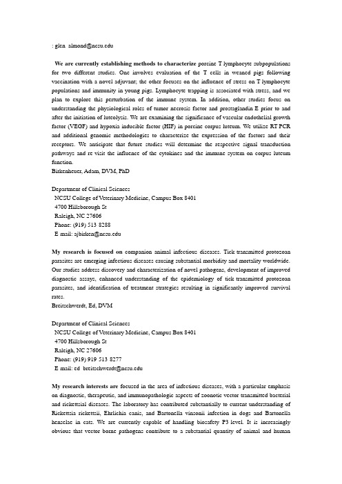
Fig. 4. Inflammatory pain behavior in littermate controls (white boxes/bars) and DTA mice (black boxes/bars). (A) FCA; thermal hyperalgesia was measured using the 足底测痛仪Hargreaves A strong difference was observed. (B) FCA; mechanical sensitivity was measured using von Frey filaments. A strong difference was observed.
J Neurobiol. 2004, 61:55–71.
Voltage-Gated Sodium Channels and Pain Pathways.
John N. Wood, James P. Boorman, Kenji Okuse, Mark D. Baker Molecular Nociception Group, Department of Biology, University College, Gower Street, London WC1E 6BT, UK
nav18通道亚型是一种主要表达于伤害感觉神经元上的海豚毒素不敏感型钠通道该通道电流占所在细胞动作电位去极化相电流的绝大部分
糖皮质激素对炎症的治疗实验报告
糖皮质激素对炎症的治疗实验报告Title: The Therapeutic Effect of Glucocorticoids on Inflammation: Experimental ReportIntroduction:Inflammation is a natural response of the body to tissue damage, infection, or injury. Although inflammation is a crucial defense mechanism, excessive or prolonged inflammation can lead to chronic diseases, such as rheumatoid arthritis, asthma, and inflammatory bowel disease. Glucocorticoids are a class of steroid hormones that regulate various physiological processes, including the immune response and inflammation. This experimental report aims to investigate the therapeutic effectof glucocorticoids in the treatment of inflammation.Materials and Methods:1. Animal models: Male Wistar rats were divided into three groups: control, inflammation, and glucocorticoid treatment.2. Induction of inflammation: Local inflammation was induced in the rats by injecting carrageenan solution into the paw.3. Glucocorticoid treatment: Rats in the glucocorticoid treatment group received daily injections of synthetic glucocorticoid, such as dexamethasone or prednisone, at a pre-determined dosage.4. Assessment of inflammation: The severity of inflammation was evaluated by measuring paw swelling, histological analysis of tissue samples, and quantification of inflammatory cytokines using ELISA.5. Statistical analysis: Data were analyzed using appropriate statistical tests, and p-values less than 0.05 were considered statistically significant.Results:2. Histological analysis: Tissue samples from the inflammation group displayed marked infiltration of inflammatory cells, edema, and tissue damage. In contrast, the glucocorticoid treatment group exhibited reduced infiltration of inflammatory cells and improved tissue architecture.3. Inflammatory cytokines: The inflammation group showed significantly elevated levels of pro-inflammatory cytokines, such as tumor necrosis factor-alpha (TNF-α) and interleukin-6 (IL-6). However, treatment with glucocorticoids led to a significant decrease in the levels of these cytokines,indicating their anti-inflammatory effect.Discussion:Glucocorticoids exert their anti-inflammatory effects through various mechanisms. They inhibit the production of pro-inflammatory cytokines, reduce the migration of inflammatorycells to the site of inflammation, and suppress the expressionof inflammatory mediators. The results of this experiment support the hypothesis that glucocorticoids have a therapeutic effect in the treatment of inflammation.Conclusion:Glucocorticoids demonstrate significant efficacy in reducing inflammation, as evidenced by the attenuation of paw swelling, histological improvement, and decreased levels of pro-inflammatory cytokines. These findings highlight the potentialof glucocorticoids as a treatment option for various inflammatory conditions. However, further studies are requiredto investigate the long-term effects, optimal dosage, and potential side effects of glucocorticoid therapy.References:2. Barnes PJ. Glucocorticoids. Chem Immunol Allergy.2024;100:311–326.。
疼痛动物模型系列
疼痛实验动物模型科研探索2007-04-25 23:11:36 阅读147 评论0 字号:大中小订阅疼痛是机制非常复杂的神经活动。
疼痛研究已经成为当前神经科学研究的重要课题之一。
由于疼痛机制的复杂性,使得在患者身上研究与疼痛有关的神经机制成为不可能的事。
因而,我们的研究需要相应的动物模型。
本章介绍了在现代神经科学研究中常用的疼痛动物模型。
在概要介绍了疼痛研究的意义及其现状之后,重点介绍了在生理痛研究和急性、慢性病理痛研究中所应用的动物模型。
生理痛的模型即常用的动物伤害性感受阈测定法;急性病理痛的模型则主要是各种急性炎症模型模型;慢性病理痛的模型则包括慢性炎症模型和慢性神经损伤模型。
前言疼痛(pain)是人们一生中经常遇到的不愉快的感觉。
它提供躯体受到威胁的警报信号,是生命不可缺少的一种特殊保护功能。
另一方面,它又是各种疾病最常见的症状,也是当今困扰人类健康最严重的问题之一。
近年来,仅在美国就有三至四千万人患有慢性痛。
据估计,美国每年用于治疗慢性痛的费用约为400~600亿美元;澳大利亚每年用于治疗疼痛的费用占全部医疗费用的40%。
随着医学的进步和人类生活水平的提高,烈性传染病逐渐得到控制,疼痛在人的身心痛苦和医疗费用消耗上的相对地位将越来越重要。
由于难以在人体对疼痛进行深入的机制研究,有必要建立疼痛的动物模型。
但疼痛是是包括性质、强度和程度各不相同的多种感觉的复合,并往往与自主神经系统、运动反应、心理和情绪反应交织在一起,它既不是简单地与躯体某一部分的变化有关,也不是由神经系统某个单一的传导束、神经核和神经递质进行传递的,所以很难将某种客观指标与疼痛直接联系起来。
因而,我们只能根据模型动物对伤害性刺激的保护反应和保护性行为来推测它们的疼痛程度。
伤害性感受(nociception)和痛觉是两个有密切关系但又不相同的概念。
前者是指中枢神经系统对由于伤害性感受器的激活而引起的传入信息的加工和反应,以提供组织损伤的信息;痛觉则是指上升到感觉水平的疼痛感觉。
蛋白激酶CK2的抗炎及抗病毒作用研究进展
蛋白激酶CK2的抗炎及抗病毒作用研究进展尤信信(综述);刘新光(审校)【摘要】蛋白激酶CK2具有组成型活化功能。
目前已知CK2作用底物超过300种,涉及细胞生长、增殖、分化、凋亡及信号转导等。
此外,CK2通过抑制细胞非正常凋亡而在细胞生命活动中扮演重要角色,确保细胞在炎症或病毒入侵环境下存活。
该文综述了CK2的抗炎和抗病毒作用。
【期刊名称】《广东医科大学学报》【年(卷),期】2019(037)001【总页数】3页(P1-3)【关键词】蛋白激酶CK2;炎症;病毒【作者】尤信信(综述);刘新光(审校)【作者单位】[1]广东医科大学衰老研究所,广东东莞523808;[1]广东医科大学衰老研究所,广东东莞523808;【正文语种】中文【中图分类】R774酪蛋白激酶Ⅱ (CK2)参与了许多细胞的变化过程,包括增殖、细胞存活、分化、转化、肿瘤的发生以及 tR NA和rR NA的合成等,部分机制与 Wnt和NF-kB信号途径的相互作用有关 [1-3]。
此外,研究发现, CK2具有组成型活性,这一活性维持了细胞机体的正常稳态,当细胞微环境发生改变后,CK2的表达或活性也随之发生改变 [4]。
近年一些研究亦发现, CK2在抗炎和抗病毒过程中起着非常重要的作用[5-6],文章对此进展作一综述。
1 CK2的基本特征CK2是一个极保守的、多效性的丝/苏氨酸蛋白激酶,表达在所有真核生物的器官和组织细胞中,是由两个催化亚基(α和α' ) 和两个调节亚基β形成的四聚体。
人的CK2α和CK2α'催化亚基在结构上具有同源性但由不同基因所编码,在 N端的3 30个氨基酸具有 88%的同源性。
人类细胞中,有第三种相似形式的亚基CK2α " 存在,它与CK2α几乎一样,但是其最后的 32个氨基酸是不同的。
通过比较催化亚基的相似性,调节亚基CK2β并不像其他已知蛋白那样具有广泛的相似性。
尽管CK2α和CK2α'并无显著的差异,但是有确凿的证据表明它们具有功能特异性,基因敲除α'催化亚基会让α失去作用,导致圆头精子症的发生,进而引发雄性不育 [7]。
- 1、下载文档前请自行甄别文档内容的完整性,平台不提供额外的编辑、内容补充、找答案等附加服务。
- 2、"仅部分预览"的文档,不可在线预览部分如存在完整性等问题,可反馈申请退款(可完整预览的文档不适用该条件!)。
- 3、如文档侵犯您的权益,请联系客服反馈,我们会尽快为您处理(人工客服工作时间:9:00-18:30)。
Chao Ma and Jun-Ming Zhang (eds.), Animal Models of Pain, Neuromethods, vol. 49, DOI 10.1007/978-1-60761-880-5_2, © Springer Science+Business Media, LLC 2011Chapter 2Animal Models of Inflammatory PainRui-Xin Zhang and Ke RenAbstractAnimal models of inflammatory pain have been widely used to study the mechanisms of tissue injury-induced persistent pain. A variety of inflammatory agents or irritants, including complete Freund’s adjuvant, carrageenan, zymosan, mustard oil, formalin, capsaicin, bee venom, acidic saline, lipopolysac-charide, inflammatory cytokines, and sodium urate crystals, have been used to produce tissue injury and hyperalgesia in such structures as cutaneous/subcutaneous tissues, joints, and muscles. Additionally, models of pain hypersensitivity have also been established with injuries produced by burning, freezing, and ultra irradiation. Although these models do not simulate every aspect of chronic pain, they do model key features of human inflammatory pain. Studies in animals give insight into certain aspects of human pain conditions and lead to improved pain management for patients.Pain perception is more complex in humans than in animals sincehuman pain perception encompasses psychosocial, cultural, devel-opmental, and environmental variables. However, human andanimal pain perceptions show parallels, and animal models par-tially mimic the persistent pain encountered in the clinic. In thelast two decades animal models of inflammatory pain have beenwidely used to study the mechanisms of tissue injury-induced per-sistent pain. Although none of the existing models can simulateall symptoms of inflammatory pain, studies in animals give insightinto certain aspects of human pain conditions and lead to betterpain management for patients. In the following paragraphs, com-monly used inflammatory pain animal models will be summa-rized. Interested readers may consult more comprehensive reviewsfor further details (1, 2).1. I ntroduction2324Zhang and RenAnimal models of tissue injury and inflammatory hyperalgesia can be induced by a number of inflammatory agents in a variety of structures, including cutaneous and subcutaneous, joint, and muscle tissues.A Mycobacterium butyricum oil suspension was initially used to inoculate the tail base of the rat to induce adjuvant arthritis and persistent pain (3). Since polyarthritis develops after the inocula-tion along with a state of generalized illness, most pain researchers have discontinued the use of this model. However, the injection of complete Freund’s adjuvant (CFA, composed of inactivated and dried Mycobacterium and adjuvant) into the footpad produces localized inflammation and persistent pain (4, 5). After a CFA injection into the footpad, cutaneous inflammation appears in minutes to hours and peaks within 5–8 h.CFA produces dose-dependent inflammatory responses, and 30–200 m g of Mycobacterium butyricum suspended in oil/saline (1:1) yield significant edema and thermal hyperalgesia in the injected hind paw (6) (Fig. 1). The edema peaks around 24 h after the injection. The hyperalgesia and allodynia peak around 5 h after injection and persist for approximately 1–2 weeks (7).CFA-induced hyperalgesia and allodynia in rats are consistent with those seen in humans receiving inadvertent injections of 2. Inflammatory Models of Persistent Pain 2.1. Cutaneous and Subcutaneous Models of Inflammatory Pain 2.1.1. CFA ModelFig. 1. Inflammation and hyperalgesia produced by intraplantar injection of complete Freund’s adjuvant in rats. (a ) Edema of the rat hind paw after injection of different doses of CFA, determined by measuring the dorsal-ventral thickness of the injected hindpaw. *P < 0.05 compared to 200 m g injected rats; **P < 0.01 compared to 20 m g injected rats; ***P < 0.001 compared to 20 m g injected rats. (b ) Changes in hind paw withdrawal latency to a noxious thermal stimulus at different time points (2 h to 3 days) after injection of different doses of CFA into the hindpaw. *P < 0.05 compared to 200 m g injected rats; **P < 0.01 compared to 20 m g injected rats; ***P < 0.001 compared to 20 m g injected rats [reproduced withpermission from Chinese Journal of Neuroanatomy (1999, 15:19–26), Chinese Society of Anatomical Science].25Animal Models of Inflammatory Pain CFA (8). The physiological and biochemical effects of CFA are limited to the affected limb (5) and there are no signs of immune response or systemic disease. It has been shown that rats with CFA-induced inflammation exhibit minimal reductions in weight and show normal grooming behavior (5). Exploratory motor behavior is normal, and no significant alterations occur in an open field locomotion test (5).Studies with three strains of rats, Lewis (LEW), Fischer 344 (FIS) and Sprague–Dawley (SD), demonstrate that, according to the difference scores computed by subtracting paw withdrawal latency (PWL) of the contralateral paw from that of the injected paw, F344 rats show significantly greater thermal hyperalgesia than do SD and LEW rats, both of which exhibit similar but rela-tively less intense hyperalgesia (9).An intraplantar injection of carrageenan is also widely used to produce a model of localized inflammatory pain. When 0.5 mg of carrageenan is injected, edema develops, mainly in two phases: the first 30 min after the injection, the second beginning at the end of the first hour and lasting until the third hour after injec-tion. The edema peaks 3–5 h after injection (10, 11). When 6 mg of carrageenan is injected, edema peaks on day 3 (5) and thermal hyperalgesia peaks around 4 h after injection and lasts for at least 96 h (5). Studies with FIS, LEW, and SD rats demonstrate that LEW rats showed the least, and FIS rats the greatest, thermal hyperalgesia after intraplantar administration of 3.5 mg of carra-geenan (12).CFA and carrageenan are also injected into the facial area to study orofacial pain (13–15). A CFA injection into the perioral (PO) skin results in orofacial thermal hyperalgesia and mechanical allodynia that peak between 4 and 24 h and persist for at least 2 weeks (15). Facial carrageenan injection in mice causes increased responses to facial stimulation with a von Frey hair (1 g force) 8 h, 1 day, and 3 days after injection (13).Other inflammatory agents such as mustard oil, a small fiberirritant, and zymosan, a glucan from the cell walls of yeast, have been used to produce behavioral hyperalgesia. Mustard oil elic-its inflammatory pain by activating transient receptor potential cation channel, subfamily A, member 1 (TRPA1), an excitatory ion channel of primary afferent nociceptors (16). Topical appli-cation of mustard oil to the ear induces dose-dependent increases in plasma extravasation and ear thickness, which peak approxi-mately 30 min after application (17). Application of mustard oilto rat paw skin induces plasma protein extravasation and slight edema, a 7–8% increase in paw volume (18). Topical application of mustard oil (20 m l, 100%) to the lateral surface of the lefthind leg induces immediate agitation with frequent biting and 2.1.2. C arrageenan Model2.1.3. Mustard Oiland Zymosan Models26Zhang and Renvocalizations. This response lasts approximately 5–7 min.Mustard oil also significantly facilitates a tail-flick reflex thatappears 5 min after application and lasts up to 60 min, peaking20 min after application (19).Intraplantar injection of zymosan (0.31–6.25 mg) producespersistent dose- and time-dependent mechanical and thermal hype-ralgesia. Edema is greatest at dosages ≥2.5 mg and peaks 30-minpostinjection irrespective of dosage. Mechanical hyperalgesia appearsat dosages ≥1.25 mg and reaches its maximum 4 h after applicationat a dosage of 5 mg. Thermal hyperalgesia is biphasic and dose-dependent. An early-phase peak occurs at 30 min at dosages≥2.5 mg; this is not apparent at lower dosages. A late-phase peakoccurs at 4 h at dosages of ≥0.0625 mg. Higher dosages (5 and6.25 mg) also cause spontaneous pain, sometimes characterized byoccasional flicking of the hind paw but more commonly by eleva-tion of the paw for extended periods of time (20).The formalin test is a popular model for studying pain mecha-2.1.4. F ormalin Modelnisms under prolonged nociception. Formalin is injected beneaththe footpad of a rat, mouse, or cat and produces two phases ofnocifensive behavior, characterized by licking and flinching of thepaw, that are separated by a short period of quiescence (21, 22).The first or acute phase occurs typically in the first 5 min; thesecond starts from 15 min and lasts about 40–60 min after injec-tion. It is generally agreed that the first phase is due to the directactivation of both low-threshold mechanoreceptive and nocicep-tive primary afferent fibers (23). There has been disagreementabout the underlying mechanisms of the second phase. Earlystudies suggested that the second phase resulted from an increasein the excitability of dorsal horn neurons. More recently, it hasbeen demonstrated that ongoing activity of primary afferent fibersis necessary for the development of the second phase (23–26). Inregard to the period of quiescence, some evidence supports theidea of an absence of activity, other evidence implicates an activeinhibitory mechanism (27).Formalin-induced pain is measured by combining scores offavoring, lifting, licking, and flinching/shaking of the injuredpaw. Power analysis indicates that using a moderate dosage (1.5%,0.05 ml) of formalin and a combined pain score gives the greatestpower to detect pain differences (22). Further, combining theformalin model with the place-conditional paradigm demon-strates that, when compared with a distinct environmental con-text, a hind paw injection of formalin induces conditioned placeavoidance, which reflects a negative affective state (28).To study orofacial pain, formalin is subcutaneously injected intothe rat upper lip or lateral face and generates similar biphasic behav-ioral responses (face rubbing), an early and short-lasting first phasefollowed, after a quiescent period, by a second prolonged (tonic)27Animal Models of Inflammatory Pain phase. The orofacial formalin test can be used to produce and quantify nociception in the trigeminal region of the rat (29–31). Study with different formalin concentrations indicates that the second phase response to formalin only occurs with dosages of 1.5% and higher (31).A subcutaneous injection of bee venom (0.2-mg lyophilized whole venom in 0.1-ml saline) into the hind paw produces persis-tent nociceptive responses (flinching and lifting/licking the injected paw) for 1–2 h, followed by a 72–96 h period of mechan-ical allodynia and thermal hyperalgesia accompanied by edema and redness of the injected paw. It also produces thermal hyperal-gesia, but not mechanical allodynia, on the contralateral hind paw although with less amplitude than that of the injected paw (32).Capsaicin, the pungent component of cayenne pepper that acti-vates transient receptor potential vanilloid type 1 (TRPV1), aheat-sensitive cation channel on nociceptor terminals, has been used in humans and animals to study neurogenic inflammation and hyperalgesia.Intradermal injection of capsaicin results inflare reaction, allodynia, and hyperalgesia, the areas of which extend beyond the injection site. Visual observation of flareresponse reveals that the area of visual flare is significantly smaller than the area of hyperalgesia to stroking stimuli and that the lat-ter issignificantly smaller than that for punctate stimuli. The heat hyperalgesia (thermode maintained at 38°C) area is thesmallest (33, 34). Thermographic detection of the flare response shows that the thermographic area is larger than the area ofvisual flare and coincides with the area of mechanical (nylon monofilament, 1.02-mm diameter exerting a bending force of 2.02 N) and heat hyperalgesia (from a 1-cm 2Peltier thermode maintained at 47°C) (35). Regarding the temporal pattern offlare, visual flarereaches its maximum within 3–5 min (33).Laser-Doppler flowmetry also shows that blood flow reaches maximum 5 min after the injection and then decreases (34).A thermographic device shows that the flare response starts as early as a few seconds after the capsaicin injection (35). The areaof hyperalgesia to stroking stimuli appears immediately afterinjection, peaks within 15 min and then gradually decreases over 1–6 h. The area of hyperalgesia to punctate stimulation is imme-diately present after injection, grows to a maximum within15–30 min, decreases gradually, and disappears at about 21 h. The area of the heat hyperalgesia reaches maximum 30 min after injection, gradually decreases, and disappears about 1.5 h after injection (33). Capsaicin (0.1, 1, 10, and 100 m g) producesdose-dependent increases in spontaneous pain, area and inten-sity of mechanical allodynia, area and intensity of pinprick hype-ralgesia, and flare area (36).2.1.5. B ee Venom Model2.1.6. C apsaicin Model28Zhang and RenIt should be noted that capsaicin may produce differential responses in different areas. For instance, peak pain intensity and duration are greater in the forehead than in the forearm, while areas of visible flare and pinprick hyperalgesia are significantly larger in the forearm than in the forehead (37).This neurogenic model of inflammation has been used in monkeys to study changes in nociceptor activity and changes in the responses of spinal dorsal horn neurons (33, 38). It has recently been adapted for behavioral studies in rats (39). Intraplantar injec-tion of capsaicin evokes nocifensive behavior characterized by lift-ing and guarding of the injected paw that lasts for about 3 min. Capsaicin dose-dependently produces thermal and mechanical hyperalgesia. Thermal hyperalgesia to heat lasts up to 45 min, whereas mechanical hyperalgesia persists up to 4 h.To study trigeminal pain, subcutaneous injection of different dosages of capsaicin into the vibrissa pad produces an immediate rubbing–scratching of the injected area. This behavior is per-formed with the ipsilateral forepaw, often accompanied by the contralateral forepaw. The rubbing–scratching response reaches its maximum during the 12- to 18-min interval and subsides about 42 min after capsaicin injection. Morphine dose-dependently reduces the capsaicin-induced rubbing–scratching (40, 41).Further, capsaicin-sensitive primary afferents play different roles in chemical irritant-induced spontaneous nociception, hype-ralgesia, and inflammatory responses (42). Local pretreatment with capsaicin significantly inhibits the two phases of formalin-induced persistent spontaneous nociception, while it only inhibits the late phase (tonic nociception; 11–60 min), not the early phase (acute nociception; 0–10 min), of spontaneous nociception in the bee venom test. Although capsaicin pretreatment prevents ther-mal hyperalgesia in the bee venom, carrageenan, and CFA models, it only prevents mechanical allodynia in the bee venom and car-rageenan models, not the CFA model. Regarding inflammatory response, it significantly inhibits bee venom-elicited paw edema but not carrageenan-, CFA-, or formalin-elicited paw edema.Rank order of the duration of the inflammatory hyperalgesia produced by these agents, is, from longest to shortest, CFA > bee venom > carrageenan > zymosan > formalin > mustard oil. See Table 1 for a comparison of onsets and durations.Adjuvant arthritis is induced in rats for a laboratory animal model of chronic pain that mimics that of human rheumatic disease. A CFA injection into the base of the rat’s tail causes polyarthritis (43). Hypersensitivity of multiple joints occurs after 10 daysand lasts up to 3 weeks. The paws and tails of arthritic rats show lower thresholds in response to noxious pressure (hyperalgesia), higher thresholds in response to noxious heat (hypoalgesia), and no change in response to noxious electrical stimulation (44).2.2. Models of JointInflammationand Hyperalgesia2.2.1. CFA-Induced JointInflammation29Animal Models of Inflammatory Pain Pain is also inferred from scratching behaviors, reduced motor activity, weight loss, vocalization when the affected limbs are pinched, and a reduction in these behaviors following the admin-istration of opioids. It should be noted that this is a systemic disease that includes skin lesions, destruction of bone and cartilage, impairment of liver function, and lymphadenopathy, which leads to ethical concerns (45). Moreover, the systemic lesions make it difficult to differentiate pain behavior from generalized malaise and debilitation. The likely presence of central nervous system changes associated with the alterations in immune function also call into question the use of this model as a correlation of pain behavior to neural activity and neurochemical alterations. Polyarthritis is also induced by type II collagen emulsified in Freund’s incom-plete adjuvant and injected into the base of the rat’s tail. Tail-flick latency significantly decreases 1 week after inoculation, peaks at 3 weeks, and lasts for at least 5 weeks (46). Like CFA-induced polyarthritis, collagen-induced polyarthritis raises ethical concerns.An alternative to the polyarthritic rat for prolonged studies, intra-knee joint injection of 250-mg suspension of heat-killed Mycobacterium butyricum in peanut oil and saline (1:1), causes ipsilateral thermal hyperalgesia of the hind paw 1 day after injec-tion, peaks on day 3, and remains at that level until day 14. By the 21st day, experimental animals recover from the hyperalgesia (47). Knee joint withdrawal threshold measurement, in which a gradually increasing squeeze is applied across the joint, shows that the average ipsilateral limb withdrawal threshold (LWT) sig-nificantly decreases by 57%, from 790 ± 39 to 316 ± 45 g, 1 dayTable 1 Comparison of cutaneous/subcutaneous inflammatory pain modelsaChemicalHyperalgesia Allodynia Time of onset Duration a Modified and reprinted with permission from the ILAR Journal, 40(3),1999, Institute for Laboratory Animal Research, The National Academics, 500 Fifth Street NW, Washington, DC 20001 (http:// w /Ilar )b Not applicable30Zhang and Renafter the CFA injection. This decrease lasts for a full 28 days, the period studied. An incapacity test shows that the ratio of weight distribution between ipsilateral and contralateral limbs signifi-cantly decreases from 0.96 ± 0.03 to 0.29 ± 0.05. This significant decrease lasts up to day 28 (48).Injection (0.05 ml) of 300 m g Mycobacterium butyricum into the tibio-tarsal joint also produces monoarthritis. As revealed by clinical observations and X-ray examinations, the arthritis is lim-ited anatomically, pronounced, prolonged, and stable from weeks 2 through 6 postinjection. The affected limb shows a marked increase in sensitivity to paw pressure. Animals gain weight, remain active, and evince little systemic disturbance in contrast to polyarthritic rats (49).CFA also produces significant thermal hyperalgesia and mechanical allodynia following its injection into the temporo-mandibular joint. Thermal hyperalgesia develops at 5 h, peaks at 24 h, and lasts 2 weeks. Mechanical allodynia starts at 2 h, peaks between 4 and 24 h and persists for at least 2 weeks after the injection (15).Another arthritis animal model is induced by injecting kaolin and carrageenan (3 mg/3 mg) into the knee joint. In rats, decrease in PWL occurs ipsilaterally to the inflamed knee as early as 4 h after an injection of the two agents and lasts about 24 h. The circum-ference ofthe ipsilateral knee joint is significantly larger than baseline between 4 and 24 h. The rats also show spontaneous pain, indicated by decrease of weight bearing by the injected limb (50, 51). Recently, the elevated plus maze test has demonstrated that arthritic rats show amygdala-involved anxiety-like behavior, evidenced by a decreased preference for the open arms (52). Intra-articular injection of carrageenan and kaolin in cats causes guarding of the leg and avoidance of movement or weight bear-ing. These symptoms begin ~2 h after the injection, are fully developed after 4 h, and last at least 15 h.Recording of saphenous nerve filament activity consistently demonstrates that almost all filament units of inflamed joints have low thresholds to passive movement of the knee joint compared to units of normal joints. The number of receptive fields per unit is significantly greater than that seen in normal joints (53). In rats, cats, and monkeys, somatosensory neurons of the spinal cord become hyperexcitable to mechanical stimuli during the develop-ment of experimental arthritis (54). Changes in joint receptors and spinal dorsal horn neuronal activity begin as soon as 1–2 h follow-ing injection and build for several hours. Noticeably, the magni-tude of hyperalgesia in this model is relatively low compared to that of the hind paw inflammation model. It should be kept in mind that the test site, the paw, in the joint inflammation model is remote from the injury site. What is measured by PWL in the knee joint inflammation model is, likely, only secondary hyperalgesia.2.2.2. Kaolin andCarrageenan-Induced JointInflammation31Animal Models of Inflammatory Pain Compared to the short-lasting effects of the kaolin and carrageenan combination, carrageenan alone produces a long-lasting effect. An intra-articular injection of 3 mg of carrageenan significantly decreases ipsilateral and contralateral PWL to heat; the decrease occurs at 4 h and lasts 6 weeks. Carrageenan at 0.3 and 1 mg produces only ipsilateral effects that are shorter-lasting: 24 h for 0.3 mg and up to 3 weeks for 1 mg. Intra-articular injection of 3 mg of carrageenan also produces significant decrease of the mechanical withdrawal threshold ipsilaterally (between 3 and 7 weeks) and contralaterally (between 3 and 6 weeks). Carrageenan at 1 mg induces a significant ipsilateral decrease between 4 h and 1 week, but 0.3 mg has no effect (55).Formalin (0.5, 3, and 5%) injected into the knee joint of rats induces dose-dependent nocifensive responses. The nociception consists of two phases (from 0 to 5 min and from 10 to 60 min) of intense guarding behavior on the affected limb with an inter-vening period of quiescence (from 5 to 10 min). Morphine (4 mg/kg, subcutaneously) pretreatment reduces the guarding behavior in both nocifensive phases (56). A formalin (0.5, 2.5, and 5%) injection into the temporomandibular joint (TMJ) region induces a dose-dependent phase of orofacial rubbing and one of flinching, alternately displayed. The rubbing responses start earlier, peak 18 min postinjection, and then decrease; the flinching responses start later, peak 27 min postinjection, and last up to 36 min. A sig-nificant correlation exists between formalin concentration and rubbing and flinching responses. The magnitude of these responses reaches its maximum at a concentration of 2.5% (57).Other models of arthritis have been developed using sodium urate crystals, which are injected into the ankle joint of a rat or cat (45, 58).The arthritis is fully developed within 24 h. These animals tend to place less weight on the treated hind limb and exhibit guarding movements of the limb. In the rat, touch, pressure and thermal stimuli applied to the affected paw result in a decrease in respon-siveness, presumably due to pain associated with the movement. There are no signs of systemic disease in the urate arthritis model other than the joint pathology secondary to tissue edema and the infiltration of polymorphonuclear leukocytes (45).Capsaicin (0.2%, 50ml) injected into the lateral aspect of the left anklejoint results in a decreased mechanical withdrawalthreshold 2 h after injection; this is maintained through a 4-h period (59). Capsaicin-sensitive primary afferents play different roles in a variety of joint inflammation models, as they do in cuta-neous/subcutaneous pain models. Pretreating the joint with 1% capsaicin (about 1 week before injection) significantly reduces the inflammatory response to carrageenan and urate but not to for-malin (60). Onsets and pain durations produced by joint inflam-mation are compared in Table 2.2.2.3. Carrageenan-Induced JointInflammation2.2.4. Formalin-InducedJoint Inflammation2.2.5. Joint InflammationInduced by Other Irritants32Zhang and RenThe bulk of available knowledge about pain mechanisms is derived from studies on cutaneous pain. However, the existing subjective differences between muscle and skin pain (e.g., muscle pain is poorly localized and shows referral) suggest that muscle pain has distinct characteristics. Models have been developed to examine mechanisms underlying the development and maintenance of chronic muscle pain.A decrease in tissue pH has been observed in response to inflammation, hematomas, and isometric exercise. Decreasing pH increases activity of nociceptors and produces a painful response in humans (61). Using an in vitro nerve-skin preparation, con-tinuous infusion of low pH (5.2–6.9) saline increases discharges of C-polymodal primary afferent fibers without adaptation (62). Repeated injection of low pH saline into the gastrocnemius mus-cle of rats produces long-lasting, widespread mechanical hyperal-gesia without motor deficits or significant tissue damage.Following the first unilateral intra-gastrocnemius muscle injec-tion of pH 4.0, 5.0, or 6.0 saline, the mechanical withdrawal threshold of the ipsilateral paw dose-dependently decreases 4 h, and returns to baseline 24 h, after injection. After a second unilat-eral injection of low pH saline on day 5, the mechanical with-drawal threshold dose-dependently decreases in both ipsilateral and contralateral hind paws. These bilateral decreases are greatest for pH 4.0 saline, persisting for 4 weeks after the second injection. Inter-injection intervals of 2 and 5 days (pH 4.0 saline) produce equivalent and significant bilateral decreases in mechanical with-drawal threshold. A 10-day inter-injection interval does not pro-duce persistent mechanical hyperalgesia. This suggests that there is a critical window in which re-injury to muscle tissue results in exaggerated, more persistent hyperalgesia. Intra-gastrocnemius muscle lidocaine 24 h after the second injection of pH 4.0 saline increases the ipsilateral mechanical withdrawal threshold during the first 10–15 min after injection. However, lidocaine has no significant effect on the decreased contralateral withdrawal threshold. 2.3. Models of MuscleInflammation andHyperalgesia2.3.1. Acidic Saline-Induced MuscleInflammationand Hyperalgesia Table 2Comparison of joint inflammatory pain modelsChemicalHyperalgesia Allodynia Time of onset Duration a Not applicableWithdrawal latencies to radiant heat average approximately 10 s at baseline and are no different than controls after either the first or second injection of low pH saline. After both the first and the second injection of low pH saline, rats show no limb guarding, have equal weight bearing and normal gait patterns, and their ability to per-form the treadmill test is unchanged (63). Interestingly, in sharp contrast to most persistent pain models intramuscular acidic saline-induced allodynia does not involve spinal glial activation or inflam-matory cytokine interleukin-1 (64).Intramuscular application of carrageenan sensitizes group III and IV muscle afferents, including nociceptors, lowering their thresh-olds to mechanical activation and increasing their background activity. Furthermore, carrageenan induces local inflammation when injected into the muscle, as evidenced by the accumulation of leukocytes that begins 2 h postinjection and continues for the next 8 h (65).Injection of carrageenan (0.5–6 mg/triceps) into the bilateral triceps muscles produces dose-dependent reduction in forelimb grip force that peaks 24 h, and returns to the control level 48 h, postinjection. Capsaicin (50 mg/kg i.p.) admi nistration to rats on the second day of life reduces carrageenan-evoked hyperalgesia by about 45%, indicating that the muscle hyperalgesia induced by carrageenan is mediated, in part, by capsaicin-sensitive afferent fibers (66).Unilateral intra-gastrocnemius muscle injection of carrageenan also dose-dependently produces thermal and mechanical hyperal-gesia. Carrageenan at 3 mg significantly decreases ipsilateral PWL to heat that occurs within 4 h and lasts 8 weeks. Contralateral PWL also decreases by the end of the first week and last 8 weeks. Carrageenan at 0.3 and 1 mg produces no significant effect on PWL to noxious thermal stimulus (55). However, a 3-mg intra-muscle injection of carrageenan significantly decreases the ipsilat-eral mechanical withdrawal threshold between 4 h and 6 weeks and the contralateral threshold between weeks 3 and 6. The ipsi-lateral mechanical withdrawal threshold significantly decreases between 4 and 8 h after a 1-mg intramuscle injection of carra-geenan. Carrageenan at a dosage of 0.3 mg produces no changes in mechanical sensitivity (55). It seems that mechanical sensation is more sensitive to carrageenan than is thermal sensation.An intra-gastrocnemius injection of tumor necrosis factor-alpha (TNF) significantly decreases mechanical withdrawal thresholds to muscle pressure in rats when measured with an algesimeter that exerts pressure on the gastrocnemius muscle. It also decreases forelimb grip strength as measured with a digital grip force meter. The hyperalgesia lasts at least 60 min (67). Similarly, an intra-gastrocnemius injection of another pro-inflammatory cytokine2.3.2. Carrageenan-Induced Muscle Inflammation and Hyperalgesia2.3.3. Cytokine-Induced Muscle Inflammation and Hyperalgesia。