小鼠内脏脂肪细胞使用说明
碧云天生物技术 Min6 (小鼠胰岛β细胞) 产品说明书

碧云天生物技术/Beyotime Biotechnology订货热线:400-168-3301或800-8283301订货e-mail:******************技术咨询:*****************网址:碧云天网站微信公众号Min6 (小鼠胰岛β细胞)产品编号产品名称包装C7406 Min6 (小鼠胰岛β细胞) 1支/瓶产品简介:Organism Tissue Morphology Culture Properties Mus musculus (Mouse) Pancreas Epithelial Adherent本细胞株详细信息如下:General InformationCell Line Name Min6 (Mouse Islet Β Cells)Synonyms Min6; MIN-6; Mouse INsulinoma 6Organism Mus musculus (Mouse)Tissue PancreasCell Type -Morphology EpithelialDisease Mouse insulinomaStrain -Biosafety Level* -Age at Sampling 13 weeksGender -Genetics -Ethnicity -Applications -Category Transformed cell line* Biosafety classification is based on U.S. Public Health Service Guidelines, it is the responsibility of the customer to ensure that their facilities comply with biosafety regulations for their own country.CharacteristicsKaryotype -Virus Susceptibility -Derivation -Clinical Data -Antigen Expression -Receptor Expression -Oncogene -Genes Expressed -Gene expressiondatabases -Metastasis -Tumorigenic -Effects -Comments -Culture MethodDoubling Time -Methods for Passages Wash by PBS once then 0.05% trypsin-EDTA solution and incubate at room temperature, observe cells under an inverted microscope until cell layer is dispersed (usually 1 minute)Medium DMEM (high glucose) 10% FBS+0.05mM 2-Mercaptoethanol MCH2 / 5 C7406 Min6 (小鼠胰岛β细胞)400-1683301/800-8283301 碧云天/BeyotimeSpecial Remarks -Medium Renewal -Subcultivation Ratio 1:5 to 1:15 Growth Condition 95% air+ 5% CO 2, 37ºC Freeze medium DMEM (high glucose)+20% FBS+10% DMSO ,也可以订购碧云天的细胞冻存液(C0210)。
OriCell TMC57BL 6 老鼠脂肪膜源性骨髓衍生性多泛性胞质胞株说明书

User ManualOriCell TM C57BL/6Mouse Adipose-derived Mesenchymal Stem CellsCatalog No.MUBMD-01001IntroductionAdipose-derived mesenchymal stem cells are a type of pluripotent stem cells that exist in adipose matrix.Because of its strong value-added ability and immune regulation function,it is widely used in the fields of tissue engineering,cell therapy and gene therapy.As a research hotspot,C57BL/6mouse adipose-derived mesenchymal stem cells are widely used in regenerative medicine and tissue engineering,especially in the fields of bone, cardiovascular and nervous system diseases.OriCell TM C57BL/6mouse adipose-derived mesenchymal stem cells are taken from the inguinal fat of C57BL/6mouse.These cells can express specific proteins of ADSCs and have strong proliferation and multidirectional differentiation capabilities.It can be used as a cell model to study proliferation,aging,immunity,differentiation and transplantation.Note:This product is only provided for further scientific research.It is not intended for diagnostic,therapeutic,clinical,household,or any other applications.Product InformationName OriCell TM C57BL/6Mouse Adipose-derived Mesenchymal Stem Cells Catalog Number MUBMD-01001Amount of Cells1×106cells/vialPassage Number P2Storage at Liquid Nitrogen(-196℃)The Shape of OriCell TM C57BL/6Mouse Bone Marrow Mesenchymal Stem CellsQC●Pass the detection of bacteria,fungi,mycoplasma,and endotoxins.●Pass the viability examination.The viable rates is higher than80%.●The cell doubling time is less than72hours.●Flow cytometry showed that,CD29、CD44、Sca-1are positive(>70%),CD117、CD31is negative(<5%).●The cells can be induced to differentiate into osteoblasts,adipocytes,chondrocytes,etc.Please reference"COA"for details.General Handing Principles1.Ensure that all equipments are kept clean and tidy.2.Please Follow the instructions.e suitable and reliable consumables and reagents.4.Adipose-derived mesenchymal stem cells have limited ability to proliferate in vitroand cannot maintain their differentiation potential for a long time.OriCell TMC57BL/6Mouse Adipose-derived Mesenchymal Stem Cells can be passaged formore than5times and still maintain all indicators qualified.But we alwaysrecommend using lower generation cells for scientific research.ually the inoculation density of mouse adipose-derived mesenchymal stem cellsis(2.5~4)×104live cells/cm2.Note:The cryopreservation solution of this product contains DMSO,which has potential risks.Please handle it carefully.Thawing and Establishing of CellsMaterials Required●OriCell TM C57BL/6Mouse Adipose-derived Mesenchymal Stem Cells●OriCell TM C57BL/6Mouse Adipose-derived Mesenchymal Stem Cells Complete Medium(MUXMD-90011)StepsNote:If the received cells are thawed within24hours,they can be stored in a refrigerator at-80°C.If more than24hours,please store them in liquid nitrogen. Please take them out10minutes early before thawing and place them at-80°C to allow the liquid nitrogen in the tube to evaporate.1.Preheat the water bath at37°C.2.Warm the complete medium to37°C.3.Add more than5mL of complete medium to a15mL centrifuge tube for use.4.Take the cells out of the-80°C refrigerator,put them in a37°C water bath andshake them quickly to thaw the cryopreservation solutionNote:During the thawing process,the cryotube must be shaken to ensurethat solution melts quickly and evenly.5.When shaking,please avoid water immersing the pipe cover to cause pollution.6.When the cryopreservation solution has thawed into ice crystal with a diameter ofabout2mm,stop the water bath.Continue to shake the cryotube until the icecrystal melts thoroughly.7.Wipe the outer surface of the cryotube with75%medical alcohol.8.Open the cryopreservation tube in the ultraclean bench,use a Pasteur pipette tosuck the cell suspension,and transfer it to the prepared centrifuge tube.9.Wash the cryotube once with1mL of complete medium to collect residual cells toreduce loss.10.Centrifuge the cell suspension at250×g for4minutes.11.Remove the supernatant after centrifugation.Add2mL of complete medium,gently pipette the cell pellet,blow and mix thoroughly.12.Inoculate the cells into a T25flask or a culture container with an equivalent bottomarea.Add enough complete medium,the total amount of medium in a T25flaskshould not less than5mL.13.Shake the cells well and incubate them in a CO2incubator at saturated humidity,37°C,5%CO2inside.Note:Do not move or observe the cells within2hours of inoculation.Thiswill seriously affect cell adhesion,resulting in poor shape,cell clumping,and uneven adhesion.14.On the next day of recovery,observe the cell status,and replace medium withfresh complete medium or passage.Note:If you find lots of floating cells or other abnormal conditions,pleaseinvestigate the cause in time and contact us.15.Then refresh the complete medium every2days until the cells have grown to90%confluence,which requires passage generarion.Passaging of CellsMaterials Required●OriCell TM0.25%Trypsin-0.04%EDTA(TEDTA-10001)●OriCell TM Phosphate-Buffered Saline(1×PBS)(PBS-10001)●OriCell TM C57BL/6Mouse Adipose-derived Mesenchymal Stem Cells Complete Medium(MUXMD-90011)Steps1.Preheat complete medium and Trypsin to37°C.2.Move the medium in the culture container.3.Wash the cells twice with PBS(approximately3mL for T25flask and6mL for T75flask).Please perform relatively slightly and wash thoroughly.Move the PBS.4.Add Trypsin(approximately1.5mL for T25flask and3mL for T75flask),spreadquickly to ensure full contact with the cells.5.Observe the cells under a microscope.After about70%~80%of the cells haveshrunk and round,tap the outer wall of the culture vessel to remove the cells fromthe culture surface.6.Add complete medium(approximately3mL for T25flask and6mL for T75flask)immediately,and then slightly shake the culture container to mix the medium and Trypsin quickly to stop the digestion.e a pipette to suck up the cell suspension,pipetting the bottom surface of theculture container several times,and pipetting down as much as possible of the cells.Note:The pipetting action should not be violent.8.Transfer the cell suspension to a centrifuge tube.Wash the container once withPBS(approximately3mL for T25flask and6mL for T75flask)to collect residual cells.9.All the collected cell suspensions are centrifuged at250×g for4minutes.10.Remove the supernatant after centrifugation.Add2mL of complete medium,gently pipette the cell pellet,blow and mix thoroughly.11.Inoculate the cells into a suitable culture container at(2.5~4)×104live cells/cm2,oradjust the passage ratio according to the actual growth of the cells.Note:OriCell TM C57BL/6Mouse Adipose-derived Mesenchymal Stem Cells usually have a passage ratio of1:3,and they will grow to reach confluence within72hours.12.Shake the cells well and incubate them in a CO2incubator at saturated humidity,37°C,5%CO2inside.13.Then refresh the complete medium every2days until the cells have grown to90%confluence,which requires passage generarion or frozen.Note:Under normal conditions,the growth time of C57BL/6Mouse adipose-derived mesenchymal stem cells does not exceed72hours per generation, and there is no need to change the medium.Frequent fluid changes will destroy the built-up cellular micro-environment.Cryopreservation of CellsMaterials Required●OriCell TM NCR Protein-Free Cryopreservation Medium For General Use(NCPF-10001)●OriCell TM Cryopreservation Medium For General Use(CYRO-10001)Steps1.If you choose OriCell TM Cryopreservation Medium For General Use,please put theFreezing Containers in the refrigerator at4°C before next process.2.The cells are cryopreserved after growing to appropriate density that can bepassaged.3.For cell digestion,please refer to OriCel lTM C57BL/6Mouse Adipose-derivedMesenchymal Stem Cells“Passaging Steps1~9”.4.The cells are uniformly suspended with an appropriate amount of cryopreservedsolution,then the supernatant is removed after centrifygation.5.The cells are divided into cryopreservation tubes based on proportion or quantity.6.If you choose OriCell TM Cryopreservation Medium For General Use,put thecryotube in the Freezing Containers,and then put the Freezing Containers in the-80°C refrigerator.If you choose OriCell TM NCR Protein-Free CryopreservationMedium For General Use,please disperse the cryopreservation tube directly intothe refrigerator at-80°C.Note:During the cryopreservation of cells,especially within4hours of thebeginning,the refrigerator door should not be opened,which will seriouslyaffect the survival rate of cells.7.After8hours,cells can be transferred to liquid nitrogen for long-term storage.Note:We suggest that the storage time in the refrigerator at-80°C shouldnot exceed48hours.Cyagen Biosciences(GuangZhou)Inc.reserves all rights to the technical documents of OriCell TM cell culture products.Without the written permission of Cyagen Biosciences(GuangZhou)Inc. any part of this document shall not be adapted or reprinted for other commercial purposes.。
小鼠肠系膜脂肪细胞染色
小鼠肠系膜脂肪细胞苏丹III染色一、实验目的1.学会用断头法处死小鼠,了解小鼠的内部结构。
2.取小鼠肠系膜用脂类染料苏丹Ⅲ染色,观察肠系膜血管周围脂肪细胞的形态和分布。
3.理解苏丹Ⅲ脂类染色的基本原理,熟悉脂类染色的操作方法。
二、实验原理脂类细胞化学的主要目的是研究细胞中脂类物质的成分变化以及分布。
在动物细胞中,脂肪是动物体主要的储能物质,很多种细胞都含有脂肪。
通常,细胞中的脂肪和类脂体混合物以游离的液滴状态悬浮在细胞质中,比如肝细胞。
在脂肪含量很高的脂肪细胞中,游离的脂肪液滴可以聚集在一起,占据大部分细胞质空间,将细胞质、细胞核挤到细胞边缘。
脂肪细胞均匀分布在微血管周围,营养物质经小肠吸收后进入小肠外围毛细血管,经肠系膜血管汇总进入肝脏,肠系膜毛细血管外围脂肪细胞与脂质的储存有关。
在小鼠肠系膜毛细血管和淋巴管周围,常有单层白色脂肪细胞(对应棕色脂肪细胞)存在。
这些脂肪细胞的作用主要是以甘油三酸酯和胆固醇酯的形式储存毛细血管从小肠中吸收的部分脂质,待机体需要时再将贮存的脂肪释放到血液中,在特定组织降解并氧化供能。
褐色脂肪细胞由于本身含有大量线粒体,可在脂肪细胞内氧化脂类供能。
细胞中的脂肪不溶于水,易溶于乙醇、氯仿、乙醚等有机溶剂,因此,对脂肪细胞的固定、染色不能使用脂溶剂。
脂肪细胞的固定常使用甲醛类固定剂如甲醛钙,染色使用脂溶性染料如苏丹Ⅲ、苏丹Ⅳ和苏丹黑。
理想脂溶性染料的溶剂应该仅能溶解染料,不溶解脂肪。
脂类染色最常用的染料是苏丹系列染料,本实验即使用苏丹Ⅲ为脂肪显色。
苏丹Ⅲ是一种橙红色偶氮染料,由于其在脂肪中的溶解度高于在乙醇中的溶解度,当用70%乙醇溶解的苏丹Ⅲ饱和溶液浸染脂肪细胞时,苏丹Ⅲ会从70%乙醇中脱离,溶解并集中在脂肪液滴中,使脂肪细胞着色(橘黄色)。
以70%乙醇作为苏丹Ⅲ的溶剂可以减少乙醇对脂肪细胞中脂肪液滴的溶解,染色较大的脂肪块。
染色的主要过程不涉及化学变化。
(苏丹Ⅲ结构式)三、实验用品小鼠,苏丹Ⅲ70%乙醇饱和溶液(室温),70%乙醇,甲醛钙固定液。
肝细胞脂肪变实验报告内容

肝细胞脂肪变实验报告内容实验目的本实验旨在研究肝细胞脂肪变的过程以及相关疾病的机制,为进一步探索治疗方法提供依据。
实验原理肝细胞内脂肪变指的是细胞内脂肪代谢紊乱,导致脂肪大量积聚。
在正常情况下,肝细胞内存在少量的中性脂肪,但在某些情况下,如肥胖、高脂血症等,肝细胞内脂肪含量会显著增加,形成脂肪变。
实验材料和方法材料准备1. 新鲜小鼠肝脏组织样本2. 甘油三酯(TG)试剂盒3. 细胞培养液4. 高糖和高脂培养液方法1. 将小鼠肝脏组织样本切割成小块。
2. 分别将肝细胞块加入细胞培养液和高糖高脂培养液中,分别作为空白对照组和实验组。
3. 将两组培养液分别在37摄氏度下孵育48小时。
4. 取出培养液,采用TG试剂盒测定培养液中的甘油三酯含量。
实验结果甘油三酯含量空白对照组:平均甘油三酯含量为X mg/dL;实验组:平均甘油三酯含量为Y mg/dL。
实验数据分析根据实验结果,我们可以看出实验组的甘油三酯含量明显高于空白对照组,说明高糖高脂培养液能够促进肝细胞内脂肪积聚。
这符合肝细胞脂肪变的特征。
实验讨论肝细胞脂肪变是多种代谢性疾病的共同病理基础,包括非酒精性脂肪性肝病(NAFLD)和酒精性脂肪性肝病(AFLD)。
本实验结果表明,高糖高脂饮食可能是导致肝细胞脂肪变的重要因素之一。
而导致肝细胞脂肪变的机制主要有以下几个方面:1. 高糖高脂饮食引起胰岛素抵抗,加速脂肪酸合成和抑制脂肪酸氧化,导致脂肪积聚。
2. 脂肪酸脱氢酶活性下降,使肝细胞内的脂肪无法完全氧化,进而沉积。
3. 肠道对葡萄糖摄取增加,使血糖水平升高,进而促使肝脏转化过多的葡萄糖为脂肪。
实验结论通过本实验,我们可以得出以下结论:1. 高糖高脂饮食可以显著促进肝细胞脂肪变。
2. 肝细胞脂肪变是多种代谢性疾病的共同病理基础。
这些结果为了解肝细胞脂肪变的发病机制提供了参考,并为进一步研究相关疾病的治疗方法奠定了基础。
参考文献1. Smith, B. W., Adams, L. A. Nonalcoholic fatty liver disease and diabetes mellitus: pathogenesis and treatment. Nature reviews. Endocrinology, 2011, 7(8), 456-465.2. Lalluvalli, R., Grandison, A., Steeples, V., et al. Inhibition of fructose-1,6-bisphosphatase by AMP-activated protein kinase in the liver. Biochimica et biophysica acta, 2010, 1802(11), 1036-1041.。
小鼠脂肪间充质干细胞的分离培养及肠道归巢

中国组织工程研究第19卷第6期2015–02–05出版C hi n ese Jo urna l o f Tis sue Engineeri ng Res earch February 5,2015Vol.19,No.6O B x ,S y RT R 5www.CRTER.org侯晓琳,女,1989年生,山西省运城市人,汉族,北京大学航天临床医学院在读硕士,主要从事干细胞移植、消化系疾病方面的研究。
通讯作者:崔梅花,主任医师,副教授,硕士生导师,北京大学航天临床医学院消化科,北京市100049d oi:10.3969/j.issn.2095-4344.2015.06.007[http://www.crter.o rg]中图分类号:R394.2文献标识码:A 文章编号:2095-4344(2015)06-00854-07稿件接受:2015-01-13Hou Xia o-lin,Stud y ing for ma ste r ’s de gree,Dep artment of Gastroe nterolo gy,Pe king Unive rsity Aero space Sch ool o f C lin ical Me dicine,Beijing 100049,ChinaC orresp ond ing a uthor:Cui Me i-hu a,C hie f ph ysicia n,Asso ciate pro f e ssor,Master ’s su pervisor,De partmen t of Gastroe nterolo gy,Pe king Unive rsity Aero space Sch ool o f C lin ical Me dicine,Beijing 100049,China 53小鼠脂肪间充质干细胞的分离培养及肠道归巢侯晓琳1,郁卫东2,崔梅花1,何湘君2,梁君1(1北京大学航天临床医学院消化科,北京市100049;2北京大学人民医院临床分子生物学研究所,北京市100044)文章亮点:1文章创新性地采用胶原酶消化法联合组织块贴壁法从小鼠脂肪组织中有效、快速分离出了脂肪间充质干细胞,其增殖活力良好,具备间充质干细胞的生物学特性,表达CD29、CD44、CD90,不表达CD45,能够向成骨细胞和脂肪细胞诱导分化。
小鼠内脏系统实验报告
小鼠内脏系统实验报告引言内脏系统是生物体的重要组成部分,对维持机体的正常生理功能起着重要作用。
为了更好地了解内脏系统的结构和功能,我们进行了小鼠内脏系统实验研究。
材料与方法1. 实验动物本实验使用十只健康的实验小鼠,年龄在6-8周之间。
2. 实验仪器和设备- 解剖刀- 显微镜- 顶针- 显微刀- 实验室用电子天平3. 实验步骤1. 用电子天平称量小鼠的体重,记录并标记每只小鼠的体重。
2. 将小鼠固定在实验台上,用顶针固定鼻口,以防止小鼠移动。
3. 用解剖刀在小鼠胸腹部进行切口,打开腹膜,暴露内脏系统。
4. 用显微镜观察和记录小鼠的内脏系统结构,包括心脏、肺、肝脏、胃、肠道等器官的形态、大小和颜色。
5. 将每个器官进行解剖,观察器官的组织结构。
6. 在显微刀和显微镜的辅助下,对器官进行细胞学观察。
结果与讨论经过实验观察,我们得到了以下结果:1. 内脏器官的形态和大小- 心脏:小鼠的心脏位于胸腔中,呈现橄榄形状,大小约为人类的拳头大小。
- 肺:小鼠的肺部位于胸腔内,分为左右两个叶,是呼吸系统的重要组成部分。
- 肝脏:小鼠的肝脏位于腹膜腔中,呈现圆形,颜色为红棕色。
- 胃:小鼠的胃位于腹腔中,具有大、小弯、贲门和幽门等不同的区域。
- 肠道:小鼠的肠道包括小肠和大肠两部分,起到消化和吸收的功能。
2. 内脏器官的组织结构- 心脏:经显微镜观察,我们发现心脏由心肌组织组成,具有明显的纤维和心脏瓣膜结构。
- 肺:肺组织由肺泡和细支气管组成,表面覆盖着血管网,起到气体交换的作用。
- 肝脏:肝脏组织富含肝细胞,形成肝小叶结构,肝细胞之间有窦oid结构。
- 胃:胃部组织覆盖有黏膜,胃壁由平滑肌组成,在胃黏膜中有许多腺体。
- 肠道:小肠内壁有许多绒毛,增加吸收面积,大肠内壁较为平整,肠道组织富含腺体。
通过本实验的观察结果,我们对小鼠内脏系统的结构和功能有了更深入的理解。
这对于进一步研究小鼠的生理机制和相关疾病的发病机理具有重要意义。
小鼠肝窦内皮细胞使用说明
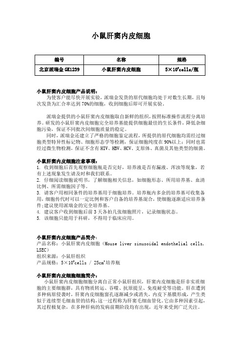
小鼠肝窦内皮细胞小鼠肝窦内皮细胞产品说明:为使客户能尽快开展实验,派瑞金发货的原代细胞均处于对数生长期,且每次发货为汇合率达到70%的细胞,收到细胞后即可开展实验。
派瑞金提供的小鼠肝窦内皮细胞取自新鲜的组织,按照标准操作流程分离培养。
研发的小鼠肝窦内皮细胞完全培养基能提供细胞最佳的生长条件,降低杂细胞污染,保证不同批次间细胞质量的稳定。
同时,派瑞金还建立了严格的细胞鉴定流程,所提供的原代细胞均需经过细胞类型特异性标记物、细胞形态学等检测,保证细胞纯度在90%以上;同时也需经过微生物检测,保证不含有HIV、HBV、HCV、支原体、真菌及其他类型的细菌。
小鼠肝窦内皮细胞注意事项:1. 收到细胞后首先观察细胞瓶是否完好,培养液是否有漏液、浑浊等现象,若有上述现象发生请及时和我们联系。
2. 仔细阅读细胞说明书,了解细胞相关信息,如细胞形态、所用培养基、血清比例、所需细胞因子等。
3. 请客户用相同条件的培养基用于细胞培养。
培养瓶内多余的培养基可收集备用,细胞传代时可以一定比例和客户自备的培养基混合,使细胞逐渐适应培养条件;建议使用派瑞金的完全培养基。
4. 建议客户收到细胞后前3天各拍几张细胞照片,记录细胞状态。
5. 该细胞只能用于科研,不得用于临床应用。
小鼠肝窦内皮细胞产品简介:产品名称:小鼠肝窦内皮细胞(Mouse liver sinusoidal endothelial cells, LSEC)组织来源:小鼠肝组织产品规格:5×10⁵cells / 25cm2培养瓶小鼠肝窦内皮细胞细胞简介:小鼠肝窦内皮细胞细胞分离自正常小鼠肝组织,肝窦内皮细胞是肝非实质细胞的主要细胞群,具有物质转运、吞噬、抗原提呈、免疫耐受等功能。
肝在遭到多种病原侵袭时,肝窦内皮细胞窗孔逐渐减少或消失,内皮下基膜形成,产生类似于连续型毛细血管的结构,这一过程称为肝窦毛细血管化。
它由多种因素引起,其过程极复杂,在多种肝病的发病前期阶段均有出现,近年来受到广泛关注。
小鼠腹腔注射的基本操作过程及注意事项
⼩⿏腹腔注射的基本操作过程及注意事项
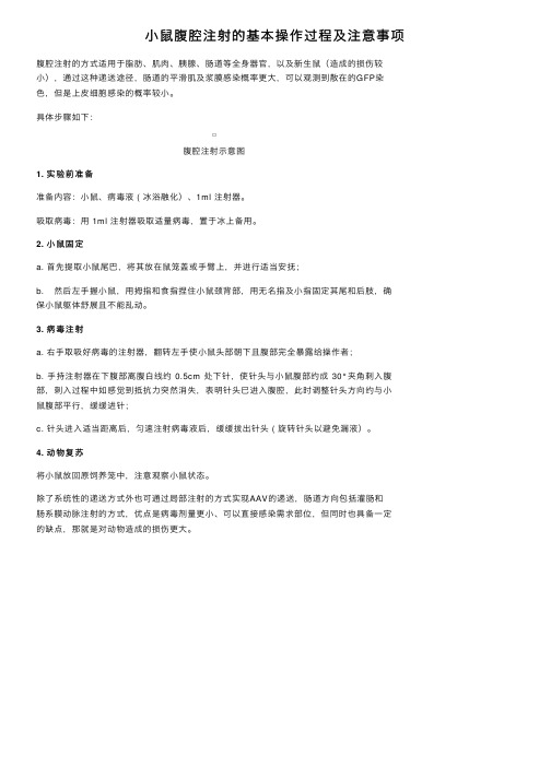
腹腔注射的⽅式适⽤于脂肪、肌⾁、胰腺、肠道等全⾝器官,以及新⽣⿏(造成的损伤较⼩),通过这种递送途径,肠道的平滑肌及浆膜感染概率更⼤,可以观测到散在的GFP染
⾊,但是上⽪细胞感染的概率较⼩。
具体步骤如下:
腹腔注射⽰意图
1. 实验前准备
准备内容:⼩⿏、病毒液 ( 冰浴融化)、1ml 注射器。
吸取病毒:⽤ 1ml 注射器吸取适量病毒,置于冰上备⽤。
2. ⼩⿏固定
a. ⾸先提取⼩⿏尾巴,将其放在⿏笼盖或⼿臂上,并进⾏适当安抚;
b. 然后左⼿握⼩⿏,⽤拇指和⾷指捏住⼩⿏颈背部,⽤⽆名指及⼩指固定其尾和后肢,确
保⼩⿏躯体舒展且不能乱动。
3. 病毒注射
a. 右⼿取吸好病毒的注射器,翻转左⼿使⼩⿏头部朝下且腹部完全暴露给操作者;
b. ⼿持注射器在下腹部离腹⽩线约0.5cm 处下针,使针头与⼩⿏腹部约成30°夹⾓刺⼊腹部,刺⼊过程中如感觉到抵抗⼒突然消失,表明针头已进⼊腹腔,此时调整针头⽅向约与⼩
⿏腹部平⾏,缓缓进针;
c. 针头进⼊适当距离后,匀速注射病毒液后,缓缓拔出针头 ( 旋转针头以避免漏液)。
4. 动物复苏
将⼩⿏放回原饲养笼中,注意观察⼩⿏状态。
除了系统性的递送⽅式外也可通过局部注射的⽅式实现AAV的递送,肠道⽅向包括灌肠和
肠系膜动脉注射的⽅式,优点是病毒剂量更⼩、可以直接感染需求部位,但同时也具备⼀定
的缺点,那就是对动物造成的损伤更⼤。
- 1、下载文档前请自行甄别文档内容的完整性,平台不提供额外的编辑、内容补充、找答案等附加服务。
- 2、"仅部分预览"的文档,不可在线预览部分如存在完整性等问题,可反馈申请退款(可完整预览的文档不适用该条件!)。
- 3、如文档侵犯您的权益,请联系客服反馈,我们会尽快为您处理(人工客服工作时间:9:00-18:30)。
小鼠内脏脂肪细胞
小鼠内脏脂肪细胞产品说明:
为使能尽快开展实验,派瑞金发货的原代细胞均处于对数生长期,且每次发货为汇合率达到70%的细胞,收到细胞后即可开展实验。
派瑞金提供的小鼠内脏脂肪细胞取自新鲜的组织,按照标准操作流程分离培养。
研发的小鼠内脏脂肪细胞完全培养基能提供细胞最佳的生长条件,降低杂细胞污染,保证不同批次间细胞质量的稳定。
同时,派瑞金还建立了严格的细胞鉴定流程,所提供的原代细胞均需经过细胞类型特异性标记物、细胞形态学等检测,保证细胞纯度在90%以上;同时也需经过微生物检测,保证不含有HIV、HBV、HCV、支原体、真菌及其他类型的细菌。
小鼠内脏脂肪细胞注意事项:
1. 收到细胞后首先观察细胞瓶是否完好,培养液是否有漏液、浑浊等现象,若有上述现象发生请及时和我们联系。
2. 仔细阅读细胞说明书,了解细胞相关信息,如细胞形态、所用培养基、血清比例、所需细胞因子等。
3. 请用相同条件的培养基用于细胞培养。
培养瓶内多余的培养基可收集备用,细胞传代时可以一定比例和自备的培养基混合,使细胞逐渐适应培养条件;建议使用派瑞金的完全培养基。
4. 建议收到细胞后前3天各拍几张细胞照片,记录细胞状态。
5. 该细胞只能用于科研,不得用于临床应用。
小鼠内脏脂肪细胞其他相关小鼠原代细胞:
小鼠小肠粘膜上皮细胞小鼠大隐静脉平滑肌细胞
小鼠肺微血管内皮细胞小鼠冠状动脉平滑肌细胞
小鼠肺血管平滑肌细胞小鼠大隐静脉内皮细胞
小鼠Ⅱ型肺泡上皮细胞小鼠冠状动脉内皮细胞
小鼠气管上皮细胞小鼠骨细胞
小鼠气管平滑肌细胞小鼠滑膜细胞
小鼠肺成纤维细胞小鼠骨骼肌细胞
小鼠支气管上皮细胞小鼠表皮细胞
小鼠支气管成纤维细胞小鼠真皮成纤维细胞
小鼠肺大静脉平滑肌细胞小鼠破骨细胞
小鼠肺大动脉平滑肌细胞小鼠皮肤肥大细胞
小鼠肺大动脉内皮细胞小鼠前脂肪细胞
小鼠肺动脉成纤维细胞小鼠成骨细胞
小鼠肺大静脉内皮细胞小鼠关节软骨细胞
小鼠气管和支气管上皮细胞小鼠胎儿表皮角质形成层细胞小鼠胰岛细胞小鼠成年表皮角质形成层细胞小鼠胰腺星状细胞小鼠皮下脂肪细胞
小鼠胰腺导管上皮细胞小鼠内脏脂肪细胞
小鼠颌下腺上皮细胞小鼠脑动脉血管内皮细胞
小鼠腮腺细胞小鼠脑动脉血管平滑肌细胞小鼠乳腺上皮细胞小鼠脑静脉血管内皮细胞
小鼠胰腺上皮细胞小鼠脑静脉血管平滑肌细胞小鼠甲状腺上皮细胞小鼠脑膜细胞
小鼠淋巴管内皮细胞小鼠神经胶质细胞
小鼠淋巴成纤维细胞小鼠海马神经元细胞
小鼠外周血白细胞小鼠脑微血管内皮细胞
小鼠骨髓基质细胞小鼠脑成纤维细胞
小鼠食管上皮细胞小鼠神经小胶质细胞
小鼠食管平滑肌细胞小鼠雪旺氏细胞
小鼠肠动脉内皮细胞小鼠小脑颗粒细胞
小鼠肠静脉内皮细胞小鼠嗅鞘细胞
小鼠肝实质细胞小鼠视网膜微血管内皮细胞小鼠肝动脉内皮细胞小鼠小梁网细胞
小鼠肝动脉平滑肌细胞小鼠视网膜色素上皮细胞
小鼠小肠血管内皮细胞小鼠视网膜muller细胞
小鼠小肠隐窝上皮细胞小鼠虹膜色素上皮细胞
小鼠肝内胆管上皮细胞小鼠晶状体上皮细胞
小鼠胃粘膜上皮细胞小鼠角膜上皮细胞
小鼠肝窦内皮细胞小鼠视网膜神经节细胞
小鼠肝星形细胞小鼠角膜成纤维细胞
小鼠直肠平滑肌细胞小鼠脉络膜血管细胞
小鼠小肠平滑肌细胞小鼠牙乳头细胞
小鼠结肠平滑肌细胞小鼠肝外胆管上皮细胞
小鼠肠上皮细胞小鼠肝Kupffer细胞
小鼠肠微血管细胞小鼠骨髓间充质干细胞
小鼠肠巨噬细胞小鼠下丘脑神经元细胞
小鼠子宫内膜上皮细胞小鼠睾丸支持细胞
小鼠卵巢颗粒细胞小鼠心肌微血管内皮细胞
小鼠子宫颈上皮细胞小鼠真皮微血管上皮细胞
小鼠子宫平滑肌细胞小鼠胚胎成纤维细胞
小鼠卵巢上皮细胞小鼠心脏干细胞
小鼠子宫成纤维细胞小鼠神经干细胞
小鼠卵巢成纤维细胞小鼠骨髓来源内皮祖细胞
小鼠肾实质细胞小鼠椎间盘髓核细胞
小鼠肾系膜细胞小鼠肾足突细胞
小鼠膀胱上皮细胞小鼠肾小管平滑肌细胞小鼠膀胱平滑肌细胞小鼠肾成纤维细胞
小鼠肾动脉内皮细胞小鼠尿道上皮细胞
小鼠肾动脉平滑肌细胞小鼠输尿管上皮细胞小鼠肾小管上皮细胞小鼠肾管状上皮细胞小鼠肾小球内皮细胞小鼠心肌细胞
小鼠前列腺上皮细胞小鼠心肌成纤维细胞小鼠肾上皮细胞小鼠主动脉内皮细胞小鼠膀胱成纤维细胞小鼠主动脉平滑肌细胞小鼠血管外膜成纤维细胞。