Caveolin-1 as Tumor Suppressor Gene in Breast Cancer
Akt1基因沉默降低小鼠乳腺癌细胞肺转移能力

谢谢
研究目标和内容
作为一种原癌基因,Akt1在许多人类肿瘤中表达显 著增高,促进肿瘤转移;但也有研究表明, Akt1的活 化可抑制乳腺癌细胞的侵袭和转移。为了深入探讨 Akt1在肿瘤发生发展过程中的作用,采用RNA干扰技术 沉默了高转移小鼠乳腺癌细胞4T1中Akt1的表达。MTT 法检测发现,Akt1沉默不影响4T1细胞的增殖能力。Transwell法检测证明,Akt1沉默可降低4T1细胞的迁移能 力。与以上结果相一致,Akt1沉默不影响乳腺癌形成原 位瘤的能力,但显著降低其体内肺转移能力。结果表 明,Akt1在小鼠乳腺癌细胞转移过程中发挥重要作用, 并提示Akt1可能成为乳腺癌治疗载体pLKO.1-Control,pLKO1.1-siAkt1-1 和pLKO.1-siAkt1-2的酶切鉴定电泳图谱
定量PCR检测4T1-control,4T1-siAkt1-1和 4T1-siAkt1-2细胞中Akt1mRNA表达量
免疫印迹检测4T1-Control,4T1-siAkt1-1和4T1siAkt1-2细胞中Akt1蛋白表达量
MTT法检测4T1-Control,4T1-siAkt1-1和4T1siAkt1-2细胞的增殖能力
4T1-Control ,4T1-siAkt1-1和4T1-siAkt1-2 细胞荷瘤小鼠的原位瘤重量
Transwell法检测4T1-Control, 4T1-siAkt11和4T1-siAkt1-2的细胞迁移能力
近年来研究发现,Akt1不但影响肿瘤细胞 的增殖、凋亡,而且对许多肿瘤的侵袭和转移 也有影响。Akt1基因在人胃癌中表达增加;活 化的Akt1促进人类胰腺癌细胞、纤维肉瘤细胞 和成纤维细胞的侵袭能力,但可抑制MDA-MB-2 31细胞的迁移能力。在乳腺癌中也发现Akt1 异常表达和活性改变,并在乳腺癌的转移过程 中发挥重要的作用。降低Akt1水平抑制Erb-2介 导的乳腺癌体内转移;而过表达myr-Akt1可抑 制人乳腺癌细胞的迁移和侵袭能力。Akt1活化 还可促进Erb-2介导的乳腺肿瘤发生但抑制其侵 袭能力。这些结果表明,Akt1在不同的细胞型 或肿瘤的不同发展阶段所起的作用也不同。
J Mol Cell Biol-2011-Ollila-jmcb_mjr016
ReviewThe tumor suppressor kinase LKB 1:lessons from mouse modelsSaara Ollila and Tomi P.Ma¨kela ¨*Institute of Biotechnology,University of Helsinki,Viikki Biocenter,Viikinkaari 9B,FIN-00014,Helsinki,Finland*Correspondence to:Tomi P.Ma¨kela ¨,E-mail:tomi.makela@helsinki.fiMutations in the tumor suppressor gene LKB 1are important in hereditary Peutz–Jeghers syndrome,as well as in sporadic cancersincluding lung and cervical cancer.LKB 1is a kinase-activating kinase,and a number of LKB 1-dependent phosphorylation cascades regulate fundamental cellular and organismal processes in at least metabolism,polarity,cytoskeleton organization,and prolifer-ation.Conditional targeting approaches are beginning to demonstrate the relevance and specificity of these signaling pathways in development and homeostasis of multiple organs.More than one of the pathways also appear to contribute to tumor growth fol-lowing Lkb 1deficiencies based on a number of mouse tumor models.Lkb 1-dependent activation of AMPK and subsequent inacti-vation of mammalian target of rapamycin signaling are implicated in several of the models,and other less well characterized pathways are also involved.Conditional targeting studies of Lkb 1also point an important role of LKB 1in epithelial–mesenchymal interactions,significantly expanding knowledge on the relevance of LKB 1in human disease.Keywords:LKB 1,tumor suppressor,mouse model,AMPKIntroductionCancer arises as a result of accumulating genetic and epige-netic changes,which compromise the cell’s ability to control its identity and proliferation.Many identified tumor suppressors play a well-established role in regulation of cell growth and div-ision (e.g.Rb,APC,p 21,PTEN)and genome maintenance (e.g.p 53,BRCA 1-2,ATM,ATR,MLH 1,MSH 2),providing a logical link between the loss of gene product and promotion of carcinogen-esis.An interesting exception is the serine /threonine kinase gene LKB 1(also known as STK 11),which has in recent years taken a prominent position among tumor suppressors.Heterozygous germline mutations in LKB 1predispose to Peutz–Jeghers syndrome (PJS)where patients develop benign polyps in the gastrointestinal (GI)tract and are in high risk of developing malignant tumors in GI tract,breast,and gyneco-logical organs (Giardiello et al .,2000).Importantly,somatic LKB 1mutations are found at least in lung (Ji et al.,2007)and cervical cancer (Wingo et al .,2009).Through phosphoryl-ation of several cellular kinases LKB 1has been implicated in control of cellular and organismal metabolism,cell polarity,and a variety of other functions ranging from proliferation and migration to senescence,apoptosis,DNA damage responseand differentiation (Vaahtomeri and Ma¨kela ¨,2011).Despite these many functions attributed to LKB 1,their specific contri-butions to the maintenance of tissue homeostasis in vivo and tumor growth are only sketchily appearing with thedevelopment of LKB 1mouse models.This work is important to enable rational treatment strategies to LKB 1-deficient tumors.The LKB 1kinase acts in a trimer with a pseudokinase STRAD and the scaffold protein MO 25to phosphorylate at least 14kinases with conserved activation sites (Katajisto et al.,2007).A well-known substrate of LKB 1is AMPK,which is the master reg-ulator of cellular and organismal metabolism,providing a putative downstream pathway to LKB 1-mediated tumor suppression (Shackelford and Shaw,2009).In mouse studies,AMPK requires LKB 1for activation in vivo in most tissues (Sakamoto et al .,2005;Shaw et al .,2005;Contreras et al .,2008;Hezel et al .,2008).AMPK senses the energy state of cells through monitoring AMP levels as a sensitive readout for ATP.AMPK is activated following exercise,hypoxia,or glucose deprivation,after which it phosphor-ylates multiple targets to increase energy uptake and catabolic processes such as glucose uptake and fatty acid oxidation,and suppress anabolic processes such as lipogenesis and cholesterol synthesis (Hardie et al.,2003).AMPK is the potential candidate to mediate LKB 1’s effects in cell growth via the mammalian target of rapamycin (mTOR)signal-ing (Corradetti et al .,2004;Shaw et al .,2004),which is the pathway monitoring the availability of nutrients in regulation of cell size and protein synthesis as well as proliferation (Zoncu et al.,2011).Increased mTOR signaling is common in cancer (Guertin and Sabatini,2007)and also present in at least some Lkb 1-deficient tumors (Shaw et al.,2004;Ji et al.,2007;Contreras et al.,2008;Hezel et al.,2008;Shackelford et al.,2009).An additional link between LKB 1and mTOR pathway#The Author (2011).Published by Oxford University Press on behalf of Journal ofMolecular Cell Biology ,IBCB,SIBS,CAS.All rights reserved.doi:10.1093/jmcb /mjr 016Journal of Molecular Cell Biology (2011),Vol no.0,1–11|1Journal of Molecular Cell Biology Advance Access published September 15, 2011 at Shihezi University on September 27, 2011 Downloaded frommay lie in regulation of PI 3K-Akt pathway inhibitor PTEN by LKB 1(Mehenni et al .,2005).Loss of cell polarity is commonly noted in cancer,and LKB 1is an important factor for cell polarity in different organisms.In C.elegans ,the orthologs for LKB 1(par-4)and MARK s (par-1)were identified in a panel of six partitioning (par )mutants which disrupted the polarity of the early embryos (Kemphues et al.,1988).In Drosophila ,Lkb 1is required for proper oocyte polarity (Martin and St Johnston,2003).In mammalian cells,in both 2D and 3D cell culture models and in vivo ,LKB 1is known to regulate polarity (Baas et al .,2004;Partanen et al .,2007;Hezel et al .,2008).Polarity defects are,however,not seen in all Lkb 1-deficient tumors (Contreras et al.,2008,2010).Several of the LKB 1substrates have been reported to mediate the regulation of cell polarity through regulating the cytoskeleton and formation of cell–cell junctions.MARK kinases are implicated in the stability of microtubules by phosphorylating and thereby dissociation microtubule-associated proteins (MAPs),for example the tau protein,from microtubules (Drewes et al .,1997;Stoothoff and Johnson,2005).Neuronal polarity and axon formation are regu-lated by LKB 1at least partially via BRSK kinases (Kishi et al.,2005;Barnes et al.,2007).To what extent LKB 1acts as a polarity protein in mammalian non-neuronal cells still remains to be deter-mined,although at least in both exo-and endocrine pancreas Lkb 1loss leads to polarity defects in vivo (Hezel et al .,2008;Granot et al .,2009).As formation of stress fibers is essential incell contractility,recent studies associate LKB 1with cell motility via NUAK 1and NUAK 2,which have been implicated in regulation of myosin light chain phosphorylation (Vallenius et al.,2010;Zagorska et al.,2010).For detailed information of the molecular signaling pathways of Lkb 1,the reader is recommended recent reviews more focused on that topic (Katajisto et al.,2007;Hezel and Bardeesy,2008;Vaahtomeri and Ma¨kela ¨,2011).Role of Lkb 1in development and tissue homeostasis in miceAlthough LKB 1is a tumor suppressor,inactivation of Lkb 1through homologous recombination or ‘knock-out’(KO)does not always lead to tumors.This is due partly to essential functions of Lkb 1in development and partly demonstrates the tissue-specificity of Lkb 1functions,where in some cell types biallelic deletion is detrimental to cells or affects specific functions in metabolism as summarized in Figure 1and discussed below.Role of Lkb 1in embryogenesisGeneration of full KO revealed that Lkb 1is essential for embry-ogenesis;no viable Lkb 12/2embryos were seen after E 11.Analysis of the E 8.5–E 9.5embryos revealed severe developmen-tal defects including impaired neural tube closure and somitogen-esis,mesenchymal tissue cell death,and defective vasculature.The extra-embryonic tissues (yolk sac and placenta)were also deformed.VEGF signaling was highly upregulated in theKOFigure 1Non-tumorigenic phenotypes following Lkb 1targeting in mice.Phenotypes (green)are grouped according to tissue type,cell typeaffected /analyzed (blue),and alleles used for targeting.When appropriate,activator of deletor is indicated in purple.Noted signaling change(s)indicated in red.Alleles as displayed in original publications except for Lkb 1flox 2h /flox 2h hypomorphic Lkb 1(Sakamoto et al,2005).(1)Londesborough et al.,2008;(2)Ohashi et al.,2010;(3)Cao et al.,2010;Tamas et al.,2010;(4)Shorning et al.,2009;(5)Woods et al.,2011;(6)Shaw et al.,2005;(7)Sun et al.,2010a ;(8)Sun et al.,2011;(9)Granot et al.,2009;Fu et al.,2009;(10)Koh et al.,2006;(11)Sakamoto et al.,2005;(12)Sakamoto et al.,2006;Jessen,et al.,2010;(13)Ikeda et al.2009;(14)Gurumurthy et al.,2010;Nakada et al.,2010;(15)Gan et al.,2010;(16)Barnes et al.,2007;(17)Ylikorkala et al.,2001.tam,tamoxifen;b -NF,b -naphtoflavone;pIpC,polyinosinic–polycytidylic acid;iv,intravenous.2|Journal of Molecular Cell Biology Ollila and Ma¨kela ¨ at Shihezi University on September 27, 2011 Downloaded fromembryos,possibly relating to the vascular phenotype (Ylikorkala et al .,2001).Embryonic lethality,no embryonic turning,and small somites were also shown in another report of Lkb 1full KO (Jishage et al .,2002).The severe developmental defect was not a result of the abnormal extra-embryonic tissues,since epiblast-specific conditional inactivation of Lkb 1using Mox 1-Cre resulted in very similar embryonic lethal phenotype to full KO (Londesborough et al .,2008).The important role of Lkb 1in development and maintenance of neurons,mesenchymal cells,and vascularization has been recapitulated in tissue-specific Lkb 1KOs.Role of Lkb 1in angiogenesisLondesborough et al .(2008)further dissected the role of Lkb 1in endothelia by deleting Lkb 1in vascular endothelial cells using Tie 1-Cre (Figure 1).The mice died at E 12.5and displayed dilated embryonic vessels and pericardial swelling.The vessels were irre-gular and distorted and suffered from inadequate supportive vas-cular smooth muscle cell layer.Since Tgf b signaling was reduced both in Lkb 1-deficient mouse yolk sacs and human umbilical vein endothelial cells (HUVECs)where LKB 1expression was silenced by siRNA,the vascular phenotype was suggested to result from a loss of supporting vascular smooth muscle cells as a conse-quence of attenuated Tgf b signaling from endothelial cells (Londesborough et al .,2008).Another report also described mice lacking Lkb 1in endothelial cells,deleted using Tie 2-Cre driver (Ohashi et al.,2010)(Figure 1).This study repeated the finding that endothelial Lkb 1is essential for proper embryonic development and no homozygous mutants were born.Analysis of heterozygous Tie 2-Cre;Lkb 1flox /+mice revealed that the mice,including vasculature,seemed phenotypically normal,but displayed reduced revascularization after hind-limb ischemia.Studies in mouse tissues,primary mouse endothelial cells,and HUVECs implemented that the phenotype was mediated via AMPK (Ohashi et al.,2010).In this study,the authors did not address the contribution of Tgf b signaling to the observed phenotype.In the Tie 2-Cre model,Lkb 1–AMPK axis seemed to mediate proangiogenetic signaling as Lkb 1heterozygosity resulted in reduction of revascularization in adult mice (Ohashi et al.,2010).In developing embryo,increased VEGF signaling upon Lkb 1loss would suggest the opposite,antiangiogenic role for Lkb 1(Ylikorkala et al .,2001).Also in the context of PJS polyps where a loss of Lkb 1leads to increased HIF 1a and vasculature,Lkb 1seems to be rather antiangiogenic (Shackelford et al .,2009).However,reduced capillary density was reported in mice where Lkb 1was conditionally deleted from the heart (Ikeda et al .,2009).In 3D culture system where endothelial cells are embedded in Matrigel,both over-expression (Xie et al .,2009)and inhibition (Ohashi et al.,2010)of Lkb 1have been reported to inhibit network formation,suggesting proper expression of LKB 1is essential for angiogenesis.Thus,the precise role of Lkb 1in angiogenesis seems to be dependent on the tissue type and /or the developmental phase,varying from inhibition to promotion.Role of Lkb 1in liverThe finding that Lkb 1functions upstream of AMPK (Shaw et al .,2004)led to interest to study its effects in liver,where many path-ways of carbohydrate and lipid metabolism,including glycogen-esis,glycogenolysis,gluconeogenesis,lipogenesis,and cholesterol synthesis take place.Tail-vain injection of Adeno-Cre to mice carrying conditional Lkb 1allele led to hepatocyte-specific Lkb 1deletion since Adeno-Cre has high tropism for hepatocytes (Shaw et al .,2005)(Figure 1).Lkb 1loss resulted in nearly complete abolishment of AMPK activation in liver,and the glucose metabolism of the mice was impaired demonstrated by elevated blood glucose.CRTC 2phosphorylation was reduced in the livers of the mice,leading to elevated CREB-mediated transcription,including expression of PGC 1a and other gluconeogenetic genes.Also lipogenetic genes were over-expressed.Metformin,the diabetes drug which reduces blood glucose levels via AMPK pathway (Zhou et al .,2001),did not lower blood glucose in the liver-specific Lkb 1KO mice,demonstrating that AMPK activity induced by Lkb 1in liver is required for the effects of metformin in vivo .In another report of liver-specific Lkb 1knockout using Alb-Cre driver,Woods et al.(2011)reported defective bile ducts in liver,leading to accumulation of bile in liver and serum (Figure 1).Bile salt export pump was not located in canalicular membrane of the bile canaliculi,indicating possible defects in cell polarity.The mice also suffered from cholestasis (Woods et al.,2011).These reports of liver-targeted deletions of Lkb 1demonstrate the critical requirement of Lkb 1in glucose,lipid,bile,and cholesterol metab-olism.Furthermore,they show that in liver,Lkb 1is the main acti-vator of AMPK,and its activity is required for the AMPK-mediated suppression of lipogenesis and gluconeogenesis to take place.Role of Lkb 1in muscleMuscles are highly energy-consuming tissues whose glucose homeostasis needs to be regulated both in response to insulin after blood sugar increase,and to exercise-mediated deficiency of glucose storage.Sakamoto et al.(2005)provided the first genetic evidence that Lkb 1is required for AMPK activation in vivo in skeletal muscle.They generated conditional Lkb 1mice in which cDNA of Lkb 1exons 5–7fused with neomycin resistance cassette,surrounded by loxP sites,was inserted between exons 4and 8in the genomic Lkb 1locus.The resulting mice were hypomorphic and expressed only 10%–20%of normal levels of Lkb 1in the absence of Cre -mediated ing MCK-Cre driver to create muscle-specific Lkb 1KO,they found that AMPK a 2(one of the two alternative catalytic subunits of AMPK)activation either by the AMP analog AICAR,muscle con-traction or phenformin,a similar blood glucose lowering drug to metformin,was lost and AMPK a 1activation greatly reduced.Upon contraction,glucose transport to muscle cells was abol-ished (Sakamoto et al.,2005).In another study using the same muscle-specific MCK-Cre with another (non-hypomorphic)con-ditional Lkb 1line,effects of Lkb 1loss in muscle to levels of blood glucose were investigated (Koh et al.,2006)(Figure 1).Interestingly,glucose metabolism seemed to be enhanced in these mice,demonstrated by reduced fasting blood glucose and blood insulin concentrations,improved glucose tolerance,and increased muscle glucose uptake.This phenotype,indicating that Lkb 1in muscle functions as a negative regulator of glucose metabolism,was suggested to be resulting from improved muscle glucose uptake,mediated by increased phosphorylation of Akt and reduced the gene expression of the Akt inhibitor TRB 3.Lkb 1loss abolished the activity of AMPK a 2,but notLessons from LKB 1mouse modelsJournal of Molecular Cell Biology |3at Shihezi University on September 27, 2011 Downloaded fromAMPK a 1in muscle cells.Also MARK 4,but not MARK 2/3activitywas significantly reduced.Based on this study,the metabolic effects mediated by Lkb 1in muscle seem to oppose those of the liver,at least in terms of blood glucose levels (Koh et al.,2006).Recently,the Lkb 1substrate NUAK 2was proposed to be a mediator of contraction-stimulated glucose transport by skel-etal muscle (Koh et al.,2010).Also cardiac muscle lacking Lkb 1has been investigated.Sakamoto et al.(2006)studied the effect of Lkb 1deficiency in heart using the MCK-Cre driver,which deletes Lkb 1in both skel-etal and cardiac myocytes and found that Lkb 1inactivation did not lead to overt cardiac dysfunction,although the weight of the heart was reduced and the atria enlarged;however,the study revealed that cardiac Lkb 1is required for activation of AMPK a 2both in basal conditions and in response to ischemia (Figure 1).Also Jessen et al.(2010)used the MCK-Cre driver but the Lkb 1allele was not hypomorphic as in the Sakamoto et al.(2006)study.They showed that ablation of Lkb 1in heart leads to impaired cardiac function both in basic conditions and post-ischemia and suggested that failure to downregulate mTOR sig-naling by AMPK a 2activation underlined the phenotypes.Ikeda et al.(2009)used a -MHC-Cre to delete Lkb 1specifically from the heart,and a more severe phenotype was observed:the mice displayed hypertrophy and impaired function of the heart,reduction of cardiac capillary density,and increased fibrosis and collagen content and died by 6months of age.The differ-ences between these phenotypes may reflect differences in the timing of Cre activity,specificity of the Cre recombination,and /or the conditional Lkb 1allele used.However,it seems clear that Lkb 1is needed for the normal function of heart both in basal and ischemic conditions.Role of Lkb 1in pancreasPancreatic b -cells secrete insulin and are thus important mediators of whole-body glucose metabolism.As Lkb 1–AMPK axis is important in regulation of liver metabolism and muscle glucose homeostasis,it is of interest to study whether Lkb 1has an effect on the insulin release.Granot et al.(2009)used the Pdx 1-CreER driver to delete pancreatic Lkb 1in 6-week-old mice by tamoxifen injection (Figure 1).In response to glucose injection,the mutant mice secreted more insulin than control mice,which carried the conditional Lkb 1allele but were not subjected to tamoxifen injection.Deletion of Lkb 1led to increased size of b -cells together with disrupted polarity.Increased mTOR signal-ing seemed to mediate the cell size increase,while the polarity defect took place at least partially through MARK 2.Increased insulin secretion was partially dependent on AMPK (Granot et al .,2009).Fu et al .(2009)used the same Pdx 1-CreER system to delete Lkb 1in adult b -cells and also found that the mice showed improved glucose tolerance,b -cells mass had increased,and mTOR pathway was activated (Figure 1).These results place Lkb 1as an important regulator of pancreatic b -cell size,polarity,and function,further highlighting its essence in regulation of organismal metabolism.Sun et al.(2010a)investigated pancreatic b -cells with the Rip 2-Cre driver,which activates Cre -mediated recombination in pancreatic b -cells and some hypothalamic neurons,and found that the mice displayed diminished food intake and weight gain,enhanced insulin secretion,and improved glucose tolerance (Figure 1).Also here,mTOR pathway was activated.However,the study by the same group where both AMPK a subunits were deleted in b -cells using the same Rip 2-Cre showed decreased insulin secretion (Sun et al.,2010b ).This suggests that Lkb 1loss regulates mTOR signaling in b -cells partially independent of AMPK,or that the hypothalamic Lkb 1and AMPK have different functions,impacting the feeding behavior and hormonal balance.Role of Lkb 1in immune systemThree recent studies elegantly demonstrated that Lkb 1regu-lates the quiescence and maintenance of hematopoietic stem cells (HSCs)using conditional Lkb 1alleles with Mx 1-Cre followed by injections of polyinosinic–polycytidylic acid (pIpC),or Rosa 26-CreERt 2followed by tamoxifen injections (Gan et al.,2010;Gurumurthy et al.,2010;Nakada et al.,2010)(Figure 1).Both approaches resulted in a similar phenotype:increased pro-liferation followed soon by decline in HSC number,resulting in loss of all immune cell types (pancytopenia)and death.Transplantation experiments demonstrated that Lkb 1-deficient HSCs were not able to reconstitute the bone marrow of irradiated wild-type (wt)mice,nor were they able to compete with wt donor cells,demonstrating that the effect was cell-autonomous;mito-chondrial defects and decreased ATP levels,as well as altered long-chain fatty acid and nucleotide metabolite levels suggested metabolic defects to underlie the phenotypes noted (Gan et al.,2010;Gurumurthy et al.,2010;Nakada et al.,2010).Interestingly,only minor similarities in mitochondrial phenotypes were found when mice defective for both AMPK a subunits were compared with Lkb 1KO mice (Nakada et al.,2010),implicating other Lkb 1substrates in these phenotypes.Consistent with this,rapamycin or AMPK activators AICAR and A 769662did not rescue the phenotype in any of the studies.Immune cell apopto-sis was increased,and Lkb 1-deficient HSCs also demonstrated increased autophagy in bone marrow,and inhibiting this further decreased immune cell survival (Gan et al.,2010;Gurumurthy et al.,2010;Nakada et al.,2010).This would suggest that Lkb 1in this context is suppressing autophagy,whereas previously it has been reported to activate it following elevation of reactive oxygen species (Alexander et al.,2010).Yet another phenotype potentially decreasing HSC viability was the noted increase in supernumerary centrosomes,aberrant mitotic spindles,and aneuploidy (Nakada et al.,2010),which could be due to compro-mised BRSK 2activity (Alvarado-Kristensson et al.,2009).Recently,two groups generated mice where Lkb 1expression is specifically abolished in the T cell progenitors using the proximal p 56lck-Cre promoter.The studies demonstrate severe deficiency in survival and proliferation of T cell progenitors and mature T cells in the absence of Lkb 1(Cao et al.,2010;Tamas et al.,2010)(Figure 1).Also the survival of isolated peripheral T cells in vitro was dependent on Lkb 1(Tamas et al.,2010).Transfection of thymo-cytes with constitutively active AMPK a 2partially rescued the thy-mocytes from cell death,indicating that thymocyte survival is mediated at least via AMPK pathway (Cao et al.,2010).Thus,the common hematopoietic cell precursors and T cell precursors seem to have different requirement for AMPK signaling,although cell sur-vival is defective in both cell types in the absence of Lkb 1.The studies in hematopoietic cells have revealed an interesting aspect4|Journal of Molecular Cell Biology Ollila and Ma¨kela ¨ at Shihezi University on September 27, 2011 Downloaded fromof Lkb 1biology:although being a tumor suppressor in some tissues,in others Lkb 1is required for survival.Role of Lkb 1in nervous systemLkb 1KO embryos exhibit severe deficiencies in development of neuronal tissues (Ylikorkala et al .,2001).Since LKB 1orthologs in nematodes and fruit flies have been identified through their indis-pensable role in establishing polarity (Kemphues et al .,1988;Martin and St Johnston,2003)and LKB 1regulates polarity also in some mammalian cells (Baas et al .,2004;Partanen et al .,2007),it was of interest to generate models which would reveal the in vivo relevance of Lkb 1in establishing the axon-dendrite polarity in neuronal cells.Barnes et al.(2007)deleted Lkb 1in cer-ebral cortex of developing mice using Emx-Cre driver and showed that Lkb 1and its substrates BRSK 1and BRSK 2are required for axon specification in the studied neurons.This finding confirmed the previously described role of BRSK kinases in neuronal polar-ization (Kishi et al .,2005),and placed Lkb 1as the upstream kinase required for the polarization to take place.Lkb 1-activated BRSKs were shown to modify the cytoskeleton by phosphorylating MAPs (Barnes et al.,2007).Studies in rat hip-pocampal neurons in vitro and developing rat cortical neurons in vivo agreed with the finding that Lkb 1is essential in establishing neuronal polarity;there,lack of either Lkb 1or STRAD prevented axon differentiation (Shelly et al.,2007).Interestingly,over-expression of Lkb 1and STRAD resulted in formation of multiple axons.PKA-mediated phosphorylation of Lkb 1Ser 431was shown to be required for the axon specification (Barnes et al.,2007;Shelly et al.,2007).Thus,Lkb 1activity is modulated by upstream factors in a tissue-and context-specific manner.Not only axon specification but also maintenance seems to be regulated via Lkb 1in some systems.Sun et al.(2011)reported,using the pancreatic and hypothalamic Rip 2-Cre ,that the mice developed hind-limb paralysis due to axon degeneration in thor-acic spinal cord neurons at about 7–8weeks of age (Figure 1).The Rip 2-Cre was found to be active also in spinal cord,especially in the thoracic segments.Deleting both AMPK a subunits did not result in axon degeneration or paralysis,and the authors specu-lated that in the absence of Lkb 1,the neuronal polarization and axon degeneration defects might be mediated by BRSK kinase pathways (Sun et al.,2011).PJS and its mouse modelsLKB 1was linked to human disease when its mutations were found to be causative for PJS (Hemminki et al .,1998;Jenne et al .,1998).A major manifestation in PJS is the appearance of large occluding hamartomatous polyps in the GI tract (Giardiello and Trimbath,2006).Mice carrying one inactivated allele of Lkb 1(Lkb 1+/2)recapitulate PJS by developing hamartomatous GI polyps which are indistinguishable from PJS patient polyps (Bardeesy et al .,2002;Jishage et al .,2002;Miyoshi et al .,2002;Rossi et al .,2002)(Figure 2),although in mice polyps appear more in the stomach and less in the small intestine.Polyps appear at 4–6months (Udd et al.,2010),and lead to lethality at an average age of 11months due primarily to obstructions.Biallelic loss of wt Lkb 1is not a prerequisite for polyp formation,indicating that Lkb 1is a haploinsufficient tumor suppressor at least in the context of PJS polyps (Jishage et al .,2002;Miyoshiet al .,2002;Rossi et al .,2002).Strong up-regulation of COX 2has been identified in the mouse and also PJS patient polyps (Rossi et al .,2002),and COX 2inhibitors have been shown to be efficient suppressors of PJS polyps (Udd et al .,2004).PJS is associated with elevated risk of cancer,especially in the GI tract,and also in breast,pancreas and gynecological cancers (Giardiello and Trimbath,2006;Hearle et al .,2006;Mehenni et al .,2006).Lkb 1+/2mice in turn have been reported to have increased frequency of cancer in liver (Nakau et al .,2002),bones (Robinson et al .,2008),and endometrium (Contreras et al .,2008)(Figure 2).Polyposis in Lkb 1+/2mice is accelerated in a p 53-deficient background (our unpublished data)(Wei et al .,2005;Takeda et al .,2006)(Figure 2),and p 53mutations are detected in the GI cancers of PJS patients (Miyaki et al .,2000).Despite these observations,progression of the benign hamarto-matous polyps to dysplasia or carcinoma is not clearly estab-lished possibly due to the rapid growth of the hamartomatous polyps leading to GI occlusions.As haploinsufficiency of Lkb 1is sufficient for polyp initiation (Jishage et al .,2002;Miyoshi et al .,2002;Rossi et al .,2002)though biallelic loss has been noted (Bardeesy et al .,2002),loss of the remaining allele of Lkb 1may represent a progression step,although it has also been suggested that the loss of Lkb 1is associated with the resist-ance to progression in this context (Bardeesy et al.,2002).Mesenchymal Lkb 1loss leads to PJS-type polyposis in mice PJS polyps are classified as hamartomatous polyps thought to contain all the cell types of the surrounding tissue.However,it was recently noted that epithelial differentiation is disrupted in gastric and small intestinal polyps in Lkb 1+/2mice (Udd et al.,2010),but the model did not enable distinguishing whether this was a cell autonomous function of Lkb 1in epithelial cells.Biallelic disruption of Lkb 1in GI epithelia lead to imbalanced differentiation and positioning of epithelial cells (Shorning et al .,2009)(Figure 1),but was not reported to be associated with tumorigenesis.Polyps in both PJS patients and Lkb 1+/2mice harbor a large component of smooth muscle tissue.Remarkably,in a mouse model,where Lkb 1deficiency was restricted to the smooth muscle lineage by using a tamoxifen-inducible SM 22-CreERt 2line,PJS type polyps appeared in stomachs of the mice with the hetero-and homozygous Lkb 1mutants (Katajisto et al .,2008)(Figure 2).The polyps appeared later than those in the Lkb 1+/2mice,suggesting either that tamoxifen-induced Lkb 1loss at 6weeks of age delayed the poly-posis,or that mesenchymal loss of Lkb 1signaling is sufficient to drive hyperproliferation of epithelial tissue,but that coexisting epithelial mutations accelerate the process.This interesting aspect of Lkb 1signaling in tissue interactions is discussed later.Other Lkb 1tumor mouse modelsInactivating LKB 1mutations are associated with the develop-ment of cancer in several tissues.Various strategies of targeted inactivation of Lkb 1in mice,sometimes in combination of other tumorigenic mutations,have led to the development of various types and grades of tumors in multiple tissues,sometimes mod-eling human cancers in very useful ways as discussed below and summarized in Figure 2.Lessons from LKB 1mouse modelsJournal of Molecular Cell Biology |5at Shihezi University on September 27, 2011 Downloaded from。
病毒包装原理

以核心基因技术为基础,汉恒更加注重技术应用与临床转化。
汉恒简史
2010成立 2012年 4年多的发展 短期目标
中期目标 2-5年内
长期目标 10年内
病毒载体为工具的基 因技术操作全平台
覆盖、并抢占国内主流科 研、临床前研究及药物研 发的AAV市场,完成基础 底层市场的教育及占领
上市1-2种自主知识产 权的基因治疗AAV药物
专注于基因技术的 研发与应用转化
--您身边的病毒载体专家
汉恒生物2017产品手册
目录
关于公司
01
汉恒简史 公司简介
02
汉恒客户 发表文献
06
一流质量标准 汉恒专利
07 公司优势
08 产品介绍
专题研究
25 悬浮细胞专用病毒
26 原代T细胞专用病毒
27 环状RNA专题研究
28 自噬专题研究
30 CRISPR-Cas9专题研究
32
探针工具 细胞器定位工具
特色服务
09 腺相关病毒AAV 14 光遗传学AAV现货工具 17 化学遗传学AAV现货工具 19 腺病毒 22 慢病毒 24 逆转录病毒
汉恒自主研发试剂
33 HB-infusionTM 无缝克隆试剂盒 34 LipoFiterTM脂质体转染试剂 36 SaveItTM 抗支原体试剂盒 37 Annexin V-FITC 细胞凋亡检测试剂盒
2017客户最新文献节选
0003碧云天PMSF本甲磺酰胺试剂盒说明书
PMSF(100mM)产品简介:PMSF即Phenylmethanesulfonyl fluoride,中文名为苯甲基磺酰氟。
分子式为C7H7FO2S,分子量为174.19,纯度>99%。
常用生化试剂,用于抑制蛋白酶,进口试剂配制。
第一次使用时把一管PMSF晶体全部倒入到PMSF溶剂中,溶解混匀,即配制成10毫升100mM PMSF。
保存条件:室温保存,配成PMSF溶液后需-20ºC保存。
注意事项:有毒,注意防护。
本产品仅限于专业人员的科学研究用,不得用于临床诊断或治疗,不得用于食品或药品,不得存放于普通住宅内。
为了您的安全和健康,请穿实验服并戴一次性手套操作。
使用本产品的文献:1.Cai H, Yin D, Zhang L, Yang X, Xu X, Liu W, Zheng X, Zhang H, Wang J, Xu Y,Cheng D, Zheng M, Han Y, Wu M, Wang Y. Preparation and biological evaluation of 2-amino-6-[18F]fluoro-9-(4-hydroxy-3-hydroxy-methylbutyl) purine (6-[18F]FPCV) as a novel PET probe for imaging HSV1-tk reporter gene expression.Nucl Med Biol. 2007 Aug;34(6):717-25.2.Zhang JQ, Zhao XK, He J, Zhu L, Wang XJ. Express and role of FGFR2 in bladdertransitional cell carcinoma. China Medical Engineering. 2007 Sep;V ol.15(9):720-5.3.Sun YG, Wang XW, Yang SM, Zhou G, Wang WQ, Wang HB, Wang RQ, Fang DC.Inhibition of nucleostemin upregulates CDX2 expression in HT29 cells in response to bile acid exposure: implications in the pathogenesis of Barrett's esophagus. J Gastrointest Surg. 2009 Aug;13(8):1430-9.4.Hua W, Jiang J, Rong X, Wu R, Qiu H, Zhang Y, Chen Q. The dual role of thecystathionine gamma-lyase/hydrogen sulfide pathway in CVB3-induced myocarditis in mice. Biochem Biophys Res Commun. 2009 Oct 23;388(3):595-600.5.Li H, Zhang L, Huang Q. Differential expression of mitogen-activated proteinkinase signaling pathway in the hippocampus of rats exposed to chronic unpredictable stress. Behav Brain Res. 2009 Dec 14;205(1):32-7.6.Cai J, Yu C, Liu Y, Chen S, Guo Y, Yong J, Lu W, Ding M, Deng H. Generation ofhomogeneous PDX1(+) pancreatic progenitors from human ES cell-derived endoderm cells. J Mol Cell Biol. 2010;2(1):50-60.7.Li R, Zhang J, Zhang L, Cui Q, Liu H. Angelica injection promotes peripheralnerve structure and function recovery with increasedexpressions of nerve growth factor and brain derived neurotrophic factor in diabetic rats. Curr Neurovasc Res.2010 Aug;7(3):213-22.8.Yu LN, Yu J, Zhang FJ, Yang MJ, Ding TT, Wang JK, He W, Fang T, Chen G, YanM. Sevoflurane postconditioning reduces myocardial reperfusion injury in rat isolated hearts viaactivation of PI3K/Akt signaling and modulation of Bcl-2 family proteins. J Zhejiang Univ Sci B. 2010 Sep;11(9):661-72.9.Wang C, Xie H, Liu X, Qin Q, Wu X, Liu H, Liu C. Role of nuclear factor-κB involatile anaesthetic preconditioning with sevoflurane during myocardial ischaemia/reperfusion. Eur J Anaesthesiol. 2010 Aug;27(8):747-56.10.Wang W, Wang L, Li XX, Chen X, Zhang HY, He Y, Wang JJ, Zhao YY, Zhang BL,Xu YX. Effect of interrupted endogenous BMP/Smad signaling on growth and steroidogenesis of porcinegranulosa cells. J Zhejiang Univ Sci B. 2010 Sep;11(9):719-27.11.Hou T, Shi Y, Cheng S, Yang X, Li L, Xiao C. Nogo-A expresses on neural stemcell surface. Int J Neurosci. 2010 Mar;120(3):201-5. 12.Wang X, Yang Q, Wang P, Luo L, Chen Z, Liao B, Li G. Derp2-mutant genevaccine inhibits airway inflammation and up-regulates Toll-like receptor 9 in anallergic asthmatic mouse model. Asian Pac J Allergy Immunol. 2010 Dec;28(4):287-93.13.Feng DD, Zhang H, Zhang P, Zheng YS, Zhang XJ, Han BW, Luo XQ, Xu L, ZhouH, Qu LH, Chen YQ. Down-regulated miR-331-5p and miR-27a are associated with chemotherapy resistance andrelapse in leukaemia. J Cell Mol Med. 2011 Oct;15(10):2164-75.14.Zhang L, Jiang H, Gao X, Zou Y, Liu M, Liang Y, Yu Y, Zhu W, Chen H, Ge J. Heatshock transcription factor-1 inhibits H2O2-induced apoptosis via down-regulation of reactive oxygen species in cardiac myocytes. Mol Cell Biochem. 2011 Jan;347(1-2):21-8.15.Ding S, Peng H, Fang HS, Zhou JL, Wang Z. Pulsed electromagnetic fieldsstimulation prevents steroid-induced osteonecrosis in rats. BMC Musculoskelet Disord. 2011 Sep 29;12:215.16.Wu J, Song R, Song W, Li Y, Zhang Q, Chen Y, Fu Y, Fang W, Wang J, Zhong Z,Ling H, Zhang L, Zhang F. Chlorpromazine protects against apoptosis induced by exogenous stimuli in the developing ratbrain. PLoS One. 2011;6(7):e21966.17.Liu ML, Wen JJ, Xu XF, Zhao DM. Neurotoxic effect of the complex of the ovineprion protein (OvPrP(C)) and RNA on the cultured ratcortical neurons. Neurochem Res. 2011 Oct;36(10):1863-9.18.Guo L, Zhang J, Yan Q, Yin M. Establishment and characterization of RNA-editedserotonin 2C receptor isoform cell models andalteration of amyloid precursor protein ectodomain secretion in HEK293 APPSwe cells. Hum Cell. 2011 Jun;24(2):104-11.19.Feng S, Cong S, Zhang X, Bao X, Wang W, Li H, Wang Z, Wang G, Xu J, Du B, QuD, Xiong W, Yin M, Ren X, Wang F, He J, Zhang B. MicroRNA-192 targeting retinoblastoma 1 inhibits cell proliferation and induces cell apoptosis inlung cancer cells. Nucleic Acids Res. 2011 Aug;39(15):6669-78.20.Tian Y, Hu Y, Wang Z, Chen K, Zhang L, Wang L, Ren M, Huang A, Tang H.Hepatitis B virus regulates Raf1 expression in HepG2.2.15 cells by enhancing its promoter activity. Arch Virol. 2011 May;156(5):869-74.21.Yang L, Ping YF, Yu X, Qian F, Guo ZJ, Qian C, Cui YH, Bian XW. Gastric cancerstem-like cells possess higher capability of invasion and metastasis in associationwith a mesenchymal transition phenotype. Cancer Lett. 2011 Nov 1;310(1):46-52.22.He Y, Zhang H, Teng J, Huang L, Li Y, Sun C. Involvement of calcium-sensingreceptor in inhibition of lipolysis through intracellular cAMP andcalcium pathways in human adipocytes. Biochem Biophys Res Commun. 2011 Jan 7;404(1):393-9.23.Jiang S, Zu Y, Wang Z, Zhang Y, Fu Y. Involvement of mitochondrial permeabilitytransition pore opening in 7-xylosyl-10-deacetylpaclitaxel-induced apoptosis.Planta Med. 2011 Jul;77(10):1005-12.24.Chen X, Wu X, Zhao Y, Wang G, Feng J, Li Q, Qian G. A novel binding protein ofsingle immunoglobulin IL-1 receptor-related molecule: Paralemmin-3. Biochem Biophys Res Commun. 2011 Jan 28;404(4):1029-33.25.Zhang D, Jin T, Xu YQ, Lu Y, Wu Q, Zhang YK, Liu J. Diurnal-and sex-relateddifference of metallothionein expression in mice. J Circadian Rhythms. 2012 Jul 24;10(1):5.26.Wang L, Qin Y, Tong L, Wu S, Wang Q, Jiao Q, Guo Z, Lin L, Wang R, Zhao W,Zhong Z. MiR-342-5p suppresses coxsackievirus B3 biosynthesis by targeting the 2C-coding region. Antiviral Res. 2012 Feb;93(2):270-9.27.Liu S, Gao S, Wang XY, Wang DB. Expression of miR-126 and Crk inendometriosis: miR-126 may affect the progression ofendometriosis by regulating Crk expression. Arch Gynecol Obstet. 2012 Apr;285(4):1065-72.28.Chen X, Gao YD, Yang J. Elevated interferon regulatory factor 4 levels in patientswith allergic asthma. J Asthma. 2012 Jun;49(5):441-9.29.Chen Y, Wang Z, Xie Y, Guo X, Tang X, Wang S, Yang S, Chen K, Niu Y, Ji W.Folic acid deficiency inhibits neural rosette formation and neuronal differentiation from rhesusmonkey embryonic stem cells. J Neurosci Res. 2012 Jul;90(7):1382-91.30.Li FF, Shen J, Shen HJ, Zhang X, Cao R, Zhang Y, Qui Q, Lin XX, Xie YC, ZhangLH, Jia YL, Dong XW, Jiang JX, Bao MJ, Zhang S, Ma WJ, Wu XM, Shen H, Xie QM, Ke Y. Shp2 Plays an Important Role in Acute Cigarette Smoke-Mediated Lung Inflammation. J Immunol. 2012 Sep 15;189(6):3159-67.31.Jiao J, Hong S, Zhang J, Ma L, Sun Y, Zhang D, Shen B, Zhu C. Opsin3 sensitizeshepatocellular carcinoma cells to 5-fluorouracil treatment by regulating theapoptotic pathway. Cancer Lett. 2012 Jul 1;320(1):96-103.32.Ye XZ, Xu SL, Xin YH, Yu SC, Ping YF, Chen L, Xiao HL, Wang B, Yi L, WangQL, Jiang XF, Yang L, Zhang P, Qian C, Cui YH, Zhang X, Bian XW. Tumor-associated microglia/macrophages enhance the invasion of glioma stem-like cells via TGF-β1 signaling pathway. J Immunol. 2012 Jul 1;189(1):444-53.33.Deng J, Huang Q, Wang F, Liu Y, Wang Z, Wang Z, Zhang Q, Lei B, Cheng Y. Therole of caveolin-1 in blood-brain barrier disruption induced by focused ultrasound combinedwith microbubbles. J Mol Neurosci. 2012 Mar;46(3):677-87.34.Yu X, Tang J, Wang Q, Ye W, Tao K, Duan S, Lu C, Yang X, Dong S, Zheng X,Wang Y. The RxLR effector Avh241 from Phytophthora sojae requires plasma membrane localization toinduce plant cell death. New Phytol. 2012 Oct;196(1):247-60.35.Yu Y, Mu J, Fan Z, Lei G, Yan M, Wang S, Tang C, Wang Z, Yu J, Zhang G. Insulin-like growth factor 1 enhances the proliferation and osteogenic differentiation of humanperiodontal ligament stem cells via ERK and JNK MAPK pathways.Histochem Cell Biol. 2012 Apr;137(4):513-25.36.Xu XY, Shi Y, Zhang PP, Zhang F, Shen YY, Su X, Zhao BL. E-cadherin mediatesadhesion and endocytosis of Aspergillus fumigatus blastospores in humanepithelial cells. Chin Med J (Engl). 2012 Feb;125(4):617-21.37.Liu M, Zhou G, Song W, Li P, Liu H, Niu X, Fan Y. Effect of nano-hydroxyapatiteon the axonal guidance growth of rat cortical neurons. Nanoscale. 2012 May 21;4(10):3201-7.38.Lv XH, Chen JW, Zhao G, Feng ZZ, Yang DH, Sun WW, Fan JS, Zhu GH. N-mycdownstream-regulated gene 1/Cap43 may function as tumor suppressor in endometrialcancer. J Cancer Res Clin Oncol. 2012 Oct;138(10):1703-15.39.Hu LF, Qian AQ, WangY, Di SM, Shang P. Inhibitory Effect of SimulatedMicrogravity on Differentiating Preosteoblasts. Advances in Space Research. 2013 Jan 1;V ol.51(1):107-114.40.Li P, Huang JJ, Ni JJ, Sun FY. VEGF evokes reactive astroglia to convert intoneuronal cells by affecting the biological functionof MeCP2 in adult rat brain after cerebral ischemia. Neurochem Int. 2012 Jul 20. pii: S0197-0186(12)00231-8.41.Du YF, Liang L, Shi Y, Long QZ, Zeng J, Wang XY, He DL. Multi-target siRNAbased on DNMT3A/B homologous conserved region influences cell cycle andapoptosis of human prostate cancer cell line TSU-PR1. Genet Mol Biol. 2012 Jan;35(1):164-71.42.Lu T, Luo Y, Sun H, Qin W, Li Y. Electroacupuncture improves behavioral recoveryand increases SCF/c-kit expression in a ratmodel of focal cerebralischemia/reperfusion. Neurol Sci. 2013 Apr;34(4):487-95.43.Chen HY, Wang JM, Wang HY, Zhang YX, Liu W, Pan L, Wang WH, Chen SF, JinWG, Wang L. Effect of short hairpin RNA-induced CXCR4 silence on ovarian cancer cell. Biomed Pharmacother. 2012 Oct;66(7):549-53.44.Jia P, Xu YJ, Zhang ZL, Li K, Li B, Zhang W, Yang H. Ferric ion could facilitateosteoclast differentiation and bone resorption through the production ofreactive oxygen species. J Orthop Res. 2012 Nov;30(11):1843-52.45.Yin C, Zhou S, Jiang L, Sun X. Mechanical injured neurons stimulate astrocytes toexpress apolipoprotein E through ERKpathway. Neurosci Lett. 2012 Apr 25;515(1):77-81.46.Zhuo C, Ji Y, Chen Z, Kitazato K, Xiang Y, Zhong M, Wang Q, Pei Y, Ju H, WangY. Proteomics analysis of autophagy-deficient Atg7-/- MEFs reveals a close relationship between F-Actin and autophagy. Biochem Biophys Res Commun.2013 Aug 2;437(3):482-8.47.Wang J, Sai K, Chen FR, Chen ZP. miR-181b modulates glioma cell sensitivity totemozolomide by targeting MEK1. Cancer Chemother Pharmacol. 2013 Jul;72(1):147-58.48.Li X, Dong X, Zheng S, Xiao J. Expression and localization of TASK-1, -2 and -3channels in MG63 human osteoblast-like cells. Oncol Lett. 2013 Mar;5(3):865-869.49.Wang J, Li J, Huang Y, Song X, Niu Z, Gao Z, Wang H. Bcl-3 suppresses Tax-induced NF-κB activation through p65 nuclear translocation blockage in HTLV-1-infectedcells. Int J Oncol. 2013 Jan;42(1):269-76.50.Shen X, Tang J, Hu J, Guo L, Xing Y, Xi T. MiR-424 regulates monocyticdifferentiation of human leukemia U937 cells by directly targeting CDX2.Biotechnol Lett. 2013 Nov;35(11):1799-806.51.Wang C, Ma W, Su Y. NF-κB pathway contr ibutes to cadmium-induced apoptosisof porcine granulosa cells. Biol Trace Elem Res. 2013 Jun;153(1-3):403-10.52.Gao C, Wang X, Chen L, Wang JH, Gao ZT, Wang H. Knockdown of Bcl-3 inhibitscell growth and induces DNA damage in HTLV-1-infected cells. Asian Pac J Cancer Prev. 2013;14(1):405-8.53.Yu ZY, Bai YN, Luo LX, Wu H, Zeng Y. Expression of microRNA-150 targetingvascular endothelial growth factor-A is downregulated under hypoxiaduring liver regeneration. Mol Med Rep. 2013 Jul;8(1):287-93.54.Yan B, Zhou Y, Feng S, Lv C, Xiu L, Zhang Y, Shi J, Li Y, Wei P, Qin Z. β-Elemene-Attenuated Tumor Angiogenesis by Targeting Notch-1 in Gastric Cancer Stem-Like Cells. Evid Based Complement Alternat Med. 2013;2013:268468.55.Li WL, Chen ML, Liu SS, Li GL, Gu TY, Liang P, Qin YM, Zhan YH, Quan Y,Zhang GH. Sweet Preference Modified by Early Experience in Mice and the Related Molecular Modulations on the PeripheralPathway. J Mol Neurosci. 2013 Sep;51(1):225-36.56.Wei Q, Shi F. Cleavage of poly (ADP-ribose) polymerase-1 is involved in theprocess of porcine ovarian follicular atresia. Anim Reprod Sci. 2013 May;138(3-4):282-91.57.Liu JH, Xu TT, Liu YJ, Zhu WY, Mao SY. A high-grain diet causes massivedisruption of ruminal epithelial tight junctions in goats. Am J Physiol Regul Integr Comp Physiol. 2013 Aug 1;305(3):R232-41.58.Tang H, Ge L, Shao W, Qiu Y, Cui D. Effect of targeted silencing of hTERT mRNAby lentivirus-mediated siRNA on A549 lung cancer cells in vitro. Mol Biol Rep.2013 Jan;40(1):605-16.59.Wang P, Wang X, Yang X, Liu Z, Wu M, Li G. Budesonide suppresses pulmonaryantibacterial host defense by down-regulating cathelicidin-related antimicrobial peptide in allergic inflammation mice and in lung epithelial cells. BMC Immunol.2013 Feb 6;14:7.60.Wang J, Wang X, Gao C, Song X, Niu Z, Gao Z, Qin Z, Chang J, Wang H. Thepyrimidine analog FNC inhibits cell proliferation and viral protein synthesis in HTLV‑1‑infected cells. Mol Med Rep. 2013 May;7(5):1656-60.61.Chen Z, Yang Y, Yang X, Zhou C, Li F, Lei P, Zhong L, Jin X, Peng G. Immuneeffects of optimized DNA vaccine and protective effects in a MPTP model of Parkinson's disease. Neurol Sci. 2013 Sep;34(9):1559-70.注:更多使用本产品的文献请参考产品网页。
乳腺癌中Caveolin-1蛋白的表达及临床意义
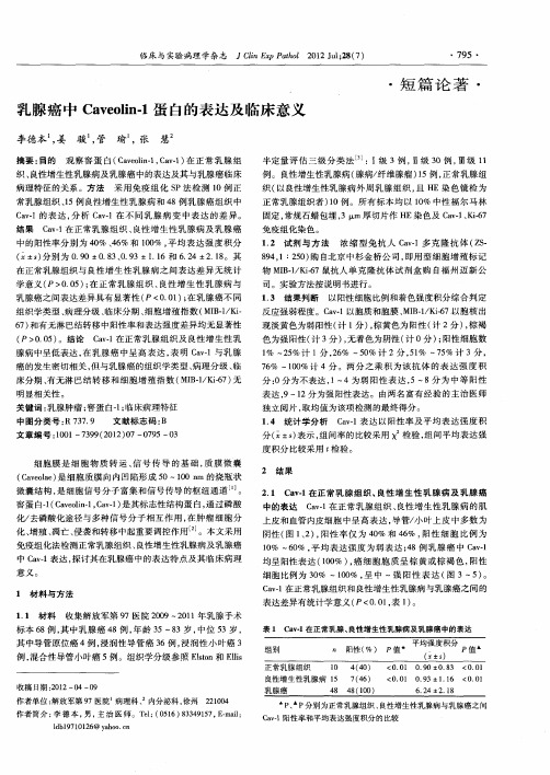
床分期 、 有无 淋 巴结 转移 和细胞 增殖 指数 ( B1 K - ) MI-/ i 7 无 6
明显相关性 。
关键词 : 腺肿瘤 ; 乳 窖蛋 白一; 1 临床病 理特征
组织学类型 、 理分级 、 病 临床 分期 、 细胞增殖 指数 ( I 一/ i M B1 K一
6) 7 和有无淋 巴结转移 中阳性 率和表达强度差异均无显著性 ( 00 ) P> .5 。结论 C v1 a. 在正常乳 腺组织及 良性增 生性乳
色为强阳性( 3 )无着色为阴性 ( 0分 ) 阳性 细胞数 计 分 , 计 ;
临床 与 实验 病 理 学 杂 志
JCi EpP to 2 1 u;8 7 l x ah l 0 2Jl2 ( ) n
・9 ・ 75
・
短篇论著 ・
乳腺 癌 中 C vo n1 白的 表 达及 临 床 意义 a el . 蛋 i
李德本 姜 ,
摘要 : 目的
骏 管 瑜 张 , ,
中图 分 类 号 : 3 . R7 7 9 文 献 标 志 码 : B
文 章 编 号 :0 1— 3 9 2 1 )7— 7 5— 3 10 79 ( 0 2 0 0 9 0
1 4 统计学分析 .
C v1表达 以阳性率 及平均 表达强 度积 a一
分( x±s表示 , ) 组间率 的比较采用 x 检验 , 组间平均 表达强 度积分 比较采用 t 检验 。
2 结 果
2 1 C v1在正 常乳腺 组织 、 . a . 良性 增 生性 乳腺病 及 乳腺癌 中的表达 C v1在正常乳 腺组织 、 a. 良性 增生性 乳腺病 的 肌 上皮和血管 内皮细胞 中呈 高表达 , 导管/ 叶上 皮 中多数 为 小 阴性( 12 , 图 、 ) 阳性率 仅 为 4 %和 4 % , 0 6 阳性 细胞 比例 为 1 % 一6 % , 0 0 平均 表达 强度 为弱表 达 ;8例乳 腺 癌 中 C v1 4 a・
cav1 综述
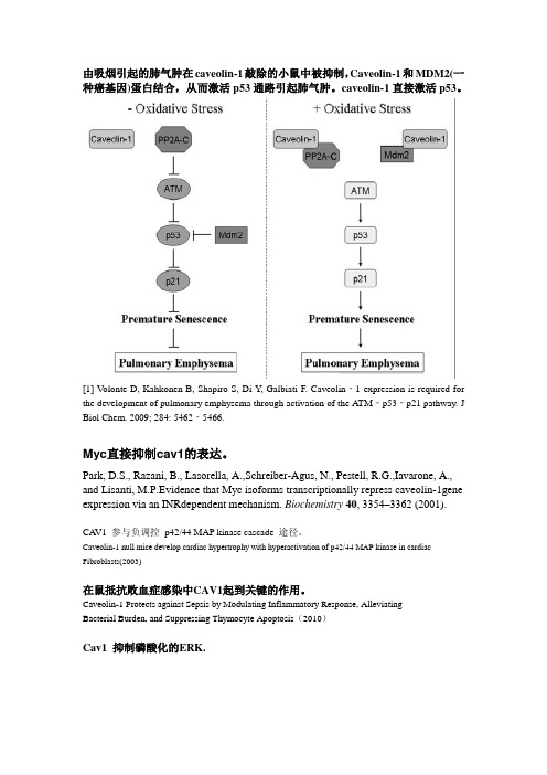
由吸烟引起的肺气肿在caveolin-1敲除的小鼠中被抑制,Caveolin-1和MDM2(一种癌基因)蛋白结合,从而激活p53通路引起肺气肿。
caveolin-1直接激活p53。
[1]V olonte D, Kahkonen B, Shapiro S, Di Y, Galbiati F. Caveolin‐1 expression is required for the development of pulmonary emphysema through activation of the ATM‐p53‐p21 pathway. J Biol Chem. 2009; 284: 5462‐5466.Myc直接抑制cav1的表达。
Park, D.S., Razani, B., Lasorella, A.,Schreiber-Agus, N., Pestell, R.G.,Iavarone, A., and Lisanti, M.P.Evidence that Myc isoforms transcriptionally repress caveolin-1gene expression via an INRdependent mechanism. Biochemistry40, 3354–3362 (2001).CA V1 参与负调控p42/44 MAP kinase cascade 途径。
Caveolin-1 null mice develop cardiac hypertrophy with hyperactivation of p42/44 MAP kinase in cardiac Fibroblasts(2003)在鼠抵抗败血症感染中CAV1起到关键的作用。
Caveolin-1 Protects against Sepsis by Modulating Inflammatory Response, AlleviatingBacterial Burden, and Suppressing Thymocyte Apoptosis(2010)Cav1 抑制磷酸化的ERK.Flotillin(浮舰蛋白)是一种脂伐标志性蛋白,参与脂伐骨架的形成,为信号传导分子提供平台并参与信号传导。
BRCA1在肿瘤中的表达与肿瘤耐药的研究进展
BRCA1在肿瘤中的表达与肿瘤耐药的研究进展摘要】乳腺癌易感基因1是一个重要的DNA损伤修复基因。
通过研究它在多种肿瘤中的表达水平及其与抗微管药物、顺铂药物敏感性的关系,发现其可作为抗微管药物、铂类药物化疗疗效的预测指标,有助于制定个体化化疗方案。
【关键词】乳腺癌易感基因1 抗微管药物顺铂【中图分类号】R730.23 【文献标识码】A 【文章编号】2095-1752(2012)06-0107-02化疗是肿瘤综合治疗的重要组成部分,临床上导致化疗失败的原因有很多,其中,肿瘤耐药是其重要因素之一。
由于分子遗传学的不同,不同个体对于同一种化疗药物敏感性也相差甚远。
因此,要想提高化疗疗效,就要寻找一种能预测化疗疗效的方法,针对每个个体合理的、准确的用药,同时也使患者免受不必要的毒副作用和经济负担。
在 2004 美国肿瘤临床学会(American Society of Clinical Oncology ASCO)年会上有人提出了个体化化疗(tailor chemotherapy TC)的概念,并指出未来 5~10 年是“标准化疗”向“个体化化疗”的过渡期[1]。
所谓“个体化化疗”即指,通过基因、蛋白等生物学指标的检测,肿瘤药敏检测筛选出最能够从化疗中获益的个体或人群。
有研究认为与肿瘤化疗相关的基因有以下六大类:药物动力学相关基因;各种生物酶相关基因;DNA损伤与修复相关基因;凋亡相关基因;致癌、抑癌基因;细胞增殖、转移相关基因等。
其中DNA损伤与修复相关基因较为关键,比如BRCA1、ERCC1、RRM1等。
随着肿瘤分子生物学的发展,发现B R C A1的表达水平与紫杉醇及顺铂化疗疗效有密切关系。
本文就这一领域的相关研究作一简单综述。
1 DNA损伤修复与肿瘤的关系DNA作为一种遗传物质,必须保持相对的稳定性。
各种物理的、化学的、生物的因素均可能造成DNA结构不同类型的损伤,如:DNA单、双链断裂;DNA链间以及DNA链和蛋白之间的交联;糖基氧化;碱基修饰等。
Caveolin-1在食管鳞癌中的表达及其意义
Caveolin-1在食管鳞癌中的表达及其意义赵醒;赵宇阳;王军;程玉;焦春敬;李春辉【期刊名称】《中华保健医学杂志》【年(卷),期】2014(016)003【摘要】目的研究正常食管黏膜和食管鳞癌组织中Caveolin-1蛋白的表达,探讨其与食管鳞癌发生发展的关系.方法采用免疫组织化学法分析50例食管鳞癌组织及10例正常食管黏膜中Caveolin-1表达情况.结果食管鳞癌中Caveolin-1的表达高于正常食管黏膜组织,Caveolin-1的表达与食管癌的淋巴结转移密切相关(P<0.05),而与年龄、肿瘤最大径、浸润深度无关(P>0.05).结论 Caveolin-1促进了食管癌的生长和转移,可作为食管癌的预后评估因子.【总页数】2页(P177-178)【作者】赵醒;赵宇阳;王军;程玉;焦春敬;李春辉【作者单位】067000 承德医学院附属医院病理科;067000 承德医学院附属医院病理科;067000 承德医学院附属医院病理科;067000 承德医学院附属医院病理科;宁波明州医院消化科;067000 承德医学院附属医院病理科【正文语种】中文【中图分类】R735.1【相关文献】1.150例食管鳞癌Caveolin-1和PCNA蛋白的表达及其生物学意义 [J], 赵醒;焦春敬;李春辉;胡潺潺2.Caveolin-1在食管鳞癌中的表达及其临床意义 [J], 葛腾飞;朱克超;于在诚;汪渊3.Caveolin-1在食管鳞癌中的表达及意义 [J], 黄小平;任必勇;朱川;熊德明;范运秀;刘华文;段松;杨杰斌4.Caveolin-1和β-catenin在食管鳞癌中的表达及临床意义 [J], 范玉宏;周海丰;侯占富;武雪亮;王立坤;侯雅雄5.Caveolin-1在内毒素性肺损伤所致的肺纤维化中的表达及临床意义 [J], 何创;王海燕;肖建斌;高巨;闫雪静因版权原因,仅展示原文概要,查看原文内容请购买。
细胞内吞作用的研究进展
Chinese-German Journal of Clinical Oncology June 2009, Vol. 8, No. 6, P360–P365 DOI 10.1007/s10330-009-0023-9In the process of cell metabolism, various kinds of ma-terials including some ions, small molecules, macromo-lecular materials and some granular matters keep in and out of cells. The macromolecular substances and granular materials can not go through the membrane. They com-plete the transfer across the cell membrane in another way, that is, materials cross in and out of cell membrane in the form of vesicle which is fused with membrane and then sciss into the cell, which is currently recognized as the main pathway of intaking biological macromolecules-endocytosis. Endocytosis is broadly divided into two cat-egories, phagocytosis and pinocytosis; the latter is divided into four species according to their different mechanisms: clathrin dependent endocytosis, caveolin dependent en-docytosis, macropinocytosis, and clathrin and caveolin independent endocytosis.The recent study reported that the abnormal expres-sion of endocytosis may be involved in the mechanism of certain diseases, such as diabetes and neurological diseases and also closely related to the malignant transformation of cells. The role of endocytosis was paid increasing atten-tion. Further research in this area will help us understand these diseases, thereby found new treatments. Here we introduce about pathway of cell endocytosis, mechanisms of regulation and endocytotic proteins. Endocytosis pathwayPhagocytosisPhagocytosis refers to endocytosing large granular matters (> 250 nm), and it provides the host a direct way digesting exogenous substances, which is one of the most important immune protecting mechanisms [1]. In mamma-lian, phagocytosis can only be completed by specific cells, such as macrophages and neutrophils which are called phagocytic cells. Phagocytic cells recognize and combine with IgFc and complement packing pathogens through IgFc receptor and complement receptor respectively. When the cell surface receptors combined with the cor-responding ligands, downstream signal transduction was activated, which caused actin polymerization under plas-ma membrane of intaking site, actin contraction makes phagocytic cell membrane form pseudo-foot fusing into vesicle to pack pathogens. In cytoplasm, dynamin assem-bled into ring at the neck of vesicle, and hydrolysed the binding GTP, dynamin contraction forced vesicle to sciss from membrane at the neck and form phagosome which integrated with the lysosome, the acid hydrolysis enzyme of lysosomal digested pathogens. In addition to phagocy-tosing pathogens, macrophages of phagocytic cells canStudy progress of cell endocytosis*Li Chen1, Hui Li1, Ren Zhao2, jianwei Zhu31Department of Pathology, Nantong University Medical College, Nantong 226001, China2Department of General Surgery, Ruijin Hospital, Shanghai Jiaotong University, Shanghai 230002, China 3Department of General Surgery, Affiliated Hospital, Nantong University, Nantong 226001, ChinaReceived: 19 February 2009 / Revised: 13 March 2009 / Accepted: 10 April 2009Abstract Endocytosis is a process through which extracellular materials are transported into cell through membrane defor-mation. This process is not a simple step-by-step process in which a series of proteins function according to the chronological order, but rather a complex process comprising many members which are regulated precisely. The role of endocytosis is broadly divided into two categories, phagocytosis and pinocytosis, the latter is divided into four species in accordance with the size of endocytosis substances: clathrin dependent endocytosis, the diameter of clathrin-coated vesicle is 100–150 nm;caveolin dependent endocytosis, the diameter of caveolin protein-coated vesicle is 50–100 nm; macropinocytosis, the diam-eter of macropinocytosis is generally 0.5–2 μm, sometimes up to 5 μm; clathrin and caveolin independent endocytosis. Many proteins including endophilin A1, A2, A3, and endocytotic proteins B, B1a, and B1b as well as dynamin, actin and Rab protein families are involved in endocytosis and play an important role in different stages. The abnormal endocytosis may be involved in the development of certain diseases.Key words endocytosis; clathrin; caveolin; endophilin; signalling pathwayCorrespondence to: Jianwei Zhu. Email: usazhujianwei@* Supported by grants from the National Natural Sciences Foundationof China (No. 30771126 and 30772106).361 Chinese-German J Clin Oncol, June 2009, Vol. 8, No. 6swallow foreign bodies through ligand-receptor binding pattern. Identify and kill tumor cells, identify and remove degenerative plasma protein, lipids, and other macromol-ecules. Remove aging and damaged cells and cell debris. PinocytosisPinocytosis refers to the intake process that endocyto-ses extracellular liquid and the dissolving materials. Clathrin dependent endocytosisClathrin plays an important role in the regulation of composition of plasma membrane proteins, and research on clathrin can help us understand how cells interact with the surrounding environment, signal transduction of mitogenic, nutrition intake of the cell, establishment of extracellular environment cell identity including the interaction with the immune system, keep a balance in the stability of the environment of cell.(1) Clathrin and adaptor protein (AP)Clathrin is the skeleton protein outside the vesicle, clathrin-coated vesicle is 100–150 nm in diameter and exists in all eukaryotic cells and it mediates the way of transportation from the plasma membrane to intracellu-lar of proteins, lipids, nutrients, antibodies and growth factors and also the vector through which proteins and lipids transport from trans Golgi net work (TGN) to endosome. Clathrin is spider-like and polymerized by three chains at the top, which is known as triskelion [1]. Adapter lies inside the clathrin-coated vesicle, which mediates membrane binding, localization, sorting signals, identification and inositol phosphate. It not only serves to combine cargo and clathrin, but also connect with polyphosphatidylinositol headgroup. It is now found four kinds of adapters (AP1–4), all of which comprise a pair of 100–130 kD subunit. These subunits are able to identify 6-phosphate mannose receptor, transferrin, low-density lipoprotein and epidermal growth factor receptor, prote-ase and de-sialic acid receptor.(2) MechanismThe process from recruitment to disassemble is very short. It comprises as follows:1) Recruitment of adapter and clathrin. Recruitment of AP2 complex to high activity, saturated, easy enzymolysis site, activate formation of clathrin-coated vesicle in the plasma membrane under the effect of sorting signal and docking protein [2].2) Invagination, scission and budding of clathrin-coat-ed vesicle. The planar clathrin protein can be transformed into curved one without nucleic acid and cytoplasm in vitro; In vivo, clathrin-coated vesicle curves to some de-gree. Invagination may be due to structure changes or rearrangement of clathrin assembling in the crystal lat-tice or between clathrin. The budding of clathrin-coated vesicle is involved with GTPase dynamin, actin tubulizes or forms ring-shape in vitro, it is the trigger that vesicle dissociated from the membrane.3) Decapsulation of clathrin-coated vesicle. This is a wasting process that needs hsc 70, auxilin and ATP. The large chain of clathrin has two sites which interact with the adapter and also with hsc 70. The interaction between clathrin and hsc 70 destroyed the interaction between clathrin and adapter. Over-expression of hsc 70 mutant blocked the cycle of transferrin receptor so that the “as-sembly – dismantle” balance moved to the assembly di-rection. Hsc 70 dissociated clathrin from the vesicle in vitro, but could not dissociate adapter. Auxilin plays an important role in decapsulation. Auxilin can not only re-cruit hsc 70 to clathrin-coated vesicle, but also stimulate activity of hsc 70 ATP enzyme.Caveolin dependent endocytosisCaveolae/caveolins mediate endocytosis of many sub-stances and is the main form of clathrin-independent en-docytosis.(1) As early as 1950, Japanese scholar Yamada used transmission electron microscopy to observe some small caveolae which was about 50–100 nm in diameter for the first time. These vesicles appeared alone or string-like, in the form of invagination of the cytoplasmic membrane, it had typical lipid bilayer structure [2]. Caveolae is currently considered to be the signal transduction center, and many signal transduction receptors, protein kinase and binding proteins are highly enriched in caveolae region. In the regulation of caveolae endocytosis, activation of tyrosine kinase-dependent signal is an important step. After the treatment of phosphatase inhibitor, the endocytosis of cell by caveolae had increased notably.(2) Caveolin is the main surface marker of a caveolae, a kind of membrane integrin family modified in the inner surface of caveolae, 21–25 kD. It consists of N-terminal region, transmembrane region and C-terminal region, N-terminal and C-terminal cytoplasmic moves inward into the cytoplasm and its peptide chains like hairpin struc-ture. Caveolin plays a critical role in the assembly process of caveolae and cholesterol and is the signaling molecule of the scaffold proteins and negative regulatory proteins and belongs to a highly conserved integrity membrane protein family [3]. Rajjayabun [4] confirmed that caveolin-1 was the key factor of caveolae endocytosis. If caveolin gene was knockout, caveolae can not be formed. So far, four kinds of caveolin isomers have been found in mammals: caveolin-1α, 1β, 2 and 3, which are products of differ-ent genes, most cells expressed caveolin-1 and caveolin-2, the two form a stable heterologous oligomers complexes, particularly rich in terminal differentiated cells, such as fat cells, endothelial cells and fibroblasts, and caveolin-3 mainly exists in various muscle cells (such as myocardial cells, rhabdomyosarcoma cells and skeletal muscle cells) and is closely related to the synthesis of muscle cells [5]. Neither caveolin protein nor caveolae structure appeared362 www. springerlink. com/content/1613-9089in peripheral blood cells and nerve cells.(3) The biological feature of caveolin. Caveolae as a sig-naling molecule plays a role as a platform in signal trans-duction. Caveolin-1 is in the center of signaling pathways at all these platforms. Caveolin, as a signaling molecule’s scaffold protein and negative regulatory protein, inhibits kinase activities of signaling molecules in the normal sig-nal transduction pathways. Caveolin-2’s skeleton region has no inhibitory activities, and other signaling molecules inhibitory regions may exit in it. Caveolin-3 is involved in energy metabolisms in muscle cells. Caveolin-1 has strong affinity to cholesterol, thus the cholesterol levels of caveolae is far higher than other biofilm. The study showed that the absence of caveolae eventually resulted in formation of foam cells, suggesting that caveolae and caveolin-1 can maintain the cholesterol balance by re-moving the excessive lipoprotein cholesterol out of cells [6].The correlation between caveolin and tumors have become one of the hot spots in tumor biology, among which, the relationship between caveolin-1 and tumor occurrence and metastasis has been studied a lot. Gene coding for caveolin-1 (CAV) locates in the suspicious tu-mor suppressive site (D7S522; 7q31.1). This site depletes or fractures in a variety of tumors (such as liver cancer, ovarian cancer, breast cancer, uterine fibroids, gastric ad-enocarcinoma, etc. [7]). The normal NIH 3T3 cell which was introduced by antisense caveolin-1 was transplanted into nude mice to observe the formation of tumor, sug-gesting caveolin-1 has tumor suppressive function. In many tumors and activated oncogene transfected cells, the expression of mRNA and its protein of caveolin-1 de-creased or lost [8]. The experiments in vivo demonstrated mutation or deletion of caveolin-1 could lead to the ex-cessive proliferation of breast epithelial cells, and then increase the occurrence of breast cancers [9]. Besides, the expression of caveolin uprehulated in diabetes and hyper-cholesterolemia and plays a significant role in Alzheim-er’s disease [10], degenerative muscle disease [11], heart and lung diseases.MacropinocytosisLarge and irregular original endocytosis vesicles are formed by folding membrane on the verge of extending cell under stimulation by certain factors, and they are known as macropinosome whose size varies with diam-eter generally 0. 5–2 μm. Macropinocytosis plays a major role in macrophages and dendritic cells, also in many tu-mor cells [11].Macropinosome has no clathrin or caveolins coating; it is closely related to actin at the early stages of formation, which provides an effective way for non-selective endo-cytosis of extracellular nutrients and liquid phase macro-molecules. Macropinocytosis and phagocytosis, regulated by a variety of proteins, such as actin, Scar protein, Ap21 complexes and RabB. Different membrane wrinkle results in different rates of developing macropinocytosis.The formation of macropinosomes can be significantly inhibited by cytochalasin D and colchicine, suggesting that microtubules and microfilaments play an important role in this process. The mechanism may be the stimulus by a variety of factors that activate corresponding recep-tor tyrosine kinase and then self-phosphorylate quickly. Phosphorylated residues recruit PI3 kinase and activate it, the activated PI3 kinase activates Rac1 which may cause microfilament reconstruction through two ways. Firstly, the activated Rac1 activates PAK1 which regulates phos-phorylation of myosin light chain. The interaction be-tween myosin and actin is regulated by the phosphory-lated myosin light chain and the interaction promotes reconstruction of microfilaments, development of the cell membrane ruffles and formation of macropinosomes; Secondly, the activated Rac1 binds with the amino-ter-minal of its target protein IRSp53, SH3 region at the car-boxyl end of IRSp53 combines with WAVE to form three molecular complexes which could activate WAVE [12]. The latter further activates actin related protein Arp2/3 complexes which stimulate microfilament nucleation, promote microfilament reconstruction, development of cell membrane ruffles and formation of macropinosomes [13]. In-depth regulation mechanism needs further study. MicrodomainMicrodomain has similar lipid composition with caveolae but not function through caveolin. It functions through dynamin and Rho A-dependent mechanism, or through clathrin-independent, dynamin-2-independent, Cdc4 mediated pathway to endocytose glycosylated phos-phatidylinositol-hexanol anchored proteins [13]. The path-way plays a role in endocytosis of IL-2. EndophilinThe biological features of endophilinThe main function of endophilin is related to the en-docytosis of neurotransmitter. Endophilin has a common special structure, whose C-terminal has a unique SH3 do-main with the unique binding ability [14], which can in-teract with a number of special proteins such as dynamin, thus affecting the functions of other proteins. While N-terminal participates in membrane invagination of vesi-cles. They were named endophilin A1, A2, A3 in 2002. Huntingtin belongs to binding protein of endophilin A1, the complex composed of it and the huntingtin binding protein 40 (HAP40) inhibited early microtubule-depen-dent endosome movement, increased connection be-tween early endosome and actin [15]. Then a new family was discovered, which was named endophilin B.363 Chinese-German J Clin Oncol, June 2009, Vol. 8, No. 6Endophilin and its role in signal transduction Endophilin can interact with various proteins through which involved in different signal transduction pathway Endophilins which interact with cell membrane re-ceptors are mainly distributed in the cytoplasm and cell membrane, it can interact with cytoplasmic membrane receptor and conduct signals, such as beta 1-adrenergic receptor. Endophilin is also involved in phospholipid sig-nal transduction. Many endophilins contain the binding sites of inositol 4,5-diphosphate (PIP2). The degradation of PIP2 is necessary for the completion of the endocytosis. Mark PIP2 connected domain by immunohistochemical fluorescent protein and track endocytosis movement of living cells, and PIP2 was found present in the cell mem-brane surface in the form of many small plaques, these plaques entered gradually into the cell, which confirmed that the shift was the endocytosis. The disappearance of PIP2 matched the recruitment of phosphoinositide phosphatase enzyme, and we can propose that the latter can degraded the former. When PIP2 degradation was blocked, abnormal invagination can be observed, shear-ing machine of endocytosis gathered around the plaque on the membrane and failed to complete endocytosis. Therefore, PIP2 degradation is the necessary step of ves-icle scission [16].Recycling of the neurotransmitter Neurotransmitter recycle is essentially a process of cell endocytosis, clathrin interact with AP2 or AP180 (adapter protein) and membrane lipid to form clathrin-coated ves-icles. The process of its formation is just that endophilin A1 drove the rupture of membrane vesicles from the do-nor membrane at the neck of vesicles, LPAAT at N-termi-nal of endophilin A1 can catalyze lysophosphatidic acid (LPA) and arachidonic ene-CoA to form lysophosphatidic acid (PA), LPA is a three-dimensional structure like in-verted cone-type and PA is cone-type, the former struc-ture is beneficial for positive membrane deformation, the later is conducive to negative membrane deformation. It is the transformation of lysophosphatidic acid that drive rupture between vesicle and the donor vesicle membrane. After endophilin mutation, it will severely affect recycle mechanism of synaptic particle [17].Function besides endocytosisEndophilin A2 is actually involved in absorbing nu-trients and growth factors and endocytosis of pathogens and receptors, participating in a variety of membrane transport mechanism . While endophilin B1 locates in the membrane of the cell and its main function is relat-ed to cell membrane dynamics. Apoptosis receptor Fas/ CD95 can not correctly located in the membrane without endophilin, resulting in inhibition of the mechanism of apoptosis [18]. Endocytosis related proteinsDynaminDynamin is a 100 kD GTPase, and its GTPase region at N-terminal can bind and hydrolyse GTP, PH (pleck-strin homology) region can bind with membrane and me-diate polymerization between dynamins, PRD (praline arginine rich) region at C-terminal mediate the interac-tion between other proteins. In addition, dynamin has a small GED (GTPase effector) region which is necessary in hydrolysis of GTP [18], and also plays an important role in some clathrin independent endocytosis. Dynamin is necessary in pinching and formation of vesicles. After its binding with ligand amphiphysin-1, dynamin-1 plays a key role in dynamin dependent endocytosis under regu-lation of cycle-dependent kinase 5 (Cdk5). Dynamin-1-dependent endocytosis occurs quickly, usually only a few seconds, while dynamin-2 mediated endocytosis is slow, usually ten minutes.Dynamin uses mechanochemical activity – specifi-cally a twisting action – to pinch off endocytic vesicles [19]. Dynamin was, early on, localized to the collar around the neck of forming endocytic vesicles. This suggested that dynamin may use the energy of GTP hydrolysis to directly pinch a membranous neck. Indeed, dynamin could tubulate lipids and break apart the tubules in vitro, although later it seemed that the breaking apart was hap-pening as the samples dried on EM grids. Meanwhile, Schmid had come up with a “regulatory GTPase” mode: that dynamin was active not as it hydrolyzed GTP but in its GTPbound form, which recruited other proteins to do the pinching. The Yale group has more evidence for the earlier “pinchase” model. They used light microscopy rather than EM to follow tubulation and fission directed by dynamin in vitro. Longitudinal tension was needed with constriction to achieve fission. In mammalian, the dynamin collars are relatively short, so cortical actin is the most likely source of tension that would help the dynamin to wrench an endocytic vesicle free [20–23]. Dynamin has intrinsic activity of GTP and is also the cerebellum Dyrk1A kinase substrate, with its own ag-gregation feature of the endometrium can help new pro-cesses from the cell membrane vesicles isolated. Dynamin is involved in tubulation by PH epsin and ENTH (epsin NH2-terminal homology) [21].Wild-type dynamin mutant block the tube, which can be restricted to speculate dynamin of these membrane protein of the ability to control grid protein coated vesi-cle endocytosis and other media invagination degree. ActinActin is the main component of microfilament. Microfilament is involved in cell shape and polarity of the maintenance of endocytosis, intracellular transport,364 www. springerlink. com/content/1613-9089cell shrinkage and movement, cell division, and many other functions. Actin has two kinds: G-actin and F-ac-tin. The importance of F-actin in endocytosis was clearly illustrated by the effect of addition of the actin-mono-mersequestering drug latrunculin A. Within 5 minutes of its addition, actin patches were no longer visible and endocytosis was completely abrogated. This was the point at which the yeast WASP orthologue Las17p and the type I myosins (Myo3p and Myo5p) began to nucleate actin filaments through activation of the Arp2/3 complex [24]. This group of proteins has been called the “actin net-work growth machinery”. Some researchers combined epifluorescence with total internal reflection microscopy (TIRF) to follow the internalisation of individual coated pits in living cells expressing a fluorescent tagged form of clathrin light chain (DsRed clathrin). They showed that transient recruitment of actin coincides with the inward movement of vesicles, they also observed recruitment of the Arp2/3 complex to clathrin-coated pits. PH-sensitive probes have been developed that allow internalisation of cargo into individual clathrin-coated vesicles to be visual-ized, allowing us to follow the time course of invagination and identify the point of scission. The recruitment of actin during invagination is thought to provide the force that drives the invagination of the coated pit [25]. It is currently identified that the vesicle scission module contains the two yeast amphiphysin proteins Rvs161p and Rvs167p. These proteins contain BAR (Bin-Amphiphysin-Rvs) domains that bind to and tabulate, actin and associated proteins recruit amphiphysins and thus mark the site at which membrane tubulation should occur. Tubulation is then suggested to facilitate the scission process. Following scission, the vesicle is uncoated and moves away from the membrane until it fuses with an endosome. There are two possible roles for actin at this final stage: vesicles could move along actin cables; alternatively actin could be nu-cleated at the vesicle surface to facilitate their movement within the cell. Then the actin depolymerized from the endocytosis site. The most representative depolymeriz-ing factor is cofilin. Cofilin is necessary for endocytosis in mamamals, but its mechanism is still unknown [26]. In the process of invagination, Sac6p/microfilament binding protein/fimbrin is also required, invagination induced by actin failed without Sac6p [27].PCH (Pombe Cdc15 homology) / F-BARPCH (Pombe Cdc15 homology) / F-BAR is another family of proteins which plays an important role in en-docytosis. The BAR region of these proteins combine with phosphoinositide. FBP17 is a member of PCH fam-ily, containing PCH domain and extended FC domain. The EFC domains show weak homology to the Bin-am-phiphysin-Rvs (BAR) domain. The EFC domains bound strongly to phosphatidylserine and phosphatidylinositol 4,5-bisphosphate and deformed the plasma membrane and liposomes into narrow tubules. Most PCH proteins possess an SH3 domain that is known to bind to dynamin and that recruited and activated neural Wiskott-Aldrich syndrome protein (N-WASP) at the plasma membrane. FBP17 contributed to the formation of the protein com-plex, including N-WASP and dynamin-2, in the early stage of endocytosis. Furthermore, knockdown of endog-enous FBP17 impaired endocytosis, suggesting FBP17 is necessary for dynamin-dependent endocytosis [28].Rab proteinsRab protein which is a small GTP enzyme is an im-portant regulator of endocytosis. Rab5 is involved in tubulization and its role in endocytosis has been clear. Rab5 protein has three isomers, Rab5A, Rab5B and Rab5C. Rab5 protein exists primarily on the plasma mem-brane, clathrin-coated vesicle and early endosome, tak-ing charge of vesicle fusion and recycling. Rab5 protein functions with ongoing GTP/GDP cycle. As molecular switch of vesicle transport, the activated state of combin-ing with GTP is “open”, the unactivated state of combin-ing with GTP is called “close”. Rab5 regulates transport of endocytotic materials between membrane and early en-dosome, and help vesicle movement along microtubules. HAP40 is an effective regulatory factor of Rab5, and Rab5 increase connections of endosome with actin un-der existence of HAP40. RAB5A protein was related to endocytosis of prostacyclin [29]. A-synuclein is the root of Parkinson’s disease, Lewy body Dementia and Alzheim-er’s disease and other central nervous system diseases. Moreover, Young found the mutant can decrease endo-cytosis of A-synuclein according to RAB5A GTP mutant enzyme research while Lewy body-like decreased in cy-toplasm, the cytotoxicity also decreased. Over-expression of RAB5A protein in immune system deficiency, which can slower macrophage phagocytosis of pathogens and decrease the rate of degradation of pathogens and IFN-C-mediated phagocytosis weakened.ConclusionWe have found many molecules involved in endocy-tosis in the past few decades, and had a preliminary un-derstanding about its process and metabolism. We now know that the process of endocytosis is not a simple step-by-step process in which a series of proteins function ac-cording to the chronological order, but rather a complex process comprising many members which are regulated precisely. Although the mechanism of regulation of en-docytosis is still unknown, it can be predicted that as more and more new research techniques applied in this area, we will be able to understand the mechanism of cell endocytosis more comprehensively. Whether tumor cells365Chinese-German J Clin Oncol, June 2009, Vol. 8, No. 6endocytose more nutrients than normal cells and wheth-er tumor cells increase sizes through endocytosing more growth factors? Can we inhibit tumor growth through inhibiting cell endocytosis or can we cure tumor by in-ducing specific drug endocytosis of tumor cells? Whether the endocytosis of 6-phosphate mannose receptor (MPR) which plays a very important role in cell death signal transduction is restrained in tumor cells? The mechanism of cell endocytosis can be further elucidated by solving all of the above problems.ReferencesGiodini A, Rahner C, Cresswell P. Receptor-mediated phagocytosiselicits cross-presentation in nonprofessional antigen-presenting cells. Proc Natl Acad Sci USA, 2009, 106: 3324–3329.Kirchhausen T. Clathrin. Annu Rev Biochem, 2000, 69: 699–727.Kiss AL, Turi A, Müller N, et al . Caveolae and caveolin isoforms in ratperitoneal macrophages. Micron, 2002, 33: 75–93.Rajjayabun PH, Garg S, Durkan GC, et al . Caveolin-1 expressionis associated with high-grade bladder cancer. Urology, 2001, 58: 811–814.Frank PG, Woodman SE, Park DS, et al . Caveolin, caveolae andendothelial cell function. Arterioscler Thromb Vasc Biol, 2003, 23: 1161–1168.Krajewska WM, Masłowska I. Caveolins: structure and function in sig -nal transduction. Cell Mol Biol Lett, 2004, 9: 195–220.Pascariu M, Bendayan M, Ghitescu L. Correlated endothelial caveolinoverexpression and increased transcytosis in experimental diabetes. J Histochem Cytochem, 2004, 52: 65–76.Fra AM, Pasqualetto E, Mancini M, et al . Genomic organization andtranscriptional analysis of the human genes coding for caveolin-1 and caveolin-2. Gene, 2000, 243: 75–83.Cameron PL, Liu C, Smart DK, et al . Caveolin-1 expression is main-tained in rat and human astroglioma cell lines. Glia, 2002, 37: 275–290.Williams TM, Cheung MW, Park DS, et al. Loss of caveolin-1 gene ex-pression accelerates the development of dysplastic mammary lesions in tumor-prone transgenic mice. Mol Biol Cell, 2003, 14: 1027–1042.Gaudreault SB, Dea D, Poirier J. Increased caveolin-1 expression inAlzheimer′s disease brain. Neurobiol Aging, 2004, 25: 753–759.Müller JS, Piko H, Schoser BG, et al . Novel splice site mutation in thecaveolin-3 gene leading to autosomal recessive limb girdle muscular dystrophy. Neuromuscul Disord, 2006, 16: 432–436.Lim JP, Wang JT, Kerr MC, et al . A role for SNX5 in the regulation ofmacropinocytosis. BMC Cell Biol, 2008, 9: 58.1.2.3.4.5.6.7.8.9.10.11.12.13.Miki H, Yamaguchi H, Suetsugu S, et al . IRSp53 is an essential in -termediate between Rac and WAVE in the regulation of membrane ruffling. Nature, 2000, 408: 732–735.Sun P, Yamamoto H, Suetsugu S, et al . Small GTPase Rah/Rab34 isassociated with membrane ruffles and macropinosomes and promotes macropinosome formation. J Biol Chem, 2003, 278: 4063–4071.Daniels RL, Takashima Y, McKemy DD. Activity of the neuronal coldsensor TRPM8 is regulated by phospholipase C via the phospholipid phosphoinositol 4,5-bisphosphate. J Biol Chem, 2009, 284: 1570–1582.Gad H, Ringstad N, Löw P, et al . Fission and uncoating of synapticclathrin-coated vesicles are perturbed by disruption of interactions with the SH3 domain of endophilin. Neuron, 2000, 27: 301–312.Petrelli A, Gilestro GF, Lanzardo S, et al . The endophilin-CIN85-Cb1complex mediates ligand-dependent downregulation of c-Met. Na-ture, 2002, 416: 187–190.Williams R. PIP2 in endocytosis. J Cell Biol, 2007, 177: 185.Guichet A, Wucherpfennig T, Dudu V, et al . Essential role of endophilinA in synaptic vesicle budding at the Drosophila neuromuscular junc-tion. EMBO J, 2002, 21: 1661–1672.Takei K, McPherson PS, Schmid SL, et al . Tubular membrane invagi-nations coated by dynamin rings are induced by GTP-gamma S in nerve terminals. Nature, 1995, 374: 186–190.Zhang X, Wang F, Chen X, et al . Post-endocytic fates of delta-opi-oid receptor are regulated by GRK2-mediated receptor phosphory-lation and distinct beta-arrestin isoforms. J Neurochem, 2008, 106: 781–792.William A. Twisting endocytosis. J Cell Biol, 2006, 173: 456.Damke H, Binns DD, Ueda H, et al . Dynamin GTPase domain mu-tants block endocytic vesicle formation at morphologically distinct stages. Mol Biol Cell, 2001, 12: 2578–2589.Tsujita K, Suetsugu S, Sasaki N, et al . Coordination between the ac-tin cytoskeleton and membrane deformation by a novel membrane tubulation domain of PCH proteins is involved in endocytosis. J Cell Biol, 2006, 172: 269–279.Sun Y, Martin AC, Drubin DG. Endocytic internalization in buddingyeast requires coordinated actin nucleation and myosin motor activity. Dev Cell, 2006, 11: 33–46.Okreglak V, Drubin DG. Cofilin recruitment and function during actin-mediated endocytosis dictated by actin nucleotide state. J Cell Biol, 2007, 178: 1251–1264.Kaksonen M, Toret CP, Drubin DG. A modular design for the clathrin-and actin-mediated endocytosis machinery. Cell, 2005, 123: 305–320.O’Keeffe MB, Reid HM, Kinsella BT. Agonist-dependent internaliza-tion and trafficking of the human prostacyclin receptor: a direct role for Rab5a GTPase. Biochim Biophys Acta, 2008, 1783: 1914–1928.14.15.16.17.18.19.20.21.22.23.24.25.26.27.28.29.。
Caveolin and caveolae in age associated cardiovascular disease
Journal of Geriatric Cardiology(2013)10:6674 2013JGC All rights reserved;Review Open Access Caveolin and caveolae in age associated cardiovascular diseaseHeidi N.Fridolfsson1,Hemal H.Patel1,21Dep artments of Anesthesio lo gy,University of Californ ia,San Diego,La J olla,California92093,USA2VA San Dieg o Healthcare Sy stem,3350La J olla V illage Drive,San Diego,CA92161,USAAbstractIt is estimated that the elderly(>65years of age)will increase from13%14%to25%by2035.If this trend continues,>50%of the United States population and more than two billion people worldwide will be“aged”in the next50years.Aged individuals face formidable challenges to their health,as aging is associated with a myriad of diseases.Cardiovascular disease is the leading cause of morbidity and mor-tality in the United States with>50%of mortality attributed to coronary artery disease and>80%of these deaths occurring in those age65 and older.Therefore,age is an important predictor of cardiovascular disease.The efficiency of youth is built upon cellular signaling scaffolds that provide tight and coordinated signaling.Lipid rafts are one such scaffold of which caveolae are a subset.In this review,we consider the importance of caveolae in common cardiovascular diseases of the aged and as potential therapeutic targets.We specifically address the role of caveolin in heart failure,myocardial ischemia,and pulmonary hypertension.J Geriatr Cardiol2013;10:6674.doi:10.3969/j.issn.1671-5411.2013.01.011Keywords:Cardiovascular disease;Caveolin;Lipid rafts;The aged1IntroductionCardiovascular disease is the leading cause of mortality in the United States and more than80%of these deaths oc-cur in those65years or older.[1,2]Advanced age is the most important predictor of poor outcome in patients with acute coronary syndromes[3]and with the projected increase in the elderly population worldwide,it is important to understand the risk factors associated with aging in order to identify appropriate treatments.The aged heart has an increased sensitivity and decreased tolerance to ischemia/reperfusion (I/R)injury.[4,5]Aging also results in a loss of the heart’s ability to respond to cardioprotective stimuli such as phar-macological and ischemic preconditioning.[6,7]It is believed that the aging cardiomyocyte develops a reduced tolerance to stress because of decreased mitochondrial function,in-creased oxidative stress,changes in gene expression,and aberrant cell signaling.[8]Lipid rafts are subcellular microdomains of the plasma membrane that consist of lipid clusters enriched in choles-terol and sphingolipids,in which particular proteins areCorrespond ence to:Hemal H.Patel,PhD,Dep artment of Anesthesiology, University of California,San Diego,V ASDHS(9125),3350La Jolla Vil-lag e Drive,San Diego,CA92161,USA.E-mail:hepatel@u Telephone:+1-858-552-8585-x3052Fax:+1-858-534-0104 Received:August7,2012Rev ised:December15,2012 Accepted:Decemb er18,2012Pu blished online:M arch28,2013concentrated.[9]Caveolae,a subset of lipid rafts,have a unique flask-like structure that is generated by caveolin and cavin proteins.[10-14]Caveolae and caveolins act to coordi-nate cellular signaling events in many cells,including those of the cardiovascular system.[15]Age-associated alterations in the composition of lipid rafts and caveolae could affect a variety of cellular functions and have been linked to dis-eases,such as Alzheimer’s,atherosclerosis,and diabetes, which are more prevalent in the elderly population.[16]The focus of this review will be on the role of lipids rafts and caveolae in the progression of age related cardiovascular diseases,such as heart failure,myocardial infarction,and pulmonary hypertension.2Caveolae and caveolinsCaveolae,or“little caves”,are cholesterol and sphingo-lipid-enriched invaginations of the plasma membrane.[10] Caveolins,structural proteins of caveolae,are present in three isoforms.[11]Caveolin-1(Cav-1)is required for caveo-lae formation in many non-muscle cell types and for the membrane localization of Cav-2,[17]while Cav-3is required for caveolae formation in striated(skeletal and cardiac)and smooth muscle cells.[18]Caveolins function as chaperones and scaffolds for signaling molecules in caveolae by pro-viding temporal and spatial regulation of signal transduc-tion.[11]Through their caveolin scaffolding domain(CSD),h ttp://www.j g c301.co m;jgc@jg c301.co m|J o u rn a l o f Ger iatric Card io lo g ycaveolins not only anchor other proteins in caveolae,but also inhibit or enhance that protein’s signaling capacity. Caveolins have a variety of other functions including ve-sicular transport,cholesterol and calcium homeostasis,and t-tubule formation.[19,20]The generation of mice lacking the caveolin genes has made it possible to better understand the significance of each caveolin isoform and its contribution to whole animal physiology and human disease.While all caveolin null mice [Cav-1,Cav-2,Cav-3single knockouts and Cav-1/3double knockout(KO)]are viable and fertile,each displays a unique phenotype.Cav-1KO mice have a complete loss of caveolae in non-muscle cells and a90%loss of Cav-2ex-pression due to protein degradation.[21,22]Thus,Cav-1/3 double KO mice lack expression of all three caveolin pro-teins and do not form visible caveolae in any cell type.[23] Caveolin protein expression and association with caveolae is decreased with age and studies of the caveolin KO mice support the hypothesis that the loss of caveolin protein causes an aged phenotype.[24,25]Caveolins are,therefore, potential therapeutic targets in the treatment of age related disorders,such as cardiovascular disease.3Heart failureDespite advances in diagnosis and treatment,heart failure remains one of the most common,costly,and deadly dis-eases.[26]The prevalence of the disease is significantly in-creased in people over65years of age and arises as a con-sequence of abnormal cardiac structure,function,rhythm,or conduction.[26]Ventricular dysfunction resulting from myo-cardial infarction and/or hypertension is frequently the cause of heart failure and each will be discussed in more detail in the sections below.Although Cav-3is the predominant caveolin in cardiac myocytes,Cav-1KO mice develop a severe cardiomyopa-thy,which appears to contribute to their significantly short-ened life span.At24months,Cav-1KO mice show a50% reduction in viability,with a major decline between27and 65weeks of age.[27]The hearts of Cav-1KO mice are struc-turally and functionally abnormal at2–4months of age. Imaging and functional studies revealed that Cav-1KO hearts have significantly enlarged ventricular chambers, abnormal ventricular wall thickness,hypertrophy,and de-creased contractility.[28,29]These defects are worse by12 months of age,which shows that loss of Cav-1causes a progressive cardiomyopathy.[27]Increased fibrosis in the heart has also been reported in Cav-1KO mice.[22,27]This could affect stiffness of the heart and contribute to impaired myocardial function.Since Cav-1is expressed in both cardiac myocytes and fibroblasts,[30]it is not clear if this decline with aging is due to altered function of a specific cardiac cell(i.e.,myocyte death and replacement by fibroblasts vs.increased activation of fibroblasts).As signaling scaffolds,caveolins have been shown to interact with receptor tyrosine kinases,Src family tyrosine kinases,endothelial nitric oxide synthase(eNOS), and members of the p42/44MAP kinase cascade(MEK1/2 and ERK1/2).[31]At the molecular level,loss of Cav-1is thought to cause cardiac hypertrophy through disruption of signaling pathways.Cav-1is thought to negatively regulate the p42/44MAP kinase cascade in cardiac fibroblasts[32]and activation of the p42/44MAPK pathway in cardiac myo-cytes can drive cardiac hypertrophy.[33]In Cav-1KO mice the p42/44MAP kinase cascade is hyperactivated,which contributes to the cardiac defects seen in these mice.[28]This finding suggests that the major effect of Cav-1loss may come from its function in the supporting cells of the heart (fibroblasts and endothelium).However,Cav-1is also found in the cardiac myocyte and its role in these cells can-not be dismissed.In heart failure,β-adrenergic signaling is reduced and this may lead to further deterioration of heart function since,with decreased contraction,the heart is un-able to meet its needs.[34]In Cav-1KO hearts,the levels of cyclic AMP(cAMP),an important second messenger of β-adrenergic signaling,and ATP are decreased.[35] Cardiac myocytes from Cav-3KO mice completely lack caveolae.[36]Cav-3KO mice develop a progressive cardio-myopathy marked by significant hypertrophy,dilation,and reduced fractional shortening.[37]Similar to Cav-1KO mice, the p42/44MAP kinase cascade is hyperactivated in the hearts of Cav-3KO mice and may partly account for these cardiac defects.[37]Similarly,a mutation in Cav-3has been found in familial hypertrophic cardiomyopathy.[38]In car-diac myocytes,t-tubules ensure rapid,uniform cell activa-tion.In heart failure there is an extensive remodeling of the t-tubule network and this change may contribute to abnor-mal calcium handling.[39]The disorganization of t-tubules in Cav-3KO skeletal muscle suggests that cardiac myocytes may display a similar phenotype that contributes to the pro-gression of heart failure.The activity of dozens of ion channel proteins and mem-brane transporters generate the flux of ions across the sar-colemmal membrane,which is responsible for activation and contraction.Many of these ion channels reside in caveolae and abnormalities in the function or regulation of these channels result in arrhythmias that can lead to heart failure.[40]Therefore,the loss of Cav-3and caveolae in the cardiac myocyte could contribute to arrhythmias as another potential source of heart failure.In fact,mutations in Cav-3http://www.jg c301.co m;jgc@mail.s cien |J o u rn al o f G eria tric Ca rd io lo g yhave been found in the inherited arrhythmogenic syndrome, long-QT congenital syndrome.[41]Mice with a constitutive overexpression ofα1-adenosine receptor demonstrate car-diac dilation and decreased left ventricular function.[42]In this model of heart failure,Cav-3expression is decreased (Cav-1and Cav-2are unaffected)and there is a direct rela-tionship between Cav-3expression and ventricular dysfunc-tion.[42]Cav-3levels are also decreased in the failing human heart.[42]Consistent with the findings that loss of Cav-3contrib-utes to heart failure,overexpression of Cav-3has been shown to attenuate heart failure.Natriuretic peptides are endogenous hormones released by the heart to modulate cardiac hypertrophy.[43]Caveolae contain natriuretic peptide receptors such as atrial natriuretic peptide(ANP),which is closely associated with Cav-3.[44]Overexpression of Cav-3 attenuates cardiac hypertrophy,at least partially,by in-creasing natriuretic peptide expression and signaling.[45] Overexpression of Cav-3may also prevent hypertrophy in cardiomyocytes by suppression of ERK1/2signaling.[46] These results suggest Cav-3may not only be a marker for heart failure,but also a therapeutic target.Cav-1/3double KO mice lack all caveolin protein ex-pression and caveolae formation.This combined loss of caveolin protein does not produce any new phenotypes and only cardiac defects were exacerbated compared to single knockout animals.[23]Cav-1/3double KO mice display a severe cardiomyopathy by two months of age in which left ventricular wall thickness,hypertrophy,ventricular dilation, and decreased fractional shortening that is more pronounced. The cardiac tissue also shows signs of interstitial inflamma-tion,fibrosis,and myocyte necrosis.These results clearly establish a role for both Cav-1and Cav-3in maintaining normal cardiac function and further suggest that a loss of caveolin proteins with age could be a major contributing factor to the development of cardiovascular disease.4Myocardial infarctionIschemic heart disease and myocardial infarction result in loss of blood flow and oxygen to part of the heart,which causes damage and reduced cardiac function.The incidence of myocardial infarction is increased in the elderly popula-tion.[1]Evidence suggests caveolae and caveolins are in-volved in the pathogenesis of ischemic injuries.Changes in caveolin protein expression have been identified in renal, cerebral,hindlimb,and myocardial ischemia.[47–49]Cerebral artery occlusion causes a marked increase in endothelial Cav-1and Cav-1KO mice display a significant increase in the volume of cerebral infarcts,as compared with wild-type mice.[50]In this model,Cav-1KO mice displayed decreased proliferation of endothelial cells and increased apoptosis.In a model of myocardial infarction,Cav-1KO mice subjected to left anterior descending coronary artery ligation display reduced survival and despite similar infarct sizes,Cav-1KO mice showed reduced cardiac function compared to wild-type mice.[51]Mechanistically,it appears that caveolins provide protection from ischemic injury through scaffolding signaling molecules and promoting cell survival.[49,51]Spe-cifically,reduced Cav-1expression altersβ-adrenergic sig-naling through decreased cAMP production and protein kinase A phosphorylation,which alters cardiac contractil-ity.[51]Recent evidence suggests that caveolins may also induce cardioprotection through epigenetic regulation.[52] Ischemic preconditioning is an innate protective mecha-nism by which brief episodes of ischemia protect the heart from the damaging effects of prolonged I/R injury.[53]In addition to sublethal ischemia,several pharmaceutical agents,such as opioids and volatile anesthetics,produce a similar preconditioning protective effect.[54]Preconditioning activates a complex signaling cascade known as the reper-fusion injury salvage kinase pathway and many of these signaling molecules associate with caveolins in caveo-lae.[55–57]Caveolae play a pivotal role in the generation of survival signals in cardioprotection and ischemic precondi-tioning increases the number of caveolae present in the sar-colemmal membrane.[58]Methyl-β-cyclodextrin disrupts lipid rafts and caveolae by removing cholesterol from the membrane.Preconditioning the heart in the presence of methyl-β-cyclodextrin abolishes the cardioprotective effects leading to decreased ventricular performance,increased myocardial infract size and cardiomyocyte apoptosis.[48,59] Additionally,aging is associated with a decrease in the pro-tective effects of preconditioning in animal models and hu-man patients.[6–8,60,61]The loss of caveolae and caveolins with age may,therefore,contribute to the loss of signal regulation and protection.Ischemic preconditioning-induced protection is lost in caveolin KO mice.[52,58,62]Caveolins also function in pre-conditioning with isoflurane,a volatile anesthetic,and opioids.Cardiac myocytes are protected from simulated I/R injury when incubated withδ-opioid receptor agonists.This effect is lost in the presence of methyl-β-cyclodextrin,indi-cating a critical role for caveolae.[48]Cav-3KO mice cannot be preconditioned withδ-opioid receptor stimulation.[63] Isoflurane-induced cardioprotection is dependent on the presence of caveolae,and is lost in both Cav-1and Cav-3 KO mice.[64–66]Perhaps the most interesting evidence for the role of caveolins in cardioprotection comes from analysis of a transgenic mouse with myocyte specific overexpression ofJ ou rn a l o f Ge riatric Card iolog y|jg c@j g c301.co m;http://www.j g c301.co mCav-3(Cav-3OE).[58]Cav-3overexpression causes en-hanced formation of caveolae on the cardiac myocyte sar-colemmal membrane.Cav-3OE mice subjected to I/R in-jury have significantly improved functional recovery,re-duced infarct size and apoptosis relative to wild-type mice. This innate cardioprotection in Cav-3OE mice is similar to that seen in wild-type mice undergoing ischemic precondi-tioning.[58]The precise molecular mechanism by which caveolins protect the heart from I/R injury remains to be determined.However,a generalized pathway has emerged in which preconditioning stimuli enhance release of agonists of one or more G-protein coupled receptor families(opioid, adenosine,bradykinin)to enhance activity of pro-survival kinase pathways(including PI3-K/Akt and MAPKs)and inhibit activity of pro-death pathways(GSK3β).[67]The abil-ity of caveolin to preserve such signaling as the heart ages may be a key feature of caveolin mediated stress adaptation. 5Pulmonary hypertensionSevere pulmonary hypertension(PH)is characterized by a progressive increase in pulmonary vascular resistance and vascular remodeling leading to right heart failure and early death.The ability of the right ventricle to respond to the increased vascular resistance is influenced by several factors, including age.Cav-1is highly expressed in the lung and is found in pulmonary endothelial cells.Disruption of the function of Cav-1in the vascular system can have profound effects on cardiac function and may serve as a marker for pulmonary hypertension,which would allow earlier detec-tion and improved treatment strategies.Cav-1KO mice show lung abnormalities,with reduced alveolar spaces,increased wall thickening,fibrosis,and hypercellularity.[22,68]Direct measurement of pulmonary artery pressure and histological analysis revealed that Cav-1 KO mice exhibit pulmonary hypertension,which may con-tribute to the right ventricle hypertrophy seen in these mice.[29]Additionally,lungs from Cav-1KO mice exhibit increased pulmonary vascular resistance associated with pulmonary vascular remodeling including increased medial thickness and muscularization of distal pulmonary vessels, which is an underlying feature of pulmonary vascular re-modeling in PH.[69]Reduced expression of Cav-1in the lungs after myocardial infarction has been suggested to ini-tiate the development of PH and lung structural remodel-ing.[70]Patients with severe pulmonary hypertension show reduced levels of Cav-1in total lung tissue and pulmonary vascular endothelial cells.[65]Cav-1polymorphisms have also been identified in pulmonary hypertension patients.[71] These findings suggest an antihypertensive function of Cav-1with respect to the pulmonary vascular architecture.Cav-1KO mice display hyperactivation of eNOS and al-tered vasoconstriction and vasorelaxation responses of iso-lated aortic rings.[22,29,68,72]Disruption of eNOS activity in Cav-1KO mice is likely involved in the development of the observed cardiopulmonary pathologies.eNOS is a critical mediator of cardiovascular homeostasis regulating multiple physiologic and pathophysiologic processes including vas-cular tone,vascular remodeling,platelet aggregation,and angiogenesis.[73]Through interactions with Cav-1,eNOS is targeted to caveolae in endothelial cells.[74]Several studies have demonstrated a role for Cav-1as a negative regulator of eNOS.Direct interaction of eNOS with Cav-1signifi-cantly inhibits its enzyme activity in vitro[75]and infusion of a membrane permeable Cav-1CSD peptide performs simi-larly in vivo.[76]The hyperactivation of eNOS in Cav-1KO mice results in increased NO production,which is believed to cause the majority of the cardiovascular defects.Double KO of Cav-1and eNOS(NOS3)completely blocks the in-crease in NO production in lung tissue.[69]These double KO mice which do not develop pulmonary hypertension,have normal pulmonary vasculature and lung morphology,and no right ventricle hypertrophy.The adverse effects of Cav-1 deficiency on lung architecture and pulmonary hypertension can also be reversed by inhibition of eNOS by L-NAME.[77] Interestingly,endothelial specific expression of Cav-1in Cav-1KO mice rescues the development of pulmonary and vascular defects.[78]These mice show no pulmonary hyper-tension or cardiac hypertrophy phenotype.These data indi-cate that loss of Cav-1in the vasculature can have profound effects on cardiac function and the progression of pulmo-nary hypertension.The effects of Cav-1may function pri-marily through eNOS,but regulation of other pathways, including p42/44and angiotensin-converting enzyme,can also influence hypercellularity and endothelial dysfunction contributing to pulmonary hypertension.[79]Cav-1expression is ubiquitous and evidence suggests that its role in pulmonary hypertension may be cell specific. Pulmonary hypertension may be characterized by a reduc-tion of Cav-1in lung tissue,but Cav-1levels in vascular smooth muscle are elevated.[80,81]Human patients with pul-monary hypertension exhibit increased Cav-1levels and also display altered Ca2+regulation.[82]Enhanced influx of Ca2+in vascular smooth muscle causes hyper-proliferation of vascular smooth muscle,which leads to hypertrophy of the pulmonary vascular wall and increased pulmonary vas-cular resistance.[80,83]Additionally,Cav-1functions within caveolae to inhibit cell proliferation,but during increased mechanical stress,Cav-1translocates out of caveolae to other sites within the plasma membrane of smooth musclehttp://www.jg c301.co m;jgc@mail.s cien |J o u rn al o f G eria tric Ca rd io lo g ycells.[84]Endothelial damage sustained during early stages of pulmonary hypertension may result in exposure of smooth muscle cells to high cyclic pressure,thus resulting in en-hanced expression of Cav-1and cell proliferation.This data suggests that Cav-1may have a cell specific dual role in pulmonary hypertension,similar to how caveolin can both promote and inhibit tumor progression in cancer.[85,86]Two stages of Cav-1expression change promote pulmonary hy-pertension;an initial loss of Cav-1in endothelial cells which is followed by subsequent increased expression of Cav-1in smooth muscle cells.Conversely,there is evidence sug-gesting an increase in Cav-1in endothelial cells can have detrimental effects.Increased Cav-1levels are associated with pathologic angiogenesis,breakdown of the blood-brain barrier,cardiac hypertrophy,and diabetic angiopathy.[87–90] This must be considered when selecting therapeutic strate-gies.6Ther apeuticsGiven the evidence presented here,it can be hypothe-sized that the aged population is vulnerable to cardiovascu-lar disease as a consequence of lost caveolin expression. Therefore,treatments focused on restoring caveolin expres-sion may reverse these effects.Up regulation or overexpres-sion of caveolins promote signaling via enhanced recep-tor-effector coupling or enhanced receptor affinity.[91]This improved signaling could prevent aging and the develop-ment of disease,or rescue impaired cardiac function.Treat-ments already in use today may have success because they positively affect caveolin expression.For example,in the failing heart,a left ventricular assist device initiates struc-tural and functional changes through mechanical unloading. This device improves cardiac adrenergic responsiveness and lipid metabolism,processes regulated by caveolae.Implan-tation of a left ventricular assist device in human patients increases expression of Cav-1and causes redistribution of Cav-3.[92]These data suggest that enhanced caveolin ex-pression in the failing heart in response to mechanical unloading leads to reverse remodeling.It has also been shown that exercise confers cardioprotection from I/R in-jury.[93]Mild exercise causes an increase in Cav-3expres-sion,which may partially account for these results.[94]Simi-larly,exercise training of spontaneously hypertensive rats, which undergo pathologic hypertrophy as a consequence of hypertension,produces a reversal of this phenotype with increased expression of Cav-3.[95]Treatments that more directly target caveolin protein lev-els may also have therapeutic potential.Infusion of a caveo-lin scaffolding domain(CSD)peptide has cardioprotective effects against I/R injury.[96]This peptide significantly at-tenuates cardiac contractile dysfunction,similar to precon-ditioning.Administration of the Cav-1CSD has also been shown to effectively prevent the development of pulmonary hypertension and right ventricular hypertrophy.[97]Success has also been found in treating the downstream effects of lost caveolin.Hyperactivation of eNOS caused by loss of Cav-1leads to an imbalance with vascular tetrahydrobiop-terin(BH4),which acts as an essential eNOS cofactor.The resultant oxidative stress contributes to the development of cardiac and pulmonary defects.Donation of BH4to Cav-1 KO mice causes improvement of both systolic and diastolic heart function and marked improvement of the impaired lung phenotype.[98]Additional therapies targeted at down-stream signaling molecules may have similar results.Statins were originally developed as lipid lowering drugs, but have additionally been shown to have vasoprotective properties through NO dependent pathways.[99,100]Studies also suggest that statins may be useful in the treatment of pulmonary hypertension.[101]Fluvastatin causes dissociation of eNOS and Cav-1,which has been shown to increase eNOS activity and improve endothelial function.[102]This suggests that decreasing Cav-1expression in some cases could potentially be therapeutic as well.In contrast,the im-portance of caveolin/caveolae in cardiac systems suggest that though there may be beneficial effects of statins on multiple cellular systems that impact proper cardiovascular function,muscle specific myopathies associated with statin use may be of concern possibly through disruption of car-diac myocyte caveolae.Further studies in aging systems looking specifically at statins and muscle pathology are necessary.7ConclusionsFrom the evidence presented above,caveolins are central players in a number of cardiovascular diseases,and caveolin based therapeutics may be a powerful approach for ad-dressing age-associated cardiovascular pathologies.Certain limitations need to be addressed as future therapy is consid-ered:as caveolins are ubiquitously expressed,can they be targeted to specific cell types;current therapies are limited to delivery of gene,is it possible to develop pharmacologic agents that enhance the expression of caveolin;what is the role of caveolin as a protein vs.its property as a structural protein for caveolae in modulating disease phenotypes;is the regulation of caveolin expression transcriptional or translational with age,and can secondary processes,such as microRNA or transcription factors,be potential targets? Consideration of these questions will be vital to advancing the therapeutic potential of caveolin.J ou rn a l o f Ge riatric Card iolog y|jg c@j g c301.co m;http://www.j g c301.co mAcknowledgementsThis work was supported by grants from AP Giannini Foundation(HNF);National Institutes of Health HL091071 (HHP)and HL107200(HHP).References1Lloyd-Jones D,Adams RJ,Brown TM,et al.Heart disease and stroke statistics--2010update:A report from the american heart association.Circulation2010;121:E46–E215.2Jahangir A,Sagar S,Terzic A.Aging and cardioprotection.JA ppl Physiol2007;103:2120–2128.3Boersma E,Pieper KS,Steyerberg EW,et al.Predictors of outcome in patients with acute coronary syndromes without persistent ST-segment elevation.Results from an international trial of9461patients.The pursuit investigators.Circulation 2000;101:2557–2567.4Headrick JP,Willems L,Ashton KJ,et al.Ischaemic tolerance in aged mouse myocardium:The role of adenosine and effects of a1adenosine receptor overexpression.J Physiol2003;549: 823–833.5Lesnefsky EJ,Gallo DS,Ye J,et al.Aging increases ische-mia-reperfusion injury in the isolated,buffer-perfused heart.J Lab Clin Med1994;124:843–851.6Mio Y,Bienengraeber MW,Marinovic J,et al.Age-related attenuation of isoflurane preconditioning in human atrial cardiomyocytes:Roles for mitochondrial respiration and sar-colemmal adenosine triphosphate-sensitive potassium channel activity.Anesthesiology2008;108:612–620.7Boengler K,Konietzka I,Buechert A,et al.Loss of ischemic preconditioning's cardioprotection in aged mouse hearts is associated with reduced gap junctional and mitochondrial levels of connexin43.AmJ Physiol Heart Circ Phy siol2007;292:H1764–H17698Boengler K,Schulz R,Heusch G.Loss of cardioprotection with ageing.Cardiovasc Res2009;83:247–261.9Simons K,Ikonen E.Functional rafts in cell membranes.Nature1997;387:569–572.10Palade G.Fine structure of blood capillaries.J A ppl Phys 1953;24:1424.11Williams TM,Lisanti MP.The caveolin proteins.Genome Biol2004;5:214.12Vinten J,Johnsen AH,Roepstorff P,et al.Identification of a major protein on the cytosolic face of caveolae.Biochim Biophys Acta2005;1717:34–40.13Liu L,Pilch PF.A critical role of cavin(polymerase i and transcript release factor)in caveolae formation and organi-zation.J Biol Chem2008;283:4314–4322.14Hill MM,Bastiani M,Luetterforst R,et al.Ptrf-cavin,a conserved cytoplasmic protein required for caveola formation and function.Cell2008;132:113–124.15Insel PA,Patel HH.Membrane rafts and caveolae incardiovascular signaling.Curr Opin Nephrol Hypertens2009;18:50–56.16Simons K,Ehehalt R.Cholesterol,lipid rafts,and disease.J Clin Invest2002;110:597–603.17Parolini I,Sargiacomo M,Galbiati F,et al.Expression of caveolin-1is required for the transport of caveolin-2to the plasma membrane.Retention of caveolin-2at the level of the golgi complex.J Biol Chem.1999;274:25718–25725.18Song KS,Scherer PE,Tang Z,et al.Expression of caveolin-3 in skeletal,cardiac,and smooth muscle cells.Caveolin-3is a component of the sarcolemma and co-fractionates with dystrophin and dystrophin-associated glycoproteins.J Biol Chem1996;271:15160–15165.19Hardin CD,Vallejo J.Caveolins in vascular smooth muscle: Form organizing function.CardiovascRes2006;69:808–815. 20Minetti C,Bado M,Broda P,et al.Impairment of caveolae formation and t-system disorganization in human muscular dystrophy with caveolin-3deficiency.A m J Pathol2002;160: 265–270.21Razani B,Combs TP,Wang XB,et al.Caveolin-1-deficient mice are lean,resistant to diet-induced obesity,and show hypertriglyceridemia with adipocyte abnormalities.J Biol Chem2002;277:8635–8647.22Drab M,Verkade P,Elger M,et al.Loss of caveolae,vascular dysfunction,and pulmonary defects in caveolin-1gene-dis-rupted mice.Science2001;293:2449–2452.23Park DS,Woodman SE,Schubert W,et al.Caveolin-1/3 double-knockout mice are viable,but lack both muscle and non-muscle caveolae,and develop a severe cardiomyopathic phenotype.A m J Pathol2002;160:2207–2217.24Ratajczak P,Damy T,Heymes C,et al.Caveolin-1and-3 dissociations from caveolae to cytosol in the heart during aging and after myocardial infarction in rat.Cardiovasc Res 2003;57:358–369.25Kawabe JI,Grant BS,Yamamoto M,et al.Changes in caveolin subtype protein expression in aging rat organs.Mol Cell Endocrinol2001;176:91–95.26McMurray JJ,Pfeffer MA.Heart ncet2005;365: 1877–1889.27Park DS,Cohen AW,Frank PG,et al.Caveolin-1null(-/-) mice show dramatic reductions in life span.Biochemistry 2003;42:15124–15131.28Cohen AW,Park DS,Woodman SE,et al.Caveolin-1null mice develop cardiac hypertrophy with hyperactivation of p42/44map kinase in cardiac fibroblasts.A m J Physiol Cell Physiol2003;284:C457–C474.29Zhao YY,Liu Y,Stan RV,et al.Defects in caveolin-1cause dilated cardiomyopathy and pulmonary hypertension in knock-out mice.Proc Natl A cad Sci USA2002;99:11375–11380. 30Scherer PE,Lewis RY,Volonte D,et al.Cell-type and tissue-specific expression of caveolin-2.Caveolins1and2 co-localize and form a stable hetero-oligomeric complex in vivo.J Biol Chem1997;272:29337–29346.31Patel HH,Murray F,Insel PA.Caveolae as organizers ofhttp://www.jg c301.co m;jgc@mail.s cien |J o u rn al o f G eria tric Ca rd io lo g y。
- 1、下载文档前请自行甄别文档内容的完整性,平台不提供额外的编辑、内容补充、找答案等附加服务。
- 2、"仅部分预览"的文档,不可在线预览部分如存在完整性等问题,可反馈申请退款(可完整预览的文档不适用该条件!)。
- 3、如文档侵犯您的权益,请联系客服反馈,我们会尽快为您处理(人工客服工作时间:9:00-18:30)。
Surg Today (2003) 33:486–490Caveolin-1 as Tumor Suppressor Gene in Breast CancerMasato Hino, Hiroyoshi Doihara, Kazuyasu Kobayashi, Motoi Aoe, and Nobuyoshi ShimizuDepartment of Surgical Oncology and Thoracic Surgery, Graduate School of Medicine and Dentistry, Okayama University,2-5-1 Shikata-cho, Okayama 700-8558, Japan
teins.3 As caveolin-1 inhibited many of these signalingmolecules which can cause cellular transformation,caveolin-1 may possess a transformation suppressor ac-tivity.4 In addition, caveolin-1 has been shown to bedownregulated in sarcoma, lung carcinoma, ovarian car-cinoma, and breast carcinoma.5–7 Antisense-mediateddownregulation of caveolin-1 in NIH-3T3 fibroblastsleads to anchorage-independent growth and shows ahyperactivation of the p42/44 mitogen-activated protein(MAP) kinase pathway.8 Lee et al. introduced thecaveolin-1 into T47D human breast cancer cell line andshowed growth inhibition.7 The p53 tumor suppressorprotein plays a critical role in regulating cell growtharrest.9 P53 prevents the accumulation of genetic alter-ations through the induction of growth arrest or senes-cence to block the replication of damaged DNA.10 T47Dcells have mutated p53, but MCF-7 have wild-typep53.11,12 We therefore introduced caveolin-1 into anotherhuman breast cancer cell line, MCF-7, which expressedno detectable basal levels of caveolin-1. Thereafter, weassessed the effect of the caveolin-1 gene by cell growth,cell viability, and anchorage-independent growth.Material and MethodsCell CultureThe human breast cancer cell lines MCF-7 and MDA-MB-231 were obtained from the American Type Cul-ture Collection. The cell lines were maintained at 37°Cin a humidified atmosphere of 5% CO2 and 95% air.MCF-7 was maintained in MEM medium containing10% fetal bovine serum and 0.01mg/ml bovine insulin.MDA-MB-231 was maintained without bovine insulin.Caveolin-1 Expression ConstructA 611bp cDNA fragment containing the complete cod-ing region of human caveolin-1 was amplified by reverseAbstractPurpose. Many articles have reported the caveolin-1gene to be downregulated thus suggesting that it mightbe a candidate tumor suppressor gene in many tumors,but in bladder tumors the caveolin-1 gene was ex-pressed and related to the pathological grade. We inves-tigated the function of caveolin-1 protein in breasttumors.Methods. We introduced the caveolin-1 gene into aMCF-7 human breast cancer cell line, and examined itscell growth, cell viability, and anchorage-independentgrowth.Results. The caveolin-1 transfectants showed less prolif-eration than the vector control transfectants on thefourth day regarding the cell growth rate. In addition,the cell viability of the caveolin-1 transfectants wasabout 55% of the vector control. A soft agar assay of thecaveolin-1 transfectants showed less growth with alower number of colonies.Conclusion. In MCF-7 cells, the caveolin-1 gene mayinfluence the tumor suppressor efficacy.
Key words Caveolin-1 · MCF-7 · Cell growth
IntroductionCaveolae are 50–100-nm invaginations of the plasmamembrane that have been implicated in endocytosis andsignal transduction.1 The caveolin family consists of fourproteins, caveolin-1, -1, -2, and -3.2 A variety of lipid-modified signaling molecules are concentrated withinthese plasma membrane microdomains, such as Srcfamily tyrosine kinase, Ha-Ras, endothelial nitricoxide synthase (eNOS), and heterotrimeric G pro-
Reprint requests to: M. Hino, Niigata Cancer Center Hospital,2-15-3 Kawagishi-cho, Niigata 951-8566, JapanReceived: May 20, 2002 / Accepted: November 19, 2002487M. Hino et al.: Caveolin-1 in MCF-7 Shows Suppressor Roletranscriptase–polymerase chain reaction (RT-PCR)using the caveolin-1-specific forward primer 5Ј-ATGTCTGGGGGCAAATACGTA and reverse primer 5Ј-TTGGAACTTGAAATTGGCACCA, and cloned intothe pCR3.1 expression vector (Invitrogen, Groningen,the Netherlands).TransfectionMCF-7 cells (3 ϫ 105) were plated onto 60-mm dishesand after 24h were transfected with 1µg of caveolin-1expression plasmid or the empty pCR3.1 vector as acontrol, according to the manufacturer’s instructions.Forty-eight hours after transfection, 4mg/ml of gene-ticin (G418) was added to the culture medium and thenwas selected for 2 weeks.RT-PCR AnalysisThe extraction of total RNA from the cell lines wascarried out following the manufacturer’s protocol forTrizol (Life Technologies, Rockville, MD, USA). Onemicrogram of total mRNA was used as a template forthe synthesis of first-strand cDNA by reverse trans-cription with oligo dT primer using Rever Tra Ace-α(Toyobo, Tokyo, Japan) based on the manufacturer’sprotocol. One microliter of reverse transcription prod-uct was then subjected to PCR with 0.4pmol caveolin-1-specific forward primer 5Ј-ATGTCTGGGGGCAAATACGTA, reverse primer 5Ј-TTGGAACTTGAAATTGGCACCA in 20µl for PCR reaction mixture withrTh DNA Polymerase XL (Perkin Elmer, Chiba,Japan). The PCR condition was one cycle at 95°C for3min, 37 cycles at 95°C for 1min, 60°C for 30s, and 72°Cfor 30s with a final extension period of 72°C for 7min,using an MJC Research PTC-200 thermal cycler (Ge-netic Research Instrumentation Braintree, Essex, UK).GAPDH was used as an internal control for ampli-fication efficiency. RT-PCR products were separated on2% agarose gel. Image of caveolin-1 gene was capturedby FAS-II (Toyobo). Intensity of caveolin-1 gene ex-pression was calculated by Kodak Digital Science Soft-ware (Kodak, Rochester, NY, USA).Western Blot AnalysisWe cultured each cell line to a density of 5 ϫ 106 cellsand scraped it from the plates. The lysates were mixedwith 2 ϫ sodium dodecyl sulfate (SDS) sample buffer(0.5M Tris-HCl (pH 6.8), 10% SDS, 5% glycerol, and5% 2-mercaptoethanol). Samples were separated onAttopagel by SDS-gel electrophoresis and transferredto a PDVF membrane (Millipore, Tokyo, Japan).Immunodetection was performed using the caveolin-1polyclonal antibody C13630 (Transduction Laborato-ries, Lexington, KY, USA) at a dilution of 1:1000 andvisualized by True Blue. Intensity was calculated byKodak Digital Science Software.