Biological therapy_ approaches in colorectal cancer. Strategies to enhance carcinoembryonic antigen
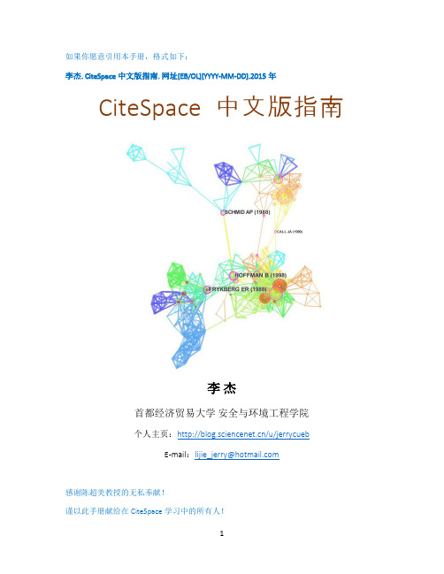
CiteSpace中文手册

5.2 打开地理可视化功能 ......................................................................................................... 41 5.3 相关参数设置 ..................................................................................................................... 41 5.4 结果的展示 ......................................................................................................................... 42 5.5 结果的编辑 ......................................................................................................................... 43 5.6 使用其他程式可视化 KML 文件 ........................................................................................ 45 6 Derwent 专利数据分析 ............................................................................................................. 47 6.1 登录 Derwent Innovations Index 数据库.
白血病(英文版)

Fusion gene formation
Model for Development of
Leukemia
Normal stem or progenitor cell
Mature blood cells
Mutalls
Normal development blocked
leukemia cells
Acute
Rapid growth of immature blood cells
Mostly in children, young adults
Needs immediate treatment
Chronic
Excessive build up of more mature blood cells
WHO Classification of Lymphoproliferative Syndromes
Precursor B Lymphoblastic Leukemia/Lymphoma (ALL/LBL) -- ALL in children (80-85% of childhood ALL); LBL in young adults and rare; FAB L1 or L2 blast morphology
CONTENTS
DEFINITION CLASSIFICATION ETIOLOGY MANIFESTATIONS LABORATORY EVALUATION DIAGNOSIS TREATMENT
What is leukemia?
Origin: Hematopoietic system Level: Malignant clonal disease of stem cell Nature: Malignancy
肿瘤生物免疫治疗宣教培训课件

➢肿瘤生物免疫治疗宣教
➢25
抗肿瘤免疫机制
➢免疫细胞如何发挥抗肿瘤作用?
➢肿瘤生物免疫治疗宣教
➢26
➢免疫系统识别“自我”与“非我” ➢肿瘤细胞表达肿瘤抗原
➢肿瘤生物免疫治疗宣教
➢27
1.什么是肿瘤抗原?
➢ 泛指在肿瘤发生、发展过程中新出现或过 度表达的抗原物质。
➢肿瘤特异性抗原 (TSA): 仅在肿瘤细胞上表达; ➢肿瘤相关性抗原 (TAA): 在某些肿瘤细胞上表达
Belagenpumatucel-L
Algenpantucel-L GV-1001 TroVAX(MVA-5T4)
IMA901
OncoVAX Abagowomab DCVax®-Brain Rindopepimut(CDX-110) BiovaxID®(Id➢-肿KL瘤H生/物GM免-疫C治SF疗)宣教
➢In 2015, the FDA approved PD-1 /PD-L1 ( Nivolumab, pembrolizumab ) immunotherapies to treat the most common forms of advanced lung and kidney cancer
➢肿瘤生物免疫治疗宣教
➢ Some biological therapies for cancer use vaccines or bacteria to stimulate the body’s immune system to act against cancer cells. These types of biological therapy, which are sometimes referred to collectively as “immunotherapy” or “biological response
生物治疗英文作文

生物治疗英文作文Biological therapy, also known as biotherapy or immunotherapy, is a type of treatment that uses substances made from living organisms to treat disease. It can involve using the body's own immune system to fight cancer, or using genetically engineered proteins to target specific cells in the body.One of the most common forms of biological therapy is the use of monoclonal antibodies, which are designed to target and destroy specific proteins on the surface of cancer cells. These antibodies can be used alone or in combination with other cancer treatments, such as chemotherapy or radiation therapy.Another form of biological therapy is the use of cytokines, which are proteins that help regulate the immune system. Cytokine therapy can be used to boost the body's natural defenses against cancer, or to help reduce the side effects of other cancer treatments.In addition to treating cancer, biological therapy can also be used to treat other diseases, such as autoimmune disorders, infectious diseases, and inflammatory conditions. For example, biological therapy can be used to target the underlying causes of rheumatoid arthritis, or to help the body fight off infections such as HIV or hepatitis.Overall, biological therapy offers a promising approach to treating a wide range of diseases, by harnessing the power of the body's own immune system and using targeted therapies to attack specific disease-causing cells. As research in this field continues to advance, the potential for biological therapy to revolutionize the treatment of disease is becoming increasingly apparent.。
Impulse Control Disorders

Kleptomania
All answers, unless otherwise stated, are from DSM-IV-TR or First and Tasman.
Kleptomania criteria
Q. The criteria for kleptomania is?
Kleptomania criteria
Ans. 1. Recurrent stealing of objects that are not needed by that person. 2. Tension before stealing. 3. Relief of tension with the stealing 4. Stealing is not the result of anger, vengeance, or another psychiatric disorder
放射医学专业英语翻译

Unit 4 Section ADNA Strand Breaks and Chromosomal Abberations Inducedby Ionizing Radiation(1)1.What is the principal target for the biologic effects of radiation on the basis of strong circumstantial evidence?基于详细的证据,辐射生物效应的主要靶点是什么?2. What is the structure of a deoxyribonucleic acid molecule?脱氧核糖核酸分子的结构是什么?DNA Strand BreaksDNA链断裂There is strong circumstantial evidence to indicate that deoxyribonucleic acid (DNA) is the principal target for the biologic effects of radiation, including cell killing mutation, and carcinogenesis. A consideration of the biologic effects of radiation therefore must begin logically with a description of the breaks in DNA caused by charged-particle tracks and by the chemical species produced.有足够多且详细的证据表明脱氧核糖核酸是辐射生物效应的主要的靶,包括细胞杀伤性突变和致癌作用。
因此,从带电粒子和化学产物导致的DNA链断裂来考虑辐射生物效应是较合乎逻辑的。
心理学专业外语翻译第12页

The nature-nurture debate in psychologyNATUREAPPROACHRoots of the approach-nativist philosophy,biology(physiology and genetics),evolutionary theory.Causes of behaviour-genetic determinism;inheritedinfluence,maturationalblueprint,neurochemical and hormonal influences,brain activity.Methods employed-gene/chromosome mapping,twin and adoption studies,brain scanning,brain stimulation or damage studies,drug testing.Implications-due to biological determinism,behaviour can only be changed through physical means,such as selective breeding(eugenics),gene therapy,brain surgery,or durgs. Criticisms-reductionist,may neglect environmental influences.AREAS OF EXPLANATION Perception-Research conducted by Fanz,Bower,and Gibson and Walk on new-born babies indicated pattern detection,size constancy and depth perception are innate abilities. Aggression-The ethologist Lorenz and psychoanalyst Freud believed aggression is an innate drive.Bio-psychologists have examined the role of hormones and brain areas in aggression.Sex-role behaviour-Bio-psychologists propose gender identity is a direct result of genetic and hormonal influences. Abnormality-the biomedical approach 心理学的先天与后天教养遗传决定论研究方法理论根源:自然主义哲学、生物学(心理与遗传学)、进化论.决定行为的因素:基因决定、遗传决定、个体成熟、神经化学与荷尔蒙的影响和大脑活动。
光热治疗

Hollow silica nanoparticles loaded with hydrophobic phthalocyanine for near-infrared photodynamic and photothermal combination therapyJuanjuan Peng a,Lingzhi Zhao a,Xingjun Zhu a,Yun Sun a,Wei Feng a,*,Yanhong Gao b, Liya Wang c,**,Fuyou Li a,*a Department of Chemistry&State Key Laboratory of Molecular Engineering of Polymers,Fudan University,220Handan Road,Shanghai200433,PR Chinab Department of Geriatrics,Xinhua Hospital of Shanghai Jiao Tong University,School of Medicine,Shanghai200092,PR Chinac College of Chemistry and Pharmaceutical Engineering,Nanyang Normal University,Nanyang473061,PR Chinaa r t i c l e i n f oArticle history:Received16May2013 Accepted8July2013Available online24July2013Keywords:PhthalocyanineHollow silica nanoparticles Photothermal therapy Photodynamic therapy a b s t r a c tOwing to the convenience and minimal invasiveness,phototherapy,including photodynamic therapy (PDT)and photothermal therapy(PTT),is emerging as a powerful technique for cancer treatment.To date,however,few examples of combination PDT and PTT have been reported.Phthalocyanine(Pc)is a class of traditional photosensitizer for PDT,but its bioapplication is limited by high hydrophobicity.In this present study,hollow silica nanospheres(HSNs)were employed to endow the hydrophobic phthalocyanine with water-dispersity,and the as-prepared hollow silica nanoparticles loaded with hy-drophobic phthalocyanine(Pc@HSNs)exhibits highly efficient dual PDT and PTT effects.In vitro and in vivo experimental results clearly indicated that the dual phototherapeutic effect of Pc@HSNs can kill cancer cells or eradicate tumor tissues.This multifunctional nanomedicine may be useful for PTT/PDT treatment of cancer.Ó2013Elsevier Ltd.All rights reserved.1.IntroductionPhototherapy has attracted much interest in recent years as a powerful technique for cancer treatment due to the convenience and minimal invasiveness.Photodynamic therapy(PDT)and pho-tothermal therapy(PTT)are two typical phototherapy approaches, which require light absorption and photosensitizer to generate reactive oxygen species and heat to kill cancer cells,respectively [1,2].Recently,nanoparticles with PTT capabilities have attracted a great deal of attention in the photothermal treatment of tumor cells.A number of inorganic nanomaterials,such as Au nano-materials(including nanoshells,nanorods,nanocubes and nanoc-ages)[3e6],carbon nanomaterials[7e10],Pd nanosheets[11], copper sulfide[12],copper selenide[13]and W18O49nanoparticles [14]have also been shown to generate photothermal heating by NIR optical illumination to destroy cancer cells.The uses of conductive polymers-based nanomaterials[15e17]as PTT agents have also attracted significant attention.However,these nano-materials have very weak ability to generate reactive oxygen spe-cies(ROS)and are incapable for PDT application.Meanwhile,the most widely used commercial PDT agents are based on porphyrin, however,the maximal absorption wavelength of porphyrin is located in visible region,which is not suitable for in-depth tumor treatment through tissue.As a result,few examples of these PDT agents based on porphyrin have been applied to PTT[18,19]. Therefore,the combined therapy of PDT and PTT has rarely been developed to date.To generate thermal and ROS effectively for combination PTT and PDT,an ideal agent should exhibit strong absorbance band in the NIR region,which is a transparency window for biological tis-sues,and possess high photothermal conversion efficiency[17].To date,only two types of compounds,that is,NIR-absorbing BODIPY dyes and phthalocyanine(Pc)can meet the aforementioned de-mands.However,organic BODIPY dyes are limited by rapid pho-tobleaching.Phthalocyanine compounds,as one of the main class of photosensitizers,have been approved by the US Food and Drug Administration for clinical applications in the treatment of cancer due to their ability to generate singlet oxygen upon irradiation of light[20].Phthalocyanine derivatives have been extensively studied for their excellent stability against heat,light and harsh*Corresponding authors.Fax:þ862155664185.**Corresponding author.E-mail addresses:fengweifd@(W.Feng),wlya@(L.Y.Wang), fyli@(F.Y.Li).Contents lists available at ScienceDirectBiomaterialsjournal homepage:w ww.elsevi/locate/biomaterials0142-9612/$e see front matterÓ2013Elsevier Ltd.All rights reserved./10.1016/j.biomaterials.2013.07.027Biomaterials34(2013)7905e7912chemical environments[21].Although phthalocyanine shows considerable adsorption band at NIR region,few efforts have been devoted to investigate its application of phthalocyanine as PTT agent.To the best of our knowledge,the potential use of the phthalocyanine parent structure for PTT of cancer has not yet been reported due to its hydrophobicity.Our interest is developing a general administration route to fabricate nanocomposite and study the dual PTT and PDT effect, utilizing phthalocyanine as a model sensitizer.In this present study, hollow silica nanospheres(HSNs)with aqueous dispersibility and high stability were employed to load the hydrophobic phthalocy-anine(denoted as Pc@HSNs).Upon irradiated with NIR(730nm) laser,the Pc@HSNs can rapidly convert optical energy into heat and generate ROS after laser irradiation to eliminate tumors.The cor-responding PDT and PTT effects were evaluated in both cell level and animal models of tumor-bearing mice.The dual therapeutic properties from a single nanoparticle will play a significant role in promoting the use of hydrophobic photosensitizers in tumor therapies.2.Experimental section2.1.Materials and characterizationAll the starting materials were obtained from commercial supplies and used as received without further purification.Pluronic F108(EO132PO50EO132,where EO is polyethylene oxide and PO is polypropylene oxide)were purchased from Sigma e Aldrich.1,3,5-Trimethylbenzene,tetraethyl orthosilicate(TEOS),dimethyldime-thoxysilane(DMDMS),HCl,and AgNO3were purchased from Shanghai Chemical Corp.Phthalocyanine(Pc)was purchased from Sigma e Aldrich.Deionized water was used throughout the experiments.The morphology of HSNs was observed on a JEOL JEM-2010F transmission electron microscope(TEM)operated at200kV.Dynamic light scattering was carried out on an ALV-5000spectrometer-goniometer equipped with an ALV/LSE-5004light scattering electronic and multiple tau digital correlator and a JDS Uniphase He e Ne laser(632.8nm)with an output power of22mW.UV e vis spectra were measured with a UV e vis spectrophotometer(Agilent,8453).Nitrogen sorption isotherms were measured at77K with a Quantachrome Quardrasorb analyzer.Before mea-surement,the sample was degassed at180 C in vacuum for6h.The specific surface area was calculated using Brumauer e Emmet e Teller(BET)method,the pore size distribution was derived from the adsorption branches of the isotherms based on the Barrett e Joyner e Halenda(BJH)model.Weight changes of the products were monitored using a Mettler Toledo TGA-SDTA851analyzer(Switzerland)from25 C to750 C with a heating rate of10 C minÀ1.2.2.Synthesis of hollow silica nanospheres(HSNs)The HSNs were synthesized according to a previously reported procedure[22]. In a typical synthesis,1.0g of pluronic F108and1.0g of1,3,5-trimethylbenzene were added to30mL2.0M HCl and stirred vigorously for6h at25 C to form a homo-geneous emulsion.Then1.0g of TEOS was added to the surfactant solution under vigorous stirring.After6h of the reaction,0.5g of DMDMS was added and the re-action was continued for another48h.The milky mixture was dialyzed with a semipermeable membrane(molecular-weight cutoff,MWCO¼14,000)in500mL water for36h,and the water was refreshed every12h.The dialysate was evaporated at80 C,and the obtained white powder was calcined at350 C for5h to get the final product HSNs.2.3.The loading of phthalocyanine to HSNs0.1g of HSNs and0.1g of phthalocyanine were added to10mL chloroform.The mixture was stirred at40 C for12h and centrifuged(12,000rpm)to allow theHSNsFig.1.(a,b)TEM images of HSNs with different magnification.1a insert:the size distribution of HSNs.(c)DLS of HSNs samples disperse in water.(d)N2adsorption isotherm and pore size distribution curve of solid HSNs calculated from the adsorption branch by the NLDFT method before and after adsorption of phthalocyanine.J.Peng et al./Biomaterials34(2013)7905e79127906load phthalocyanine(called as Pc@HSNs).The obtained Pc@HSNs was washed with ethanol for several times until the supernatant was almost transparent after centrifugation to remove the unloaded phthalocyanine completely.Finally,the Pc@HSNs was dried at80 C.The loading ratio of the phthalocyanine inside HSNs was determined by UV-Vis absorption spectrum.2.4.Investigation of the release and photostability of Pc@HSNsTo investigate the possible release of phthalocyanine from Pc@HSNs in the physiological environment,2mg/mL Pc@HSNs was soaked in phosphate buffer so-lution(PBS)or RMPI1640nutrition medium for one week.Then the solution was centrifuged and the concentration of the phthalocyanine in the supernatant was analyzed using UV e vis spectroscopy.To investigate the photostability of Pc@HSNs, another group of2mg/mL Pc@HSNs was socked in PBS or RMPI1640nutrition medium for one week,irradiated by a730nm laser with a power of1.5W/cm2for 10min every day.The concentration of the phthalocyanine in the supernatant was monitored using UV e vis spectroscopy.2.5.Examination of photothermal effect for Pc@HSNs in aqueous solutionThe Pc@HSNs at the concentration of1mg/mL was irradiated using a730nm laser with a power density of1.5W/cm2.The temperature of solution was measured with infrared imaging devices(FLIR E40of FLIR Systems,Inc.,United States)at20ms intervals for a total of10min.Each solution was measured three times.2.6.ROS generation of Pc@HSNsThe generation of ROS was monitored by Image-iT LIVE Reactive Oxygen Species Kit(Molecular Probes/Beyotime)based on5-(and-6)-carboxy-20,70-dichlorodihy-drofluoresceindiacetate(carboxy-H2DCFDA),following the manufacturer’s protocol. H2DCFDA is afluorogenic marker for ROS,which permeates live cells and is deacetylated by intracellular esterases.In the presence of ROS,the reducedfluo-rescein compound is oxidized and emits bright greenfluorescence.Pre-seeded KB cells were incubated with Pc@HSNs suspension(250m g/mL)for3h at37 C with5% CO2.After washing the cells thoroughly with PBS,the cells were irradiated with a 730nm laser for3min H2DCFDA(10m M)was added and the cells were incubated at 37 C for30min.Fluorescence intensity was measured on an OLYMPUS FV1000 confocalfluorescence microscope.2.7.Cytotoxicity assessmentsCell-viability following PTT was determined by a methyl thiazolyl tetrazolium (MTT)assay.Briefly,cells were plated in96-wellflat-bottomed plates with a con-centration of5Â104cells per well and allowed to grow overnight prior to the exposure to HSNs or Pc@HSNs with different concentrations,and another group exposure to HSNs or Pc@HSNs with different concentrations for3h to allow the cell phagocytize nanoparticles,followed by irradiation with laser.After24h(including the phagocytosis time)of further incubation,MTT(20m L,5mg/mL)was added to each well and the plate was incubated for an additional4h at37 C under5%CO2to allow the conversion of MTT into a purple formazan product by active mitochondria. Then the formazan product was dissolved in dimethyl sulfoxide(DMSO)and quantified by the absorbance at570nm,with background subtraction at690nm, which was measured by means of a Tecan Infinite M200monochromator-based multifunction microplate reader.The following formula was used to calculate the inhibition of cell growth:Cell viability(%)¼(mean of Abs.value of treatment group/ mean Abs.value of control)Â100%.2.8.In vivo photothermal effect of Pc@HSNsThe S180mice sarcoma were inoculated to the male BALB/c mice(n¼4,6-week-old).When the tumor length reached50e70mm,the mice were intratumorally injected with Pc@HSNs suspension(100m L,2mg/mL).For control groups,mice were treated with the same volume of saline.Mice with and without Pc@HSNs injection were irradiated with the730nm laser(Hi-Tech Optoelectronics Co.,Ltd.Beijing,China) at power densities of1.5W/cm2for10min every day.The length(l)and width(w)of the tumors were measured by a vernier caliper,and the volume(V)of the tumor was estimated by the following formula:V¼4p/3Â(lw)2.Relative tumor volumes were calculated as V/V0(V0is the tumor volume when the treatment was initiated).2.9.Histological assessmentIn the test group,Kunming mice(n¼3)were intravenously injected with Pc@HSNs at a total dose of5mg/mL(0.4mL).And Kunming mice(n¼3)with no injection were selected as the control group.Blood samples and tissues were har-vested from test and control group after96h.Blood was collected from the orbital sinus by quickly removing the eyeball from the socket with a pair of tissue forceps. Upon completion of the blood collection,mice were sacrificed.The heart,liver,Fig.2.UV e vis spectra of Pc@HSNs solutions at a concentration of1mg/mL Inset:Photo of water(left)and Pc@HSNs(right)solutions at a concentration of1mg/mL in water.(b) Heating curves of water and Pc@HSNs(1mg/mL,2mL)under730nm laser irradiation at a power density of1.5W/cm2.(c)IR thermal images of water and Pc@HSNs solution exposed to the NIR laser at power densities of1.5W/cm2recorded at different time intervals,1:water;2:Pc@HSNs.J.Peng et al./Biomaterials34(2013)7905e79127907spleen,lung,and kidney were removed,and fixed in paraformaldehyde,embedded in paraf fin,sectioned,and stained with hematoxylin and eosin.3.Results and discussion3.1.Synthesis and characterization of Pc@HSNsHSNs were synthesized as carriers according to the method reported before [22].The as-prepared HSNs are hollow nano-spheres with uniform particle size,as shown in the transmission electron microscopy (TEM)image (Fig.1a).No apparent aggrega-tion of HSNs can be observed (Fig.1b).A statistic mean size of 35nm with a narrow size distribution (inset of Fig.1a)of HSNs is obtained by measuring the diameters of 100individual hollow nanospheres from the TEM image,demonstrating good mono-dispersity of the hollow nanospheres.Dynamic light scattering (DLS)measurements showed that HSNs exhibited a mean hydrated diameter of 37nm (Fig.1c),which agrees well with the statistical result observed from TEM.The nitrogen adsorption isotherm and the corresponding calculated pore size distribution curve of HSNsafter calcination at 350 C are shown in Fig.1d.A type IV adsorp-tion e desorption isotherm can be observed (Fig.1d).A uniform BJH pore size distribution centered at 24nm can be deduced for HSNs,which also in good accordance with the diameters of the cavity observed in the TEM image.The pore volume is 1.10cm 3/g,and the BET surface area is 579m 2/g.The wall thickness of the HSNs is calculated to be w 6.5nm.After loaded with phthalocyanine,the pore volume and surface area decrease signi ficantly to 0.28cm 3/g and 143m 2/g,respectively,proving that the phthalocyanine was adsorbed inside the hollow of the HSNs.The loading ratio of phthalocyanine in HSNs is measured to be 83.1%(W/W,Fig.S1),which is in accordance with thermal gravimetric analysis (TGA)results (Fig.S2).To testify the in vivo stability of Pc@HSNs,the material was soaked in PBS or RMPI1640nutrition medium for one week or with laser irradiation (730nm,1.5W/cm 2for 10min)every day.As shown in Fig.S3,even with 730nm irradiation,no obvious signal of phthalocyanine can be detected from the adsorption spectrum of the supernatants in both PBS and RPMI 1640,sug-gesting that the photosensitizer phthalocyanine was stably loaded inside the hollow cavities of HSNs withoutleakage.Fig.3.Confocal fluorescence images of KB cells to detect oxidative stress using Image-iT LIVE Reactive Oxygen Species (ROS)Kit.From top to down:untreated,irradiation with NIR (730nm,1W/cm 2,3min),treated with Pc@HSNs solution (250m g/mL),treated with Pc@HSNs solution (250mg/mL)followed by irradiation with NIR (730nm,1W/cm 2,3min).The cells showing green fluorescence color represent oxidatively stressed cells affected with ROS.FL images were collected at 510e 560nm,under excitation at 488nm.The scale bar is 50m m.(For interpretation of the references to colour in this figure legend,the reader is referred to the web version of this article.)J.Peng et al./Biomaterials 34(2013)7905e 79127908Fig.4.Confocalfluorescence images and brightfield images of KB cells.From top to bottom:untreated,irradiation with NIR(730nm,1.5W/cm2,10min),treated with Pc@HSNs solution(250m g/mL),treated with Pc@HSNs solution(250m g/mL)followed by irradiation with NIR(730nm,1.5W/cm2,10min).The cells are stained with PI,and the dead cells are observed in red.FL images were collected at600e680nm,under excitation at543nm.The scale bar is50m m.(For interpretation of the references to colour in thisfigure legend,thereader is referred to the web version of this article.)(b)Cells viabilities after phototherapy treatment under different Pc@HSNs concentration.Quantified KB cell viability from various groups.Data are expressed as meanÆs.d.(n¼3).3.2.Photothermal effect of Pc@HSNsPc@HSNs exhibited a high optical absorption coef ficient in the NIR range with a peak centered at w 760nm (Fig.2a).In order to verify the ef ficacy of Pc@HSNs as a photothermal agent,Pc@HSNs suspension with a concentration of 1mg/mL was exposed to 730nmlaser at a power density of 1.5W/cm 2.An obvious temperature in-crease was observed for Pc@HSNs suspension from 25 C to 50 C under laser irradiation (Fig.2b);the suspension was heated to over 45 C within 6min,which is capable for thermal treatment of tumor.While under the same conditions,pure water only showed a slight temperature raise of 3.5 C in 10min,demonstrating the raise of temperature is mainly contributed by Pc@HSNs rather than the ab-sorption of light by water.Following Roper ’s reported calculation method [23],the photothermal conversion ef ficiency (h )was calcu-lated to be 37.1%,using the following equation (1),the detailed cal-culations was supplied in Supporting Information .And Fig.2c shows the thermal images of Pc@HSNs suspension and pure water exposed to the 730nm laser recorded at different time intervals,where the difference in temperature can be visually observed.Xim i C p ;id Td t¼Q NP þQ DIS ÀQ Surr (1)where m and C p are the mass and heat capacity of water,respec-tively,T is the solution temperature,Q NP is the energy inputted by Pc@HSNs,Q Dis is the baseline energy inputted by the sample cell,and Q Surr is heat conduction away from the system surface by air.3.3.Intracellular ROS generated by Pc@HSNsAs designed,the Pc@HSNs will have PDT activity together with PTT ability upon 730nm illumination.The PDT activity to generate ROS of Pc@HSNs is evaluated by the detection of ROS generated within human epidermoid mouth carcinoma KB cells containing Pc@HSNs,upon irradiation with 730nm laser.The ROS production was assessed using the Image-iT LIVE Reactive Oxygen Species Kit.This assay is based on 5-(and-6)-carboxy-20,70-dichlorodihydro-fluoresceindiacetate (carboxy-H 2DCFDA)as a fluorogenicmarkerFig.6.IR thermal images of tumor-bearing mice with Pc@HSNs injection exposed to the NIR laser at power densities of 1.5W/cm 2recorded at different time intervals.As the control,IR thermal images of mice without Pc@HSNs injection exposed to the NIR laser at the power of 1.5W/cm 2weretaken.Fig.7.(a)Representative photos of mice bearing S180murine sarcoma after various different treatments indicated.The scale bar was 2cm.(b)Growth of S180tumors in different groups of mice after treatment.The relative tumor volumes were normalized to their initial sizes.Error bars were based on standard deviations.(c)Survival curves of mice after various treatments as indicated in (c).Pc@HSNs-injected mice after PTT treatment showed 100%survival ratio over 46days.J.Peng et al./Biomaterials 34(2013)7905e 79127910for ROS-permeated viable cells,and deacetylated by nonspecific intracellularesterase.In the presence of ROS,the reduced carboxy-H2DCFDA emits bright greenfluorescence when oxidized.There-fore,greenfluorescence can be observed from the cells oxidatively stressed by ROS.As shown in Fig.3,the test groups of untreated cells,laser-treated cells and Pc@HSNs-treated cells exhibited negligible production of ROS.However,Pc@HSNs-treated cells with 730nm irradiation showed substantially high greenfluorescence intensity.These results clarify that Pc@HSNs can generate ROS when excited with730nm light,therefore,Pc@HSNs can be applied for PDT therapy.3.4.In vitro cell viability assay of Pc@HSNsAs above-mentioned discussing,Pc@HSN has combination ability of both PTT and PDT effect.Thus,anti-cancer performance of Pc@HSNs in biological systems could be expected and was proved by in vitro cell experiments.KB cancer cells were incubated with Pc@HSNs at a concentration of250m g/mL for3h,and then irra-diated by the730nm laser with a power density of1.5W/cm2for 8min.After irradiation,the cells were stained by propidium iodide (PI)to indicate dead(red)cells,and imaged by a confocalfluores-cence microscope.As shown in Fig.4,significant cell death indi-cated by redfluorescence can only be observed in the test group of cells incubated with Pc@HSNs and followed by NIR irradiation.Standard cell viability tests were carried out to test the potential cell toxicity of Pc@HSNs.It was found that Pc@HSNs,even at a high concentration up to0.5mg/mL,exhibited no appreciable negative effect on the viability of cells after24h exposure(Fig.5a).MTT assay was also performed to quantitatively measure the relative cell via-bilities after phototherapy treatment under different concentrations of Pc@HSNs.As the concentration of Pc@HSNs was increased,more cells were killed by the laser irradiation(Fig.5b).The majority of cells were destroyed after being incubated with400m g/mL of Pc@HSNs and exposed to the730nm laser at1.5W/cm2for8min.In contrast, cells treated by only laser or HSNs with different concentration were not affected(Fig.S4).3.5.Photo-therapeutic efficacy of Pc@HSNs in vivoIn vivo phototherapy tests using Pc@HSNs as photosensitizer were performed on tumor-bearing mice.To monitor the photo-thermal effect in vivo of Pc@HSNs,the changing of temperature in the tumor region was recorded using a thermal imaging apparatus. After being intratumorally injected with Pc@HSNs suspension (100m L,2mg/mL)for10min,S180tumor-bearing mice were exposed to a730nm laser at a power density of1.5W/cm2.Under irradiation,the temperature of the tumor increased significantly from w30 C to w47 C within5min(Fig.6).In comparison,the temperature of tumor without Pc@HSNs injection had almost no changes under same730nm irradiation conditions.As tumor cells and tissue are sensitive to heat,these results proved the promise of phthalocyanine as a PTT agent.Although phthalocyanine has been used as PDT agents in a number of pathological indications[24,25], the potential use of phthalocyanine for photothermal treatment of cancer has not yet been reported.The phototherapeutic efficacy of Pc@HSNs was further investi-gated.A highly malignant murine tumor cell line S180was selected as the tumor model.After the length of tumors on the back of the mice reached approximately50e70mm,Pc@HSNs suspension (100m L,2mg/mL)was intratumorally injected.The tumors of each mouse in the treatment group were then exposed to a730nm laser at a power density of1.5W/cm2for10min every day.Three other groups including untreated mice(control,n¼4),mice exposed to the730nm laser(laser only,n¼4),and Pc@HSNs-injected mice without laser irradiation(Pc@HSNs,n¼4)were used as control groups.Tumor sizes were measured every2days after treatment.It is remarkable that for the mice group treated with both Pc@HSNs and730nm laser,the solid tumor shrinked gradually and was completely eradicated from the mice after5days of treatment (Fig.7b).In contrast,neither laser irradiation at the current power density nor only Pc@HSNs injection can affect the tumor growth (Fig.7a).Meanwhile,mice in the three control groups showed mean life spans of20e32days,by contrast,mice in the treated group(Pc@HSNsþlaser)were tumor-free after treatment,and survived over45days without a single death(Fig.7c).The above results proved that Pc@HSNs was a powerful agent for in vivo antitumor phototherapy.3.6.Histology and hematology results of the mice injected withPc@HSNsTo further evaluate the biosafety of Pc@HSNs,a serum biochem-istry assay and complete blood panel test were carried out using Pc@HSNs-injected(10mg/kg)healthy Balb/c mice at7days post in-jection.Notably,no detectable lesion(e.g.necrosis,hydropic degen-eration,inflammatory infiltrates,pulmonaryfibrosis,gastroenteritisFig.8.Histochemical study of the organs harvested from mice injected with Pc@HSNs (5mg/mL,0.4mL)for one week.The left and right rows are the control and experi-mental groups,respectively.J.Peng et al./Biomaterials34(2013)7905e79127911or infectious diarrhea)was observed for mice (Fig.8).Established serum biochemistry assays were also performed to further evaluate the toxicity to organs,especially the liver and kidney.No signi ficant fluctuations of the three important hepatic function indicators,alanine aminotransferase (ALT),aspartate aminotransferase (AST)and total bilirubin can be observed (Fig.9).The enzyme level of mice injected with Pc@HSNs was in the normal range.Two other indicators related to kidney function,creatinine and urea were also normal.The results collectively show that Pc@HSNs possess no noticeable toxic to mice in vivo .However,more effort is still required to systematically examine the potential long-term toxicity of our nanoparticles at various doses in animals.4.ConclusionWe demonstrated hollow silica nanoparticles loaded with hy-drophobic phthalocyanine Pc@HSNs for near-infrared photodynamic and photothermal combination therapy of cancer in vitro and in vivo .Using HSNs with good water dispersity and stability as host,Pc@HSNs are stable in physiological environments and nontoxic to cells at the tested concentrations.By intratumoral injection of Pc@HSNs,we further realized excellent tumor treatment ef ficacy using a 730nm laser irradiation at 1.5W/cm 2and achieved tumor elimination,without observing signi ficant toxic effects after treat-ment.Although the original phthalocyanine photosensitizer has been recognized only as a PDT agent,Pc@HSNs exhibit dual PDT and PTT properties,as both in vitro and in vivo phototherapeutic exper-iments against cancer cells demonstrated successful eradication of cancer cells.We believe that our results will open a new strategy for designing next-generation photosensitizers for cancer phototherapy.Con flict of interestNo financial con flict of interest was reported by the authors of this paper.AcknowledgmentsThis work was financially supported by NSFC (21231004and 21201038),MOST (2011AA03A407and 2013CB733700).Appendix A.Supplementary dataSupplementary data related to this article can be found at /10.1016/j.biomaterials.2013.07.027.References[1]Lal S,Clare SE,Halas NJ.Nanoshell-enabled photothermal cancer therapy:impending clinical impact.Acc Chem Res 2008;41:1842e 51.[2]Bardhan R,Lal S,Joshi A,Halas NJ.Theranostic nanoshells:from probe designto imaging and treatment of cancer.Acc Chem Res 2011;44:936e 46.[3]Cheng L,Yang K,Li Y,Chen J,Wang C,Shao M,et al.Facile preparation ofmultifunctional upconversion nanoprobes for multimodal imaging and dual-targeted photothermal therapy.Angew Chem Int Ed 2011;123:7523e 8.[4]Wang J,Zhu G,You M,Song E,Shukoor MI,Zhang K,et al.Assembly ofaptamer switch probes and photosensitizer on gold nanorods for targeted photothermal and photodynamic cancer therapy.ACS Nano 2012;6:5070e 7.[5]Wu X,Ming T,Wang X,Wang P,Wang J,Chen J.High-photoluminescence-yield gold nanocubes:for cell imaging and photothermal therapy.ACS Nano 2009;4:113e 20.[6]Gao L,Fei J,Zhao J,Li H,Cui Y,Li J.Hypocrellin-loaded gold nanocages withhigh two-photon ef ficiency for photothermal/photodynamic cancer therapy in vitro .ACS Nano 2012;6:8030e 40.[7]Wang X,Wang C,Cheng L,Lee S-T,Liu Z.Noble metal coated single-walledcarbon nanotubes for applications in surface enhanced Raman scattering imaging and photothermal therapy.J Am Chem Soc 2012;134:7414e 22.[8]Chu M,Peng J,Zhao J,Liang S,Shao Y,Wu ser light triggered-activatedcarbon nanosystem for cancer therapy.Biomaterials 2013;34:1820e 32.[9]Robinson JT,Tabakman SM,Liang Y,Wang H,Sanchez Casalongue H,Vinh D,et al.Ultrasmall reduced graphene oxide with high near-infrared absorbance for photothermal therapy.J Am Chem Soc 2011;133:6825e 31.[10]Hu S-H,Chen Y-W,Hung W-T,Chen IW,Chen S-Y.Quantum-dot-taggedreduced graphene oxide nanocomposites for bright fluorescence bioimaging and photothermal therapy monitored in situ.Adv Mater 2012;24:1748e 54.[11]Tang S,Huang X,Zheng N.Silica coating improves the ef ficacy of Pd nano-sheets for photothermal therapy of cancer cells using near infrared laser.Chem Commun 2011;47:3948e 50.[12]Zhou M,Zhang R,Huang M,Lu W,Song S,Melancon MP,et al.A chelator-freemultifunctional [64Cu]CuS nanoparticle platform for simultaneous micro-PET/CT imaging and photothermal ablation therapy.J Am Chem Soc 2010;132:15351e 8.[13]Hessel CM,Pattani VP,Rasch M,Panthani MG,Koo B,Tunnell JW,et al.Copperselenide nanocrystals for photothermal therapy.Nano Lett 2011;11:2560e 6.[14]Chen Z,Wang Q,Wang H,Zhang L,Song G,Song L,et al.ultrathin PEGylatedW 18O 49nanowires as a new 980nm-laser-driven photothermal agent for ef ficient ablation of cancer cells in vivo .Adv Mater 2013;25:2095e 100.[15]Zha Z,Yue X,Ren Q,Dai Z.Uniform polypyrrole nanoparticles with highphotothermal conversion ef ficiency for photothermal ablation of cancer cells.Adv Mater 2013;25:777e 82.[16]Liu Y,Ai K,Liu J,Deng M,He Y,Lu L.Dopamine-melanin colloidal nano-spheres:an ef ficient near-infrared photothermal therapeutic agent for in vivo cancer therapy.Adv Mater 2013;25:1353e 9.[17]Yang K,Xu H,Cheng L,Sun C,Wang J,Liu Z.In vitro and in vivo near-infraredphotothermal therapy of cancer using polypyrrole organic nanoparticles.Adv Mater 2012;24:5586e 92.[18]Lovell JF,Jin CS,Huynh E,Jin H,Kim C,Rubinstein JL,et al.Porphysomenanovesicles generated by porphyrin bilayers for use as multimodal bio-photonic contrast agents.Nat Mater 2011;10:324e 32.[19]Lovell JF,Jin CS,Huynh E,MacDonald TD,Cao W,Zheng G.Enzymatic regio-selection for the synthesis and biodegradation of porphysome nanovesicles.Angew Chem Int Ed 2012;124:2479e 83.[20]Triesscheijn M,Baas P,Schellens JHM,Stewart FA.Photodynamic therapy inoncology.The Oncologist 2006;11:1034e 44.[21]Karan S,Mallik B.Templating effects and optical characterization of copper (II)phthalocyanine nanocrystallites thin film:nanoparticles,nano flowers,nanocabbages,and nanoribbons.J Phys Chem C 2007;111:7352e 65.[22]Zhu J,Tang J,Zhao L,Zhou X,Wang Y,Yu C.Ultrasmall,well-dispersed,hollow siliceous spheres with enhanced endocytosis properties.Small 2010;6:276e 82.[23]Roper DK,Ahn W,Hoepfner M.Microscale heat transfer transduced by surfaceplasmon resonant gold nanoparticles.J Phys Chem C 2007;111:3636e 41.[24]Li LS,Luo ZP,Chen Z,Chen JC,Zhou SY,Xu P,et al.Enhanced photodynamicef ficacy of zinc phthalocyanine by conjugating to heptalysine.Bioconjug Chem 2012;23:2168e 72.[25]Xu P,Chen JC,Chen Z,Zhou SY,Hu P,Chen XY,et al.Receptor-targetingphthalocyanine photosensitizer for improving antitumor photocytotoxicity.PLoS ONE 2012;7:e37051.Fig.9.Serum biochemistry results obtained from mice injected with Pc@HSNs (5mg/mL,0.4mL)at the 7th day and mice receiving no injection.J.Peng et al./Biomaterials 34(2013)7905e 79127912。
- 1、下载文档前请自行甄别文档内容的完整性,平台不提供额外的编辑、内容补充、找答案等附加服务。
- 2、"仅部分预览"的文档,不可在线预览部分如存在完整性等问题,可反馈申请退款(可完整预览的文档不适用该条件!)。
- 3、如文档侵犯您的权益,请联系客服反馈,我们会尽快为您处理(人工客服工作时间:9:00-18:30)。
MECHANISMS OF TUMOUR ESCAPE FROM IMMUNOLOGICAL RECOGNITION
The variability of tumours permits their escape from immune recognition. An improvement in the understanding of the immunobiology of cell-mediated anti-tumour defences as well as a better knowledge of tumour recognition molecules expressed on the surface of many tumours has permitted the development of new anti-tumour strategies to stand alongside conventional chemotherapy and radiotherapy in colorectal cancer. Isolated tumour cells are able to be eliminated by several conventional immunological mechanisms, most notably antibody dependent cellular cytotoxicity (ADCC) (Steplewski et al, 1983; Adams et al, 1984) complement-dependent cytolysis (Herlyn and Koprowski, 1981) and apoptosis (Trauth et al, 1989). Knowledge of cell surface regulatory molecules expressed on tumour cells may enhance natural apoptosis and tumour regression (Wyllie et al, 1980).
British Journal of Cancer (1998) 77(5), 683-693 © 1998 Cancer Research Campaign
Review
Biological therapy: approaches in colorectal cancer
Strategies to enhance carcinoembryonic antigen (CEA)
AP Zbar,' NR Lemoine,2 M Wadhwa,3 H Thomas,4 D Snary5 and WA Kmiot'
'Academic Department of Colorectal Surgery, Hammersmith Hospital, London, UK; 2Molecular Pathology Laboratory, Imperial Cancer Research Fund, Imperial College School of Medicine, Hammersmtih Campus, London, UK; 3National Institute for Biological Standards and Control, Hertfordshire, UK; 4Department of Clinical Oncology, Imperial College School of Medicine, Hammersmtih Campus, London, UK; 5Applied Development Laboratories, Imperial Cancer Research Fund
Received 11 June 1997 Revised 9 September 1997 Accepted 12 September 1997
advanced colorectal cancer and in an adjuvant setting. The adjuvant use of the murine monoclonal IgG2a antibody, 17- A directed against the CO 17-1A surface epitope of CEA (found in up to 80% of colorectal carcinomas) has resulted in an improvement in disease-free survival and a reduction in locoregional recurrence rates of almost 30% in patients with Dukes' C carcinoma compared with untreated controls (Riethmuller et al, 1994). Carcinoembryonic antigen (CEA), a surface-expressed tumourassociated antigen, is a well-characterized glycoprotein represented in high density on most malignant tumours of the gastrointestinal tract (Muraro et al, 1985). The immunogenicity of CEA as a potential target antigen in colorectal cancer is at present unclear, with variable reports of inducible humoral and cellmediated responsiveness to CEA epitopes in patients with different stages of disease. There is much that remains unknown regarding the natural immunological response to a native antigen such as CEA both in terms of its antigenic processing and its potentially immunodominant epitopes. This review assesses the role of CEA as a 'natural' autoantigen along with strategies that render epitopes of CEA potentially immunogenic. This may be achieved by the use of xenogeneic, chimaeric, humanized or wholly human monoclonal and polyclonal antibodies and with anti-idiotypic therapy. The advantages and limitations of each strategy and their potential role in the treatment of advanced colorectal cancer are discussed.
as an
imthe UK, almost 20 000 people die each year from colorectal cancer. Despite a potential curability rate of 70% or greater, the overall survival at 5 years is just over 30%; a figure that has changed little over the last 4 decades despite advances in adjuvant and therapeutic chemotherapy and radiotherapy (King's Fund Forum, 1990). Moreover, there is evidence that over half the patients operated upon for cure have occult metastatic disease at the time of initial surgery (August et al, 1994). Recent novel immunocytochemical techniques using immunobead polymerase chain reaction (PCR) for detection of the tumour-associated antigen carcinoembryonic antigen (CEA) have permitted the identification of single malignant cells in peripheral blood samples, bone marrow aspirates and peripheral stem cell harvests through the recognition of unique hybrid gene transcripts. (Schlimok et al, 1990; Lindeman et al, 1992; Hardingham et al, 1993; Johnson et al, 1995). Although the presence of such cells in the bone marrow at the time of preliminary colon resection appears to be associated with a worse prognosis (Riethmuller and Johnson, 1992) the number of cells that correlates with a poor outcome is unknown, and the relationship between the development of secondary disease and circulating tumour cells remains poorly understood (Osborne et al, 1991). Phage cloning and hybridization have taken advantage of the limited but specific genetic alterations in developing large bowel neoplasms to detect ras mutations in colorectal cancer cells in the stool (Sidransky et al, 1992). The demonstration of small tumour burdens of this type in which cells are exposed in unshielded mesenchymal locations may provide relatively novel immunotherapeutic and chemoimmunotherapeutic targets and identify surrogate end points in treatment that may prove superior to crude survival time. Immunotherapeutic strategies in advanced colorectal cancer have generally met with little success. Conventional treatments have largely relied on either interleukin 2 (IL-2) or adoptive IL-2stimulated tumour infiltrating lymphocytes (TILs), with only sporadic reports of tumour regression (Rosenberg et al, 1989; Kradin et al, 1989). Recently, there has been a resurgence of interest in immunotherapy (and the potential of gene therapy) in
