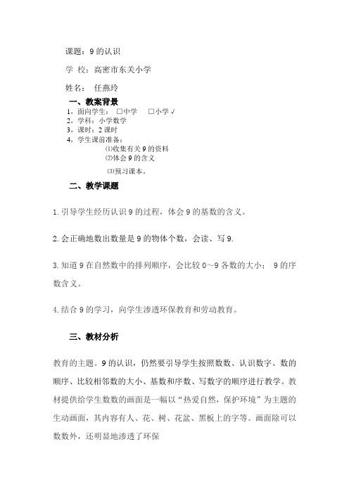
9
9的认识

课题:9的认识学校:高密市东关小学姓名:任燕玲一、教案背景1,面向学生:□中学□小学√2,学科:小学数学3,课时:2课时4,学生课前准备:⑴收集有关9的资料⑵体会9的含义⑶预习课本。
二、教学课题1.引导学生经历认识9的过程,体会9的基数的含义。
2.会正确地数出数量是9的物体个数,会读、写9.3.知道9在自然数中的排列顺序,会比较0~9各数的大小; 9的序数含义。
4.结合9的学习,向学生渗透环保教育和劳动教育。
三、教材分析教育的主题。
9的认识,仍然要引导学生按照数数、认识数字、数的顺序、比较相邻数的大小、基数和序数、写数字的顺序进行教学。
教材提供给学生数数的画面是一幅以“热爱自然,保护环境”为主题的生动画面,其内容有人、花、树、花盆、黑板上的字等。
画面除可以数数外,还明显地渗透了环保教学重点:1.掌握9的组成和9的写法.2.知道9在自然数中的排列顺序3、会比较0~9各数的大小; 9的序数含义教学难点:正确书写数字9教学准备:多媒体课件四、教学方法教学数数时要充分利用教材所提供的资源和线索,先引导学生数树、花、黑板上的字和花坛周围的蝴蝶等数量是9的物体,让学生在头脑里形成有关9的具体数量,然后引导学生数学生和老师的人数、花盆的数量,让学生在头脑里形成与9有关的数量。
关于9的序数含义,可先让学生按照要求涂色,然后分组适当讨论9只和第9只的区别,让学生正确理解9的基数含义和序数含义。
数字9比较难写,首先让学生搞清楚9的字形,然后着重说明写9的笔顺,最后进行书写练习。
五、教学过程一、情景导入:1、动画媒体:9只小动物,先出8只,又来了一只.师:一共几只?你喜欢哪一只,从左数排在第几,从右数排在第几?2、森林运动会:师:你看见了什么?生:9棵小树,9只小袋鼠,9面小红旗……师:出示“9”,这是几?生:“9”。
师:你是怎么知道的?生:幼儿园学的。
师:真是个好学生。
师在黑板上张贴板书:9 的认识二、探究新知:1、摆小棒:师:用9 根小棒摆成你最喜欢的图案.(2人到磁力黑板上摆,其余在下面摆)生:有的摆成太阳,有的摆成电视,有的摆成电脑,有的摆成房子,有的摆成机器人,有的摆成花儿,有的摆成0 ,有的摆成9 ------,2、用9说一句话。
九的部首和偏旁结构

九的部首和偏旁结构九的部首是一种汉字的结构组成部分,它位于字的左上角,用来表示该字的意义或音。
九的部首和偏旁结构在汉字中起到非常重要的作用,它们能够帮助我们更好地理解和记忆汉字,同时也体现了汉字的独特之处。
九的部首是“丿”,它是由一条斜线组成的。
在汉字中,九部首通常用来表示与九相关的事物或含义。
例如,“九”字本身就是由九的部首和“口”字组成的,表示数字9。
另外,九的部首还可以在其他字中表示与九相关的意义,比如“九月”、“九宫格”等。
除了九的部首,汉字还有许多其他的部首和偏旁结构。
这些部首和偏旁结构的存在,使得汉字具有了丰富的内涵和多样的意义。
例如,“人”字的部首是“亻”,表示与人有关的事物;“木”字的部首是“木”,表示与木材或木制品有关的事物;“水”字的部首是“氵”,表示与水有关的事物。
通过了解和记忆这些部首和偏旁结构,我们可以更好地理解和使用汉字。
部首和偏旁结构在学习汉字的过程中起到了重要的作用。
通过学习部首和偏旁结构,我们可以通过一定的规律推测出字的意义或音,帮助我们更好地记忆和理解汉字。
此外,部首和偏旁结构还可以帮助我们扩大汉字的词汇量,提高我们的阅读和写作能力。
除了在学习汉字中的应用,部首和偏旁结构在汉字的字典编纂和字形规范中也起到了重要的作用。
汉字字典按照部首和偏旁结构进行编排,使得查找汉字变得更加方便和快捷。
同时,部首和偏旁结构也规范了汉字的字形,使得字形更加统一和规范。
九的部首和偏旁结构是汉字中的一部分,它们在汉字的学习和应用中起到了重要的作用。
通过了解和记忆部首和偏旁结构,我们可以更好地理解和使用汉字,提高我们的汉字水平。
同时,部首和偏旁结构也体现了汉字的独特之处,使得汉字成为世界上独一无二的文字系统。
让我们一起学习和探索汉字的奥秘吧!。
九字开头的成语_

九字开头的成语九字开头的成语:九回肠、九宗七祖、九州四海、九蒸三熯、九折臂、九五之尊、九天九地、九死未悔、九死不悔、九世之仇、九仞一篑、九衢三市、九年之储、九流十家、九流宾客、九流百家、九九归原、九江八河、九间大殿、九间朝殿、九阍虎豹、九合一匡、九关虎豹、九故十亲、九天揽月、九鼎大吕、九鼎不足为重、九儒十丐、九牛一毛、九霄云外、九曲回肠、九牛二虎之力、九死一生、九流人物、九回肠断、九年之蓄、九原可作、九九归一、九转功成、九牛拉不转、九洲四海、九烈三贞、九垓八埏、九行八业、九天仙女、九泉之下、九牛一毫、九流三教、九鼎一丝、九变十化【成语解释】(1)九回肠:形容回环往复的忧思。
回肠:形容内心焦虑不安。
(2)九宗七祖:泛指祖宗。
(3)九州四海:犹言天下。
泛指全中国。
(4)九蒸三熯:热气上升为蒸,火干为熯。
喻久经熬炼。
(5)九折臂:九:泛指多次;折:断。
多次折断胳膊,经过反复治疗而熟知医理。
比喻阅历多,经验丰富。
(6)九五之尊:九五:指帝位。
旧指帝王的尊位。
(7)九天九地:原指天上的最高层和地的最深处。
后比喻两者相差极远。
1 / 5(8)九死未悔:九:表示极多。
纵然死很多回也不后悔。
形容意志坚定,不认经历多少危险,也决不动摇退缩。
(9)九死不悔:九:表示极多。
纵然死很多回也不后悔。
形容意志坚定,不认经历多少危险,也决不动摇退缩。
(10)九世之仇:九世:九代,形容历时久远。
指久远的深仇。
(11)九仞一篑:“为山九仞,功亏一篑”的略语。
喻功败垂成。
(12)九衢三市:指繁华的街市。
(13)九年之储:九年的储备。
指国家平时有所积蓄,以备非常。
(14)九流十家:先秦到汉初各种学说派别的总称。
(15)九流宾客:先秦到汉初有法、名、墨、儒、道、阴阳、纵横、杂、农九种学术流派。
指上中下各品的人才和各种人物。
(16)九流百家:泛指各种学术流派。
参见“九流一家”。
(17)九九归原:犹言归根到底。
(18)九江八河:泛指所有的江河。
数字9的分解教案幼儿园

数字9的分解教案幼儿园教学目标通过数字9的分解教学,让幼儿园的小朋友能够了解数字9的组成方式,并能够简单的进行加减运算。
教学内容1.数字9的分解2.数字9的加减运算教学步骤步骤一:引入数字9老师向幼儿园小朋友展示数字9,问小朋友们是否知道数字9,并让他们数数,数到哪个数字。
步骤二:数字9的分解1.老师向小朋友们介绍数字9的分解,让他们知道数字9可以由多组数字相加得到。
2.老师拿出现成的数字牌或棋子,让小朋友们把它们组合成数字9。
教师可以通过以下几组数字进行练习:– 1 + 8 = 9– 2 + 7 = 9– 3 + 6 = 9– 4 + 5 = 93.让小朋友们自己找出数字9的组合方式,并用数字牌或棋子进行展示,教师可以通过点评的方式引导他们正确的进行组合,并纠正他们的错误。
步骤三:加减运算1.老师可以用数字牌让小朋友们进行数字9的加减练习,例如:–9 + 1 = ?–9 - 3 = ?2.通过以上的练习,让小朋友们了解到数字9可以用于加减运算,以此为基础,进行数字1~10的加减运算,需要注意的是,教师需要适应小朋友的认知水平及学习进展。
教学反思数字9的分解教学在幼儿园中是一种较为基础且有效的教学方法,但同时也需要教师对幼儿进行耐心地引导和指导。
在课程中,因为小朋友们的注意力很难集中,教师可以通过形象生动的方式来吸引小朋友们的注意力,例如使用卡通形象或游戏的方式来进行数字9的分解展示,从而将教学效果最大化。
在课后教师可以通过评价和鼓励小朋友们的方式来增强他们的学习信心,激发他们的学习兴趣。
9的笔顺详解

9的笔顺详解如下:
“9”的笔画顺序是“竖折钩”和“竖弯钩”。
首先,从上到下写一个“竖折钩”,这是“9”的第一部分。
这个笔画需要注意竖直,并且折钩处要稍微弯曲,形成一个自然的弧度。
接着,在竖折钩的下方,写一个“竖弯钩”。
这个笔画从上到下逐渐变弯,然后向右上方延伸。
这个笔画也需要保持一定的弧度,让整个数字看起来更加自然。
在书写“9”时,要注意笔画的力度和角度。
竖折钩需要有一定的力度,显得挺拔有力;而竖弯钩则需要有一定的弧度,显得柔和自然。
同时,两个笔画的衔接处也要注意协调,让整个数字看起来更加和谐。
此外,书写“9”时还需要注意字体的端正和美观。
要保持字体的中心对称,避免出现偏斜或扭曲的情况。
同时,也要注意字体的间距和大小,让整个数字看起来更加整齐、美观。
总之,“9”的笔顺虽然简单,但需要注意细节和技巧。
只有掌握了正确的笔画顺序和书写方法,才能写出美观、大方的数字“9”。
九开头的四字成语大全

九开头的四字成语大全九牛一毛、九霄云外、九牛二虎之力、九五之尊、九死一生、九间朝殿、九州八极、九鼎不足为重、九攻九距、九牛拉不转、九曲回肠、九宗七祖、九年之蓄、九变十化、九天使者、九流宾客、九关虎豹、九儒十丐、九万欲抟空、九仞一篑、九垓八埏、九故十亲、九泉无恨、九回肠断、九五之位、九转功成、九死不悔、九十其仪九棘三槐、九天仙女、九天九地、九鼎一丝、九战九胜、九鼎大吕、九蒸三熯、九旋之渊、九十春光、九流十家、九九归一、九原可作、九泉之下、九折成医、九烈三贞、九行八业、九原之下、九春三秋、九年之储、九江八河、九折回车、九天揽月、九经三史、九衢三市九鼎大吕:九鼎:古传说,夏禹铸九鼎,象征九州,是夏商周三代的传国之宝;大吕:周庙大钟。
比喻说得话力量大,分量重。
九鼎不足为重:形容说话有分量,比较起来九鼎也不算重。
九儒十丐:儒:旧指读书人。
元代统治者把人分为十等,读书人列为九等,居于末等的乞丐之上。
后指知识分子受到歧视和苛待。
九曲回肠:形容痛苦、忧虑、愁闷已经到了极点。
九流人物:指社会上的各种人物。
九回肠断:形容痛苦、忧虑、愁闷已经到了极点。
同“九回肠”。
九年之蓄:蓄:积聚,储藏。
九年的储备。
指国家平时有所积蓄,以备非常。
九原可作:九原:春秋时晋国卿大夫的墓地在九原,因称墓地;作:起,兴起。
设想死者再生。
九九归一:归根到底。
九转功成:转:循环变华。
原为道家语,指炼得九转金丹。
后常比喻经过长期不懈的艰苦努力而终于获得成功。
九牛拉不转:形容态度十分坚决。
九洲四海:九洲:指中国;四海:古人认为,中国九州之久是一望无际的大海,此指中国以外的地方。
指中国及四周以外的地方。
九烈三贞:贞:贞操;烈:节烈。
封建社会用来赞誉妇女的贞烈。
九垓八埏:垓:通“陔”,重,层;九垓:即九重天,天之极高处;埏:边际;八埏:指边际远之地。
指天地的终极之处,即天涯海角。
九行八业:指各种行业。
九天仙女:指天上的仙女,比喻绝色美女。
九牛一毫:九条牛身上的一根毛。
9的拆字谜题

9的拆字谜题
1、扦离别拭泪珠(打一数字) 答案:九。
2、几回改变劝酒声(打一字)答案:九。
3、有头无尾字(打一字)答案:九。
4、不要杂木(打一字)答案:九。
5、仇人已除(打一字)答案:九。
6、旭日升空(打一字)答案:九。
7、热点提前消失(打一字) 答案:九。
8、列车出轨(打一数字) 答案:九。
9、旭日东升(打一字)答案:九。
10、六神无主(打一字)答案:九。
11、因九助加的一番会城动。
答案:解都
12、因九九的一城市名。
答案:双阳。
13、旭日不出时打一字。
答案:九
14、旭日临空打一字。
答案:九。
15、因抛弃两边的打一字。
答案:九
16、国八一重直的一数学用固靠。
答案:表
17、百岁少年不少年打一股票名答案:九九众。
18、图一比一百一三字俗调。
答案:小九九。
19、左九仇九,九十九。
(打一字)答案:伯。
20、九十九克(打一《水浒传》人名)答案:白胜。
21、九团九营(打一古代称谓二) 答案:军师,统制。
22、九月初九(打一节日)答案:重阳节。
23、九十九对(打一成语) 答案:呒一是。
24、九九归(打一字)答案:本。
25、九九重阳(打一字)答案:相。
26、连载九纨十九期(打一歌名)答案:千等一回。
九的罗马数字

九的罗马数字
9的罗马数字是Ⅸ。
解析:
罗马数字1至9的写法:Ⅰ、Ⅱ、Ⅲ、Ⅳ、Ⅴ、Ⅵ、Ⅶ、Ⅷ、Ⅸ。
在阿拉伯数字使用前,罗马数字是常使用的数字,罗马数字采用七个
罗马字母作数字、即Ⅰ(1)、X(10)、C(100)、M(1000)、V(5)、L(50)、D(500)。
罗马数字组数规则:为基本数字Ⅰ、X、C中的任何一个、自身连用
构成数目、或者放在大数的右边连用构成数目、都不能超过三个;放在大
数的左边只能用一个。
不能把基本数字V、L、D中的任何一个作为小数放
在大数的左边采用相减的方法构成数目;放在大数的右边采用相加的方式
构成数目、只能使用一个。
罗马数字记数方法:
相同的数字连写,所表示的数等于这些数字相加得到的数,如:Ⅲ=3。
小的数字在大的数字的右边,所表示的数等于这些数字相加得到的数,如:Ⅷ=8;Ⅻ=12。
小的数字,(限于Ⅰ、X和C)在大的数字的左边,所表示的数等于
大数减小数得到的数,如:Ⅳ=4;Ⅸ=9。
正常使用时,连写的数字重复不得超过三次。
(表盘上的四点钟“IIII”例外)(注:现使用IV代表4)。
在一个数的上面画一条横线,表示这个数扩大1000倍。
- 1、下载文档前请自行甄别文档内容的完整性,平台不提供额外的编辑、内容补充、找答案等附加服务。
- 2、"仅部分预览"的文档,不可在线预览部分如存在完整性等问题,可反馈申请退款(可完整预览的文档不适用该条件!)。
- 3、如文档侵犯您的权益,请联系客服反馈,我们会尽快为您处理(人工客服工作时间:9:00-18:30)。
ReviewA critical evaluation of the ubiquitin –proteasome system in Parkinson's diseaseCasey Cook,Leonard Petrucelli ⁎Department of Neuroscience,Mayo Clinic,4500San Pablo Road,Jacksonville,FL 32224,USAa b s t r a c ta r t i c l e i n f o Article history:Received 15October 2008Received in revised form 12January 2009Accepted 27January 2009Available online 3February 2009Keywords:Ubiquitin proteasome system Parkinson's disease Alpha-synuclein Ubiquitin ligase Substantia nigra Aggregation ParkinLewy bodies DJ-1Leucine-rich repeat kinase 2The evidence for impairment in the ubiquitin proteasome system (UPS)in Parkinson's disease (PD)is mounting and becoming increasingly more convincing.However,it is presently unclear whether UPS dysfunction is a cause or result of PD pathology,a crucial distinction which impedes both the understanding of disease pathogenesis and the development of effectual therapeutic approaches.Recent findings discussed within this review offer new insight and provide direction for future research to conclusively resolve this debate.©2009Elsevier B.V.All rights reserved.1.IntroductionThe evidence for impairment in the ubiquitin proteasome system (UPS)in Parkinson's disease (PD)is mounting and becoming increasingly more convincing.However,it is presently unclear whether UPS dysfunction is a cause or result of PD pathology,a crucial distinction which impedes both the understanding of disease pathogenesis and the development of effectual therapeutic approaches.Thus recent findings speci fically regarding the role of the UPS in PD are discussed within this review,and offer new insight and provide direction for future research to conclusively resolve this debate.2.Parkinson's disease (PD)PD is a progressive neurodegenerative disease clinically character-ized by bradykinesia,gait disturbances,resting tremor,muscular rigidity,and postural instability [1].Pathological hallmarks of the disease include loss of dopaminergic neurons in the substantia nigra (SN),as well as the presence of eosinophilic cytoplasmic inclusions and dystrophic neurites in remaining neurons,first described by Friederich Heinrich Lewy in 1912and termed Lewy bodies (LB)and Lewy neurites (LN)in his honor [2].The identi fication of α-synuclein as the major,filamentous protein component of LBs [3],in addition tothe linkage of missense mutations (A53T,A30P,E46K)and genomic duplication and triplication of the α-synuclein gene with autosomal dominant PD [4–8],is indicative of a key role for α-synuclein in disease pathogenesis.However,the detection of LBs in clinically normal individuals upon postmortem analysis,frequently called incidental Lewy body disease (iLBD),brings into question the pathological signi ficance of α-synuclein aggregation.Utilizing a unique brain donation program to control for the inherent biases associated with more conventional case control studies,a recent population-based study estimates the prevalence of synuclein pathology in people over 70years of age is approximately 37%,with synuclein burden a poor predictor of clinical status/diagnosis [9].Despite these findings,Dickson and colleagues demonstrate that iLBD cases exhibit a decrement in tyrosine hydroxylase,a marker of dopaminergic and noradrenergic neurons and a characteristic feature of PD,in both striatal and epicardial nerve fibers that is intermediate to control and PD patients [10].The authors conclude that the absence of parkinsonian symptoms is the result of a subthreshold-level of pathology,thus further solidifying the pathogenic role of α-synuclein in PD progression.3.Ubiquitin proteasome system (UPS)The UPS regulates the degradation of key regulatory proteins that control signal transduction,cell cycle progression,apoptosis,as well as cellular differentiation [11].In addition to involvement in these processes,the UPS also degrades misfolded and damaged proteins,Biochimica et Biophysica Acta 1792(2009)664–675⁎Corresponding author.Tel.:+19049532855;fax:+19049537370.E-mail address:petrucelli.leonard@ (L.Petrucelli).0925-4439/$–see front matter ©2009Elsevier B.V.All rights reserved.doi:10.1016/j.bbadis.2009.01.012Contents lists available at ScienceDirectBiochimica et Biophysica Actaj ou r n a l h o me pa g e :ww w.e l s ev i e r.c o m/l o c a t e /b ba di sthus collectively implicating the UPS in a wide range of conditions, including neurodegenerative diseases,cancer,inflammation,and autoimmunity[12,13].Given the detrimental consequences of unregulated protein degradation,the UPS utilizes a class of enzymes to covalently link ubiquitin polypeptide chains to proteins,marking those proteins as substrates for the proteasome and allowing for targeted and selective degradation(reviewed in[14,15]).Initially,the carboxyl end of ubiquitin is activated in an ATP-dependent process by the ubiquitin-activating enzyme(E1),which results in a highly reactive ubiquitin thiolester that is transferred to a ubiquitin-carrier protein(E2).The E3class of enzymes,which are also called ubiquitin protein ligases,recognize and bind proteins to be marked for degradation,subsequently catalyzing the transfer of ubiquitin chains from the E2to lysine residues on protein substrates,which can serve as a signal for proteasome-mediated degradation.The proteasome is a large,multisubunit complex containing a common proteolytic core,the20S proteasome,which is composed of 28subunits arranged in four,heptameric rings(reviewed in[15,16]. The two outer rings are each composed of seven alpha-type subunits (α1–α7),while the two inner rings each contain seven beta-type subunits(β1–β7).The proteolytic activity is enclosed within the inner rings,with onlyβ1,β2,andβ5subunits possessing caspase-like, trypsin-like,and chymotrypsin-like cleavage specificity,respectively [17,18].These active sites have been shown to allosterically regulate one another through substrate binding or cleavage,leading to a proposed model in which single polypeptide chains are successively hydrolyzed by a structured and coordinated activation of these catalytic subunits[19,20].However,Liu and colleagues present evidence that a disordered polypeptide loop,such as aβ-hairpin structure,can also be permitted entry into the inner canal of the20S proteasome,allowing for the endoproteolytic cleavage and partial degradation of unstructured proteins that is not dependent upon ubiquitination[21].This describes a novel function of the proteasome, liberating active peptides from precursor proteins,as well as correcting folding defects in internal domains of large proteins.The activity of the20S proteasome is modulated by a variety of regulators,including the19S/PA700complex,PA200,as well as PA28α/βand PA28γ[22–24].The most common regulator,the19S/PA700 complex,contains six AAA-family ATPases and is capable of binding both ends of the20S proteasome in an ATP-dependent manner, forming the26S proteasome,which is involved in the degradation of ubiquitinated proteins[20,25,26].Given that only the19S/PA700 complex possesses ATPase activity and binds to polyubiquitin chains, alternative regulators of the20S proteasome are believed to modulate ubiquitin-independent functions of the proteasome.However,hybrid proteasomes have also been described,in which the19S/PA700and PA28α/βcomplexes bind opposite ends of the20S proteasome[27]. The specific function of these hybrid proteolytic complexes is unclear, and studies evaluating the cellular localization of the20S proteasome, which has been detected in both nuclear and cytosolic compartments, have failed to distinguish between free and bound20S proteasomes [28,29].A recent study has further investigated the modulation of20S proteasome activity and/or localization,demonstrating that20S proteasomes associated with PA28γcomplexes are localized to nuclear speckles and implicated in the intranuclear trafficking of SR proteins[30].Additional research of this nature will be needed to more fully characterize the precise cellular functions of these alternate proteasome-regulator complexes,as well as to decipher the specific physiological signals that regulate proteasome-regulator composition.4.UPS and PDThe evaluation of human postmortem brain tissue has provided a considerable amount of evidence implicating proteasomal dysfunc-tion in PD ing enzymatic assays to measure proteasome activity,a significant decrement in chymotrypsin-like,trypsin-like,and caspase-like activity was detected in the SN of PD patients when compared to age-matched controls[31–34].However, no deficits in proteasomal activity were detected in extranigral regions,and Furukawa and colleagues actually observed an increase in proteasomal activity in unaffected regions,specifically the cerebral cortex and striatum,of PD patients compared to age-matched controls [31].In line with thesefindings,immunoblotting and histological techniques revealed a decrease in subunits of the20S proteasome and the PA700/19S complex in the SN of PD patients,while protein levels where unchanged or increased in extranigral brain regions[31,32,35]. In addition,the accumulation of ubiquitinated proteins,heat shock proteins/chaperones,and components of the UPS within LBs provides further support for a central role of UPS dysfunction in the etiopathogenesis of PD[36–41].However,thesefindings must be interpreted with caution,as the above-mentioned studies do not take into account neuronal loss,nor do they identify the affected cell type (i.e.neuronal vs glial).The link between proteasomal inhibition and the pathogenesis of PD was further solidified by the demonstration that treatment with the proteasomal inhibitor lactacystin dose-dependently leads to the degeneration and the formation of synuclein and ubiquitin-positive inclusions in rat ventral mesencephalic primary neurons[42,43].In vivo,McNaught and colleagues reveal that systemic administration of proteasomal inhibitors in Sprague–Dawley rats produced both a behavioral and pathological phenotype reminiscent of PD[44].In addition to the progressive nature of the motor impairment exhibited by treated rats,administration of dopamine agonists alleviated behavioral symptoms.Postmortem analysis revealed loss of dopamine in the striatum,as well as neuronal loss and the presence of eosinophilic,synuclein/ubiquitin-positive inclusions in remaining neurons of the SN[44,45].However,this model has since been viewed with great scrutiny due to the inability of different laboratories to replicate thesefindings[46–49].Although two additional labora-tories were able to replicate dopaminergic cell loss following systemic administration of proteasome inhibitors,only Zeng and associates detected the presence of synuclein aggregates in the SN,while neither group observed a progressive motor impairment[50,51].It is hypothesized that extraneous variables due to differences in formula-tion of the proteasomal inhibitors,strain background differences in treated rats and mice,as well as environmental factors,could account for this variability infindings.However,the extensive variability in consequences of in vivo proteasomal inhibition casts significant doubt on the utility of this approach as an accurate model of PD.Despite the failure of in vivo administration of proteasome inhibitors to consistently produce a parkinsonian phenotype,an exciting new report from Bedford and associates provides striking evidence establishing a link between26S proteasome dysfunction and the development ofα-synuclein neuropathology[52].In this study, Bedford and colleagues develop and characterize a novel mouse model expressing a conditional deletion of the Rpt2/PSMC1subunit, an ATPase of the19S regulatory complex,spatially restricted to neurons of the forebrain,or a second model in which the Rpt2/PSMC1 subunit is ablated in TH-positive neurons.As the Rpt2/PSMC1subunit is required for both the assembly and activity of the26S proteasome, conditional knockdown of Rpt2/PSMC1expression produced a specific impairment of26S proteasome activity,while20S proteasome activity was unaffected.Intriguingly,synuclein and ubiquitin-positive inclusions resembling LBs were observed in either neurons of the forebrain region or the nigrostriatal pathway,with the localization of pathology coincident with Rpt2/PSMC1knockdown,and thus26S dysfunction[52].Although no motor impairment or parkinsonian phenotype is reported in this study,genetic ablation of Rpt2/PSMC1in the forebrain did produce a learning deficit,as well as progressive neurodegeneration of forebrain regions.As restriction of Rpt2/PSMC1 knockdown to TH-positive neurons is particularly relevant to PD pathology,it is disappointing that autonomic dysfunction leading to665C.Cook,L.Petrucelli/Biochimica et Biophysica Acta1792(2009)664–675premature death by1month of age prevents a full behavioral assessment of these mice[52].The relationship between UPS impairment and sporadic PD has also been strengthened by a number of in vitro studies demonstrating a decrease in proteasome activity following exposure to pesticides and environmental toxins linked to PD,including rotenone,paraquat,and maneb[53–55].Consistent with in vitrofindings,the in vivo administration of rotenone led to a reduction in proteasome activity specifically in the ventral midbrain of rats[53].Intriguingly,utilization of osmotic minipumps to continually deliver the PD-linked toxin MPTP to mice for one month produced a PD-like phenotype,including depletion of striatal dopamine levels and neuronal loss in both the SN and locus coeruleus,which was accompanied by the formation ofα-synuclein and ubiquitin-positive inclusions[56].These mice also exhibited a decrease in proteolytic activity of the proteasome in striatal extracts as assessed by enzymatic assays,as well as a progressive decline in motor activity that was rescued by administra-tion of dopamine agonists.Surprisingly,when experiments were replicated in mice lackingα-synuclein,neuronal loss,behavioral impairments,and the formation of ubiquitin-positive inclusions were alleviated[56].Perhaps most telling was the demonstration that impairments in proteolytic activity following MPTP administration were also alleviated in the absence ofα-synuclein,suggesting thatα-synuclein exacerbates the deleterious effects of PD-linked environ-mental toxins on UPS function.Furthermore,thesefindings imply that α-synuclein,and possibly UPS dysfunction,is critically involved in the manifestation of a PD phenotype.The demonstration that MPTP treatment alters proteasomal activity has also been replicated in non-human primates[57]. Specifically,both proteolytic activity and expression of proteasomal subunits is decreased in extracts from the SN of MPTP-treated marmoset monkeys similarly to alterations observed in PD patients, though synuclein pathology,neuronal loss,and behavioral impair-ments were not assessed in this cohort of monkeys.However,an earlier study performed by Kowall and colleagues revealed an initiation ofα-synuclein aggregation upon MPTP treatment in baboons,with regrettably no evaluation of either proteasomal activity or expression performed in this study[58].Thus the precise involvement ofα-synuclein pathology and UPS impairment in MPTP-linked PD and parkinsonism remains to be more conclusively established.5.Genetic links to PD and association with UPSAlthough the majority of PD cases are sporadic,a number of genetic loci have been identified and linked to the inheritance of familial PD.The relationship between these genes is still presently unclear,as is the connection between familial-linked genes and the etiology of idiopathic PD.However,the clinical and pathophysiological similarities between familial and idiopathic forms of PD suggest they may share a common pathogenic mechanism[59].Given the considerable evidence implicating a central role for UPS impairment in the development and progression of sporadic PD,it is intriguing that a number of genetic mutations linked to PD are also involved in the regulation of UPS function.The direct and indirect relationship(s) between these PD-linked genes and modulation of the UPS will be discussed below.5.1.α-Synucleinα-Synuclein is a natively unfolded presynaptic protein initially cloned from the electric lobe of Torpedo californica[60].Although the function ofα-synuclein is still unknown,it adopts anα-helical structure upon binding to phospholipids[61],and has been shown to modulate synaptic transmission through the regulation of synaptic vesicle recycling and the compartmentalization of neurotransmitters [62–66].In addition,Fortin and colleagues have demonstrated that lipid rafts are required for the presynaptic localization ofα-synuclein, and further that both synaptic localization and membrane association ofα-synuclein are modulated by neuronal activity[67,68].These findings,in concert with evidence that BDNF-TrkB signaling acts upstream of the UPS to regulate the expression level of key synaptic proteins in response to neuronal activity,could have significant implications for PD pathogenesis[69].In particular,given the suspected link between PD and UPS dysfunction,a local impairment of the UPS within the synapse theoretically could promote the accumulation of ubiquitinated proteins irrespective of BDNF-TrkB signaling,thereby preventing BDNF-TrkB-mediated synaptic remodel-ing and leading to a decrease in neuronal activity.A reduction in neuronal activity would not only decrease BDNF expression and synaptic release[70–73],but based on thefindings of Fortin and associates,would also be expected to increase the amount of membrane-boundα-synuclein localized to the synapse[67,68]. Taking into consideration the higher propensity of membrane-bound synuclein to aggregate and seed the aggregation of the more abundant,cytosolic form ofα-synuclein[74],a decrease in neuronal activity and subsequent increase in membrane-bound synuclein, further exacerbated by a decrement in BDNF expression,could effectively establish a pathogenic,positive-feedback mechanism linking neuronal activity and UPS function with synuclein aggregation (Fig.1).In addition,the demonstration by Dluzen and colleagues that targeted deletion of a BDNF allele potentiates the age-dependent decline in nigrostriatal dopaminergic function in mice provides a potential explanation for susceptibility of the nigrostriatal dopamine system to synuclein pathology with aging[75].5.1.1.Modulation of aggregation potential ofα-synucleinThe precipitating basis ofα-synuclein aggregation in synucleino-pathies is controversial,though broadly speculated to arise from an increase inα-synuclein protein expression(either through gene triplication or altered transcriptional or translational activities), excessive posttranslational modifications(including phosphorylation, ubiquitination,oxidation,nitration,truncation),or throughincreasedFig.1.A putative mechanism by which the UPS acts downstream of BDNF/TrkB signaling to ultimately regulateα-synuclein aggregation.According to this hypothetical model,synuclein-mediated inhibition of the UPS interferes with stimulatory effects of BDNF on synaptic activity,as well as the inhibitory influence of BDNF signaling on parkin cleavage and inactivation by the Fas/FADD death receptor pathway.666 C.Cook,L.Petrucelli/Biochimica et Biophysica Acta1792(2009)664–675interaction with other proteins,all of which could modulate the propensity ofα-synuclein tofibrillize[76–88].In addition,the negatively-charged C-terminus ofα-synuclein,which has also been shown to bind dopamine derivatives[89],appears to act as a negative regulator of aggregation[82,84,90].Thus it is highly likely that posttranslational modifications to this region,including phosphoryla-tion,ubiquitination,oxidation,nitration,and truncation[77,78,91], influence the propensity ofα-synuclein to aggregate.Critical evaluation of the variousα-synuclein species observed in LBs demonstrates thatα-synuclein is selectively and extensively phosphorylated at Ser129(pSer129)in these lesions,and further that this is the predominant modification ofα-synuclein in LBs [77,92,93].In addition to phosphorylation at Ser129,α-synuclein in LBs is also N-terminally acetylated and ubiquitinated,as well as C-terminally truncated[92].Given that both normal and diseased brains contain trace amounts of solubleα-synuclein pSer129,as well as species that are truncated at Asp119,it is believed these forms of α-synuclein are generated through normal metabolism[92].How-ever,in postmortem brain tissue from synucleinopathy patients,the majority of pSer129is detected in insoluble fractions.Because the main ubiquitinatedα-synuclein species found in LBs is also pSer129α-synuclein,it is hypothesized that an excess of pSer129may actually serve as the priming event which ultimately culminates in the formation of LBs.Anderson and colleagues further posit that pSer129may serve as a signal for proteolysis,supported by their observation that allα-synuclein species truncated at Tyr133were also pSer129[92].Given the potential ramifications of modulating phosphorylation at Ser129,a number of laboratories have investigated prospective kinases that phosphorylate this site,leading to the identification of casein kinase1and2,as well as G-protein coupled receptor kinases[94–97]. In support of a pathogenic role of pSer129,overexpression ofα-synuclein and GRK5(G-protein coupled receptor kinase5),which colocalize in LBs,promotes GRK5-mediated phosphorylation at Ser129 and leads to the formation of soluble oligomers and aggregates ofα-synuclein[94].In addition,pSer129has also been shown to increase the propensity ofα-synuclein to aggregate following exposure to mitochondrial or oxidative stressors[98,99].In contrast,Paleologou and colleagues demonstrate that in vitro phosphorylation ofα-synuclein at Ser129inhibitsfibrillogenesis,but does not perturb the overall conformation of synuclein and its ability to adoptα-helical conformations upon membrane-binding to synthetic vesicles[96]. Perhaps most importantly,Paleologou and associates reveal a discrepancy in the structural and aggregation properties ofα-synuclein phosphorylated in vitro and the phosphorylation mimics S129E,S129D[96],which could explain the inconsistentfindings evaluating the consequences ofα-synuclein phosphorylation.Speci-fically,Gorbatyuk and coworkers report that the phosphorylation mimic S129D is protective against dopaminergic cell loss when injected into the SN of rats,while Chen and Feany demonstrate an enhanced toxicity associated withα-synuclein phosphorylation in Drosophila[76,100].However,Chen and Feany substantiate their findings by demonstrating that both the phosphomimetic and kinase-phosphorylated wild-type synuclein produce a similar pheno-type[76].In addition,Chen and Feany observe an inverse correlation betweenα-synuclein phosphorylation and aggregation potential, which is consistent with the report by Paleologou and colleagues [76,96].In addition to phosphorylation,α-synuclein present in LBs is also ubiquitinated[77,92].Recently,the RING-type E3ubiquitin ligase SIAH (seven in absentia homolog)has been shown to interact with and monoubiquitinateα-synuclein in vitro and in vivo,thereby increasing the propensity ofα-synuclein to aggregate[101,102].Although there was no difference in the ability of SIAH to monoubiquitinate wild-type or mutantα-synuclein,significantly more inclusions were observed in cells overexpressing the A53T mutant[102].This suggests that despite an increased tendency ofα-synuclein to aggregate upon SIAH-mediated monoubiquitination,additional factors further modulate this tendency.In addition,ubiquitination ofα-synuclein by SIAH increases cell susceptibility to proteasome impairment and promotes apoptotic cell death,suggesting that SIAH activity plays a crucial role in determining the toxicity ofα-synuclein under conditions of proteasome dysfunction[101,102].An additional substrate of SIAH,synphilin-1,is a synuclein-interacting protein that colocalizes withα-synuclein in LBs[103–105]. Intriguingly,overexpression of synphilin-1inhibits proteasomal func-tion,and also leads to the formation of ubiquitinated cytoplasmic inclusions positive for both synphilin-1andα-synuclein[103,105]. SIAH-mediated ubiquitination has been shown to target synphilin-1for degradation by the UPS[104],though phosphorylation of synphilin-1 on serine556by GSK3βprevents SIAH-mediated ubiquitination and the subsequent degradation of synphilin-1[106].Prevention of this phosphorylation by either GSK3βinhibition or mutation of the phospho-residue(S556A)promotes the formation of synphilin-positive ubiquitinated inclusions that colocalize with increased expression of the UPS reporter,GFPμ,which may indicate that phosphorylation determines inhibitory potential of synphilin-1on proteasome activity [106].However,the effect of synphilin-1phosphorylation or ubiquiti-nation on ability to bind and interact withα-synuclein could be a confounding variable in these studies,in particular with the discovery thatα-synuclein is also a substrate for SIAH[101,102].Although Anderson and colleagues state that truncatedα-synuclein does not appear to be highly enriched in LBs in comparison to pSer129α-synuclein[92],earlier studies report that approximately 15%ofα-synuclein in LBs is truncated[34,107,108].Based on these earlierfindings,as well as the demonstration that truncated humanα-synuclein(amino acid residues1–120)fibrillizes faster than either wild-type or mutant protein[84,90,109],a mouse model was generated expressing truncated humanα-synuclein(1–120)on a synuclein null background[88].Surprisingly,synuclein-positive inclusions were detected in dopaminergic neurons in the substantia nigra and olfactory bulb,and decrements in striatal dopamine levels correlated with motor impairment[88].The susceptibility of dopa-minergic neurons to synuclein toxicity could be explained by the observation that dopamine has been shown to inhibitα-synuclein fibrillization in vitro,leading to the proposal that either dopamine or its metabolites kinetically stabilize oligomericα-synuclein intermedi-ates[89,110–113].This hypothesis is supported by a significantly higherα-synuclein oligomer to monomer ratio in the SN compared to cortical tissue of symptomatic A53T mice[114].In addition,Mazzulli and associates demonstrate that increasing catechol levels in SH-SY5Y cells that overexpress mutant A53Tα-synuclein by cotransfecting with tyrosine hydroxylase prevents the formation of insolubleα-synuclein aggregates and increases the concentration of soluble oligomers[114].As the amino acid residues125–129in the C-terminus ofα-synuclein have been shown to be required for catechol-mediated inhibition of synuclein aggregation[89,115],it is interesting that a mouse model overexpressing truncated wild-typeα-synuclein (1–120),which lacks the catechol-interaction site,develops insoluble synuclein aggregates in dopaminergic neurons[88].Alternatively,a decrease in degradation of theα-synuclein protein could serve as the basis for pathogenic overexpression.To characterize the degradative pathway forα-synuclein,Bennett and colleagues demonstrate that both wild-type and mutant A53Tα-synuclein are substrates of the proteasome in SH-SY5Y neuroblastoma cells[116]. However,Ancolio and associates were unable to observe proteasome-mediated degradation of either wild-type or mutant synuclein in HEK293cells,though an effect of calpain inhibition on synuclein expression was also not detected[117],despite the fact that a number of investigators have observed calpain-mediated cleavage ofα-synuclein[83,118,119].Given the natively unfolded structure ofα-synuclein,it has now been shown that wild-typeα-synuclein can be667C.Cook,L.Petrucelli/Biochimica et Biophysica Acta1792(2009)664–675。
