【蔡司ZEISS】Start_AxioImager
ZEISS Axio Imager Light Manager手册说明书
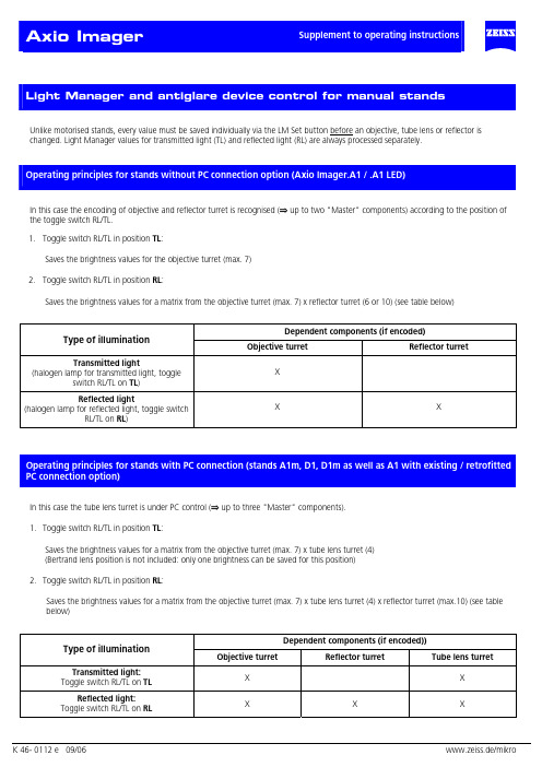
Unlike motorised stands, every value must be saved individually via the LM Set button before an objective, tube lens or reflector ischanged. Light Manager values for transmitted light (TL) and reflected light (RL) are always processed separately.In this case the encoding of objective and reflector turret is recognised (⇒ up to two "Master" components) according to the position ofthe toggle switch RL/TL.1. Toggle switch RL/TL in position TL:Saves the brightness values for the objective turret (max. 7)2. Toggle switch RL/TL in position RL:Saves the brightness values for a matrix from the objective turret (max. 7) x reflector turret (6 or 10) (see table below)Dependent components (if encoded)Type of illuminationObjective turret Reflector turretTransmitted lightX (halogen lamp for transmitted light, toggleswitch RL/TL on TL)Reflected lightX X (halogen lamp for reflected light, toggle switchRL/TL on RL)In this case the tube lens turret is under PC control (⇒ up to three "Master" components).1. Toggle switch RL/TL in position TL:Saves the brightness values for a matrix from the objective turret (max. 7) x tube lens turret (4)(Bertrand lens position is not included: only one brightness can be saved for this position)2. Toggle switch RL/TL in position RL:Saves the brightness values for a matrix from the objective turret (max. 7) x tube lens turret (4) x reflector turret (max.10) (see tablebelow)Dependent components (if encoded))Type of illuminationObjective turret Reflector turret Tube lens turretTransmitted light:X X Toggle switch RL/TL on TLReflected light:X X X Toggle switch RL/TL on RLDefault setting of the stand after switching on:Transmitted light:• Toggle switch RL/TL on TLButton TL on (shutter open or lamp on)Button RL offReflected light:• Toggle switch RL/TL on RLButton TL offButton RL on (shutter open or lamp on)Saving LM value:• To save the current lamp voltage for the current objective turret position press the LM Set button brieflySaving 3200K:This function determines whether the stand is set at 3200K when it is switched on.• To set 3200K to be active on switching on: activate 3200K and press LM Set button.• To set 3200K to be inactive on switching on: deactivate 3200K and press LM Set button.The 3200K setting is saved globally and is independent of other LM values that have already been saved. The normal LM values are available at any time as soon as 3200K is deactivated.Overwriting the LM values:• To save the new value at the relevant position press the LM Set buttonDeleting of the LM values:This is not possible.Activating an LM value:This is done by switching on and changing the position of a "Master" component.To permanently deactivate/activate Light Manager (LM) & antiglare device (AG)• Keep the "RL" button pressed down when you switch on:• One beep signifies deactivation.• Two beeps signify activation.To permanently deactivate/ activate Light Manager only• Keep "3200"button pressed down when you switch on:One beep signifies deactivation. Two beeps signify activation.To permanently deactivate/ activate antiglare device only• Keep “TL” button pressed down when you switch on:One beep signifies deactivation. Two beeps signify activation• If button "RL" is pressed when you switch on and only one of the two functions is activated, that function will be deactivated:Starting condition OutcomeLM AG LM AG⇒0 01 1⇒0 01 0⇒0 00 1⇒ 1 10 0These parameters can also be set via MTB 2004 for motorised stands.Antiglare device:If there is a shutter in the TL optical path, the lamp voltage remains constant when the objective is changed and the shutter takes over thefunction of the antiglare device.If no shutter is present, the lamp is switched off.Safety function:If the reflector turret flap is opened or the reflector turret is completely removed, the safety switch-off device automatically closes thereflected light shutter. In addition, the shutter can no longer be opened by pressing a button as long as the reflected light path is "open".The shutter also closes automatically when the stand is switched off.The brightness of the Light Control LEDs can be adjusted by the user.Manual stands:• Keep SET button pressed down for about 3 seconds until a long beep is heard.All LEDs go on.The brightness of the LEDs can now be adjusted by the brightness control (control knob).However, the brightness cannot be completely extinguished!Activating the control knob in this mode has no effect on the lamp voltage!This mode is exited automatically by releasing the LM Set buttonThe setting is saved permanently!Motorised stands:The adjustment of the LED brightness is linked with the brightness control of the TFT Display.All motorised reflector turrets can be mounted on the stands D1 and D1m. The reflector turrets for the D1 stands have been incorporated into the MTB 2004 in the same way as those for the motorised stands.The motorised reflector turrets can be operated either by the AxioVision Software or by the keyring in Z drive. If a motorised reflector turret is recognised when the microscope is switched, keys are assigned automatically in the following order:Refl.turret to the right (Pos. +), Refl.turret to the left (Pos. -), RL shutter, lamp voltage +, lamp voltage -.Otherwise the default assignment of keys applies:TL shutter, RL shutter, unassigned, lamp voltage +, lamp voltage -.The position indication is shown by the LED Bargraph. As soon as a motorised reflector turret is recognised, the LED Bargraph indicates the reflector turret position, if ”RL on” was set (by pressing button on the Light Control or on the keyring or via Software). If TL is switched on as well (only possible if the toggle switch HAL is on TL), the reflector position will continue to be displayed, overriding the display of the lamp brightness.。
ZEISS Axio Imager 2 开放微观系统,适用于自动材料分析说明书
ZEISS Axio Imager 2Your Open Microscope System for Automated Material AnalysisProduct Information Version 1.2Axio Imager 2 from ZEISS is your system platform tailored to demanding materials analysis tasks, development of new materials as well as quality control.You always profit from crisp images and high optical performance. This applies in particular to sophisticated contrasting techniques, e.g. like the Circular Differential Interference Contrast (C-DIC) and polarization contrast.Use the motorized stand to achieve reproducible illumination settings and, consequently, constant image quality. You always obtain comparable results and high productivity by automating your workflow. Axio Imager 2 offers a high degree of adaptability in line with your future requirements. The stands are open to expand and cover a wide range of applications.Your Open Microscope System for Automated Material Analysis› In Brief › The Advantages › The Applications › The System› Technology and Details ›ServiceAnimation20 µmSimpler. More Intelligent. More Integrated.Profit from an Open Microscope System Whether in research, testing or failure analysis, materials microscopy faces quite various challenges. With Axio Imager 2 from ZEISS you will be able to meet and win these challenges. Attach application-specific components and perform e.g. particle analysis, investigate non-metallic inclusions (NMI), liquid crystals or semiconductor-based MEMs. Axio Imager 2 supports the correlative workflow to electron microscopic investigations, too.Achieve Reliable, Reproducible ResultsStability is essential if you want to obtain good results. You will appreciate the stable imaging conditions of Axio Imager 2, especially when working with high magnifications and performing time dependent studies. Due to the motorization of Axio Imager 2 you will achieve quick and repro-ducible results while you always work under constant conditions. For instance, the motorized apertures and the illumination control, which automatically adjusts the color temperature via filter wheels. Experience Competence in all Contrasting TechniquesChoose from a variety of contrasting techniques to achieve an optimum image quality for your dedicated applications. Examine your samples in reflected light in brightfield, darkfield, Differential Interference Contrast (DIC), Circular Differential Interference Contrast (C-DIC), polarization or fluorescence contrast. For transmitted light you can choose between brightfield, darkfield, Differential Interference Contrast (DIC), polarization or circular polarization. Minimized stray light enables homogenous illumination. You achieve outstanding image contrast, even at high magnifications.Carbon fiber-reinforced polymer (CFRP), Differential Interference Contrast (DIC); Objective: EC Epiplan-NEOFLUAR 50×/0.8Stage insert with correlative sample holder for a big variety of specimen.Appreciate the stable imaging conditions with Axio Imager 2.› In Brief› The Advantages › The Applications › The System› Technology and Details › ServiceBrightfield and Darkfield: Maximum Homogeneity and a Stray Light Free Image Background In brightfield Axio Imager 2 provides homoge-neous illumination and exceptional contrast. By minimizing disturbing stray light and reducing the longitudinal color aberration of the illumination optics, the darkfield illumination contrast is suitable for the most challenging samples and impresses even when faced with finest structures. Switching between the techniques only requires a simple turn. The motorized stands allow you to work particularly quickly and conveniently.C-DIC: Perfect for All StructuresCircular Differential Interference Contrast (C-DIC) is a polarization-optical technique which, in contrast to ordinary Differential Interference Contrast (DIC), uses circularly polarized light. This technique has a number of decisive advantages for the contrasting of differently aligned object structures. The speci-men no longer has to be rotated for best imageExpand Your PossibilitiesCopper casting, brightfield.Objective: EC Epiplan-NEOFLUAR 20×/0.5Copper casting, darkfield.Objective: EC Epiplan-NEOFLUAR 20×/0.5contrast and quality, as it is the case in basic DIC. With C-DIC it is simply enough to adjust the position of the C-DIC prism to achieve best image quality whether it is for contrast and/or resolution independent of sample orientation. And all this is possible using one C-DIC prism for a homoge-neous unsurpassed quality image.Copper casting, C-DIC.Objective: EC Epiplan-NEOFLUAR 20×/0.5Experience Competence in all Contrasting Techniques › In Brief› The Advantages › The Applications › The System› Technology and Details › Service200 µm Experience Competence in All Contrasting TechniquesExpand Your PossibilitiesBrightfieldDarkfieldC-DICSample: pure aluminum; Objective: EC Epiplan-NEOFLUAR 10×/0.25, same position acquired with different contrasting techniquesPolarization Contrast Polarization with Additional Lambda Plate› In Brief› The Advantages › The Applications › The System› Technology and Details › ServiceTailored Precisely to Your Applications› The Advantages› The Applications› The System› Technology and Details› ServiceTailored Precisely to Your Applications› The Advantages› The Applications› The System› Technology and Details› Service20 µm20 µm20 µm20 µm200 µm200 µmZEISS Axio Imager 2 at WorkAviation and Space IndustryCarbon fiber-reinforced polymer (CFRP), brightfield, objective: EC Epiplan-NEOFLUAR 50×/0.8Carbon fiber-reinforced polymer (CFRP), darkfield, objective: EC Epiplan-NEOFLUAR 50×/0.8Carbon fiber-reinforced polymer (CFRP), DIC, objective: EC Epiplan-NEOFLUAR 50×/0.8Raw iron, brightfield,objective: EC Epiplan-NEOFLUAR 50×/0.8Aluminium, polarization,objective: EC Epiplan-NEOFLUAR 10×/0.25Metal Producing and Processing IndustryAluminium, polarization with Lambda plate, objective: EC Epiplan-NEOFLUAR 10×/0.25› In Brief › The Advantages › The Applications › The System› Technology and Details › Service10 µm20 µm50 µmZEISS Axio Imager 2 at WorkOil, Gas and Mining IndustryVitrinite,objective: EC Epiplan-NEOFLUAR 50×/1.0 Oil PolCast iron, brightfield,objective: EC Epiplan-APOCHROMAT 50×/0.95Particle analysis, brightfield,objective: EC Epiplan-NEOFLUAR 20×/0.5Automotive IndustryParticle Analysis› In Brief › The Advantages › The Applications › The System› Technology and Details › ServiceExpand Your PossibilitiesAnalyze Tiny Particles: Accurately and ReproduciblyParticle Analyzer is a milestone for your quality control. With the fully motorized light microscope Axio Imager 2 you measure particles down to 2 µm.Particle Analyzer software supports the standards for cleanliness testing ISO 16232, VDA 19, and oil analysis ISO 4406, ISO 4407, and SAE AS 4059. With the system solutions from ZEISS, you ensure that the required microscope settings are always selected correctly. You receive reliable, reproducible results nearly independent of the user carrying out the analysis. By carrying out correlative particle analyzes, you expand the depth of information contained within your findings to include the results of element and materials characterization.› In Brief › The Advantages › The Applications › The System› Technology and Details › Service100 µmExpand Your PossibilitiesCompletely characterize residual dirt particles with Correlative Automated Particle Analysis from ZEISS. Detect particles with your Axio Imager 2 and relocate preselected particles automatically, using your SEM from ZEISS. Perform an EDX analyisis to reveal information of their elemental composition. Correlative Particle Analyzer automatically documents the results from both, the light microscopic and electron microscopic analysis. You receive a combined, informative report at the touch of a button.As an experienced user, you can inspect the results of the combined light microscopic and electron microscopic analysis on an interactive overview screen. Retrieve particles at the touch of a button, automatically start new EDX analyzes, and auto-matically generate a report. With Correlative Particle Analyzer, your results will be available up to ten times faster than first conducting an analysis with a light microscope and then sub-sequently with an electron microscope. You can systematically focus on potentially process-critical particles.The complementary material characterization from both microscopic worlds gives you added security.Correlative Automated Particle Analysis (CAPA): More Knowledge. Higher Quality.Image of a metallic particle from a light microscopeImage of the same metallic particle from an electron microscopeOverlay of the images from both systems; chemical element composition via EDX analysis; graphical EDX overlay prepared with Bruker Esprit softwareCorrelative sample holder for efficient relocation of particles in your ZEISS scanning electron microscope.› In Brief › The Advantages › The Applications › The System› Technology and Details › Service20 µm 20 µm20 µm20 µmExpand Your PossibilitiesCLEM (Correlative Light and Electron Microscopy) image of a region of interest from an aged Li-ion battery with different contrasts of brightfield (a) and polarized light (b) in LM as well as BSE signal (c) and EDS mapping (d) in SEM.Correlative Microscopy withZEISS Axio Imager 2: Bridging the Micro and Nano WorldAre you looking for a way to combine imaging and analytical methods effectively?Shuttle & Find offers precisely this: An easy-to-use, highly productive workflow from a light to an electron microscope – and vice versa.The workflow between the two systems has never been so easy. The precise recall of regions of interest enhances productivity. Instead of wasting valuable time searching, you now gain new insights into your samples with a few mouse clicks. Regions of interest, marked on one system, you can instantely relocate on the other system. Open up new dimensions of information in numerous material analysis applications. Absolutely reproducible.› In Brief › The Advantages › The Applications › The System› Technology and Details › ServiceExpand Your PossibilitiesExaminations in the fields of research and industrial production (e.g. surface examinations of reflective, low-contrast specimens such as metallographic specimens and polished or textured wafers) require a fast focusing system that ensures high precision levels of max. 0.3 times the objective’s depth of field. This requirement can be easily met by com-bining your Axio Imager 2 with the Auto Focus system to benefit from fast and accurate focusing across a wide capture range of up to 12,000 µm. The Auto Focus system is designed to work with reflected light and transmitted light in brightfield, darkfield, polarized light and DIC.How it WorksThe objective guides the structured light produced by an LED in the Auto Focus system onto the specimen, with the specimen’s surface reflecting it back. During this process, Auto Focus permanently analyses the signal and derives the appropriate control signals for the focus drive, to bring the surface into focus. The Auto Focus sensor detects changes and deviations in the focus position and compensates them automatically. The Auto Focus system comes with three different modes corre-sponding to different specimen characteristics (reflective/partially reflective/diffuse) and with three different precision levels (precision/balance/speed).How the Auto Focus system works: 1) LED 2) Sensor module 3) Sensor 4) Beam splitter 5) O bjective 6) Specimen› In Brief › The Advantages › The Applications › The System› Technology and Details › Service5362141 Microscope• Axio Imager.A2m (encoded)• Axio Imager.D2m (encoded, partly motorizable)• Axio Imager.M2m (motorizable, TL manual)• Axio Imager.Z2m (motorizable, TL motorized)2 Objectives Reflected Light • EC EPIPLAN• EC Epiplan-NEOFLUAR • EC Epiplan-APOCHROMAT Transmitted Light • N-ACHROPLAN • EC Plan-NEOFLUAR • Plan-APOCHROMAT • C-APOCHROMAT • FLUARLong Working Distance • LD EPIPLAN• LD EC Epiplan-NEOFLUAR 3 Illumination Reflected Light • MicroLED • VisLED • Halogen • HBO / HXP Transmitted Light • MicroLED • VisLED • HalogenYour Flexible Choice of Components4 Cameras • Axiocam 105 • Axiocam 305• Axiocam 506• Axiocam 705• Axiocam 7125 Software • ZEN core • ZEN starter6 Accessories • Auto Focus• Linkam heating- and cooling stages • Focus Linear Sensor • Correlative Microscopy› In Brief › The Advantages › The Applications › The System› Technology and Details › ServiceSystem Overview› The Advantages› The Applications› The System› Technology and Details› ServiceSystem Overview› The Advantages› The Applications› The System› Technology and Details› ServiceSystem Overview› The Advantages› The Applications› The System› Technology and Details› ServiceTechnical Specifications› The Advantages› The Applications› The System› Technology and Details› ServiceTechnical Specifications› The Advantages› The Applications› The System› Technology and Details› ServiceTechnical Specifications› The Advantages› The Applications› The System› Technology and Details› ServiceTechnical Specifications› The Advantages› The Applications› The System› Technology and Details› ServiceBecause the ZEISS microscope system is one of your most important tools, we make sure it is always ready to perform. What’s more, we’ll see to it that you are employing all the options that get the best from your microscope. You can choose from a range of service products, each delivered by highly qualified ZEISS specialists who will support you long beyond the purchase of your system. Our aim is to enable you to experience those special moments that inspire your work.Repair. Maintain. Optimize.Attain maximum uptime with your microscope. A ZEISS Protect Service Agreement lets you budget for operating costs, all the while reducing costly downtime and achieving the best results through the improved performance of your system. Choose from service agreements designed to give you a range of options and control levels. We’ll work with you to select the service program that addresses your system needs and usage requirements, in line with your organization’s standard practices.Our service on-demand also brings you distinct advantages. ZEISS service staff will analyze issues at hand and resolve them – whether using remote maintenance software or working on site. Enhance Your Microscope System.Your ZEISS microscope system is designed for a variety of updates: open interfaces allow you to maintain a high technological level at all times. As a result you’ll work more efficiently now, while extending the productive lifetime of your microscope as new update possibilities come on stream.Profit from the optimized performance of your microscope system with services from ZEISS – now and for years to come.Count on Service in the True Sense of the Word>> /microservice› In Brief › The Advantages › The Applications › The System› Technology and Details › ServiceCarl Zeiss Microscopy GmbH 07745 Jena, Germany******************** /axioimager-mat Notfortherapeuticuse,treatmentormedicaldiagnosticevidence.Notallproductsareavailableineverycountry.ContactyourlocalZEISSrepresentativeformoreinformation.EN_42_11_31|CZ11-219|Design,scopeofdelivery,andtechnicalprogresssubjecttochangewithoutnotice.|©CarlZeissMicroscopyGmbH。
ZEISS Axio Imager 2研究显微镜说明书
Pilidium larvae of the Nemertean ribbon worm,Cerebratulus lacteus. Courtesy of the Marine BiologyLaboratory & Development journalEsophagus (guinea pig)ZEISS Axio Imager 2Your Upright Research Microscope for Advanced ImagingAxio Imager 2 combines everything you’veever asked for in your research environment:brilliant optics, bright fl uorescence and vari-ous methods of transmitted light contrast.The contrast manager and light manager en-sure defi ned conditions and reproducible re-sults at all times. With your Axio Imager.Z2you can use ACR to automatically detect andconfi gure objectives and fi lter sets.Choose your system out of six different standversions. Whether you are simply observingand recording, or performing highly compleximaging experiments, it’s easy to adapt thesystem components to the application athand. The research microscope Axio Imager2 resolves fi nest details of your samples -from tissue sections in pathology to brainspecimens in neuroscience to multi-colorFISH samples.Highlights• Encoding: read-out magnifi cation,illumination and contrast settings, andautomatic transfer to ZEN Imagingsoftware• Motorization for reproducible compo-nent settings and automated processes• Excellent optics and homogeneousillumination in both transmitted lightand fl uorescence applications• Research microscope with high perfor-mance focus for maximum precisionand 24/7 operation• Smart control concept for ergonomicworking conditions and multi-usermanagement for up to ten differentusers or working scenarios.• Extend your Axio Imager 2 with high endfl uorescence imaging systems, such asApotome.2, LSM 800 and LSM 880Ease of UseUse your motorized Axio Imager 2 withequipment tailored precisely to yourneeds - and upgrade your system easilyat any time.The smart automated contrast settingslet you work quickly and effi ciently. Themotorized aperture and luminous fi elddiaphragm and light intensity areautomatically adjusted. Achieve repro-ducible results and control all componentsvia optional TFT monitor.***************/axioimagerN o t a l l p r o d u c t s a r e a v a i l a b l e i n e v e r y c o u n t r y . U s e o f p r o d u c t s f o r m e d i c a l d i a g n o s t i c , t h e r a p e u t i c o r t r e a t m e n t p u r p o s e s m a y b e l i m i t e d b y l o c a l r e g u l a t i o n s . C o n t a c t y o u r l o c a l Z E I S S r e p r e s e n t a t i v e f o r m o r e i n f o r m a t i o n .E N _41_012_113 | C Z 08-2015 | D e s i g n , s c o p e o f d e l i v e r y a n d t e c h n i c a l p r o g r e s s s u b j e c t t o c h a n g e w i t h o u t n o t i c e . | © C a r l Z e i s s M i c r o s c o p y G m bHPerformance:The motorized reflector turret accommo-dates either six or ten Push & Click filtermodules. Auto-configure all motorized components with Smart Setup of the ZEN imaging software.Acquire fluorescence images with an excellent signal-to-noise ratio. The fluorescence beam path and high efficiency fluorescence filter sets of this research microscope deliver exposure times that are up to 50 percent shorter.Stand Versions:• Axio Imager.A2 LED • Axio Imager.A2• Axio Imager.D2• Axio Imager.M2p • Axio Imager.M2• Axio Imager.Z2 Suitable Applications:• Cell biology • Neuroscience • Molecular genetics • PathologyNote: This product is primarily for research use. Only Axio Imager.M2p is for use in diagnostic procedures or patient management.ZEISS Axio Imager 2Your Upright Research Microscope for Advanced Imaging。
最新AxiIMAGER说明(中英对照)汇总
A x i I M A G E R说明(中英对照)蔡司图像分析系统Carl Zeiss Imaging Analysis Systems北京普瑞赛司仪器有限公司BEIJING PRECISE INSTRUMENT CO., LTDAxio ImagerA1M技术说明Axio ImagerA1M Technical Specifications用户名称:Username:日期:2009-4-30Date: 2009-4-30尊敬的杜工您好!首先感谢贵方的询问及询价!我方根据贵方对材料检验研究的最新要求,推荐ZEISS顶级研究级倒置万能材料显微镜Axio ImagerA1MDear Manager Du,Hello!First of all, thank you for your Inquiries!According to your refreshed requirements on Materials analysis, we recommend Axio Imager A1M to you.品牌介绍:世界顶级品牌,可见光及电子光学领导企业-----蔡司是一家致力於应用研究,对於光学、玻璃技术、精密技术以及电子等高品质的产品开发、制造、销售有着突出贡献的德国军工企业。
自1846 年开始,carl zeiss已有160多年的传统与创新。
百年历史缔造了蔡司在光学领域不可撼动的领导地位,至今显微镜的生产标准中的83%是以蔡司厂标为基准。
国际物镜的检测标准是以蔡司物镜为基准。
蔡司显微镜以其不断领先的技术和可靠的质量推动了世界材料科学的发展同时也受到知名科学家和诺贝尔奖得主的青睐!作为显微镜的鼻祖国际标准的缔造者,蔡司公司将以更新的技术延续carl zeiss成功的传奇故事!Brand Introduction:The world's top brand, the leading enterprises of visible light and electron optical——Carl Zeiss is a German military enterprises with outstanding contributions to the development, manufacturing and sales of optics, glass technology, microtechnic and electronic products. Since 1846, Carl Zeiss has a history of tradition and innovation for 160 years. The nearly I00 years history has established the leadership of Carl Zeiss. Up to now, 83% of the microscope production use Zeiss criterion and the international objective testing standard are based on Carl Zeiss. Zeiss microscopes have also win favor of famous scientists and Nobel Prize winner by its technology and quality. As the originator of the microscope international standards, Carl Zeiss will update its technology constantly and carry on the legend!设备名称:德国蔡司金相显微镜Device Name: ZEISS Metallographic Microscopy (Germany)规格型号: Axio ImagerA1MModel: Axio ImagerA1M用途:对钢铁有色金属等材料的显微组织观察和分析。
激光共聚焦系统使用说明 Imager Vario LSM
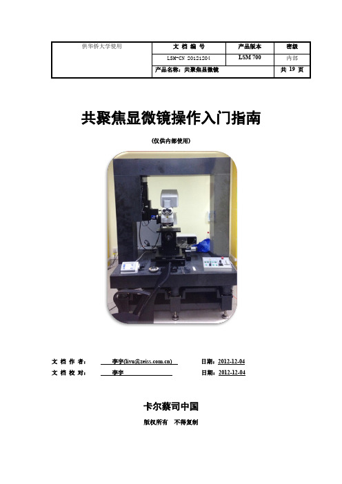
共聚焦显微镜操作入门指南(仅供内部使用)文档作者:李宇(liyu@) 日期:2012-12-04文档校对:李宇日期:2012-12-04卡尔蔡司中国版权所有不得复制目录1开机 (1)1 .1接通总电源 (1)1 .2打开激光器 (1)1 .3打开控制器、主控电脑 (1)2使用激光共聚焦扫描软件Zen 2010 (2)2 .1打开软件 (2)2 .2切换到明场观察模式(目镜筒) (2)2 .3放入样品并在明场模式下找到焦平面 (3)2 .4切换到共聚焦扫描模式 (6)2 .5设置激光扫描参数,找到样品最亮的焦平面位置 (6)2 .6设置Z-stack扫描上下限 (8)2 .7开始扫描 (10)2 .8分析扫描结果,进行三维观测 (11)3关机 (15)4附:目镜中,使用明场、暗场和偏光模式观察样品 (16)4 .1明场模式 (16)4 .2暗场模式 (17)4 .3偏光模式 (17)5附:使用相机(CCD)拍摄明场、暗场和偏光图 (18)5 .1拍摄明场图 (18)5 .2拍摄暗场图 (19)5 .3拍摄偏光图 (19)1 开机1 .1接通总电源图 1 从左至右依次为:墙体总电源、稳压器电源、激光器和电脑电源1 .2打开激光器图 2 转动激光器钥匙,打开激光器,LED指示灯亮1 .3打开控制器、主控电脑图 3 依次打开左图显微镜主机控制器电源、右图电脑主机电源提示:当仅使用CCD拍图,或者长时间不用机器时,建议关闭激光器,以延长其寿命。
2 使用激光共聚焦扫描软件Zen 20102 .1打开软件双击图标,然后点击“Start System ”进入软件。
2 .2切换到明场观察模式(目镜筒)2 .2.1 在共聚焦软件中切换为明场观察模式:点击“Locate ”标签,选择“Online ”,点击“BF ”(Bright Field 的缩写)。
此时系统打开卤素灯,并将明场光学模块转入光路。
图 4 切换为明场观察模式提示:如果出现硬件通讯问题,软件左下角会弹出信息对话框,此时一般的解决方法是:1)重启Zen 软件;2)如果仍无效,关闭整个系统,过5分钟后再重启系统。
Axio Imager M2显微镜使用手册
8、调节荧光光强
ZeiSsseitWe angying
9、调节焦距
显微镜主机的左右两侧都有调焦旋钮
左侧调焦旋钮 外圈为粗调,里圈为微调
微调
粗调
ZeiSsseitWe angying
右侧调焦旋钮 外圈为粗调,里圈为微调
粗调
微调
ZeiSsseitWe angying
10、移动载物台
通过此手柄移动电动载物台
荧光观察
ZeiSsseitWe angying
1、打开电源
按下开关,打开显微镜总电源
ZeiSsseitWe angying
2、打开显微镜开关
按下显微镜主机体左下方开关,打开显微镜
ZeiSsseitWe angying
1、打开荧光灯源
ZeiSsseitWe angying
1、打开显微镜开关后,显微镜右侧电子 显示屏会启动,
2、直到出现此界面,显微镜启动完成 3、白色为激活状态,如Home等
注意:准备好样本和选择好滤 光块前,此状态一定为off
ZeiSsseitWe angying
3、降低载物台
点击触摸屏上“Load position”,载 物台下降
ZeiSsseitWe angying
4、放入样本
1,将样品放入样本夹并夹紧
ZeiSsseitWe angying
明场观察
ZeiSsseitWe angying
1、打开电源
按下开关,打开显微镜总电源
ZeiSsseitWe angying
2、打开显微镜开关
按下显微镜主机体左下方开关,打开显微镜
ZeiSsseitWe angying
1、打开显微镜开关后,显微镜右侧电子 显示屏会启动,
“一枝独秀”之馆藏曲柄铜盉的科学分析与考古初探
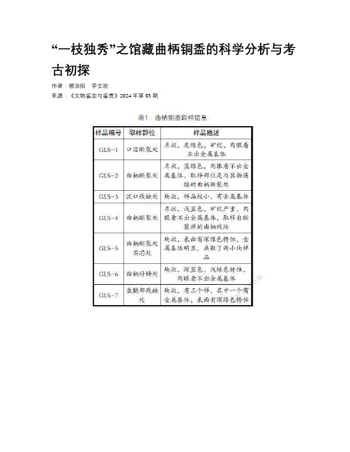
“一枝独秀”之馆藏曲柄铜盉的科学分析与考古初探作者:穆浴阳李文欢来源:《文物鉴定与鉴赏》2024年第03期摘要:利用掃描电镜-能谱仪(SEM-EDS)及金(矿)相显微镜等研究手段,对江西九江共青城市博物馆馆藏唯一一件曲柄铜盉进行金相组织观察和成分检测分析,并初步进行考古研究。
结果表明:此曲柄铜盉的材质为铜-锡-铅(Cu-Sn-Pb)三元合金,制作工艺为铸造而成,其成熟的铜锡铅配比和加工方法佐证了商周青铜器的铸造技术。
虽然曲柄铜盉的出土地为江西地区,但是通过形制初步判断其为群舒文化典型青铜盉,在一定程度上说明了吴、楚、越文化和群舒文化在先秦时期的融合情况。
截至目前,此曲柄铜盉在江西地区尚属首次和唯一出现,填补了江西此类型青铜器研究的空白,对于揭示江西先秦青铜器冶金技术内涵及探讨江西与其周边地区的交流传播具有重要的价值意义。
关键词:江西先秦古史;西周春秋时期;吴楚越文化;群舒文化DOI:10.20005/ki.issn.1674-8697.2024.03.0270 引言在人类技术发展过程中,自使用青铜器伊始,称为青铜时代。
中国的青铜时代始于公元前2000年左右,历经夏商周时期。
青铜器的使用随着商周时期青铜冶铸业的发展达到鼎盛,其产生的青铜艺术亦是亚洲大陆上一颗璀璨的明珠①。
江西的考古自中华人民共和国成立后逐步发展,商周考古取得了较大的成绩,这些大量古文化遗存和丰富的文化遗物作为“无字地书”对于探寻江西先秦古史的意义重大②。
随着江西考古的发展,出土青铜器等文物逐步问世,其种类繁多、遍布全境,年代上溯商周下至汉代。
在江西省九江市共青城市博物馆中,馆藏的唯一一件青铜器—曲柄铜盉较为特殊,其为截至目前该地区出土青铜器中首次且唯一出现的青铜盉类型,不仅填补了江西地区此类型青铜器的空白,更是对于研究江西先秦古史具有重要的价值。
本文拟通过对江西省九江市共青城市博物馆馆藏曲柄铜盉进行科学分析,初步揭示其制作工艺及冶金技术内涵,同时为先秦时期江西与其周边地区交流和发展的研究提供实物证据。
Axio Imager M2显微镜使用手册

Seite
Zeiss Wangying
明场观察
Seite
Zeiss Wangying
1、打开电 源
按下开关,打开显微镜总电源
Seite
Zeiss Wangying
2、打开显微镜开 关
按下显微镜主机体左下方开关,打开显微镜
Seite
Zeiss Wangying
1、打开显微镜开关后,显微镜右侧电子 显示屏会启动,
Seite
Zeiss Wangying
2、打开显微镜开 关
按下显微镜主机体左下方开关,打开显微镜
Seite
Zeiss Wangying
1、打开显微镜开关后,显微镜右侧电子 显示屏会启动,
2、直到出现此界面,显微镜启动完成
3、白色为激活状态,如Home等
Seite
Zeiss Wangying
3、降低载物 台
点击触摸屏上“Load position”, 载 物台下降
Seite
Zeiss Wangying
4、放入样 本
1,将样品放入样本夹并夹紧
Seite
Zeiss Wangying
5、升高载物 台
点击触摸屏上红色箭头所示 按钮,载物台会自动上升到 原来位置
Seite
Zeiss Wangying
6、选择物 镜
左侧调焦旋钮 外圈为粗调,里圈为微调
微调
粗调
Seite
Zeiss Wangying
右侧调焦旋钮 外圈为粗调,里圈为微调
粗调
Seite
微调
Zeiss Wangying
10、移动载物 台
通过此手柄移动电动载物台
Zeiss Wangying