Mesenchymal stem cells and innate tolerancebiology and clinical applications
晚期糖基化终产物通过氧化应激引起人骨髓间充质干细胞凋亡

[文章编号]1007-3949(2011)19-05-0399-06·实验研究·晚期糖基化终产物通过氧化应激引起人骨髓间充质干细胞凋亡刘钊,边云飞,肖传实(山西医科大学第二医院心内科,山西省太原市030001)[关键词]晚期糖基化终产物;人骨髓间充质干细胞;氧化应激;细胞凋亡[摘要]目的观察不同剂量晚期糖基化牛血清白蛋白(AGE-BSA)对体外分离培养的人骨髓间充质干细胞增殖、凋亡及氧化应激的影响,为临床上提高干细胞移植治疗糖尿病心肌病后供体干细胞的存活率提供新的依据。
方法采用全骨髓法从人的骨髓标本中分离培养骨髓间充质干细胞,以含10%FCS的L-DMEM培养细胞,0.25%的胰酶消化后按1ʒ2比例传代接种培养,对第3代细胞应用流式细胞仪检测细胞表面标志CD44、CD105和CD34。
在骨髓间充质干细胞中加入不同浓度的AGE-BSA,作用24h后加入CCK-8溶液,37ħ5%CO培养箱培养2 1h,用酶标仪在450nm处测定吸光度值。
采用Annexin V/PI双染法进行染色,避光作用20min后,将细胞置于流式细胞仪上检测细胞凋亡率。
同时对细胞内活性氧水平进行测定,并且测定细胞内的丙二醛含量和超氧化物歧化酶活性。
结果传代后的骨髓间充质干细胞呈鱼群样或漩涡状排列,细胞为长梭形,贴壁紧密,形态较为一致。
细胞表面标志CD105(间充质干细胞相对特异性标志)及CD44(黏附分子,基质细胞表达)呈阳性表达,阳性率分别为98.9%、97.8%;CD34(造血干细胞/祖细胞及内皮细胞阳性)呈阴性表达,表达百分率为0.8%。
与对照组相比,20、50、100和200mg/L AGE-BSA均不同程度地抑制骨髓间充质干细胞的增殖,促进其凋亡,随着作用浓度的增加,细胞内活性氧含量、丙二醛含量明显增加,而细胞匀浆中超氧化物歧化酶的活性却受到了抑制,具有剂量依赖效应。
结论晚期糖基化终产物通过促进骨髓间充质干细胞内活性氧生成、减少抗氧化酶生成,增强氧化应激,破坏细胞内环境稳定性,从而抑制骨髓间充质干细胞增殖,促进细胞凋亡。
间充质干细胞-是在中胚层的早期发展中形成的多能干细胞,具有不断的自我更新、多向分化潜能
间充质干细胞-是在中胚层的早期发展中形成的多能干细胞,具有不断的自我更新、多向分化潜能间充质干细胞-是在中胚层的早期发展中形成的多能干细胞,具有不断的自我更新、多向分化潜能、低免疫原性、免疫抑制作用和修复作用。
学术术语来源---无动物源性成分培养基体外扩增的间充质干细胞文章亮点:1 此问题的已知信息:传统培养基中含有胎牛血清,可能携带动物细菌、病毒、蛋白传染性疾病或朊病毒,用其培养的间充质干细胞输入人体后,具有潜在传播疾病的风险。
2 文章增加的新信息:在间充质干细胞的体外扩增过程中,为了避免培养基中的动物源性成分,本文着重讨论了替代胎牛血清的其他添加剂的优缺点以及应用前景。
3 临床应用的意义:间充质干细胞体外大规模扩增培养是实验研究和临床应用的基础,还需在此基础上构建一个立体全面、有效、安全的扩增间充质干细胞的评价体系,保证间充质干细胞临床治疗安全化和规范化。
关键词:干细胞;间充质干细胞;人血清;脐血清;富血小板血浆;血小板裂解液;化学成分明确无血清培养基主题词:间质干细胞;培养基;血清;富血小板血浆摘要背景:间充质干细胞是组织工程、再生医学和免疫抑制治疗领域重要的种子细胞来源,在体外大量扩增成为其进入临床应用的关键环节。
目的:对无动物源性成分培养基体外扩增间充质干细胞的研究进展做一综述。
方法:由第一作者应用计算机检索2004年1月至2014年4月中国期刊全文数据库(CNKI)及PubMed数据库,英文检索词为“mesenchymal (stromal) stem cells”,“animal serum-free media”,“humanized media”,“human serum”,“umbilical cord blood serum”,“platelet rich plasma”,“platelet lysate”,“defined medium”,中文检索词为“间充质干细胞、无动物血清培养基、人源性培养基、人血清、脐血清、富血小板血浆、血小板裂解液、化学成分明确无血清培养基”,最终保留41篇文献。
《临床肝胆病杂志》推荐使用的规范医学名词术语

临床肝胆病杂志第40卷第3期2024年3月J Clin Hepatol, Vol.40 No.3, Mar.2024[3]XIA SL, LIU ZM, CAI JR, et al. Liver fibrosis therapy based on biomi⁃metic nanoparticles which deplete activated hepatic stellate cells[J]. J Control Release, 2023, 355: 54-67. DOI: 10.1016/j.jconrel.2023.01.052.[4]LIU YW, DONG YT, WU XJ, et al. The assessment of mesenchymalstem cells therapy in acute on chronic liver failure and chronic liver disease: A systematic review and meta-analysis of randomized con⁃trolled clinical trials[J]. Stem Cell Res Ther, 2022, 13(1): 204. DOI:10.1186/s13287-022-02882-4.[5]ZHANG ZL, SHANG J, YANG QY, et al. Exosomes derived from hu⁃man adipose mesenchymal stem cells ameliorate hepatic fibrosis by inhibiting PI3K/Akt/mTOR pathway and remodeling choline me⁃tabolism[J]. J Nanobiotechnology, 2023, 21(1): 29. DOI: 10.1186/ s12951-023-01788-4.[6]ZHAO T, SU ZP, LI YC, et al. Chitinase-3 like-protein-1 function andits role in diseases[J]. Signal Transduct Target Ther, 2020, 5(1): 201. DOI: 10.1038/s41392-020-00303-7.[7]YANG H, ZHAO LL, HAN P, et al. Value of serum chitinase-3-likeprotein 1 in predicting the risk of decompensation events in patients with liver cirrhosis[J]. J Clin Hepatol, 2023, 39(7): 1578-1585. DOI:10.3969/j.issn.1001-5256.2023.07.011.杨航, 赵黎莉, 韩萍, 等. 血清壳多糖酶3样蛋白1(CHI3L1)对肝硬化患者发生失代偿事件风险的预测价值[J]. 临床肝胆病杂志, 2023, 39(7): 1578-1585. DOI: 10.3969/j.issn.1001-5256.2023.07.011.[8]MA L, WEI J, ZENG Y, et al. Mesenchymal stem cell-originated exo⁃somal circDIDO1 suppresses hepatic stellate cell activation by miR-141-3p/PTEN/AKT pathway in human liver fibrosis[J]. Drug Deliv, 2022, 29(1): 440-453. DOI: 10.1080/10717544.2022.2030428. [9]NISHIMURA N, DE BATTISTA D, MCGIVERN DR, et al. Chitinase 3-like 1 is a profibrogenic factor overexpressed in the aging liver and in patients with liver cirrhosis[J]. Proc Natl Acad Sci U S A, 2021, 118(17): e2019633118. DOI: 10.1073/pnas.2019633118.[10]WANG CG, LI SZ, SHI JM, et al. Research progress in differentia⁃tion, identification, and purification methods of human pluripotent stem cells to mesenchymal-like cells in vitro[J]. J Jilin Univ Med Ed, 2023, 49(6): 1655-1661. DOI: 10.13481/j.1671-587X.20230634.王成刚, 李生振, 史嘉敏, 等. 体外人多能干细胞向间充质样细胞分化、鉴定和纯化方法的研究进展[J]. 吉林大学学报(医学版), 2023, 49(6): 1655-1661. DOI: 10.13481/j.1671-587X.20230634.[11]LI TT, WANG ZR, YAO WQ, et al. Stem cell therapies for chronicliver diseases: Progress and challenges[J]. Stem Cells Transl Med, 2022, 11(9): 900-911. DOI: 10.1093/stcltm/szac053.[12]YANG X, LI Q, LIU WT, et al. Mesenchymal stromal cells in hepaticfibrosis/cirrhosis: From pathogenesis to treatment[J]. Cell Mol Im⁃munol, 2023, 20(6): 583-599. DOI: 10.1038/s41423-023-00983-5. [13]ZHAO SX, LIU Y, PU ZH. Bone marrow mesenchymal stem cell-derived exosomes attenuate D-GaIN/LPS-induced hepatocyte apop⁃tosis by activating autophagy in vitro[J]. Drug Des Devel Ther, 2019, 13: 2887-2897. DOI: 10.2147/DDDT.S220190.[14]LEE CG, HARTL D, LEE GR, et al. Role of breast regression protein39 (BRP-39)/chitinase 3-like-1 in Th2 and IL-13-induced tissue re⁃sponses and apoptosis[J]. J Exp Med, 2009, 206(5): 1149-1166.DOI: 10.1084/jem.20081271.[15]HIGASHIYAMA M, TOMITA K, SUGIHARA N, et al. Chitinase 3-like 1deficiency ameliorates liver fibrosis by promoting hepatic macro⁃phage apoptosis[J]. Hepatol Res, 2019, 49(11): 1316-1328. DOI:10.1111/hepr.13396.收稿日期:2023-06-09;录用日期:2023-08-17本文编辑:邢翔宇引证本文:LIU PJ, YAO LC, HU X, et al. Effect of human umbilical cord mesenchymal stem cells in treatment of mice with liver fibrosis and its mechanism[J]. J Clin Hepatol, 2024, 40(3): 527-532.刘平箕, 姚黎超, 胡雪, 等. 人脐带间充质干细胞(hUC-MSC)对肝纤维化小鼠模型的治疗作用及其机制分析[J]. 临床肝胆病杂志, 2024, 40(3): 527-532.读者·作者·编者《临床肝胆病杂志》推荐使用的规范医学名词术语有关名词术语应规范统一,以全国自然科学名词审定委员会公布的各学科名词为准。
人脐带来源间充质干细胞分离培养方法的优化

中国组织工程研究与临床康复 第15卷 第23期 2011–06–04出版Journal of Clinical Rehabilitative Tissue Engineering Research June 4, 2011 Vol.15, No.23P .O. Box 1200, Shenyang 110004 4220Department of Biochemistry and Molecular Biology, Shanxi Medical University, Taiyuan 030020, Shanxi Province, ChinaQi Kai ★, Studying for master’s degree, Department of Biochemistry and Molecular Biology, Shanxi Medical University, Taiyuan 030020, Shanxi Province, China******************Correspondence to: Niu Bo, Doctor, Professor, Doctoral supervisor, Department of Biochemistry and Molecular Biology, Shanxi Medical University, Taiyuan 030020, Shanxi Province, China ****************Supported by: the National Natural Science Foundation of China, No. 30600226*, 30472251*Received: 2011-01-04 Accepted: 2011-03-16人脐带来源间充质干细胞分离培养方法的优化**★齐 凯,董丽媛,陈显久,赵 婕,薛国芳,陈 彦,牛 勃Optimized study on isolation and cultivation of human umbilical cord mesenchymal stem cellsQi Kai, Dong Li-yuan, Chen Xian-jiu, Zhao Jie, Xue Guo-fang, Chen Yan, Niu BoAbstractBACKGROUND: There are different methods to isolate and culture human umbilical cord mesenchymal stem cells (hUC-MSCs), so how to fast and efficiently harvest hUC-MSCs have become a research hotspot.OBJECTIVE: To optimize digestive enzymes components for the preparation of hUC-MSCs, in order to lay the foundation of preparation for hUC-MSCs.METHODS: Human umbilical cords were collected from full term deliveries under aseptic conditions. According to different mixed enzyme concentration ratio, the samples were divided into three groups: mixed enzyme Ⅰ group, mixed enzyme Ⅱ group, mixed enzyme Ⅲ group. Then according to digest time each group into three subgroups: 1 hour, 2 hours, and 3 hours. Finally, suspended cell volume was decided as 4 mL to count cells. Dulbecco’s Modified Eagle Medium with fetal bovine serum was used for cell culture.RESULTS AND CONCLUSION: Based on the three digestion enzyme concentration, at the duration of 1 hour, 2 hours and 3 hours, mixed enzyme Ⅲ group had the highest total cell number and the total cell rate was statistically different from the other groups (P < 0.05). At the duration of 3 hours, live cell rate was the lowest in the mixed enzyme Ⅲ group, and there wasstatistically significant differences among these groups (P < 0.01). The optimal isolation for hUC-MSCs is 0.3% collagenase Ⅱ, 0.1% trypsin, 0.02% EDTA, 0.1% hyaluronidase and 0.1% DNA enzyme Ⅰ digestion for 2 hours.Qi K, Dong LY, Chen XJ, Zhao J, Xue GF, Chen Y, Niu B. Optimized study on isolation and cultivation of human umbilical cord mesenchymal stem cells.Zhongguo Zuzhi Gongcheng Yanjiu yu Linchuang Kangfu. 2011;15(23): 4220-4224. [ ]摘要背景:关于人脐带间充质干细胞的分离培养方法不一,如何提高人脐带来源间充质干细胞获得效率的问题尚未解决。
间充质基质细胞对脊髓损伤的神经保护机制

·综述·间充质基质细胞对脊髓损伤的神经保护机制张桂通1,2张建政2李连华2孙天胜2【摘要】间充质基质细胞(MSCs)以其取材方便、自体移植、免疫原性低等独特的生物学特征,成为包括脊髓损伤在内的多种疾病治疗领域的研究热点及具有良好的临床应用前景。
MSCs可通过释放营养因子、抑制炎症反应等多条途径发挥神经保护作用,在脊髓损伤的治疗领域有很广阔的应用前景。
【关键词】间质干细胞;脊髓损伤;细胞凋亡;炎症反应Neuroprotection mechanism of mesenchymal stromal cells on spinal cord injury Zhang Guitong1,2,Zhang Jianzheng2,Li Lianhua2, Sun Tiansheng2. 1Chinese PLA Medical School, Beijing 100853, China;2Department of Orthopaedics, PLA Army General Hospital, Beijing 100700, ChinaCorresponding author: Sun Tiansheng, Email: suntiansheng-@【Abstract】Mesenchymal stromal cells (MSCs) with its unique biological characteristics and greatprospects for clinical application, become a hot research topic in many therapeutic areas including spinalcord injury. MSCs can play a neuroprotective role in the treatment of spinal cord injury by releasing trophicfactors, inhibiting inflammatory response and so on, and has a broad application prospects in the treatmentof spinal cord injury.【Key words】Mesenchymal stem cells; Spinal cord injuries; Apoptosis; Inflammatory response脊髓损伤(spinal cord injury,SCI)是脊柱外科常见的急重症,是青壮年致残的主要原因,已经成为全球性的医疗难题[1]。
【doc】骨髓经羟乙基淀粉(HES)处理后分离和培养MSCs的初步探讨

骨髓经羟乙基淀粉(HES)处理后分离和培养MSCs的初步探讨2l8?丛型壁笙旦笙堂笙兰塑』垦oFPRcTIcALHANDSURGERYDec2006,V ol20,No.4======!====:=!= ===2111=====!====————————-———=——————————————————————————————二二:==__,…,组织工程?文章编号:1671—2722(2006)04—0218-02骨髓经羟乙基淀粉(HES)处理后分离和培养MSCs的初步探讨王宁,安贵林,魏侃,张艳,徐媛(沈阳医学院奉天医院手外科研究所,辽宁沈阳110024)中图分类号:R6文献标识码:B种予细胞,支架材料和生长因子是组织工程的三大要素,而寻找合适的种子细胞是成功的重要因素l1l.骨髓间充质干细胞(MesenchymalStemCells,MSCs),因其具有多向分化潜能,如形成骨,软骨,肌组织,皮肤等,成为最具代表性的种了细胞_21.目前,国内外已分离培养出人,小鼠,大鼠,兔等的骨髓MSCs.并诱导分化出多种组织H,但由于它在骨髓内含量极少,只占0.01%,-0.001%,而组织工程需要短期内有大量的种子细胞再生组织,故需进行体外培养,分化和扩增日.本实验对骨髓MSCs的两种分离方法进行比较,以期获得更好的方法.1材料与方法1.1材料动物:以两只狗(雄性,体重18kg左右)骨髓为观察材料.药品:DMEM-LG培养基(Gibco),胎牛血清(Gibco),Ficoll淋巴细胞分离液,羟乙基淀粉40氯化钠注射液(HES).1.2方法在同一条件抽取狗的骨髓,经两种方法多次体外分离和培养的MSCs,主要对MSCs数量与MSCs存活和生长等进行对比观察.方法一:当骨髓经羟乙基淀粉(HES)处理后离心分离MSC,其具体操作程序如下:(1)取健康狗在无菌条件下,以穿刺针垂直穿刺髂骨骨髓腔.刺入骨髓腔2~3cm,骨穿针后套20mL注射器,(注射器事先吸取lmL肝素润湿管壁),反复抽吸两次分别抽得骨髓30mL,18mL,立即注入50mL离心管内(事先加肝素2mL). (2)拿回实验室,加入7.5mL,4.5mL(骨髓量的1/4)羟乙基淀粉,吹打均匀,静置2~3h,血细胞沉降后,吸取上清液8mL和1OIIlL.(3)分别装入离心管,离心1200r/min,8min,弃上清液,加入生理盐水5mL,制成细胞悬浮液.(4)将细胞悬浮液缓慢地加入事先盛有3mLFico~淋巴细胞分离液的离心管内液面上,离心3000r/min,15min,离心管液体呈现出4层,最下面为血细胞层,其上面为淋巴细胞分离液层,离心管最上面为生理盐水层,生理盐水与淋巴细胞分离液之间呈浑浊状薄层即单个核细胞层(MSCs存在处).收稿日期:2006—04—12作者简介:王宁(1979一).女.医帅.(5)用吸管轻缓的吸取单个核细胞存在的混浊层,装入已盛有DMEM培养基的离心管内,混匀后离心1200r/min, 5min.(6)弃上清液,再加入5mL培养基,制成细胞悬浮液.混匀后细胞计数和胎盘兰染色计数活细胞占有的百分比. (7)调竹MSCs密度,接种培养观察细胞长势.方法二:为常规应用的密度梯度离心分离法,作为方法一分离MSCs效果的对照.其具体操作程序,除抽取的骨髓不经羟乙基淀粉处理外,其余操作和条件均与方法一相同.观察指标:2只狗抽取的骨髓,经两种方法多次分离MSCs和原代培养的效果,对比观察的指标如下:(1)两法经梯度离心分离后,观察含单个核细胞的混浊层的差异,厚而清晰者表明MSCs居多,反之相反.(2)细胞计数:用血细胞计数板,分别计算两种方法分离出的MSCs数量,具体操作如下:在血细胞计数板中央放置专用的盖玻片:用虹吸管吸取细胞悬液,滴入计数板使盖片下充满悬液为止;在200倍镜下,计数四角大方格内的细胞(包括压线细胞,成团细胞按单个细胞计数);按下式计算:细胞密度=(4大方格细胞总数/4)×10000个/mL.(3)活细胞百分比:用苔盼兰染色法,显示活细胞与死细胞(死细胞着蓝色,活的不显色)具体操作如下:①将细胞悬液1滴与胎盘兰染液2滴混合后,注入血细胞计数板小室内1滴,静置2分钟;②在200倍镜下,计数500个细胞中着色细胞数量;③按下式计算:细胞活力=(总细胞数一着色细胞数)/总的细胞数×100%.(4)细胞长势:将两法经梯度离心分离出来的MSCs接种于盛有DMEM培养基的培养瓶内,主要观察两法在细胞贴壁和铺展(细胞占培养基瓶底面积的百分比)中的差异.2结果经两种方法分离骨髓MSCs的效果:(1)方法一密度梯度离心分离后,离心管内液体显示混浊(乳白)色膜状部位较方法二未经羟乙基淀粉处理的略厚而清晰;(2)从乳白色膜层吸取的单个核细胞制成细胞悬液,经多次记数,方法一平均为2.0×10/mL,明显较未经羟乙基淀粉处理的方法二平均2.Ox10/mL多;(3)经苔盼染色,方法一分离出来不着色的活细胞占总计数的89%,而方法二的只占78%,即经羟乙基淀粉处理后分离出来的着兰色的死细胞较不经处理方法二少;(4)MSCs在原代培养中,可见方法一的贴壁细胞较未经羟乙基淀粉处理方法二的早和快,接种48h,方法一细胞贴壁占实用手外科杂志2006堡旦箜萱箜!塑jQ些Q!型竺坠培养瓶底面积2/3,而方法_的占1/3,7天时,方法一细胞扩增铺展可达培养瓶底面积的90%,而方法二I79%,表明前者细胞长势优于后者方法二.3讨论骨髓经羟乙基淀粉处理的密度梯度离心分离MSCs(法一)的效果比不经过处理(法二)的好,本文观察经羟乙基淀粉处理,然后进行密度梯度离心分离MSCs,无论在分离出来MSCs的数量和存活细胞的数量以及原代培养细胞贴壁和扩增铺展速度等均优于未经羟乙基淀粉处理的,表明了羟乙基淀粉的优越性.羟乙基淀粉在骨髓分离MSCs中的作用:据记载羟乙基淀粉是血容量扩充剂,中分_f量羟乙基淀粉有较好的血液流变学特性,适用于临床使用,它可使促凝粗酶原激酶时间快速降低,使血小板功能降低,对红血细胞集聚性沉降率有影响,有抑制红血细胞集聚作用,能增加细胞膜负电荷,使已集聚细胞解聚等作用.由此本实验将含有肝素的骨髓经tiES 处理后,使骨髓内细胞成分,能更充分处于游离单个细胞状态,能更有利于从骨髓内分离出更多的MSCs,并为MSCs提供充足的贴壁及生长空间,虽然仍沉淀有少量的红细胞,但是这些细胞呈悬浮的状态,随着不断的换液,逐渐被淘汰,间充质干细胞迅速增殖,原代培养杂质细胞并未见明显增加.219?由此奉文初步提,羟乙基淀粉物理沉降法,简单易行,对细胞无任何损伤,培养成功率高,是值得推荐的方法.参考文献:[1】JenkinsDD,Y angGD,LorenzHP,et,a1.Tissueen~neering andregenerativemedicine[11.ClinHastSurg,2003,3O(4):581-588.I21BruderSP,Jainswa1N,HaynesworthSE.Growthkinet—ics,self-renewalandtheosteogenicpotentialofpunfied humanmesenclwmstemcellsduringextensivesubculti—vationandfollowingcryopreservation[J]jCellBiochem1997,64(2):278—94[3】黄海霞,汤雪明.成体干细胞多能性研究进展卟生命科学, 2002,14(3):129134.【4】李思源,李军,姜玉峰.人骨髓间充质干细胞的分离和体外培养卟中国临床康复.2004,8,(17):3264—3266.15l土少I【j.李宝峰,王楠骨髓基质干细胞体外培养及临床应用研究卟中国矫形外科杂志,2004,12(14):1086—1088l61刘卫东,张启祥.人工胶体血浆代用品的分类和临床应用比较Ⅲ中国药业,2006,15(12):6263.【7l迟作华,张洹,陆琰.人骨髓间充质干细胞分离方法比较中国临床康复.2006,10(1):20-22文章编号:16712722(2006)04—0219~O1按压促进静脉回流治疗静脉危象的护理宋安秀(朝阳市中心医院骨科,辽宁朝阳122000)中图分类号:R6文献标识码:B我院自1999年开展显微外科以来,对收治的患者术后出现静脉危象者,早期采取积极措施,使断指及皮瓣血运转为正常,现将护理体会报道如下.1临床资料本组18例,男12例,女6例:年龄17-45岁,平均28岁.损伤原因;砸伤8例,皮带绞伤4例,门挤伤4例,动物咬伤2例.2护理术后消除恐惧心里,增强治疗疾病的信心.避免疼痛,寒冷,吸烟,饮酒等诱因,积极配合护理工作.密切观察手指及皮瓣的皮肤颜色,张力,皮温,肿胀程度,毛细血管的充盈度并做好记录.18例患者均于早期发现皮肤颜色变暗,毛细血管反应明显加快,立即向医乍报告后,马上按摩手指及皮瓣以促进静脉回流,同时给予肝素12500单位加入0.9%NS500mL中, 以每分钟1O滴静点按压,3-10min不等,皮肤转为红色后停止.问隔5-30min不等,皮肤颜色又出现变暗,再行按压,皮肤又转红色,这样重复进行,其中断指6例,皮瓣4例需要按收稿日期:2006—07—27作者简介:宋安秀(1972一),女,主管护师.手外科护理?压问隔时间逐渐延长,分别按压8-12次之后,不再需要按压,皮肤颜色不再变暗,经过临床严密观察,结果10例断指及皮瓣都顺利成活,其余8例通过按压虽然皮肤颜色也转为红色,但间隔最长只有15-20分钟,并且间隔时间逐渐缩短, 伤口周围还有部分暗红色血性渗出,向医生报告后急诊行手术探查,术中均发现有血栓形成.按压方法及注意事项:按压时从远端向近端进行,手法要轻柔.对断指的按要保持手指不要晃动,按压一次后要间隔2-3秒后再行第二次按压,不能连续进行,给动脉供血一个时间间隔.对皮瓣按压也要由远向近进行,用食,中两指或单一食指滚动擀.3讨论护理人员要有高度的责任心,密切观察手指及皮瓣的血运情况,及时发现,采取措施.吻合血管或岛状转移皮瓣,术后出现静脉危象,’部分是因为静脉条件及吻合手术质量原凶,另部分则因为术后其它因素,如痉挛或岛状皮瓣蒂部水肿等,血液通过静脉吻合口或蒂部变得缓慢,给血栓形成提供机会,而通过按压促进静脉回流,使血液快速通过吻合口,减少血栓形成机会.同时静脉血被”挤”走后,也会促进动脉供血.。
Mesenchymal stem cell
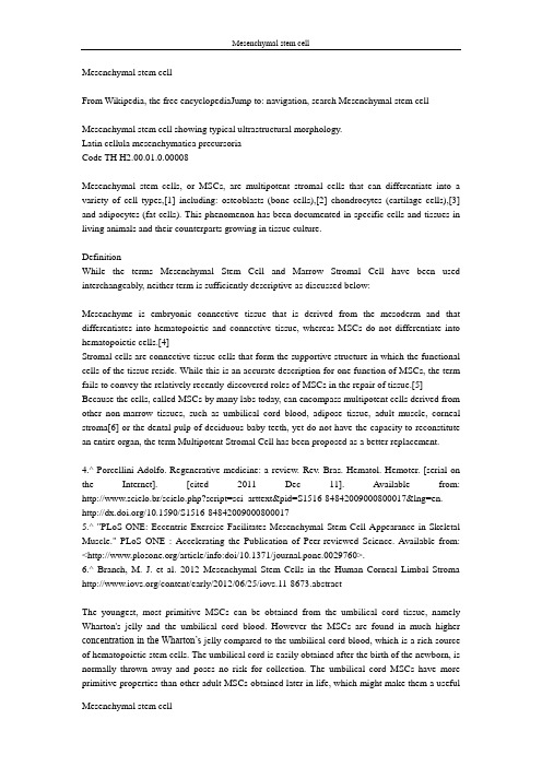
Mesenchymal stem cellFrom Wikipedia, the free encyclopediaJump to: navigation, search Mesenchymal stem cellMesenchymal stem cell showing typical ultrastructural morphology.Latin cellula mesenchymatica precursoriaCode TH H2.00.01.0.00008Mesenchymal stem cells, or MSCs, are multipotent stromal cells that can differentiate into a variety of cell types,[1] including: osteoblasts (bone cells),[2] chondrocytes (cartilage cells),[3] and adipocytes (fat cells). This phenomenon has been documented in specific cells and tissues in living animals and their counterparts growing in tissue culture.DefinitionWhile the terms Mesenchymal Stem Cell and Marrow Stromal Cell have been used interchangeably, neither term is sufficiently descriptive as discussed below:Mesenchyme is embryonic connective tissue that is derived from the mesoderm and that differentiates into hematopoietic and connective tissue, whereas MSCs do not differentiate into hematopoietic cells.[4]Stromal cells are connective tissue cells that form the supportive structure in which the functional cells of the tissue reside. While this is an accurate description for one function of MSCs, the term fails to convey the relatively recently-discovered roles of MSCs in the repair of tissue.[5] Because the cells, called MSCs by many labs today, can encompass multipotent cells derived from other non-marrow tissues, such as umbilical cord blood, adipose tissue, adult muscle, corneal stroma[6] or the dental pulp of deciduous baby teeth, yet do not have the capacity to reconstitute an entire organ, the term Multipotent Stromal Cell has been proposed as a better replacement.4.^ Porcellini Adolfo. Regenerative medicine: a review. Rev. Bras. Hematol. Hemoter. [serial on the Internet]. [cited 2011 Dec 11]. Available from: http://www.scielo.br/scielo.php?script=sci_arttext&pid=S1516-84842009000800017&lng=en. /10.1590/S1516-848420090008000175.^ "PLoS ONE: Eccentric Exercise Facilitates Mesenchymal Stem Cell Appearance in Skeletal Muscle." PLoS ONE : Accelerating the Publication of Peer-reviewed Science. Available from: </article/info:doi/10.1371/journal.pone.0029760>.6.^ Branch, M. J. et al. 2012 Mesenchymal Stem Cells in the Human Corneal Limbal Stroma /content/early/2012/06/25/iovs.11-8673.abstractThe youngest, most primitive MSCs can be obtained from the umbilical cord tissue, namely Wharton's jelly and the umbilical cord blood. However the MSCs are found in much higher concentration in the Wharton’s jelly compared to the umbilical cord blood, which is a rich source of hematopoietic stem cells. The umbilical cord is easily obtained after the birth of the newborn, is normally thrown away and poses no risk for collection. The umbilical cord MSCs have more primitive properties than other adult MSCs obtained later in life, which might make them a usefulsource of MSCs for clinical applications.An extremely rich source for mesenchymal stem cells is the developing tooth bud of the mandibular third molar. While considered multipotent, they may prove to be pluripotent. The stem cells eventually form enamel, dentin, blood vessels, dental pulp, nervous tissues, including a minimum of 29 different unique end organs. Because of extreme ease in collection at 8–10 years of age before calcification and minimal to no morbidity they will probably constitute a major source for personal banking, research and multiple therapies. These stem cells have been shown capable of producing hepatocytes. Additionally, amniotic fluid has been shown to be a very rich source of stem cells. As many as 1 in 100 cells collected from and genetic amniocentesis has been shown to be a pluripotent mesenchymal stem cell.[citation needed]Adipose tissue is one of the richest sources of MSCs. When compared to bone marrow, there is more than 500 times more stem cells in 1 gram of fat when compared to 1 gram of aspirated bone marrow. Adipose stem cells are currently actively being researched in clinical trials for treatment in a variety of diseases.HistoryIn 1924, Russian-born morphologist Alexander A. Maximow used extensive histological findings to identify a singular type of precursor cell within mesenchyme that develops into different types of blood cells.[7]Scientists Ernest A. McCulloch and James E. Till first revealed the clonal nature of marrow cells in the 1960s.[8][9] An ex vivo assay for examining the clonogenic potential of multipotent marrow cells was later reported in the 1970s by Friedenstein and colleagues.[10][11] In this assay system, stromal cells were referred to as colony-forming unit-fibroblasts (CFU-f).Subsequent experimentation revealed the plasticity of marrow cells and how their fate could be determined by environmental cues. Culturing marrow stromal cells in the presence of osteogenic stimuli such as ascorbic acid, inorganic phosphate, and dexamethasone could promote their differentiation into osteoblasts. In contrast, the addition of transforming growth factor-beta (TGF-b) could induce chondrogenic markers.[citation needed]7.^ Sell, Stewart (Stem cell handbook). Humana Press. p. 143.8.^ Becker, A. J.; McCulloch, E. A.; Till, J. E. (1963). "Cytological Demonstration of the Clonal Nature of Spleen Colonies Derived from Transplanted Mouse Marrow Cells". Nature 197 (4866): 452–4. doi:10.1038/197452a0. PMID 13970094.9.^ Siminovitch, L.; McCulloch, E. A.; Till, J. E. (1963). "The distribution of colony-forming cells among spleen colonies". Journal of Cellular and Comparative Physiology 62 (3): 327–36. doi:10.1002/jcp.1030620313. PMID 14086156.CulturingThe majority of modern culture techniques still take a colony-forming unit-fibroblasts (CFU-F) approach, where raw unpurified bone marrow or ficoll-purified bone marrow Mononuclear cell are plated directly into cell culture plates or flasks. Mesenchymal stem cells, but not red blood cells or haematopoetic progenitors, are adherent to tissue culture plastic within 24 to48 hours. However, at least one publication has identified a population of non-adherent MSCs that are not obtained by the direct-plating technique.[22]Other flow cytometry-based methods allow the sorting of bone marrow cells for specific surface markers, such as STRO-1.[23] STRO-1+ cells are generally more homogenous, and have higher rates of adherence and higher rates of proliferation, but the exact differences between STRO-1+ cells and MSCs are not clear.[24]Methods of immunodepletion using such techniques as MACS have also been used in the negative selection of MSCs.[25]22.^ Wan, Chao; He, Qiling; McCaigue, Mervyn; Marsh, David; Li, Gang (2006). "Nonadherent cell population of human marrow culture is a complementary source of mesenchymal stem cells (MSCs)". Journal of Orthopaedic Research 24 (1): 21–8. doi:10.1002/jor.20023. PMID 16419965.23.^ Gronthos, S; Graves, SE; Ohta, S; Simmons, PJ (1994). "The STRO-1+ fraction of adult human bone marrow contains the osteogenic precursors". Blood 84 (12): 4164–73. PMID 7994030. /cgi/pmidlookup?view=long&pmid=7994030.24.^ Oyajobi, Babatunde O.; Lomri, Abderrahim; Hott, Monique; Marie, Pierre J. (1999). "Isolation and Characterization of Human Clonogenic Osteoblast Progenitors Immunoselected from Fetal Bone Marrow Stroma Using STRO-1 Monoclonal Antibody". Journal of Bone and Mineral Research 14 (3): 351–61. doi:10.1359/jbmr.1999.14.3.351. PMID 10027900.25.^ Tondreau, T; Lagneaux, L; Dejeneffe, M; Delforge, A; Massy, M; Mortier, C; Bron, D (1 January 2004). "Isolation of BM mesenchymal stem cells by plastic adhesion or negative selection: phenotype, proliferation kinetics and differentiation potential". Cytotherapy 6 (4): 372–379. doi:10.1080/14653240410004943.。
不同无血清培养体系对人脐带间充质干细胞生物学功能的影响

864新医学 2023年12月第54卷第12期DOI: 10.3969/j.issn.0253-9802.2023.12.005研究论著不同无血清培养体系对人脐带间充质干细胞生物学功能的影响林惠珠 李翠平 陈晓燕 阳莉【摘要】目的 探讨3种不同无血清培养体系对人脐带间充质干细胞(hUC-MSC)体外培养的生物学功能的影响。
方法 取3株脐带组织,各分3组实验组,经贴壁法分别置于3组不同无血清培养体系(A组:纯化学成分合成的无血清培养体系,B组:血小板裂解物+基础培养基的无血清培养体系,C组:纤维蛋白原包被+含重组人蛋白质的无血清培养体系)中培养至第3代,比较其细胞增殖能力、细胞表型、三系分化能力以及免疫调节功能。
结果 3组实验组细胞均呈长而扁平的梭形,贴壁生长,大小均一。
其增殖曲线趋势相似,A组和B组P2、P3代次的增殖能力优于C组(P均< 0.05)。
3组实验组细胞均符合国际干细胞协会对hUC-MSC细胞表面标志物鉴定的要求,A组和B 组P3代次的CD29和CD90的表达量均高于C组(P均< 0.05),并且3组实验组均具有三系分化能力。
3组实验组中P3代次的T淋巴细胞增殖率、Th1和Th17细胞亚群分泌的细胞因子比例均低于对照组,调节性T细胞(CD4+CD25+ FOXP3+)比例均高于对照组,而3组实验组之间两两比较差异无统计学意义(P均> 0.05)。
结论 3种不同无血清培养体系的hUC-MSC均能保持其基本的生物学特性和免疫调节活性,纯化学成分合成的无血清培养体系和血小板裂解物+基础培养基的无血清培养体系的增殖能力和细胞表面标志物表达均优于纤维蛋白原包被+含重组人蛋白质的无血清培养体系。
【关键词】人脐带间充质干细胞;无血清培养体系;生物学功能Effects of different serum-free culture systems on the biological function of hUC-MSCs Lin Huizhu, Li Cuiping, Chen Xiaoyan, Yang Li. Biotherapy Center, the Third Affiliated Hospital of Sun Yat-sen University, Guangzhou 510630, China Corresponding author, Yang Li, E-mail:******************【Abstract】Objective To evaluate the effects of three different serum-free culture systems on the biological function of human umbilical cord mesenchymal stem cells (hUC-MSC) cultured in vitro. Methods Three strains of umbilical cord tissues were obtained,divided into 3 experimental groups and cultured to the 3rd generation (P3) in three different serum-free culture systems (Group A: serum-free culture system synthesized by pure chemical components,group B: serum-free culture system consisting of platelet lysate+basic culture medium,group C: serum-free culture system consisting of fibrinogen coating+recombinant human protein). The cell proliferation,cell phenotype,three-line differentiation and immunomodulatory function were compared. Results All the cells in three groups were long and flat fusiform,adherent to the wall and uniform in size. The proliferation curve trend was similar,and the proliferation ability of P2 and P3 generations in groups A and B was better than that in group C (both P < 0.05). The cells in three experimental groups all met the requirements of the International Stem Cell Society for identification of hUC-MSC cell surface markers. The expression levels of CD29 and CD90 in P3 generations in groups A and B were higher than those in group C (all P < 0.05),and all three experimental groups had the ability of three-line differentiation. Compared with the control group,the proliferation rate of T lymphocytes in P3 generation and the proportion of cytokines secreted by Th1 and Th17 cell subsets were significantly decreased in the three experimental groups,and the proportion of regulatory T cells (CD4+ CD25+ FOXP3+) was significantly increased in the three experimental groups,but there was no statistical significance among three experimental groups (all P > 0.05). Conclusions The hUC-MSC in three groups can maintain basic biological characteristics and immunomodulatory activity. The proliferation ability and expression levels of cell surface markers in groups A and B are superior to those in group C.【Key words】Human umbilical cord mesenchymal stem cell; Serum-free culture system; Biological function基金项目:广东省药品监督管理局科技创新项目(2021TDB22)作者单位:510630 广州,中山大学附属第三医院生物治疗中心通信作者,阳莉,E-mail:******************2023年12月第54卷第12期865新医学人脐带间充质干细胞(hUC-MSC)是一类具有自我更新能力和多向分化潜能的成体干细胞,因来源广泛、取材方便、低免疫原性、增殖能力强等优点,已被广泛应用于基因治疗等研究领域[1-3]。
- 1、下载文档前请自行甄别文档内容的完整性,平台不提供额外的编辑、内容补充、找答案等附加服务。
- 2、"仅部分预览"的文档,不可在线预览部分如存在完整性等问题,可反馈申请退款(可完整预览的文档不适用该条件!)。
- 3、如文档侵犯您的权益,请联系客服反馈,我们会尽快为您处理(人工客服工作时间:9:00-18:30)。
Review article| Published26 November 2010, doi:10.4414/smw.2010.13121Cite this as:Swiss Med Wkly. 2010;140:w13121Mesenchymal stem cells and innate tolerance: biology and clinical applicationsCristina Trento, Francesco DazziStem Cell Biology, Haematology Centre, Department of Medicine, Imperial College London, United KingdomCorrespondence:Prof. Francesco DazziStem Cell Biology, Haematology CentreHammersmith HospitalDu Cane RoadLondon W12 0NNf.dazzi@SummaryThe properties of mesenchymal stem cells (MSC) have been widely investigated during the last decade, from theirdifferentiation capacity to their immunosuppressive effect on any type of immune cell. These properties have beensuccessfully harnessed for the treatment of inflammatory diseases such as graft versus host disease (GvHD). Differentmechanisms have been proposed for their immunosuppressive properties, although it seems likely that they are used inconcert. The inflammatory environment to which MSC are exposed plays a pivotal role in activating their functions.Conversely, the interplay of MSC with the immunoregulatory networks recruited during inflammation is fundamental to the delivery of immunosuppression. Since other types of terminally differentiated stromal cells share these properties, itis plausible that stemness is not a required feature. Therefore these functions may be involved in the physiological controlof acute inflammation in various tissues. These notions highlight the importance of investigating the role of stromal cellsas modulators of immune responses.Mesenchymal stem cells (MSC) are multipotent progenitor cells of stromal origin, originally isolated from adult bone marrow and subsequently from other tissues including in both adult and foetal life [1–9]. Even though MSC are definedaccording to their ability to differentiate into various tissues of mesodermal origin (osteocytes, chondrocytes, adipocytes),there are unconfirmed reports that they can also differentiate into endothelial cells [10], as well as neural cells [11], andcells of endodermal origin [12].The identification of MSC with the use of specific markers remains elusive. They are commonly described as expressing CD73, CD105, CD90 and negative for the haematopoietic (CD45) and vascular (CD31) markers [13]. Inmouse MSC markers of embryonic origin such as SSCA-1 [14] and SSCA-4 [15] have been identified, but more recentlythe co-expression of PDGFRα and Sca-1 [16] appears to be particularly effective at selectively identifying MSC becausethe vast majority of cells with progenitor activity resides in this subset. The efforts at detecting markers of human MSChave not delivered consistent results, but have indicated that they may preferentially express markers of neuronal lineagelike low-affinity nerve growth factor receptor-1 (LNFGR1) [17] and ganglioside GD2 [18]. Although these markers havenot been entirely confirmed, the notion of the neuroepithelial origin of MSC has recently been supported by an elegantstudy showing that Sox1+neuroepithelial cells supply the earliest wave of MSC differentiation during embryogenesis[19].Pre-MSC type cells with characteristics of pluripotency have been isolated in the bone marrow or in foetal/perinatal tissues. Good examples are multipotent adult progenitor cells (MAPC), which differentiate into various lineages in vitrousing defined cytokine combinations, and when transplanted they directly contribute to haematopoiesis in vivo andgenerate long-term repopulating haematopoietic stem cells and the full repertoire of haematopoietic progenitors [20].The relative ease with which MSC can be isolated from adult tissues and the lack of ethical concern have probably been the main reason for their popularity. MSC have been successfully tested for their ability to protect from a variety oftissue injuries both in experimental [21–23] and clinical [24] settings.Key words:Mesenchymal; stem cells; immune responses; immunoregulation; graft-versus-host disease; autoimmunediseasesThe immunosuppressive activity of MSCA further aspect that makes MSC of particular interest is the finding that they exert immunoregulatory activities. MSC from various species (humans, rodents and primates) can suppress the response of T cells to mitogenic and polyclonal stimuli [25,26] and to their cognate peptide [27]. Such an effect is not cognate dependent because it can still be observed using MSC from third-party donors fully mismatched for the MHC haplotype of the responder T cells [28] or MSC which are constitutively negative for MHC molecule expression [27]. MSC-induced unresponsiveness lacks any selectivity, as it similarly affects memory and naïve T cells [27] as well as CD4+and CD8+subsets [29].The characterisation of MSC-induced anergic T cells showed that the inhibitory effect of MSC is directed mainly at the level of T cell proliferation. T cells stimulated in the presence of MSC are arrested at the G1 phase as a result of cyclin D2 downregulation. The expression of CD25 and CD69 markers of T cell activation is completely unaffected by MSC co-culture, and inhibition of T cell effector functions can be reversed by MSC removal [29]. Whilst MSC induce an unresponsive T cell profile, they can prevent the apoptosis of activated T cells [30], indicating that MSC-mediated immunosuppression results from an induced division arrest anergy.The effects of MSC on immune responses are not confined to T cells. Although they are susceptible to recognition and lysis by IL-2 activated cells and natural killer (NK) cells in vitro, due to their low expression of HLA class I [31,32], MSC have been demonstrated to be capable of inhibiting the proliferation of interleukin-2 (IL-2) or IL-15 stimulated NK cells [31–33]. Whilst there is agreement on the immunosuppressive ability of MSC on NK cells, their influence on NK cell-mediated cytotoxicity remains controversial. Initial data suggested that MSC could inhibit the cytolytic activity ofIL-2 activated NK cells [33], but more recent studies have shown that lysis of HLA I positive allogeneic targets by freshly isolated NK cells is not inhibited by MSC [31]. NK cells’ cytokine production is also influenced by MSC, which are able to induce the release of IFN-γ [32,34] and TNF-α [34].The effect of MSC on B cell proliferation remains controversial. Studies in the mouse [29] and humans [35] showed that MSC inhibit B cell proliferation, inducing a block in G0/G1 phase of the cell cycle. MSC have also been shown to inhibit the differentiation of B cells to antibody secreting cells [35,36] as well as downregulating CXCR4, CXCR5 and CCR7 chemokine receptors [35]. In contrast, other studies have suggested that human MSC promote the proliferation and differentiation of B cells from healthy donors and patients with systemic lupus erythematosus [37]. Although apparently in contradiction, the opposing results of these studies can be reconciled by the different conditions in which B cells have been stimulated. As a result of different B cell stimulation, the secreted cytokines could in fact polarise MSC towards a proinflammatory phenotype. This concept is well established for other cell types with regulatory functions, such as monocytes/macrophages [38].The immunosuppressive properties of MSC can also target antigen-presenting cells (APC). The same effects exerted on cell cycle progression in T cells have been documented to affect monocytes [39]. MSC inhibit the differentiation of monocytes or CD34+haematopoietic progenitors into mature dendritic cells (DC) [40]. DC precursors exposed to MSC lose their ability to stimulate alloresponses and acquire regulatory features producing large amounts of interleukin-10 [41].Besides their effect on immune cells, MSC exhibit an antiproliferative activity in vitro on different tumour cell lines, whereas in vivo they facilitate tumour engraftment and growth [42]. Since MSC contribute to the stem cell niche, these findings indicate that MSC can also provide the niche for cancer stem cells [43] and may influence the course of malignant diseases not only by creating an immunosuppressive environment within the tumour but also protecting tumour cells from apoptosis and facilitating its spread [44].Mechanisms involved in the immunosuppressive effectThe mechanisms by which MSC exert their antiproliferative effect have still to be fully elucidated, although several candidate molecules have been proposed that are likely to act in concert and/or in alternate fashion depending on the environmental conditions to which MSC are exposed. Studies in both animal and human systems have shown that, although the effect requires an initial cell contact phase, the ultimate signal is mediated by several factors, which include transforming growth factor β-1 (TGF-β1) [26], indoleamine 2,3-dioxygenase (IDO) [45], prostaglandin E2(PGE2) [41], nitric oxide (NO) [46], heme oxygenase-1 (HO-1) [47], and insulin-like growth factor-binding proteins [48].The role of these molecules is different in the mouse and in humans, as in human MSC the effect of IDO is prominent, whereas in murine MSC NO seems to play a major role.Transforming growth factor β-1 (TGF-β1) and hepatocyte growth factor (HGF) were the first molecules to be described as mediators of the immunosuppressive properties of MSC [26]. Recently it has been proposed that TGF-β gene expression is modulated in a contact-dependent mechanism by MSC [49].Indoleamine 2,3-dioxygenase (IDO) is one of the immunosuppressive mechanisms believed to control T cell responses to autoantigens and alloantigens [50,51], because its activity causes tryptophan depletion and kynurenine synthesis, capable of inhibiting the growth and function of immune cells by depleting nutrients and/or direct toxic activity of their catabolites [52]. IDO has been observed to be produced by MSC under inflammatory conditions such as exposure to IFNγ, and has been implicated in the inhibition of T-cell [45], NK-cell [32] and activated B-cell [33] proliferation.Another mediator with immunosuppressive potential secreted by MSC is prostaglandin E2(PGE2). Inhibitors of PGE2 synthesis mitigate the overall human MSC suppressive effects [32,41], with IDO as a synergistic partner [32]. It has been shown that MSC-derived PGE2is involved in skewing an inflammatory environment into an antiinflammatoryenvironment, altering the cytokine secretion profile of dendritic cell subsets (DC1 and DC2) and T-cell subsets (Th1, Th2, or T regs) [41].Nitric oxide (NO) is synthesised by the inducible isoform of the NO synthase (iNOS), which is induced in MSC by contact with activated CD4+or CD8+lymphocytes [46]. The proliferation of T cells is inhibited by NO-mediated suppression of phosphorylation of Stat5, a transcription factor crucial for T cell activation and proliferation [53]. Sato et al.have shown that iNOS–/–MSC are less effective in suppressing T cell proliferation than wild type MSC, and this is demonstrated also by the blocking effect of N-nitro-L-arginine methyl ester, a specific inhibitor of the iNOS, on MSC immunomodulatory potential [46].It has also been shown that HLA-G, a nonclassical MHC class I, is involved in immunomodulation by MSC [54]. Dendritic cells, NK, and T cells present inhibitory receptors that interact with both the membrane-bound and the soluble isoform of HLA-G, which are expressed by human MSC. The soluble isoform HLA-G5, secreted by MSC after IL-10 stimulation, can inhibit the cytolitic activity of NK and CD8+T cells, shift the T cell response to a Th2 cytokine profile and induce the expansion of regulatory T cells [55]. Taken together these data suggest that all these factors may be involved, depending on the environmental conditions to which MSC are exposed, and are crucial for their antiproliferative activity.Stem/progenitor cells exhibit a particularly active transcriptional activity that might account for the several properties described in undifferentiated cells, including immunosuppression [56]. However, it has been shown that the immunosuppressive effect of MSC is not a property confined to mesenchymal progenitor cells, but is rather a function exerted by most mesenchymal cells including those terminally differentiated. This has been demonstrated on primary articular human chondrocytes [57] and fibroblasts from synovial joints, lung and skin [30,58].All the immunosuppressive activities described so far are not a constitutive property of MSC. For the MSC to inhibit immune responses, they need to be “licensed”. In fact, Jones and colleagues showed that only the supernatants obtained from co-cultures of stromal cells and activated T cells displayed an immunosuppressive effect when added to secondary cultures of proliferating T cells [30]. The immunosuppressive function of MSC is elicited by IFN-γ [33] and other proinflammatory cytokines such as TNF-α, IL-1α, or IL-1β [59].MSC-mediated immunosuppression is not exclusively the result of a direct inhibitory effect but involves the recruitment of other regulatory networks. MSC act in concert with monocytes because the magnitude of the effect seems to be proportional to the number of monocytes in culture [60,61]. Furthermore, MSC can also activate and expand regulatory T cells (T regs) [62], although T regs themselves are not required as a unique component to effect MSC immunosuppressive activity [27].These findings suggest a crosstalk between MSC and the environment whereby first inflammatory monocytes‘license’ MSC to acquire their immunosuppressive properties, and in turn MSC skew the inflammatory environment into an antiinflammatory environment both directly and through the effect on immunoregulatory circuits involving monocytes and T regs.Clinical applicationsThe tissue repair function and the inhibitory effect on the cell cycle on immune and nonimmune cells are properties that lend themselves to therapeutic exploitation, and there are a variety of disorders for which the use of MSC has been proposed. One of the first preclinical studies in baboons transplanted with allogeneic skin grafts showed that the in vivo administration of donor MSC to MHC-mismatched recipient prolonged the survival of third-party skin grafts [25]. On the same lines are the findings in islet transplantation. The use of MSC has been tested in a rat model of streptozotocin-induced diabetes with a view to producing haematopoietic chimerism in concomitance with allogeneic islet transplantation [63].Further preclinical studies have shown that MSC could be successfully exploited in autoimmune diseases. MSC can ameliorate experimental autoimmune encephalomyelitis (EAE), reducing central nervous system inflammation and demyelination through the induction of peripheral T cell tolerance [64], and reduce the relapse rate of EAE impairing pathogenic T and B cell responses directed against the immunising antigen [65]. Whilst a first report on cell therapy for collagen induced arthritis (CIA) using MSC showed that their use was not beneficial in curing arthritis, suggesting that activation of the TNFα inflammatory pathway in the injured tissues might reverse their immunomodulatory effect [66], it has recently been demonstrated that a single injection of allogeneic MSC in a CIA model can prevent the occurrence of irreversible damage to bone and cartilage [67]. On the basis of their ability to skew the inflammatory environment to an antiinflammatory environment, MSC have also been investigated in sepsis [68] and colitis [69]. Activated MSC can induce in vivo the production of higher amounts of IL-10 from macrophages [68,69] by releasing PGE2, thus preventing neutrophils from migrating into tissues and causing oxidative damage [68].The most studied therapeutic application for MSC is graft-versus-host disease (GvHD), a severe condition that develops after allogeneic haematopoietic stem cell transplantation (HSCT). The clinical efficacy of MSC in GvHD was initially observed in a 9-year-old boy suffering from steroid-resistant grade IV acute GvHD who received haploidentical third-party MSC [70]. Subsequently a phase II trial involving 55 patients with the same condition demonstrated that the infusion of MSC could significantly improve overall survival [71]. A previous multicentre phase I/II clinical trial in which MSC were given at the time of HSCT before any sign of GvHD produced a different outcome, no difference being observed in the incidence of GvHD between the group receiving MSC and the controls [72]. The discrepancies might beexplained by the findings of the preclinical studies. Initial studies reported that a single infusion of MSC at the time of the transplant did not prevent the development of GvHD in MHC-mismatched donor-recipient pairs [73]. The work of Tisato and colleagues, although confirming the ineffectual activity of MSC injected in a single dose at the beginning of HSCT, showed that GvHD could be totally prevented by multiple doses [74]. Polchert et al.observed that MSC could significantly increase the survival rate of recipient mice only when given at day +2 or +20 when IFN-γ levels are at their peak, MSC efficacy being dependent on the presence of IFN-γ in the environment [75]. Timing is therefore essential for MSC to exert their inhibitory effect, due to the need for the appropriate inflammatory environment to ‘licence’ the MSC. The role of IFN-γ [33,59] and of the inflammatory environment [30] in activating MSC had also been previously described in vitro, as already discussed. Another suggested role for the inflammation derived from in vivo studies [23,74, 76] is the recruitment of MSC. IFN-γ might be able to cause the accumulation of antigen-specific T cells at the site of inflammation by inducing MHC molecule expression on the endothelium [77]. IFN-γ could therefore promote accumulation of MSC and recruitment of antigen-specific T cells at the same site, retaining suppressive and effector cells in the same anatomical compartment [78].Besides their use to modulate immune responses, MSC have been employed to promote tissue repair. It is likely that the antiproliferative and antiapoptotic activity of MSC on parenchymal cells [42] effect a cytoprotective action that preserves residual stem cells from further destruction thus favouring their recovery and spontaneous tissue repair. The anti-inflammatory activity also favours the generation of antiinflammatory macrophages, which are crucial for promotion of tissue repair [79]. MSC has therefore been used to promote the expansion and development of islet β-cells in diabetes therapy. Repeated transplantation of human MSC-induced repair of pancreatic islets and renal glomeruli in NOD/scid mice suffering from STZ-induced diabetes [80]. By coinjecting sex mismatched bone marrow cells (BMC) and syngeneic or allogeneic MSC it was possible to demonstrate that tissue repair was not the result of trans-differentiation but rather the consequence of an endogenous repair process initiated by the graft and the suppression of T cell-mediated immune response against newly formed β-cells by donor MSC [81]. A recent field of investigation is represented by the therapeutic potential of MSC in acute renal failure [82] in which a paracrine activity mediated by MSC appears to play the main role in tissue repair [23]. Similarly, in a model of bleomycin-induced pulmonary fibrosis, inflammation and collagen deposition were significantly reduced after MSC administration via a mechanism involving IL-1 receptor antagonist [22]. ConclusionsThe immunosuppressive properties of MSC have aroused keen interest in the last few years. Current data indicate that MSC utilise a number of synergistic mechanisms to nonspecifically control immune responses and activate further immunosuppressive circuits to boost MSC action.In vitro and in vivo studies have suggested that the inflammatory environment is crucial to enable MSC to exert their antiproliferative and antiapoptotic effects, thus highlighting the importance of dissecting the molecular features of the microenvironment to maximise the therapeutic impact. This notion is also important in improving our understanding of the function of tissue stroma as effector of innate tolerance to rapidly modulate immune response and, with overlapping mechanisms, to protect tissue progenitors from dying, activate their self-renewal programme and contribute to tissue repair.Funding / potential competing interestsThis work was supported by the leukaemia lymphoma research and leuka.References1 Campagnoli C, Roberts IA, Kumar S, Bennett PR, Bellantuono I, Fisk NM. Identification of mesenchymal stem/progenitor cells in human first-trimester fetal blood, liver, and bone marrow. Blood. 2001;98(8):2396–402.2 Erices A, Conget P, Minguell JJ. Mesenchymal progenitor cells in human umbilical cord blood. Br J Haematol.2000;109(1):235–42.3 Igura K, Zhang X, Takahashi K, Mitsuru A, Yamaguchi S, Takashi TA. Isolation and characterization of mesenchymalprogenitor cells from chorionic villi of human placenta. Cytotherapy. 2004;6(6):543–53.4 in ’t Anker PS, Noort WA, Scherjon SA, Kleijburg-van der Keur C, Kruisselbrink AB, van Bezooijen RL, et al.Mesenchymal stem cells in human second-trimester bone marrow, liver, lung, and spleen exhibit a similarimmunophenotype but a heterogeneous multilineage differentiation potential. Haematologica. 2003;88(8):845–52.5 O’Donoghue K, Chan J, de la Fuente J, Kennea N, Sandison A, Anderson JR, et al. Microchimerism in female bonemarrow and bone decades after fetal mesenchymal stem-cell trafficking in pregnancy. Lancet. 2004;364(9429):179–82.6 O’Donoghue K, Choolani M, Chan J, de la Fuente J, Kumar S, Campagnoli C, et al. Identification of fetalmesenchymal stem cells in maternal blood: implications for non-invasive prenatal diagnosis. Mol Hum Reprod.2003;9(8):497–502.7 Tsai MS, Lee JL, Chang YJ, Hwang SM. Isolation of human multipotent mesenchymal stem cells from second-trimester amniotic fluid using a novel two-stage culture protocol. Hum Reprod. 2004;19(6):1450–6.8 Villaron EM, Almeida J, Lopez-Holgado N, Alcoceba M, Sanchez-Abarca LI, Sanchez-Guijo FM, et al. Mesenchymalstem cells are present in peripheral blood and can engraft after allogeneic hematopoietic stem cell transplantation.Haematologica. 2004;89(12):1421–7.9 Zvaifler NJ, Marinova-Mutafchieva L, Adams G, Edwards CJ, Moss J, Burger JA, et al. Mesenchymal precursor cellsin the blood of normal individuals. Arthritis Res. 2000;2(6):477–88.10 Reyes M, Dudek A, Jahagirdar B, Koodie L, Marker PH, Verfaillie CM. Origin of endothelial progenitors in humanpostnatal bone marrow. J Clin Invest. 2002;109(3):337–46.11 Woodbury D, Schwarz EJ, Prockop DJ, Black IB. Adult rat and human bone marrow stromal cells differentiate intoneurons. J Neurosci Res. 2000;61(4):364–70.12 Sato Y, Araki H, Kato J, Nakamura K, Kawano Y, Kobune M, et al. Human mesenchymal stem cells xenografteddirectly to rat liver are differentiated into human hepatocytes without fusion. Blood. 2005;106(2):756–63.13 Dominici M, Le Blanc K, Mueller I, Slaper-Cortenbach I, Marini F, Krause D, et al. Minimal criteria for definingmultipotent mesenchymal stromal cells. The International Society for Cellular Therapy position statement.Cytotherapy. 2006;8(4):315–7.14 Anjos-Afonso F, Bonnet D. Nonhematopoietic/endothelial SSEA-1+ cells define the most primitive progenitors in theadult murine bone marrow mesenchymal compartment. Blood. 2007;109(3):1298–306.15 Gang EJ, Bosnakovski D, Figueiredo CA, Visser JW, Perlingeiro RC. SSEA-4 identifies mesenchymal stem cells frombone marrow. Blood. 2007;109(4):1743–51.16 Morikawa S, Mabuchi Y, Kubota Y, Nagai Y, Niibe K, Hiratsu E, et al. Prospective identification, isolation, andsystemic transplantation of multipotent mesenchymal stem cells in murine bone marrow. J Exp Med.2009;206(11):2483–96.17 Quirici N, Soligo D, Bossolasco P, Servida F, Lumini C, Deliliers GL. Isolation of bone marrow mesenchymal stemcells by anti-nerve growth factor receptor antibodies. Exp Hematol. 2002;30(7):783–91.18 Martinez C, Hofmann TJ, Marino R, Dominici M, Horwitz EM. Human bone marrow mesenchymal stromal cellsexpress the neural ganglioside GD2: a novel surface marker for the identification of MSCs. Blood.2007;109(10):4245–8.19 Takashima Y, Era T, Nakao K, Kondo S, Kasuga M, Smith AG, et al. Neuroepithelial cells supply an initial transientwave of MSC differentiation. Cell. 2007;129(7):1377–88.20 Jiang Y, Jahagirdar BN, Reinhardt RL, Schwartz RE, Keene CD, Ortiz-Gonzalez XR, et al. Pluripotency ofmesenchymal stem cells derived from adult marrow. Nature. 2002;418(6893):41–9.21 Li TZ, Kim JH, Cho HH, Lee HS, Kim KS, Lee SW, et al. Therapeutic Potential of Bone-Marrow-DerivedMesenchymal Stem Cells Differentiated with Growth-Factor-Free Coculture Method in Liver-Injured Rats. Tissue Eng Part A. 2010.22 Ortiz LA, Dutreil M, Fattman C, Pandey AC, Torres G, Go K, et al. Interleukin 1 receptor antagonist mediates theantiinflammatory and antifibrotic effect of mesenchymal stem cells during lung injury. Proc Natl Acad Sci. USA.2007;104(26):11002–7.23 Togel F, Hu Z, Weiss K, Isaac J, Lange C, Westenfelder C. Administered mesenchymal stem cells protect againstischemic acute renal failure through differentiation-independent mechanisms. Am J Physiol Renal Physiol.2005;289(1):F31–42.24 Horwitz EM, Prockop DJ, Fitzpatrick LA, Koo WW, Gordon PL, Neel M, et al. Transplantability and therapeuticeffects of bone marrow-derived mesenchymal cells in children with osteogenesis imperfecta. Nat Med.1999;5(3):309–13.25 Bartholomew A, Sturgeon C, Siatskas M, Ferrer K, McIntosh K, Patil S, et al. Mesenchymal stem cells suppresslymphocyte proliferation in vitro and prolong skin graft survival in vivo. Exp Hematol. 2002;30(1):42–8.26 Di Nicola M, Carlo-Stella C, Magni M, Milanesi M, Longoni PD, Matteucci P, et al. Human bone marrow stromalcells suppress T-lymphocyte proliferation induced by cellular or nonspecific mitogenic stimuli. Blood.2002;99(10):3838–43.27 Krampera M, Glennie S, Dyson J, Scott D, Laylor R, Simpson E, et al. Bone marrow mesenchymal stem cells inhibitthe response of naive and memory antigen-specific T cells to their cognate peptide. Blood. 2003;101(9):3722–9.28 Le Blanc K, Tammik C, Rosendahl K, Zetterberg E, Ringden O. HLA expression and immunologic properties ofdifferentiated and undifferentiated mesenchymal stem cells. Exp Hematol. 2003;31(10):890–6.29 Glennie S, Soeiro I, Dyson PJ, Lam EW, Dazzi F. Bone marrow mesenchymal stem cells induce division arrest anergyof activated T cells. Blood. 2005;105(7):2821–7.30 Jones S, Horwood N, Cope A, Dazzi F. The antiproliferative effect of mesenchymal stem cells is a fundamentalproperty shared by all stromal cells. J Immunol. 2007;179(5):2824–31.31 Sotiropoulou PA, Perez SA, Gritzapis AD, Baxevanis CN, Papamichail M. Interactions between human mesenchymalstem cells and natural killer cells. Stem Cells. 2006;24(1):74–85.32 Spaggiari GM, Capobianco A, Becchetti S, Mingari MC, Moretta L. Mesenchymal stem cell-natural killer cellinteractions: evidence that activated NK cells are capable of killing MSCs, whereas MSCs can inhibit IL-2-induced NK-cell proliferation. Blood. 2006;107(4):1484–90.33 Krampera M, Cosmi L, Angeli R, Pasini A, Liotta F, Andreini A, et al. Role for interferon-gamma in theimmunomodulatory activity of human bone marrow mesenchymal stem cells. Stem Cells. 2006;24(2):386–98.34 Poggi A, Prevosto C, Massaro AM, Negrini S, Urbani S, Pierri I, et al. Interaction between human NK cells and bonemarrow stromal cells induces NK cell triggering: role of NKp30 and NKG2D receptors. J Immunol.2005;175(10):6352–60.35 Corcione A, Benvenuto F, Ferretti E, Giunti D, Cappiello V, Cazzanti F, et al. Human mesenchymal stem cellsmodulate B-cell functions. Blood. 2006;107(1):367–72.36 Deng W, Han Q, Liao L, You S, Deng H, Zhao RC. Effects of allogeneic bone marrow-derived mesenchymal stemcells on T and B lymphocytes from BXSB mice. DNA Cell Biol. 2005;24(7):458–63.37 Traggiai E, V olpi S, Schena F, Gattorno M, Ferlito F, Moretta L, et al. Bone marrow-derived mesenchymal stem cellsinduce both polyclonal expansion and differentiation of B cells isolated from healthy donors and systemic lupus erythematosus patients. Stem Cells. 2008;26(2):562–9.38 Mantovani A, Sica A, Sozzani S, Allavena P, Vecchi A, Locati M. The chemokine system in diverse forms ofmacrophage activation and polarization. Trends Immunol. 2004;25(12):677–86.39 Ramasamy R, Fazekasova H, Lam EW, Soeiro I, Lombardi G, Dazzi F. Mesenchymal stem cells inhibit dendritic celldifferentiation and function by preventing entry into the cell cycle. Transplantation. 2007;83(1):71–6.40 Nauta AJ, Kruisselbrink AB, Lurvink E, Willemze R, Fibbe WE. Mesenchymal stem cells inhibit generation andfunction of both CD34+-derived and monocyte-derived dendritic cells. J Immunol. 2006;177(4):2080–7.41 Aggarwal S, Pittenger MF. Human mesenchymal stem cells modulate allogeneic immune cell responses. Blood.2005;105(4):1815–22.42 Ramasamy R, Lam EW, Soeiro I, Tisato V, Bonnet D, Dazzi F. Mesenchymal stem cells inhibit proliferation andapoptosis of tumor cells: impact on in vivo tumor growth. Leukemia. 2007;21(2):304–10.43 Orimo A, Weinberg RA. Stromal fibroblasts in cancer: a novel tumor-promoting cell type. Cell Cycle.2006;5(15):1597–601.44 Karnoub AE, Dash AB, V o AP, Sullivan A, Brooks MW, Bell GW, et al. Mesenchymal stem cells within tumourstroma promote breast cancer metastasis. Nature. 2007;449(7162):557–63.45 Meisel R, Zibert A, Laryea M, Gobel U, Daubener W, Dilloo D. Human bone marrow stromal cells inhibit allogeneicT-cell responses by indoleamine 2,3-dioxygenase-mediated tryptophan degradation. Blood. 2004;103(12):4619–21. 46 Sato K, Ozaki K, Oh I, Meguro A, Hatanaka K, Nagai T, et al. Nitric oxide plays a critical role in suppression of T-cellproliferation by mesenchymal stem cells. Blood. 2007;109(1):228–34.47 Chabannes D, Hill M, Merieau E, Rossignol J, Brion R, Soulillou JP, et al. A role for heme oxygenase-1 in theimmunosuppressive effect of adult rat and human mesenchymal stem cells. Blood. 2007;110(10):3691–4.48 Gieseke F, Schutt B, Viebahn S, Koscielniak E, Friedrich W, Handgretinger R, et al. Human multipotent mesenchymalstromal cells inhibit proliferation of PBMCs independently of IFNgammaR1 signaling and IDO expression. Blood.2007;110(6):2197–200.49 Nasef A, Chapel A, Mazurier C, Bouchet S, Lopez M, Mathieu N, et al. Identification of IL-10 and TGF-betatranscripts involved in the inhibition of T-lymphocyte proliferation during cell contact with human mesenchymal stem cells. Gene Expr. 2007;13(4-5):217–26.50 Hwu P, Du MX, Lapointe R, Do M, Taylor MW, Young HA. Indoleamine 2,3-dioxygenase production by humandendritic cells results in the inhibition of T cell proliferation. J Immunol. 2000;164(7):3596–9.51 Munn DH, Zhou M, Attwood JT, Bondarev I, Conway SJ, Marshall B, et al. Prevention of allogeneic fetal rejection bytryptophan catabolism. Science. 1998;281(5380):1191–3.52 Frumento G, Rotondo R, Tonetti M, Damonte G, Benatti U, Ferrara GB. Tryptophan-derived catabolites areresponsible for inhibition of T and natural killer cell proliferation induced by indoleamine 2,3-dioxygenase. J Exp Med. 2002;196(4):459–68.53 Bingisser RM, Tilbrook PA, Holt PG, Kees UR. Macrophage-derived nitric oxide regulates T cell activation viareversible disruption of the Jak3/STAT5 signaling pathway. J Immunol. 1998;160(12):5729–34.54 Nasef A, Mathieu N, Chapel A, Frick J, Francois S, Mazurier C, et al. Immunosuppressive effects of mesenchymalstem cells: involvement of HLA-G. Transplantation. 2007;84(2):231–7.。