Correlation spectroscopy in cold atoms(arXiv 文档)
原子物理原子核物理概论总结
原子物理原子核物理概论总结英文回答:Introduction:In the field of atomic and nuclear physics, we study the fundamental properties and behaviors of atoms and atomic nuclei. This branch of physics explores the structure, composition, and interactions of these microscopic particles. By understanding the principles of atomic and nuclear physics, we gain insights into the nature of matter and the forces that govern the universe.Atomic Physics:Atomic physics focuses on the study of atoms, which are the building blocks of matter. It investigates the behavior of electrons within atoms and the interactions between atoms and electromagnetic radiation. One of the key concepts in atomic physics is the energy levels ofelectrons in atoms. These energy levels are quantized, meaning that electrons can only occupy specific energy states. The study of atomic physics has led to the development of various technologies, such as lasers, atomic clocks, and atomic spectroscopy.Nuclear Physics:Nuclear physics, on the other hand, deals with the structure and behavior of atomic nuclei. It explores the properties of protons and neutrons, which are the constituents of atomic nuclei. Nuclear physics investigates nuclear reactions, such as nuclear fission and fusion, which release vast amounts of energy. It also examines the stability and decay of atomic nuclei, including radioactive decay. Nuclear physics has applications in fields such as energy production, medicine (e.g., nuclear medicine), and nuclear weapons.Connection between Atomic and Nuclear Physics:Atomic and nuclear physics are closely related fields,as they both study the fundamental particles that make up matter. Atomic physics provides a foundation for understanding the behavior of electrons, which play a crucial role in determining the properties of atoms. Nuclear physics, on the other hand, delves into the structure and properties of atomic nuclei, which are composed of protons and neutrons. The study of atomic and nuclear physics together allows us to comprehend the complex interactions between electrons and atomic nuclei, leading to a deeper understanding of matter and the universe.Conclusion:In conclusion, atomic and nuclear physics are essential branches of physics that explore the properties and interactions of atoms and atomic nuclei. Atomic physics focuses on the study of electrons and their behavior within atoms, while nuclear physics investigates the structure and behavior of atomic nuclei. These fields are interconnected, providing a comprehensive understanding of the fundamental particles that make up matter. The knowledge gained fromatomic and nuclear physics has led to numerous technological advancements and applications in various fields. By continuing to study and explore these areas, we can further unravel the mysteries of the universe.中文回答:介绍:原子物理和核物理是研究原子和原子核的基本性质和行为的领域。
核磁共振氢谱4组峰
核磁共振氢谱4组峰English Answer:In nuclear magnetic resonance (NMR) hydrogen spectroscopy, the chemical shift of a proton is influenced by the electronegativity of the atoms or groups of atoms that are bonded to it. The more electronegative the atoms or groups, the greater the deshielding of the proton, and the further downfield the proton will resonate.The four sets of peaks in the NMR hydrogen spectrum correspond to the four different types of protons in the molecule:The first set of peaks, which resonate at the lowest field, corresponds to the protons that are bonded to carbon atoms that are in turn bonded to electronegative atoms or groups, such as oxygen or nitrogen.The second set of peaks, which resonate at a slightlyhigher field, corresponds to the protons that are bonded to carbon atoms that are not in turn bonded to electronegative atoms or groups.The third set of peaks, which resonate at a still higher field, corresponds to the protons that are bonded to hydrogen atoms.The fourth set of peaks, which resonate at the highest field, corresponds to the protons that are bonded to oxygen atoms.The relative intensities of the four sets of peaks are proportional to the number of protons of each type in the molecule.中文回答:在核磁共振氢谱中,质子的化学位移受与其相连的原子或原子团的负电性的影响。
CCD与PMT的区别
CCD与PMT的区别CCD vs. PMT技术特点描述(光电倍增管简称PMT,以下用PMT简称)1、 PMT与CCD都是光谱仪的检测器。
PMT使用1000V的高压作为工作电压,而CCD使用42V低压作为工作电压。
在检测高纯物质,如%的电解铝或者电解铜的时候,PMT的优势是显而易见的,CCD检测器是无法检测此类高纯物质的。
但是在各种合金分析方面,这两种检测器是基本一致的。
2、 CCD检测器由于使用了全谱技术,能够将全部的谱线接收,所以能够做到实时的波峰校正,省去了PMT型光谱仪所必须的波峰校正工作,大大提高了工作效率。
实际是CCD型光谱仪在激发样品的第一秒自动完成波峰校正工作。
3、 CCD型光谱仪由于接收了全谱的谱线,所以为以后增加元素和基体打下了完善的硬件基础。
客户以后要增加元素或者基体,不需要改动硬件,只需使用标准样品建立工作曲线即可。
为客户的以后发展提供了方便。
型光谱仪能够显示所有的谱图,所以能够实现高端用户的定性需求。
5、分析精度方面,我们可以达到甚至由于如下国标:《GB-T 7999-2007 铝合金光电直读光谱分析法》;《GB11170-2008 不锈钢光谱分析方法》;《GB-T 4336-2002碳钢和中低合金钢光谱分析方法》。
甚至可以根据用户的技术要求,协商技术协议中的分析精度要求,和验收标准。
下面是一个高工总结的CCD检测器的优势,从他的描述中也能够看到,CCD检测器是一个发展趋势。
CCD vs. PMTCCD的优势(一)•整个波长范围内的所有谱线均可利用,我们可以选择所有的最佳线来进行分析,不会因为空间有限而被迫放弃某些最佳线•对于任何一个元素,都有许多谱线可供选择,能够覆盖完整的含量范围。
对于某个特定的含量范围,我们也可以同时选择几条谱线进行分析,对这些谱线的结果进行平均,这样可以提高分析结果的再现性•根据用户的需要,可以添加额外的谱线(针对不常见的元素)。
这可以在仪器生产时完成,或者在用户现场完成•在用户现场可以添加新的基体,而且无须对硬件做任何改动CCD的优势(二)•仪器整机的价格不再取决于谱线的数目;仪器的测量范围更宽;某些特殊的元素(如铁基里的Zr或者铝基里的Sr)已包含在标准配置里面。
NMR中自旋耦合与裂分
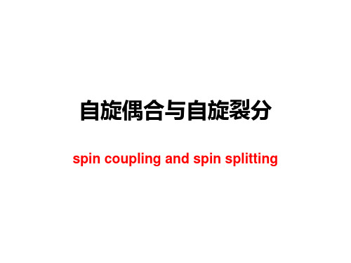
Htotal is the magnetic field (including the effect of the neighboring groups) influencing proton Ha and H^y is the magnetic moment of Hb
• Ha proton resonance will occur at two different values of the applied field, and proton Ha will have two different resonance frequencies. Therefore, the signal of proton Ha will split into a doublet. This phenomenon is called spin-spin coupling. We can also say that Ha couples with proton Hb and resonates as a doublet.
• In looking at these factors, we have neglected the point of how the alignment (parallel or antiparallel) of the magnetic moment of proton Hb in the external magnetic field can affect the chemical shift of the neighboring proton Ha. • Different alignment of the magnetic moment of the protonห้องสมุดไป่ตู้Hb, will cause fine splitting in the signal of proton Ha. • As a consequence, the number of signals in the spectrum will increase and the appearance of the spectrum will be complex. At the same time, we will obtain more information from the spectrum for interpretation.
Spectroscopy and Spectral Analysis

Spectroscopy and Spectral AnalysisSpectroscopy is a branch of science that deals with the study of the interaction between matter and electromagnetic radiation. Spectral analysis is a technique that is used to identify and measure the properties of substances based on the electromagnetic radiation that they emit, absorb, or scatter. The study of spectroscopy and spectral analysis is essential to many fields, including chemistry, physics, environmental science, and biomedical research.Types of SpectroscopyThere are several types of spectroscopy, each based on the type of electromagnetic radiation used. The most common types of spectroscopy include:1. Absorption SpectroscopyAbsorption spectroscopy is a technique that measures the amount of radiation absorbed by a sample. This type of spectroscopy is used to identify the chemical composition and concentration of a substance. Absorption spectroscopy can be used in the ultraviolet, visible, and infrared regions of the electromagnetic spectrum.2. Emission SpectroscopyEmission spectroscopy measures the amount of radiation emitted by a substance. This type of spectroscopy is used to identify the chemical composition of a substance and the temperature and pressure of the environment. Emission spectroscopy can be used in the ultraviolet, visible, and infrared regions of the electromagnetic spectrum.3. Fluorescence SpectroscopyFluorescence spectroscopy is a technique that measures the amount of radiation emitted by a substance when it is excited by light of a particular wavelength. This type of spectroscopy is used to identify the presence of certain substances in a sample, such as proteins and DNA molecules. Fluorescence spectroscopy can be used in the ultraviolet and visible regions of the electromagnetic spectrum.4. Raman SpectroscopyRaman spectroscopy is a technique that measures the scattered radiation produced when a sample is irradiated with a laser beam. This type of spectroscopy is used to identify the chemical composition and structure of a substance. Raman spectroscopy can be used in the visible and near-infrared regions of the electromagnetic spectrum.Applications of Spectroscopy and spectral analysis have a wide range of applications in various fields, including:1. ChemistrySpectroscopy is used extensively in chemistry to identify the chemical composition and properties of substances. Spectroscopy is used to determine the purity of a substance, study chemical reactions, and analyze the structure of molecules.2. PhysicsIn physics, spectroscopy is used to study the properties of materials, such as their electronic and magnetic properties. Spectroscopy is used to study the interactions between atoms and molecules and to investigate the behavior of quantum systems.3. Environmental ScienceSpectroscopy is used in environmental science to study the properties of soil, water, and air. Spectroscopy can be used to identify pollutants in the environment and to monitor the quality of drinking water and industrial wastewater.4. Biomedical ResearchIn biomedical research, spectroscopy is used to study the properties of biological molecules, such as proteins and DNA. Spectroscopy is used to image and diagnose diseases, such as cancer, and to monitor the effectiveness of treatments.ConclusionSpectroscopy and spectral analysis are powerful tools for studying the properties of matter and electromagnetic radiation. There are several types of spectroscopy, each with its own strengths and applications. Spectroscopy and spectral analysis are used in many fields, including chemistry, physics, environmental science, and biomedical research, and have a wide range of applications.。
分析仪器相关英文简称
分析仪器相关英文简称Analytical Instrumentation Related Acronyms1. GC: Gas Chromatography2. HPLC: High-Performance Liquid Chromatography3. FTIR: Fourier Transform Infrared Spectroscopy4. UV-Vis: Ultraviolet-Visible Spectroscopy5. AA: Atomic Absorption Spectroscopy6. ICP-MS: Inductively Coupled Plasma Mass SpectrometryICP-MS is an analytical technique used to determine the concentration of elements in a sample. It involves ionizing the sample using an inductively coupled plasma and then analyzing the resulting ions using a mass spectrometer. It is highly sensitive and widely used in environmental, food, and pharmaceutical analysis.7. AAS: Atomic Absorption SpectrometryAAS is a technique used to determine the concentration of elements in a sample by measuring the absorption of light at specific wavelengths. It is similar to AA spectroscopy but typically utilizes a flame or a graphite furnace as a vaporization and atomization source.8. XRD: X-Ray DiffractionXRD is a technique used to analyze the crystal structure of a material by measuring how X-rays are diffracted by the atomic lattice. It provides information about the arrangement of atoms in a solid, allowing the identification and characterization of crystalline materials.9. NMR: Nuclear Magnetic Resonance Spectroscopy10. MS: Mass Spectrometry11. SEM: Scanning Electron Microscopy12. TEM: Transmission Electron Microscopy13. AFM: Atomic Force Microscopy。
活细胞核浆粘滞系数的荧光相关谱研究
活细胞核浆粘滞系数的荧光相关谱研究活细胞核浆粘滞系数的荧光相关谱研究摘要:细胞核浆粘滞系数直接反映一定生理状态下核内微环境的特性,是判断病变细胞和研究核内分子功能机制的重要依据.为了实时、灵敏和无损地测定单个活细胞的.核浆粘滞系数,提出了一种基于荧光相关谱(fluorescence correlation spectroscopy,FCS)技术的新型检测方法.研究中利用FCS技术测定绿荧光蛋白分子(EGFP)在细胞核内的扩散系数,并且根据Stokes.Einstein关系式计算出核浆粘滞系数,同时实现了对特定生理条件下核浆粘滞系数的跟踪测量.结果表明:在pH 7.4以及37℃的条件下,人肺腺癌细胞(ASTC-a-1)和人宫颈癌细胞(HeLa)的核浆粘滞系数分别是(2.55±O.61)cP和(2.04±O.49)cP,与传统方法的测量结果一致,并且发现核浆粘滞系数大于细胞质的粘滞系数.以上研究表明RCS技术可精确无损地检测单个活细胞内达飞升量级的微观环境,是探索细胞内生物分子动态行为的有力工具. 作者:梁丽芳邢达陈同生裴沂辉 Author: LIANG Li-fang XING Da CHEN Tong-sheng PEI Yi-hui 作者单位:华南师范大学激光生命科学研究所,激光生命科学教育部重点实验室,广东,广州,510631 期刊:光谱学与光谱分析ISTICEISCIPKU Journal:SPECTROSCOPY AND SPECTRAL ANALYSIS 年,卷(期): 2009, 29(2) 分类号: Q632 关键词:荧光相关谱(FCS) 细胞核浆细胞质粘滞系数机标分类号: TH7 R45 机标关键词:细胞核粘滞系数荧光相关谱 correlation spectroscopy 单个活细胞细胞内人宫颈癌细胞人肺腺癌细胞技术测定生物分子无损微观环境生理条件生理状态扩散系数检测方法功能机制跟踪测量动态行为蛋白分子基金项目:国家自然科学基金,广东省自然科学基金。
第十八届全国凝聚态理论与统计物理学术会议
目录
1. 会议日程简表 2. 会议详细日程安排 3. 大会邀请报告、分会邀请报告、分会一般报告和张贴报告编码规则 4. 大会邀请报告: 题目与摘要 5. 分会邀请报告和一般报告: 题目与摘要 6. 张贴报告: 题目与摘要 7. 通讯录
第十八届全国凝聚态理论与统计物理学术会议
日
日期 2014 年 7 月 26 日 2014 年 7 月 27 日 时间 全天 签到、注册
午餐&小憩 地点:重庆大学学生第 1 食堂 2 楼大厅(见校园引导标识、或会 议相关地图)
2014 年 7 月 27 日第二单元:分会报告(第一分会场) 地点:民主湖报告厅 13:30-14:00 主题:1 主持人:罗洪刚(兰州大学)
分会邀请报告:Three Component Ultracold Fermi Gases Under Spin-orbit Coupling 报告人:易为(中国科技大学)
14:00-14:20
口 头 报 告 : Braiding of Majorana Edge States in One-dimensional Decorated
XY-model
报告人:董召阳(南京大学) 14:20-14:40 口头报告:Exotic Fractional Topological States in a Two-dimensional Organometallic
12:00-13:30 13:30-14:40
午餐、小憩 分会报告(含分会邀请报告)
14:40-15:00 15:00-16:10
茶歇 分会报告(含分会邀请报告)
16:10-17:30 17:30-18:30 18:30 20:00-21:00 2014 年 7 月 29 日 8:30 9:00-10:30 10:30-11:00 11:00-12:00
- 1、下载文档前请自行甄别文档内容的完整性,平台不提供额外的编辑、内容补充、找答案等附加服务。
- 2、"仅部分预览"的文档,不可在线预览部分如存在完整性等问题,可反馈申请退款(可完整预览的文档不适用该条件!)。
- 3、如文档侵犯您的权益,请联系客服反馈,我们会尽快为您处理(人工客服工作时间:9:00-18:30)。
Correlation spectroscopy in cold atoms:light sidebands resonances inelectromagnetically induced transparency conditionH.M.Florez 1,A.Kumar 1,K.Theophilo 1,P.Nussenzveig 1and M.Martinelli 11Instituto de F´ısica,Universidade de S˜a o Paulo,05315-970S˜a o Paulo,SP-BrazilThe correlation spectroscopy has been successfully employed in the measurement of the intrinsiclinewidth of electromagnetically induced transparency (EIT)in time and frequency domain.We study the role of the sidebands of the intense fields in the measured spectra,analyzing the infor-mation that can be recovered working with different analysis frequencies.In this case,the non-zero one-photon detuning appears as a necessary condition for spectrally resolving the sideband reso-nances in the correlation coefficient.Our experimental findings are supported by the perturbative model defined in the frequency domain.One of the widely used mechanisms that provide co-herent mapping between light and matter is electromag-netically induced transparency (EIT)[1–3].Such light-matter interface finds applications in quantum memories and quantum repeaters [4–6].It can also be employed for quantum non-demolition measurements [7]and as a source of intense quantum correlated light beams [8,9].Recently,it was shown that the correlation spec-troscopy can be used as a technique for measuring the intrinsic linewidth of the EIT resonance [10–12],which is directly related to the decoherence lifetime of the in-volved ground states.This linewidth is narrower than the broadened EIT linewidth usually measured by the stan-dard transmission spectroscopy.Therefore,it was shown to be a useful spectroscopic tool to estimate the decoher-ence limitations in quantum memories protocols based on EIT.These intensity correlation techniques could also be of interest in precision measurements in atomic clocks relying on coherent population trapping [13].Different research groups have been investigating how intensity correlation of two fields are affected by a medium,with special interest in the EIT process.Noise spectroscopy have shown to be a powerful method for this investigation [14].The analysis of the intensity correla-tion can be done either in time [11,15,16]or in frequency domains [9,10,12,17–19],using respectively the g (2)(τ)function or its Fourier transform C (ω).Felinto et al .[11]proposed a heuristic model for g (2)(0),and applied it to cold atomic systems where Doppler broadening is negligible.It was demonstrated that cor-relation noise spectroscopy is power broadening free in this case,and that the linewidth of the g (2)(0)function is determined by the ground state decoherence lifetime.In the frequency domain,the power broadening free features of the intrinsic EIT linewidth have been shown in a study of intensity correlation spectra C (ω)obtained with cold alkali atoms (Rubidium and Cesium)[12],with the advantage that with proper selection of analysis fre-quency the contribution from electronic noise to the mea-surement is either negligible or can be easily subtracted.Moreover,with the recent developments of quantum op-tics in frequency domain (specially multimode quantum optics [20],and the observation of multi-mode coherent effects [21])stimulates the investigation of the atomic be-havior in similar situations.Careful study of correlations in frequency domain shows the contributions from the sidebands of the main field [22].While they don’t affect the narrow structure related to the intrinsic linewidth,the whole profile of the correlation inside the typical EIT linewidth is broadened by the sidebands resonances,a major difference between analysis in time [11]and frequency [12]domains.In this work,we explore the role of the sideband res-onances and the effect of the detuning in the correlation profile.We show that the interplay between one-photon detuning and analysis frequency ωopens the possibility of detecting atomic response to sidebands in correlation spectra.We also show that in such condition the cor-relation linewidth measured in the frequency domain is exactly the same as the one in the time domain for the g 2(0)described in [11].Moreover,it is shown experimen-tally that when one of the beams is kept in resonance with the atomic transition,independent correlation informa-tion of the sidebands and the carriers is unavailable [22],therefore demonstrating the richer structure contained in C (ω)in comparison with g 2(0).In addition,the perturbative method we use provides a physical insight on the mapping of the atomic response with the correlation functions g (2)(0)and C (ω).Unlike the resonant case,where the dispersive response seems responsible for the main contribution in the intensity cor-relation,in the non-resonant case there is a balance be-tween the absorptive and dispersive response.Such a bal-ance makes the heuristic model for g (2)(0)a valid approx-imation of C (ω)only inside the typical EIT linewidth,failing at higher two-photon detunings .Our presentation is organized as follows.In section I,we briefly describe the technique of correlation spec-troscopy and the level scheme used in our study.We also describe the theoretical model used to define the corre-lation coefficient in frequency domain.In section II,we discuss the details of our experiment,starting from a cold cloud of 87Rb,and present the control of the involved pa-rameters.In section III,we show the results of correlation spectroscopy with different values of one photon detun-ing,and compare it to the theory.We also show how the presence of the sidebands is revealed in the correlation spectrum obtained at different one photon detunings.Ina r X i v :1508.04858v 1 [q u a n t -p h ] 20 A u g 20152section we discuss our results V wedonevalues of one photon detuning.I.THEORY:CORRELATION SPECTROSCOPYInspired in the Hanbury-Brown and Twiss’s experi-ments,the intensity correlation between two light fieldswith intensities I 1(t )and I 2(t +τ)can be quantified by the g (2)(τ)function [15].Our interest is to analyze such intensity correlation between two light fields induced by cold atomic media in EIT condition.Yabuzaki et al .[14]showed that the atomic medium converts excess phase noise of the input light sources into intensity noise at the output field.Therefore,to model the intensity correla-tion induced by the atoms in our bipartite system,the transformation of phase noise to amplitude noise has to be considered.Our scheme for correlation spectroscopy in a Λ-EIT condition is shown in Fig.1(a).Two fields,E 1(t )and E 2(t )are coupled to two different transitions of a three-level atom,with different one-photon detunings ∆1and ∆2respectively.The intensity correlation between the two fields is measured after interacting with the atomic ensemble as presented in Fig.1.(b).The previously re-ported correlation spectroscopy [10–12,23]has been done by setting one of the fields in resonance (e.g.∆1=0),while scanning the detuning of the second field around the resonance.However,in the present work,we set dif-ferent values of detuning ∆1i.e.∆1=0while ∆2is scanned around ∆1.Atoms (a)(b)FIG.1:(Color online)(a)Levels scheme in Λ-type configura-tion for EIT.(b)Basic setup for measuring the intensity cor-reletion spectra in time (g (2)(τ))or frequency domain (C (ω)).In what follows,we present a brief description of the semiclassical approach for the intensity correlation spec-trum based on the conversion of phase noise to amplitude noise.We consider two electromagnetic fields described byE i (t )=E i exp [i (ωi t +φi (t ))]e i ,(1)with a stochastic phase fluctuation φi that models the excess of noise in diode lasers.In the expression for the fields,i =1,2denotes the two beams,E i and ωi are re-spectively the amplitude and the frequency of each beam.Photodetectors are not sensitive to the phase noise of light beams.However,the interaction of light fields through an atomic medium makes it detectable by map-ping the phase noise into amplitude noise (PN-AN).Under the thin sample limit [24],the field after prop-agation through atomic media is given by E out i (t )=E i (t )+iκP i (t ),where polarization P i (t )represents the atomic response induced by the incident fields E i (t )and κi is a real constant that depends on the atomic den-sity.The induced polarization term is responsible for transforming the phase noise φi (contained in E i (t ))into amplitude noise detected in E out i (t ).In a Λ-EIT con-figuration (Fig.1a),the polarization induced in atomic medium by two incident fields can be given as P i =d i 3ρi 3,where d i 3and ρi 3represent the electric dipole moment and the atomic coherence associated to the fields i =1,2.The output intensity of the beam is given by I out i (t )=|E out i (t )|2.In order to describe the correlation between two intense light beams,we average correlations of the fluctuations inboth beams,δI i (t )=I out i (t )− I outi with i ={1,2}for different instants of time separated by τ.There are two possible approaches to calculate the intensity correlation:in time domain approach adopted in [10,11,16,17],the intensity correlation between two light fields can be easily described in terms of the atomic variables as [11],g (2)(0)= δI 1(t )δI 2(t )δ2I 1(t ) δ2I 2(t )=Re p 1Re p 2+Im p 1Im p 2(Re 2p 1+Im 2p 1)(Re 2p 2+Im 2p 2),(2)where polarization terms are related to atomic coher-ences as p i =ρss i 3and the notations Im 2p i =(Im p i )2and Re 2p i =(Re p i )2.The ss index stands for steady-state.The atomic response induces correlation (g (2)(0)>0)or anticorrelation (g (2)(0)<0)between the light fields,de-pending on two photon detuning δ=∆1−∆2[10,16,17].At low power regime,absorption dominates and the medium induces correlation between the light fields.However,as the power of the beams are increased,the dispersion term Re p 1 Re p 2 <0overcomes the con-tribution from the absorption term Im p 1 Im p 2 >0,leading to anti-correlated light fields.Other possible approach is the analysis of intensity fluctuations δI i (t +τ)in their different spectral compo-nents by a Fourier transform.Therefore,the correlation in the frequency domain description is defined asC (ω)=S 12(ω)1122,(3)where S ij (ω)represents the symmetrical intensity cor-relation spectrum for i and j fields at a given analysis3ij =4πdτe −iωτ[ δI i (t )δI j (t +) + δI j (t )δI i (t +τ) ].(4)Similar to g (2)(0),the function C (ω)is also normalizedsuch that for correlated fields 1≥C (ω)>0,and for anticorrelated fields 1≤C (ω)<0.A perturbative approach is proposed in ref.[22]to de-scribe the correlation coefficient of eq.(3)in terms of the absorption and dispersion of the fields,similar to the case of g (2)(0)function in eq.(2).Taking the laser linewidth γas perturbative parameter in the expansion of the spectral components of the fluctuations,we can obtain a direct expression for the correlation C (ω),at the lowest order.The resulting expression,C (ω)=νIm (ω)˜ΠIm +νRe (ω)˜ΠRe +˜Πcross (ω)+C 1 S 11(ω)S 22(ω),(5)where the absorptive,dispersive and cross terms are re-spectively defined by the polarization terms associated with the atomic coherences˜ΠIm =2 2Im p 1Im p 2+O ( 4)(6a)˜ΠRe =2 2Re p 1Re p 2+O ( 4)(6b)˜Πcross (ω)=νRI (ω)[−2 2Im p 1Re p 2+O ( 4)](6c)+νIR (ω)[−2 2Im p 2Re p 1+O ( 4)]=νRI (ω)˜ΠRI (ω)+νIR (ω)˜ΠIR (ω)(6d)Two main diferences will show up when we compare C (ω)(eq.5)with g (2)(0)(eq.2).One is the presence of a cross term involving the product of dispersive and absorptive contributions,which is absent in g (2)(0).The other is the need of weighting coefficients νIm (ω),νRe (ω)and νRI (ω)(νIR (ω))distorting the main contributions for the correlation (absorption,dispersion and Stokes tran-sitions respectively)with a dependence on the analysis frequency ω.A description of the calculation,as well as the definition for the smaller term C 1,and the explicit expressions for S 11(ω)and S 22(ω)are given in Appendix A.Thanks to this perturbative approach,we will un-derstand the observed anticorrelation for |δ|>Γwhen ∆1=0as a consequence of dispersion overcoming ab-sorption.Such situation is very different from the be-havior of g (2)(0)functions where the absorptive and dis-persive properties contribute equally to the correlation [11].It can also be understood the role of the mixing of sidebands resonances in the PN-AN conversion when the carrier frequency |δ|<Γ.This approach is an interesting resource for finding situations where the sidebands reso-nances are independent of the PN-AN mechanism when the carriers are near the two-photon resonance,and com-pare it to the experiment.II.EXPERIMENTWe used an ensemble of cold atoms of 87Rb releasedfrom a Magneto-Optical Trap (MOT).Diode lasers in 5S 1/2(F =2)↔5P 3/2(F =3)transition were used for cooling the atoms.Our atomic cloud has nearly 107atoms at 800µK.The optical depth of the cloud in the cooling transition is ∼2.The Λ-type three level sys-tem considered in the experiment is shown in Fig.2a.The correlation spectroscopy is done in the transition F G =5S 1/2(F =1)→F E =5P 3/2(F =1)where the two fields with orthogonal polarizations σ∓couple the Zeeman levels m =±1respectively,as shown in Fig.2(a).To bring the atomic population to 5S 1/2(F =1),we used an additional optical light field coupling 5S 1/2(F =2)→5P 3/2(F =2)transition.Moreover,the proposed atomic transitions obey the necessary condition F G ≥F E for the observation of EIT in the atomic medium [25].(a)F'=1Transimpedance (b)FIG.2:(Color online)(a)Levels scheme in Λ-type configura-tion for EIT,detunings ∆1(fixed)and ∆2(scanned around ∆1).(b)Basic setup for EIT spectroscopy in cold atoms,where DC and HF signal are detected separately for each beam.The two beams with orthogonal polarization compo-nents come from the same external cavity diode laser.Although the laser has a linewidth of ∼1MHz (con-sistent with the stochastic phase noise),the fields are phase correlated to the limit of the Standard Quantum Level (SQL).Each light beam is switched and frequency controlled by independent Acousto Optical Modulators (AOM)in double pass configuration.The frequency de-tuning of each beam is controlled by a computer con-trolled Digital-to-Analog interface acting on the AOM drivers through voltage controlled oscillators.The op-tical power for each beam was P 1=112(1)µW and P 2=107(1)µW,with spot sizes of w 1=1.8(4)mm and w 2=1.6(4)mm respectively (with w as the spot radius at 1/e 2intensity level of a Gaussian beam).Therefore,the beams are below saturation,with I 1/I sat =0.18and I 2/I sat =0.22.As shown in Fig.2b (not to scale),the two light beams interact with the atomic ensemble at an angle of ∼2◦to avoid any leakage from one beam to other in the detection process.We used p-i-n photodetectors where the photocurrent signals are divided into a DC signal and a High Frequency (HF)signal that is connected to a trans-impedance am-plifier.Both the signals are sent to an Analog-to-Digital4signal,is as-The(ω)forand˜Πel-ω)mul-6).Alltwo-photonvertical(a)(b)(c)FIG.3:(Color online)Spectroscopic results with three different features:DC signal(first row),correlation(second row),˜Πmatrix elements(third row)andν(ω)˜Πcoefficients(fourth row).Spectroscopy for(a)detuned case∆1=−6MHz(b) resonant case∆1=0MHz and(c)detuned case∆1=5MHz.The experimental correlation spectra in(a),(b)and(c)are shown in solid thick lines.The perturbative result for C(ω)and heuristic model for g(2)(0)are trace in dotted-dashed and solid thin lines respectively(second row).According to eqs.(6)the matrix elements of˜Πare˜ΠIm=2 2Im p1Im p1+···,˜ΠRe=2 2Re p1Re p1+···,˜ΠIR=−2 2Re p1Im p2+···and˜ΠRI=−2 2Re p2Im p1+···.The parameters used for the atomic medium are:decoherence rateγd/2π=150kHz,analysis frequencyω/2π=2MHz,phase noise¯γ/2π=1MHz.The Rabi frequencies corresponding to the experimental values of intensity areΩ1=0.30ΓandΩ2=0.34Γ.The one photon detunings used for the theoretical spectra are∆1=−6MHz,0.2MHz and5MHz respectively from left to right.Let usfirst discuss the resonant case i.e.∆1=0, shown in Fig.3b.In this case,we observe a typical EIT profile in the DC signal,which allows the measurement of a linewidth∆ωEIT from the full width at half maximum of the peak.As for the correlation C(ω),we notice that the twofields are anti-correlated for|δ|>5MHz and also for|δ|<2MHz,with a narrow peak at exact two-photon resonance.As it was discussed in[11,12],this structure is insensitive to power broadening and can be associated to the coherence between the ground states.The g(2)(0) function from the heuristic model in eq.(2)is also plotted in solid thin line.Although it presents a similar shape, the result is mostly correlated,with anticorrelation only for|δ|∼1MHz.This is a consequence of the combination5we2π,we (˜ΠRe )fac-again sate IR the other eq.(5).That Γ/2π,the absorption fixed beam 2in the second beam (Re p 1)dominates in C representedby ˜ΠIR =2 2Re p 1Im p 2+···i.e.Stokes transitions lead to anticorrelation.Similar reasoning can be applied to non-resonant cases (Figs.3a,c),as we can see in the spectroscopic results for different one-photon detunings (∆1=−6(1)MHz and 5(1)MHz respectively).We can start by observing an interesting feature in the change from correlation to an-ticorrelation at exact resonance of field 2(∆2=0MHz).The understanding is relatively simple.Fluctuations in the frequency (or equivalently their phase diffusion)are common to both beams,since they are issued from the same laser.These fluctuations are converted into inten-sity fluctuations by a variation in the absorption rate of the fields,as the frequencies approach or recede from resonance.Therefore when the detuning for both beams have the same sign,intensity fluctuations will be corre-lated.On the other hand,if detunings have opposite sign,the same correlated fluctuation in phase will lead to anticorrelated response of the medium,resulting in anticorrelated fluctuations in intensity.This behavior isclosely followed by the dispersive component ˜ΠRe ,that when is weighed by the νRe term becomes the leading contributor for most of the spectra.But contribution of ˜ΠRI is also important,specially in the anticorrelated part.Only the contributions of ˜ΠIm and ˜ΠRe ,present in the plot of g (2)(0),cannot justify the observed anticorre-lation.Another curious situation shows up in EIT regime,where contribution of dispersion is also reduced:a strong reduction in the correlation occurs in a range of ±3MHz around the EIT condition δ=0.This structure,carved on the maximized correlation we have just discussed,can-not be completely described only by g (2)(0),since instead of a pair of dips involving a narrow peak we have also an-other pair of dips close to the analysis frequency (2MHz),composing the complete structure of correlation around EIT condition.This case is pretty different from the one photon resonant case (∆1 0),seen in Fig.3b.Absorp-tive and dispersive components (˜ΠIm and ˜ΠRe ),after the compensation by ν,are not the leading terms,and thecontributions from ˜ΠIR and ˜ΠRI dominate.Anticorrela-tion is far from saturation.Unfortunately,the structures predicted in theory arenot so clearly resolved in the experimental results.There-fore we need a better resolution and a more careful anal-ysis,as we perform next.Resolving the sideband resonances in the correlationspectraLooking for a better insight into the role of the side-bands of the fields,we show the correlation spectra for different analysis frequencies ω,at the resonant case ∆1=0in Figs.4(a-c),and also for detuned case ∆1=6MHz in Figs.4(d-f).In the first row (Figs.4(a,d)),we have shown the spectra for ω/2π=2MHz dis-cussed above.In the second (Figs.4(b,e))and third row (Figs.4(c,f))the spectra for ω/2π=3MHz and 4MHz are shown respectively.FIG.4:(Color online)Correlation spectroscopy for different analysis frequencies (ω/2π)with ∆1=0MHz (first column)and ∆1=6MHz (second column).(a,d)ω/2π=2MHz,(b,e)ω/2π=3MHz and (c,f)ω/2π=4MHz.Experimental correlation C (ω)is in solid line,theoretical result for C (ω)is traced with dotted-dashed line and g (2)(0)is traced with dashed line.In (c,f)the first order term of the perturbative model is plotted in dotted line.The perturbative result of C (ω)is plotted (dotted-dashed line in Fig.4)together with the experimental data (solid thick line).The lowest order term of correlation in 2is also plotted in dotted line for ω/2π=4MHz.In the resonant case ∆1=0(see Figs.4(a-c),there is no spectral independence for the sidebands in the correla-tion C (ω)as discussed in ref.[22].The intrinsic linewidth of the correlation spectrum (147(9)kHz according to the central peak width)is consistent with the decoherence rate used in the theoretical calculations (150kHz).Al-though the sideband resonance at δ∼−ω/2πseems to be resolved for the first order term (dotted line,Fig.4c)in the perturbative model,the contribution from higher6sideband res-resonance the phase-by Thus the ex-amplitude varying case (∆1=that the spectrally re-∆ωEIT /2π,correlation with where The ω)and g 2(0)in the range at for increas-order term for g (2)(0).only at first by perturba-differently for the casemain ˜Πel-together with correlation spectra in top row Figures 5(c,d)that extra term C in eq.(A11).resonant case Fig.4(c),in non-resonant case νIm Re (see Fig.5(a)).Therefore,the absorptive and dispersive response contribute equally to the intensity correlation,while theterm ˜Πcross (eq.6)nearly cancels due to the opposing be-havior of ˜ΠRI and ˜ΠIR .This coincides with the heuristic model where the two terms have the same contribution for the g (2)(0)function in eq.(2).The coeficients νij (ω)for the extra term C 1have almost no contribution to the correlation for |δ|<3MHz,as it is shown in Figs.5(c,d).Hence,the two functions describe the same intensity correlation for |δ|<3MHz in the non-resonant case for higher analysis frequency ω>∆ωEIT .On the other hand,the coefficients ν(ω)present reso-nances exactly at the analysis frequency δ∼±ω/2π=±4MHz.The main coefficients νIm (ω)and νRe (ω)present opposite behaviors near the analysis frequency,(Fig.5a).The coefficient νRe in solid line shows an incre-ment of the dispersive response while νIm (ω)decreases.However,for the sidebands resonance,the contribution of the extra terms in Figs.5(c,d)for C 1has also to be considered,leading to C (ω)∼0.Curiously,these side-band structures are broad,with nearly half of the width of the EIT peak observed in the DC signal.As we will see,they are also sensitive to power broadening in thisFIG.5:(Color online)Mapping between the intensity corre-lation C (ω)from Fig.4.(f)and:(a)the ˜Πelements and (b)the main ν(ω)coefficients.In (c)and (d),the extra ν(ω)co-efficients that determine C 1defined in eq.(A11)are plotted.This analysis is done for ∆1=5MHz and ω=4MHz.The two vertical dotted lines indicate the EIT linewidth ∆ωEIT .case.Power broadening of the resolved sidebandstructuresWe further investigated the correlation spectra for dif-ferent power of the beams at the non-resonant case.Fig-ure 6a shows the correlation spectra for two different powers of the beams.For beam power of 110µW,i. e.I/I sat =0.23(solid line),the sideband resonances are well resolved.However,on increasing the power to 190µW (dotted line)which corresponds to I/I sat =0.40,the resonance at δ=−ω/2πnear the atomic resonance is7(a)(b)FIG.6:(Color online)Correlation spectra power of the beams non-resonant case ∆at 4MHz analysis frequency.(a)Experimental correlation spectra for 110µW (solid and W (dotted their the-oretical counterparts.Theoretical results the correla-tion spectra solid lines)for used in (a),and for powers,3×110µline)and 4×110µW lines).Further increase in the power leads to a significative push of the atoms during the run of the acquisition.Nevertheless,we can still consider what are the conse-quences relying on our theorethical model.Figure 6b shows the theoretical correlation results for the two cases of power of Fig.6a,and also the calculations for higher beam powers,3×110µW (dashed line)and 4×110µW (dotted-dashed lines)which correspond to I/I sat =0.67and I/I sat =0.90,respectively.With increasing power,the central EIT peak broadens and engulfs the sideband structure,now hidden in the anticorrelation flat signal.IV.DISCUSSIONA linearized approach for the EM field described by eq.(1)can give useful hints for the mechanisms involved in the spectroscopy studied here.First,we may regard the phase diffusion as a phase modulation process in the limit of small diffusion coefficient γ.When we measure the spectral components of the photocurrent I (t )(gener-ated by an intense,nearly monochromatic field)by their Fourier transform I (ω),we are in fact investigating the beatnotes of the sidebands with the most intense field.For a specific analysis frequency ωthe field of interest will involve three components:the carrier at optical fre-quency ωi and two sidebands shifted by ±ω.From eq.(1)we have the relevant part in calculation of I (t )asE i (t )=E(t )i exp(iωi t )e i ,(7)with the complex amplitude given byE (t )i =E i +E u i exp(iωt )+E l i exp(−iωt ).(8)in a linearized approach for phase diffusion.Pure am-plitude modulation will occur if upper and lower side-bands are symmetric and conjugated (E u i =[E l i ]∗),andpure phase modulation will take place if they are ro-tated by π/2with respect to the carrier amplitude E i (E u i =−[E l i ]∗).Phase and amplitude fluctuations can be converted to each other upon asymmetric shifts of the amplitudes of the sidebands (E u i ,E l i )or by a phase shift of the carrier amplitude E i [26].In our system,the atoms induce such asymmetric shifts in amplitude and phase.Photodetection is sensitive to amplitude modu-lation,therefore we observe in our experiment the con-version of random phase modulation (phase noise)into amplitude (intensity)modulation.With that in mind,let us analyse the feature we have observed.23_2(b)FIG.7:(Color online)Description for the correlation spec-troscopy.Three main situations of interest are:(process 1)the EIT condition when δ∼0,(process 2)when the carrier of the field E 2is resonant with the upper sideband of the field E 1(δ∼ω)and (process 3)when carrier is resonant with the lower sideband (δ∼−ω).These processes can be analyzed for two different cases of one-photon detuning:(a)Resonant case ∆1=0,(b)Non-resonant case ∆1∼Γ.For the case of small single photon detuning (∆1 0),the central structure of C (ω)that we observe at EIT condition (|δ|<∆ωEIT /2π),which is very similar to the g (2)(0)described in [11],is a consequence of the phase shifts given to the central carriers while we scan the EIT resonance (see Fig.7.a).Those shifts convert phase to amplitude noise with opposite signals for fields 1and 2(fixed and scanned),leading to a growing an-ticorrelated response as the intensity is increased,over-whelming the faint intensity correlation that the laser beams may originally present.Only close to EIT reso-8nance (process 1)those initial intensity correlations are recovered,since the dispersive contribution goes to zero.Therefore we have a narrow central peak in the corre-lation spectroscopy,either in frequency or time domain.In this case,sidebands are nearly unaffected in the pro-cess,or symmetrically affected:upper (lower)sidebands of both beams have similar dephasing and attenuation(similar transformations for E u 1and E u2),leading to cor-related contributions.The sidebands resonances in the processes 2and 3are not resolved due to the stimu-lated and spontaneous emission,since the fields couple the atoms resonantly with detunings no bigger than the natural linewidth |δ|∼ω/2π<Γ/2π(see figures 4(a-c)).The resonances of the sidebands will be evident in the case when single photon detuning is of the order of the atomic linewidth (|∆1| Γ/2π,see an example in Fig.7.b).Once again,close to EIT condition (process 1),sidebands of both beams will suffer similar ampli-tude and phase changes.Strong correlations are then expected,leading to a maximum of the normalized cor-relation at exact EIT condition (|δ| 0)for the same reasons presented in the previous case.The novelty here is the fact that when detuning matches analysis frequency (|δ|=ω/2π),correlation is reduced.Consider a simple model of phase fluctuations,given in eq.(8),where E u i =−[E l i ]∗=α.Phase transfor-mation for the fixed beam will be given by:E (t )1=αe iωt +E 1−α∗e −iωt →(9)E (t )1=αe i (ωt +ϕ3)+E 1e iϕ2−α∗1e −i (ωt −ϕ1)== αe i (ωt +ϕ3−ϕ2)+E 1−α∗e −i (ωt +ϕ2−ϕ1)e iϕ2Therefore,different phase shifts will convert phase to am-plitude fluctuations.Applying the same transformation for the second field when its carrier is resonant with the upper band of the fixed field (process 2)E (t )2=αe iωt +E 2−α∗e −iωt →(10)E (t )2=αe i (ωt +ϕ4)+E 2e iϕ3−α∗e −i (ωt −ϕ2)== αe i (ωt +ϕ4−ϕ3)+E 2−α∗e −i (ωt +ϕ3−ϕ2)e iϕ3.Notice that when we discard the overall phase (unob-served upon intensity detection),the effective shift of theupper band of field 1is the conjugate of the phase shift of the lower sideband of field 2.Fluctuations will rotate in opposite directions in a Fresnel diagram,and resulting intensity correlations will be anticorrelated.Now if the carrier of the second field is resonant with the lower band of the fixed field (process 3),transforma-tion will be given byE (t )2=αe iωt +E 2−α∗e −iωt →(11)E (t )2=αe i (ωt +ϕ2)+E 2e iϕ1−α∗e −i (ωt −ϕ0)== αe i (ωt +ϕ2−ϕ1)+E 2−α∗e −i (ωt +ϕ1−ϕ0)e iϕ1.The phase shift of the upper sideband of field 2will be the conjugate of the phase shift of the upper sideband of field 1.Once again,anticorrelation will contribute to the result of C (ω).This conversion depends on the phase gained by each mode.Under power broadening of the EIT resonance [11,12],anticorrelation will be affected by the reduction on the diference of phase shifts,as is the case in Fig.6.V.CONCLUSIONSCorrelation spectroscopy for two light fields interacting with cold atomic medium at different one-photon detun-ings has been explored.When the one-photon detuning is of the order of the natural linewidth and beam intensities are much smaller than the saturation limit,the correla-tion spectroscopy presents resonances at the sidebands frequency.If the analysis frequency is larger than the typical EIT linewidth,the intensity correlation spectra in the EIT range can be approximately described by the g (2)(0)function [11].Therefore,the intrinsic linewidth measured with the g (2)(0)function in time domain cor-responds exactly to the one observed with the correlation spectrum C (ω)in frequency domain.Sidebands resonances are auxiliar spectroscopic tools for the study of EIT process,and can be resolved for anal-ysis frequencies higher than the EIT linewidth.However,in such a condition,the sidebands resonances are sensi-tive to power broadening.It was also shown that for fields close to atomic resonance,the contribution from the side-bands and the carriers are mixed,disabling any spec-tral independence of sidebands in the correlation spec-troscopy.The resolution of the sidebands resonances is only possible with the use of cold atoms.Next step is to measure correlation spectra using a coherent source to verify if it is possible to measure a narrower intrinsic linewidth for resonant conditions as it is predicted by the first order term in the perturbative model [22].This work was supported by grant #2010/08448-2,S˜a o Paulo Research Foundation (FAPESP),CNPq and CAPES (Brazilian agencies),through the programs PROCAD,PRONEX,and INCT-IQ (Instituto Nacional de Ciˆe ncia e Tecnologia de Informa¸c ˜a o Quˆa ntica).The authors would like to thank Prof.Dr.Vanderlei S.Bag-nato and Dr.Kilvia M.F.Magalh˜a es,from IFSC-USP,for gently providing the Rb for our MOT.Appendix A:Solution for Noise spectraThe intensity correlation result in eq.(5)is demon-strated in detail in ref.[22].Shortly,to describe therole of the atomic response in the PN-AN conversion,we should begin by writing the density matrix elements x =(ρ11,ρ22,ρ13,ρ31,ρ23,ρ32,ρ12,ρ21).It can be ex-panded on the parameter,that we will eventually asso-。
