生物医学工程专业英语及其翻译讲课稿
生物医学工程专业英语 Unit08[69页]
![生物医学工程专业英语 Unit08[69页]](https://img.taocdn.com/s3/m/595641b0aeaad1f346933fe6.png)
comfort zone
舒适地带
Text: Biomedical Sensors
This is an equivalent of monitoring. Monitoring is a necessary activity in risky environments such as mining, diving, mountain climbing, and especially in all sorts of military and security actions. All of these broad application fields have common requirements. The biomedical sensor should be compact and should not force the wearer to leave the comfort zone. These common requirements suggest the smart (intelligent) textiles along with the notion of wearable.
Text: Biomedical Sensors
Biomedical sensors have a vital importance in modern life. We live in an epoch of computerization for every field of life. As we all know, computers can only process the data. Data must be collected, stored if necessary, and transferred to a computer. Biomedical sensors are designed for collecting data. It might be necessary to collect data for inpatients in hospital environment, in home for homebound patients, or for outpatients.
生物医学工程技术外文文献翻译、中英文翻译、外文翻译

生物医学工程技术外文文献翻译、中英文
翻译、外文翻译
本文旨在提供关于生物医学工程技术的外文文献翻译、中英文翻译和外文翻译的指导和技巧。
以下是一些简要说明:
外文文献翻译
- 外文文献翻译需要准确地传达原文的内容,同时确保译文自然流畅。
- 翻译时应注意专业术语的准确使用,避免将其误译或过度解释。
- 翻译人员应具备扎实的外语水平和对生物医学工程技术领域的了解。
中英文翻译
- 中英文翻译需要准确传达中文原文的内容,并使其在英文环境下具有流畅性和可读性。
- 翻译时应注意中英文表达方式的差异,确保翻译后的文本符合英文语法和惯表达惯。
- 翻译人员应具备中英文双语能力和对生物医学工程技术领域的了解。
外文翻译
- 外文翻译是将外文文本翻译为母语的译文。
- 翻译要保证译文准确、流畅,并符合目标语言的语法和惯表达方式。
- 翻译人员应具备对目标语言的熟悉和对生物医学工程技术领域的了解。
请注意,以上是针对文献翻译的一些基本指导,实际翻译过程中还需根据具体文献的特点和要求进行适当调整。
谢谢!。
生物医学工程的英语
生物医学工程的英语Biomedical engineering is the application of engineering principles and techniques to the field of medicine and biology. It encompasses a broad range of areas such as drug delivery, medical imaging, tissue engineering and electronics. In this article, we will discuss the key steps involved in biomedical engineering and the relevant terminology used inthis field.1. Research: The first step in biomedical engineering is research. This involves investigating the problem that needsto be solved and identifying the best possible solutions. It includes conducting experiments, developing models and prototypes, and testing them.2. Design: Once the research is complete, the next stepis design. This involves creating a blueprint of the solution that was identified during the research phase. It includes creating detailed plans and drawings, identifying materials and components required, and creating a mock-up or prototypeof the device.3. Development: The third step is the development phase, where the actual product or device is created. This involves assembling the components, testing the device, and making any necessary modifications. It also includes obtainingregulatory approvals and patents required for commercialization.4. Implementation: The final step is the implementation phase, where the product is launched and made available for use. This involves training the users, monitoring theperformance of the device, and providing ongoing support and maintenance.Now let's look at some key terminology used inbiomedical engineering:1. Biomaterials: These are materials that are used to create medical devices or implants, which interact with biological systems. Examples include metals, polymers, ceramics, and composites.2. Biomechanics: This is the study of the mechanics of biological systems, such as bones, muscles, and tissues. It includes analyzing the structure and function of these systems, and developing models to predict their behaviorunder different conditions.3. Biomedical imaging: This involves the use ofdifferent imaging techniques to visualize the internal structures of the body. Examples include X-rays, CT scans, MRI, and ultrasound.4. Bioprocessing: This is the use of biological systemsor their components to produce drugs or other products. Examples include fermentation, chromatography, and cell culture.In conclusion, biomedical engineering is a rapidly growing field that combines the principles of engineering and medicine to improve healthcare outcomes. It involves research, design, development, and implementation of devices and technologies that can enhance diagnostics and treatment of diseases. Understanding the key steps and terminology used in this field is vital for anyone interested in this excitingarea of healthcare.。
生物医学工程专业英语 Unit10[66页]
![生物医学工程专业英语 Unit10[66页]](https://img.taocdn.com/s3/m/c4b0d9a3c8d376eeaeaa31e6.png)
Text: Biomedical Instrumentation
Note, for example, that most voltages are in the microvolt range and that pressures are low (about 100 mm Hg=1.93 psi =13.3 kPa). Also note that all the signals listed are in the audio-frequency range or below and that many signals contain direct current (DC) and very low frequencies. These general properties of medical parameters limit the practical choices available to designers for all aspects of instrument design.
Text: Biomedical Instrumentation
The major difference between medical instrumentation and conventional instrumentation systems is that the source of signals is living tissue or energy applied to living tissue. The principal measurement and frequency ranges for each medical and physiological parameter are major factors that affect the design of all the instrument components. Most of the medical parameter measurement ranges are quite low compared with nonmedical parameters.
生物医学工程专业英语课件

5.8 微电极
electrolytic solution 电解液 electrolytic etching 电解腐蚀
storage condition储存条件 input impedance输入阻抗
phasor矢量
steady-state sinusoidal稳态正弦
effort input variable作用力输入变量
flow input variable流速输入变量
velocity速度
flow流速
第二章 基本传感器及其原理
manufacturing tolerance制造公差
electrocardiograph 心电图
input ranges输入信号的量程
minimal resolvable 最小可分辨
normal linear operating range 额定的线性工作范围
maximal operating range最大的工作量程
1.5 生物医学仪器的分类
quantity that is sensed 转换参数
organ system 生理系统
clinical medicine 临床医生 resistive 电阻的
inductive 电感的
capacitive 电容的
ultrasonic 超声波的
electrochemical 电化学的
cardiovascular 心血管
pulmonary 肺
nervous 神经
endocrine 内分泌
Pediatrics 儿科学
Obstetrics 产科学
Cardiology 心脏病学
Radiology 放射学
blood pressure 血压
可作为生物医学工程专业英语简易教程
ContentsHistory 2Radiation Physics 5X-radiography 6CT 7Nuclear Medicine 9PET 11Radiotherapy 13Radiation Protection 16Ultrasound 1919imagingUltrasoundDoppler Ultrasound 21Optical Methods 23Endoscopy 23Pulse Oximetry 25Laser Surgery 26MRI (magnetic resonance imaging) 2831 ECG(electrocardiogram)Bioengineering 3232PacemakersMedical Engineering 33Cochlear Implants 34The Future 37Glossary 38Acknowledgements 40History Most people think that Medical Physics started in 1895 when Wilhelm Roentgen discovered x-rays but, in practice, physics has been used to investigate the body for much longer than this.Hippocrates (460-377 BC), the “Father of Medicine”, may have been the first medical physicist . Over two thousand years ago, he wanted to know where an infection was on a patient’s back. He smeared mud over the patient’s back as he knew that infected tissue is warmer and would therefore dry the mud faster.Technology has improved since then, and modernthermography , which looks at heat coming from the bodyusing an infrared camera, is very different fromHippocrates' methods.When doctors wanted to be able to see inside thestomach and intestines , they first simply used a thin t with light provided by a candle. In 1868 a metal tube w passed down the throat into the stomach. This was anearly prototype of an endoscope . Large early modelswere tested on sword swallowers , and were very intimidating, with one man quoted as saying 'I'll swallow a sword, but I'll be damned if I’ll swallow a trumpet'. Modern endare smaller and more flexible, making endosopy a less unpleasant examination.ubeasoscopesIn the future, we will use camera pills that can be swallowed and travel all the way through the digestive system without any discomfort.The most common medical imaging examination today is an x-ray , or radiograph. X-rays were discovered in 1895 by Wilhelm Roentgen , who was passing an electric current through a glass tube with a vacuum inside, when he noticed a screen nearby start to glow. He realised that some invisible rays from the tube were causing the glow, and called them x-rays as he didn't know what they were. He set to work, trying to find out more about these strange rays.His wife became worried that he was spending so much time in hislab, and wasn't eating properly or talking to anyone. She finallypersuaded him to tell her what he was working on, and he took herinto the lab. She tried out his equipment by putting her hand in the x-ray beam for fifteen minutes, and saw an image of her hand appearon film behind it. This was the first medical x-ray image. Roentgenwas awarded the first ever Nobel prize in Physics in 1901.Soon after Roentgen’s discovery, Henri Becquerel , a Frenchman, wasexperimenting with salts that fluoresced when exposed to sunlight tosee if they would emit x-rays. One cloudy day he left a photographicplate and a uranium compund in a drawer, and when he developed the film he found that it had been exposed to something, even though it hadn’t seen any light. He realised that uranium gave off invisible rays that could ionise atoms and blacken film. He called this radioactivity.Marie Curie , along with her husband Pierre , continued the research into radioactive materials, discovering new radioactive elements Polonium (named after her homeland,Poland) and Radium. Bequerel and the Curies shared a Nobel prize for their work.For a while radiation was hugely fashionable, with people putting radium in water and thorium in toothpaste. This health craze was dangerous though, and ill effects were soon noticed. The girls who painted radium onto the dials of watches developed thoat and mouth cancers from licking their brushes, while people using radioactive products suffered symptoms such as burns, hair loss, bone diseases and various types of cancer. Marie Curie herself died from a blood disease linked to radiation exposure (Pierre, though suffering from radiation sickness, died when run over by a horse and cart) .By this time, it had been realised that, although harmful inlarge doses, small amounts of radiation could be used to treatdiseases such as cancer. The radioisotope cobalt-60 becameused in radiotherapy machines (see left) and todayradiotherapy uses high energy x-ray beams to treat tumours.Radiation PhysicsIonising radiation can be either alpha or beta particles, or high energy electromagnetic waves with enough energy to completely remove an electron from an atom.X-RadiographyX-ray radiography is one of the most commonly used methods of diagnosis. It can be used to examine broken or fractured bones, teeth, the digestive system, the lungs and to detect breast cancer.X-rays are produced when electrons hit a metal, which in hospital x-ray tubes, is usually tungsten. The x-rays then pass through the body and onto either a film cassette or digital detector (like in a digital camera).Structures in the body like bones are very dense and contain elements such as calcium thathave a high atomic number. This makes bone absorb a highproportion of the x-rays. Soft tissues like fat and muscle allow Array more x-rays to pass though. The body casts an x-ray shadow ontothe film. Where the x-rays have passed though bone, the film isless exposed so it looks white; where they have not passedthough anything the film is exposed and turns black; and wherethe x-rays have passed through soft tissues the film has differentlevels of grey.In order to make some parts of the body show up better,contrast media with a high atomic number can be used. This canbe a 'barium meal', where the patient drinks a liquid containingbarium (atomic number 56) which makes the digestive tract showup clearly on x-rays, or the patient can have an injection ofiodine (atomic number 53) which makes the blood vessels standout (this is called angiography).CT (computed tomography)A CT scan (sometimes called computed axial tomography, or a CAT scan) also uses x-rays.In a CT scan the patient lies on a table and is movedthough a doughnut-shaped machine. It creates imagesthat are slices through the patient.It does this by moving the x-ray tube and detector in acircle taking x-ray images of the slice from all anglesaround the body.A computer then processes these images to produce across sectional image (a picture of a slice through thebody).CT scans are useful as they can show a range of very different tissue types clearly: lung tissue, bone, soft tissue and blood vessels.By adding together CTslices, 3-D images can begenerated.They are often used toplan radiotherapytreatments.CT is useful for diagnosing internal inuries in trauma victims. Because a scan takes only a couple of minutes it can find problems quickly and save their lives.One problem with x-ray CT is the radiation dose to the patient. A scan of the abdomen gives a dose of 10mSv, which is equivalent to the natural background radiation exposure over 4 years. This is about 100 times more than a standard chest x-ray.Nuclear MedicineNuclear medicine uses radioactive isotopes (radioisotopes ) to image the body. X-ray images show only the structure of the body, so they can be used to see things like broken bones and some tumours. Unlike x-ray images, nuclear medicine can show the function of the body. It follows what happens to certain chemicals so it can be used to see if an organ is doing its job properly. The chemicals, called tracers , are 'labelled' with a radioactive isotope and their path followed through the body.The radioisotopes are produced in generators whereisotopes with long half-lives (e.g. molybdenum-99, half-life 67 hours) decay to isotopes with shorter lives (e.g.technetium-99m, half-life 6 hours). The shorter half-livesare necessary so that the radioactivity of the patientdoes not remain much above its normal background levelfor longer than necessary.The isotope with the shorter half life is drawn out of thegenerator in a solution and can be made into a range ofdifferent drugs (radiopharmaceuticals) that are absorbedby different parts of the body. The radiopharmaceutical isdrawn up into a syringe shielded with lead and its dosechecked before it is injected into the patient.akenThe gamma rays given offby the radioisotope are detected by a gamma-camera (adetector that takes images with gamma rays) which isconnected to a computer and gives an image of where theisotope is in the patient. The image shows where the drugis absorbed.If several pictures are t over a period of time it can also show how quickly theisotope is absorbed.These three images show the build up of a tracer in thekidneys over time. We can tell that the left kidney isblocked, as the tracer hasn’t been able to reach it.PET (Positron Emission Tomography)Positron Emission Tomography (PET) scanning uses beta+ emitting isotopes.The isotope decays emitting a positron (which is a positive electron, also called a beta+ particle, and is a particle of antimatter). The positron can only travel about 1mm before losing its energy and slowing down. When it slows down enough, it will meet a negativeelectron from a nearby atom, and they will 'annihilate', leaving noparticles. Their energy is converted into two gamma rays whichtravel in opposite directions so that momentum is conserved.A PET scanner has a ring of detectors so that both gamma rays areseen, and is connected to a computer which can work out where thegamma rays came from and produces an image.Not all hospitals have PETscanners as they needlarge, expensive machinescalled cyclotrons nearbyto produce the positron-emitting isotopes. The isotopes have a shorterhalf-life than the gamma emitters used intraditional nuclear medicine (e.g. Carbon-11,which has a half-life of 20.5mins).PET imaging is often used to detect tumours. As cancers are growing quickly they need a large supply of energy, which they get from glucose. A chemicalcalled fluoxyglucose can be labelled with positron emittingfluorine-15, which then collects in the tumour and shows up as abright spot in the PET scan (like in the rib in the picture on theright).Some PET scanners now have a CT scannernext to them so both types of scan can bedone at the same time. This can easily bedone as both types of scanner are shaped.This image is a combined PET/CT image. Theexcellent contrast from the PET scan, inwhich the brain and bladder show up asetail from the CT (shown in bright red, is combined with the anatomical dgrey).RadiotherapyRadiation is not just used for diagnosis, but for treating cancer as well. This is called radiotherapy .Radiotherapy uses the fact that ionising radiation damages cells, and high enough doses can kill them. The cells in cancerous tissue divide very rapidly. This makes them more susceptible to damage by radiation than healthy cells, so there is a higher chance that they will be killed. Even so, care has to be taken to ensure that only the malignant cancer cells, and not the surrounding healthy tissue, receive a high dose.This is done by mounting the system on a ring so itcan rotate around the patient, with the tumour atthe centre of the rotation. In this way the tumourgets a higher dose of radiation than thesurrounding healthy tissue.Originally, radiotherapy machines consisted of acobalt-60 source which emitted gamma rays whichirradiated the tumour. Modern hospitals use linearaccelerators (linacs for short) instead to producevery high energy x-ray beams, with a higher e than the Cobalt-60 gamma rays. In the UK, medical physicists are required by law tocalibrate the linacs to ensure that the best possible treatment is given.nergyEach treatment requires careful planning . This involves deciding which directions toirradiate the tumour from, what dose to give and, in new machines, what shape region toexpose.The size, shape and location of the tumour are worked outusing CT or MRI scans. Isodose curves, which join points thatwill receive the same dose, are drawn onto this CT scan.BrachytherapyIn Brachytherapy (meaning short-distance therapy), radioactive material is inserted into the body, inside or near to the tumour. This means the tumour receives a high dose while the surrounding tissues have a smaller exposure.Here, tiny pellets of radioactive iodine-125 have been implanted intothe prostate gland.These pellets will not be removed, but have a fairly short radioactivehalf-life so that after a while they will become inactive.The Gamma KnifeThe gamma knife is not really a knife, but a way ofperforming brain surgery without cutting through the skin,muscle or skull. It uses 201 radioactive cobalt-60 sourcesto irradiate the brain. Cobalt-60 emits gamma rays and hasa half-life of 5.26 years.The first stage in treatment is to fit a metal frame to theskull, which is done using four screws under localanaesthetic. The rigid frame allows the radiotherapy to beperformed very precisely.Then, the treatment is planned using CT or MRI images, so that the sources are correctly targeted, to irradiate the tumour and avoid healthy tissue, especially sensitive regions around the eye and cochlea.The Cobalt-60 sources are positioned in a hemisphere. The patient’s head, held in the frame, is held inside a helmet with 201 holes to precisely target the radiation. When treatment starts, the patient’s head is moved inside the unit.The gamma knife is used to treat benign and malignant tumours, blood vessel malformations, some pain conditions and some movement and psychiatric disorders. In 2006, there were three in the UK (two in London and one in Sheffield).Radiation ProtectionWhy do we use radiation?The doses of radioactivity used in medicine are small, and the benefit of being able to find out what is wrong with a patient and then treat them often outweighs the increased risk of possibly developing cancer later in life.We all receive a dose of radiation from background sources such as radioactive rocks, radon gas and cosmic rays. This can be between 1.5 and 7.5 mSv per year on average, depending on where you live. Compare this to the dose from a dental x-ray, which is about 0.01 mSv, the equivalant of about 1½ days background radiation.A chest CT scan gives a radiation dose of about 8 mSv, which isabout the same as 3½ years background exposure, but you wouldreceive the same dose from a four hour flight, about the time ittakes to fly to Greece from London, as you are higher up and haveless atmospheric protection from cosmic rays. This dose increasesyour risk of developing cancer by one in 2500, though your riskwithout ever having had an x-ray is already 1 in 3.How can we protect ourselves?There are many ways to reduce the dangers from radiation. The first is only to use it when necessary. Before people realised it was dangerous, shoe shops used to x-ray people’s feet to check that new shoes fitted properly. This no longer happens as the benefit did not justify the risk. However, the benefits of seeing where a bone is broken so it can be safely and properly mended are considered worth the small extra risk.Every x-ray examination has a strict controls about the maximum radiation dose a patient can be given, and the patient can be covered with lead-rubber shield to protect the parts of them not being examined from the radiation. This is especially used to protect reproductive organs so there is less risk of a mutation being passed on.People such as radiographers and nurses who work with radiation every day will leave the room or stand behind a lead shield when a procedure takes place, as the risk of developing problems due to radiation exposure increases with total dose. They also wear film badges which are developed regularly to check the dose they have recieved.Ultrasound Ultrasound imagingUltrasound uses sound waves with frequencies between 1 and 10MHz to look inside the body. These frequencies are too high to be heard by humans. The ultrasound waves, like all waves, can be reflected, refracted or transmitted at boundaries. It is the reflections , or echoes, which are used to produce ultrasound images.A gel is used so the probe makes good contact with the skin. Itsends out pulses of ultrasound, and measures the time taken todetect the echo and the strength of the signal. The time takenindicates how deep in the tissue the ultrasound wave is beingreflected.Ultrasound imaging isparticularly good at detectingcysts, which are pockets of fluid, in the liver, glandsand ovaries and breasts, and can be used to identifygallstones and kidney stones, which are deposits ofminerals. Large blood vessels also show up clearly.Ultrasound is commonly used during pregnancy to checkthe development of the foetus . It can show the size of the foetus which indicates how faralong the pregnancy is, check that the heart is beating and identify problems.It is thought to be safe as it doesn’t use ionising radiation.Computers can now generate 3-D ultrasound images, and 4-D (3-D over time) ultrasound scans can be made into videos for parents.This image is processed to show the skin. This is calledsurface rendering .Doppler UltrasoundThe Doppler effect is the change in the frequency of a sound due to the person listening moving relative to the source of the sound. If you move towards a source (or stand still and it moves towards you) the pitch, or frequency of the sound, increases. Likewise, if you move away from the source, the pitch of the sound decreases and it sounds lower. This can be easily noticed in the pitch of an ambulance siren as it gets closer, passes you and then moves away.The Doppler effect is used in medicine to study blood flow . It can tell you if blood is moving towards or away from the probe.This is a Doppler ultasound probe. It is being used to examine bloodflow in the radial artery , the same one that you would use tomeasure your pulse.There is some gel on the skin to make sure the probe makes a goodcontact so the sound can pass easily into the body.probe gelThis is the Doppler ultrasound signal measured from a healthyartery. When the heart beats it pushes blood at a velocity ofover 100cm/s away from the probe.The information can be colour coded and combined withconventional ultrasound images, which is particularly usefulin diagnosing blockages in blood vessels.This combined image shows the blood is all flowing in thesame direction. This indicates that the blood vessel ishealthy .In this image some blood is moving away from the probe andsome blood towards it. This is turbulent flow , like rapids in a river, and is caused by a blockage in the blood vessel.Optical methodsEndoscopyEndoscopy is a way of looking inside the human bodythrough a narrow, flexible scope. It is mostly used todiagnose problems in the oesophagus, stomach andintestines, including ulcers, bleeding and tumours. Ifsomething suspicious is seen, a biopsy(a small sample oftissue) can be taken and examined later by a pathologistto see what it is. Typically optical fibres are used totransfer light to the end of the endoscope and aminiature video camera records the image.They alsohave a biopsy channel (along which tissue can be takenor other surgical instruments can be passed) and waterpipe for washing the field of view clear.Laparoscopy is an extension of this technique where thescope is used to look inside the abdomen and pelvisthrough a small cut, or incision.In endscopic surgery, commonly known as keyhole surgery, the endoscope is passed through an incision into the patient and the surgeon, who uses knives or lasers also passed though the scope, watches what he is doing on a video screen. Keyhole surgery can be used to treat hernias and remove tumours and is often used on sportstars' injuries as the recovery time is faster than in normal open surgery.Endoscopes for remote robotic surgery are currently being tested. In 2001, doctors in New York removed the gall bladder of a woman in France using an endoscope remotely. Endoscopic pills which include a camera and transmitter are currently being developed. This would allow the whole digestive tract to be examined painlessly.Pulse OximetryOxygenated and deoxygenated blood are slightly different colours and so absorb different frequencies of light differently. By looking at the absorption of two different frequencies of light, we can distinguish between blood carrying oxygen and blood not carrying oxygen.This principle is the basis of one of the most commonlyused instruments for monitoring the body - the pulseoximeter. A pulse oximeter clips onto a finger (or ababy's foot) and has inside it one red light source, onenear-infrared light source and a detector. As theblood pulses, more blood enters the finger and theamount of light detected decreases. However, thedecrease in the amount of red light differs from thedecrease in the amount of near-infrared light. The sizeof this difference depends on the amount of oxygen inthe blood. At the same time, the blood moves through the finger in pulses, allowing the heart rate to be measured.The pulse oximeter is often used to monitor the well being of patients in intensive care and anaesthetised patient.Laser SurgeryLasers produce light that has only one wavelength, rather than a range of wavelengths like most other light sources. They are very useful in surgery as they can be focussed to a small point, enabling them to vaporise, seal or cut tissue.Eye surgeryLaser eye surgery can be used to correct long or short sight, and astigmatism (distorted vision). A surgeon cuts a thin layer of the cornea off to create a flap. A laser is then used to cut and reshape the cornea behind the flap. The flap is then closed and grows back naturally.Hair removalWhen laser light is shone onto the surface of the skin it is absorbed by melanin, the pigment that gives hair and skin their colour, and is converted into heat. If enough energy is absorbed, the part of the hair follicle that causes hair growth is destroyed, and the hair cannot grow back.As the skin contains melanin as well, it also heats up and can be damaged.Two things allow laser hair removal to be done safely:1)the hair follicle contains more melanin than the skin2)the surface area of skin is larger so it cools down faster than the hair follicleLaser hair removal therefore works best on people with pale skin and dark hair. A tan is caused by extra melanin being produced, so you should wait for a tan to fade before having laser treatment.Mole RemovalMole removal works in a similar way to hair removal.laser light and is broken up. It is then carried away bythe body and when the skin heals its colour is the sameas the surrounding skin.Port Wine StainsBlood vessels can widen (dilate) to allow more bloodthrough, so when you exercise and your body needs toget rid of heat, the blood vessels in your skin dilate andyou look red.A port wine stain is an area of red or purple skin. Theyare caused by blood vessels always being dilated, so theskin always looks red as it permanently contains a lot of blood. About 3 in 1000 babies are born with a port wine stain.Lasers can be used to destroy the tiny, dilated blood vessels, without harming the surrounding skin. Treatment works better on children than on adults.Magnetic Resonance Imaging (MRI) MRI is a way of looking inside the body and is especially good at producing images of soft tissues such as muscle, fat, cartilage and the brain. It does this by producing a map which depends on the density of hydrogen in the body.MRI uses a very strong superconducting magnet with a magnetic field strength of around 40 000 times that of the Earth. The nucleus of a hydrogen atom is a single proton, and is like a little bar magnet.When a person is lying in the magnetic field of the MRI scanner the nuclei of the hydrogenatoms in their body line up, like compassneedles in the Earth's magnetic field, eitherpointing in the direction of the field or opposite to it.Magnetic FieldThe hydrogen nuclei (protons)don’t stay still though, butmove like a spinning top aroundthe direction of the magneticfield.A radiofrequency field , an alternatingmagnetic field that has the same frequency asradio waves, is then applied. This flips some ofthe protons round and makes them all moveround together. This produces a changingmagnetic field at right angles to the largemagnetic field, which can induce a voltage in acoil of wire. This signal can be used to produce an image which, which depends on thenumber of protons and how tightly they are held by surrounding molecules.A third magnetic field has a gradient so it is stronger at one end than the other. This allows the scanner to select a slice of the body to look at, by selecting the required field strength. The gradient fields change rapidly and make the scanner very noisy.MRI is used for diagnosing many problems. It can be used toidentify tumours, diagnose multiple sclerosis (MS) and is oftenused on sportspeople to see problems with ligaments insidejoints like the knee and ankle. It can also be used to show theanatomy of the brain and how it works.MRI scan of a knee, courtesyof GE healthcare Ltd.MRI doesn't use radiation, and magnetic fields are thought to besafe. However, MRI scanners are very big and expensive. Also,because of the strong magnetic fields, all metal objects have to bekept out of the room or they would get pulled into the scanner.People with pacemakers or other implanted devices can't have MRIscans as the magnetic field would stop them working. An MRI scancan take up to 20 minutes to complete and you have to be still thewhole time as any movement would blur the image. The changingmagnetic fields also produce a lot of noise, which can be scary,and as you are inside the scanner during the scan, people withclaustrophobia can find the processupsetting.MRI produces images that are 2-D slices through the body andthey have excellent spatial resolution (i.e. you can see verysmall details in the images), making it an important tool fordoctors.ECG (Electrocardiogram)If a patient complains of chest pains or shortness of breath it is important to check that their heart is working properly. The heart is a muscle made up of four chambers which contract to push blood and around the body. The messages that tell the muscle to contract are electrical signals, and by measuring these signals we can see if there is anything wrong with the heart. This is done using an ECG.To take an ECG, three electrodes are placed on the surface of the skin: one on the right arm, one on the left arm, and one on the left leg. These contacts are connected to a machine called an electrocardiograph. This draws lines on graph paper showing the electric potential between each electrode over time.Each voltage spike is due to an electrical signal in the heart and is represented by a letter. A healthy heart should give a trace that looks something like this.Changes to this pattern could indicate a problem such as missed beats, no P-wave (the atria not contracting) or very high spikes (the ventricles working too hard due to high blood pressure).。
生物医学工程专业英语 Unit02[73页]
![生物医学工程专业英语 Unit02[73页]](https://img.taocdn.com/s3/m/e46b99f4d1f34693daef3ee6.png)
Bioinformatics represents a new field at the interface of the ongoing revolutions in molecular biology and computers. Bioinformatics is defined as the use of computer databases and computer algorithms to analyze proteins, genes, and the complete collection of deoxyribonucleic acid (DNA) that comprises an organism (the genome).
bioinformatics
n. 生物信息学
molecular biology 分子生物学
computer algorithm 计算机算法
genome
n. 基因组
computer database 计算机数据库
deoxyribonucleic acid 脱氧核糖核酸
Text: Genomics and Bioinformatics
序列数据
structural data
biochemical pathway 生化途径
disease process
genome-sequencing projects
基因组测序计划
结构化数据 疾病过程
Text: Genomics and Bioinformatics
While the discipline of bioinformatics focuses on the analysis of molecular sequences, genomics and functional genomics are two closely related disciplines. The goal of genomics is to determine and analyze the complete DNA sequence of an organism, that is, its genome. The DNA encoding genes can be expressed as ribonucleic acid (RNA) transcripts and then, in many cases, further translated into protein.
生物工程生物专业技术专业英语课文翻译完整版
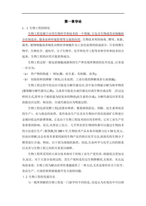
第一章导论1.1 生物工程的特征生物工程是属于应用生物科学和技术的一个领域,它包含生物或其亚细胞组分在制造业、服务业和环境管理等方面的应用。
生物技术利用病毒、酵母、真菌、藻类、植物细胞或者哺乳动物培养细胞作为工业化处理的组成部分。
只有将微生物学、生物化学、遗传学、分子生物学、化学和化学工程等多种学科和技术结合起来,生物工程的应用才能获得成功。
生物工程过程一般包括细胞或菌体的生产和实现所期望的化学改造。
后者进一步分为:(a)终产物的构建(例如,酶,抗生素、有机酸、甾类);(b)初始原料的降解(例如,污水处理、工业垃圾的降解或者石油泄漏)。
生物工程过程中的反应可能是分解代谢反应,其中复合物被分解为简单物质(葡萄糖分解代谢为乙醇),又或者可能是合成代谢反应或生物合成过程,经过这样的方式,简单分子被组建为较复杂的物质(抗生素的合成)。
分解代谢反应常常是放能反应过程,相反的,合成代谢反应为吸能过程。
生物工程包括发酵工程(范围从啤酒、葡萄酒到面包、奶酪、抗生素和疫苗的生产),水与废品的处理、某些食品生产以及从生物治疗到从低级矿石种进行金属回收这些新增领域。
正是由于生物工程技术的应用多样性,它对工业生产有着重要的影响,而且,从理论上而言,几乎所有的生物材料都可以通过生物技术的方法进行生产。
据预测,到2000年,生物技术产品未来市场潜力近650亿美元。
但也应理解,还会有很多重要的新的生物产品仍将以化学方法,按现有的生物分子模型进行合成,例如,以干扰为基础的新药。
因此,生命科学与化学之间的联系以及其与生物工程之间的关系更应阐释。
生物工程所采用的大部分技术相对于传统工业生产更经济,耗能低且更加安全,而且,对于大部分处理过程,其生产废料是经过生物降解的,无毒害。
从长远角度来看,生物工程为解决世界性难题提供了一种方法,尤其是那些有关于医学、食品生产、污染控制和新能源开发方面的问题。
1.2 生物工程的发展历史与一般所理解的生物工程是一门新学科不同的是,而是认为在现实中可以探寻其发展历史。
- 1、下载文档前请自行甄别文档内容的完整性,平台不提供额外的编辑、内容补充、找答案等附加服务。
- 2、"仅部分预览"的文档,不可在线预览部分如存在完整性等问题,可反馈申请退款(可完整预览的文档不适用该条件!)。
- 3、如文档侵犯您的权益,请联系客服反馈,我们会尽快为您处理(人工客服工作时间:9:00-18:30)。
生物医学工程专业英语及其翻译1 Unit 1 Biomedical Engineering Lesson 1A History of Biomedical EngineeringIn its broadest sense, biomedical engineering has been with us for centuries, perhaps even thousands of years. In 2000, German archeologists uncover a 3,000-year-old mummy from Thebes with a wooden prosthetic tied to its foot to serve as a big toe. Researchers said the wear on the bottom surface suggests that it could be the oldest known limb prosthesis. Egyptians also used hollow reeds to look and listen to the internal goings on of the human anatomy. In 1816, modesty prevented French physician Rene Laennec from placing his ear next to a young woman’s bare chest, sohe rolled up a newspaper and listened through it, triggering the idea for his invention that led to today’s ubiquitous stethoscope.广义上来说,生物医学工程与我们已经几个世纪以来,甚至数千年。
2000年,德国考古学家发现一个3000岁高龄的木乃伊从底比斯木制假肢与作为大脚趾的脚。
研究人员说,穿底部表面上表明它可能是最古老的下肢义肢。
埃及人也用空心的芦苇外观和听人类解剖学的内部行为。
1816年,谦虚阻止法国医生雷奈克把他的耳朵旁边一个年轻女人的裸胸,所以他卷起报纸和听它,引发他的发明的想法,导致今天无处不在的听诊器。
No matter what the date, biomedical engineering has provided advances in medical technology to improve human health. Biomedical engineering achievements range from early devices, such as crutches, platform shoes, wooden teeth, and the ever-changing cache of instruments in a doctor’s black bag, to more modern marvels,including pacemakers, the heart-lung machine, dialysis machines, diagnostic equipment, imaging technologies of every kind, and artificial organs, implants and advanced prosthetics. The National Academy of Engineering estimates that there are currently about 32,000 bioengineers working in various areas of health technology. 无论什么日期,生物医学工程提供了先进的医疗技术来改善人类健康。
生物医学工程成就范围从早期设备,如拐杖,松糕鞋,木制的牙齿,和不断变化的缓存工具在医生的黑包,更现代的奇迹,包括心脏起搏器、人工心肺机,透析机器,诊断设备,各种成像技术,和人造器官,移植和先进的假肢。
美国国家工程学院的估计,目前大约有32000生物各领域工作的卫生技术。
As an academic endeavor, the roots of biomedical engineering reach back to early developments in electrophysiology, which originated about 200 years ago. An early landmark in electrophysiology occurred in 1848 when DuBois Reymond published the widely recognized Ueber die tierische Elektrizitaet. Raymond’s contemporary,Hermann von Helmholtz, is credited with applying engineering principles to a problem in physiology and dentifying the resistance of muscle and nervous tissues to direct current.作为一个学术努力,生物医学工程的根源及早期电生理学的发展,起源于约200年前。
电生理学的早期具有里程碑意义的发生在1848年当杜布瓦Reymond发表了公认Ueber死tierische Elektrizitaet。
赫尔曼·冯·雷蒙德•当代亥姆霍兹因应用工程原则问题在生理学和dentifying电阻直流的肌肉和神经组织。
In 1895, Wilhelm Roentgen accidentally discovered that a cathode-ray tube could make a sheet of paper coated with barium platinocyanide glow, even when the tube and the paper were in separate rooms. Roentgen decided the tube must be emitting some kind of penetrating rays, which he called “X” rays for unknown. This set off aflurry of research into the tissue-penetrating and tissue-destroying properties of X-rays, a line of research that ultimately produced the modern array of medical imaging technologies and virtually eliminated the need for exploratory surgery.1895年,威廉伦琴偶然发现,阴极射线管可以与氰亚铂酸盐钡一张纸涂布发光,即使管和纸是在单独的房间。
伦琴决定管必须发出某种穿透光线,他称为“X”光线不明。
这引发了一系列tissue-penetrating和专治属性的研究x射线,一系列的研究,最终得出了现代医学影像技术和几乎消除了探索性手术的必要性。
Biomedical engineering’s unique mix of engineering, medicine and science emerged2 alongside biophysics and medical physics early this century. At the outset, the three were virtually indistinguishable and none had formal training programs.生物医学工程的独特工程、医学和科学出现2与生物物理学和医学物理学在本世纪初。
开始的时候,三人几乎无法区分,没有正式的培训计划。
Between World War I and World War II a number of laboratories undertook research in biophysics and biomedical engineering. Only one offered formal training: the Oswalt Institute for Physics in Medicine, established in 1921 in Frankfurt, Germany, forerunner of the Max Planck Institute for Biophysics.在第一次世界大战和第二次世界大战的实验室进行了生物物理学和生物医学工程的研究。
只有一个提供正式的培训:Oswalt物理医学研究所,成立于1921年在法兰克福,德国马克斯普朗克生物物理学的先驱。
The Institute’s founder, Friedrich Dessauer, pioneered research into the biologicaleffects of ionizing radiation. The Oswalt Institute and the University in Frankfurt soon established formal ties that led to a Ph.D. program in biophysics by 1940. Research topics included the effects of X-rays on tissues and the electrical properties of tissues. The staff of 20 included university lecturers, research fellows, assistants and technicians.研究所的创始人,弗里德里希·德绍,率先研究电离辐射的生物效应。
Oswalt研究所和大学在法兰克福很快建立了正式的关系,在1940年导致了生物物理学博士学位项目。