The hydrophobic nanoparticles adsorption layer on core surface and its properties
聚乙烯亚胺纳米纤维吸附剂用于高效吸附水溶液中的阴离子型染料(英文)
mater.scБайду номын сангаас
SCIENCE CHINA Materials
Published online 29 January 2016 | doi: 10.1007/s40843-016-0117-y Sci China Mater 2016, 59(1): 38–50
Keywords: nanofibrous adsorbent, anionic dye, electrospinning, polyethylenimine
INTRODUCTION
Industrial wastewater, a legacy of the industrial revolution and its conflict between environmental capacity and human expansion, continues to be a problem for governments, businesses, the scientific community, and ordinary people worldwide [1,2]. Reducing wastewater discharge and decontaminating water resources are the two acknowledged strategies for prevention and remediation of water pollution, respectively [3,4]. Technology for removing contaminants from water is crucial to the success of either strategy. Wastewater containing organic dyes is one of the
海洋软体动物蓝斑背肛海兔抗肿瘤活性成分研究(Ⅱ)
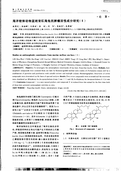
wee eu ia e s 3 - y r x 一 a 6 - p x c oe t 一一 r - n (I)a d 日 7 —i y r x 一 a 6- p x c oet 一 ( 4 一r r lcd td a 1h d o y 5 , a e o yh lsa 8e e 7o e 3 i n 3 , adh d o y 5 , a e o y h ls a8 1 )e e i
从 蓝 斑 背 舡 海 兔 中得 到 , 外 研 究 均 显 示 明 显 的 抗 瘟 活 性 体
[ 美t词] 蓝斑 背舡 海兔{ 抗肿瘤药 ; 甾醇类 [ 中圈分类 号] R 2 2 7 03 R 99 1 8 . 4 . : 7. [ 文献标识码] A [ 文章 编号] 0 5 7X(0 20 2 30 2 88 9 2 0 )30 4 —3
(I) Co cu in: o o n 1 i an w au a r d c .n o o n Ⅱ i ioae r m hsa i lfrt efrtt . n lso C mp u d s e n t rlp o u t a dc mp u d s s ltdfo t i nm o h is i b t a me,o h
摘要] 目的 : 究蓝斑背肛 海兔 Noacu t ci/ 研 trh s e hi a
的抗瘟活性成分 方 击 : 用溶 媒萃取 和多 种层析 手段 , 利 以稻瘟霉
模 型 监 测 跟 踪 , 海 兔 中抗 癌 活性 成 分 进 行 追 踪 分 离 , 用 波 谱技 术 鉴 定 化 台 物 的结 构 蛄 果 : 到 2千 活 性 化舍 物 : 羟 对 应 得 3 基一a 6一 氧 一一 甾烯 一一 (1 和 3 .口二 羟 基 5 . 环 氧 8 1 )胆 甾烯 ( 。 培 论 : 5 ,a环 8胆 7酮 ) 口 7一 u6 (4 Ⅱ) 化台 物 I为 新 天然 产物 , I为 首 次
β-环糊精修饰交联聚硅氧烷对水体中苯酚的吸附

β-环糊精修饰交联聚硅氧烷对水体中苯酚的吸附孟建强;丁欣;尹健【摘要】In order to effectively treat the organic pollutants in water, β-cyclodextrin modified cross-linked polymer (KHCD-BTEE) was prepared by siloxane coupling condensation reaction. The adsorption property of the polymer was investigated with phenol as simulated pollutant. The results showed that the KHCD-BTEE can effectively adsorb phenol from water and the adsorption capacity of adsorbent increased with the initial concentration of phenol. When the initial concentration of phenol was 0.2 g / L, the adsorption capacity was 0.015 mmol/g. When the initial concentration of phenol was constant, with the increase of adsorption time, the adsorption capacity showed a trend of first increase and then gentle and the adsorption equilibrium was reached within 3 h. The adsorption thermodynamics followed Langmuir isotherm and the adsorption for phenol was a single layer adsoption. The adsorption kinetics followed pseudo-second-order kinetics, mainly through the hydrogen bond interaction and hydrophobic interaction between theβ-cyclodextrin and the phenol molecule. Regeneration conditions were mild and the phenol can be desorbed by the methanol solution at room temperature.%为有效处理水体中的有机污染物,通过硅氧烷偶联缩合反应制备了β-环糊精修饰交联聚合物(KHCD-BTEE),并以苯酚为模拟污染物,研究了聚合物的吸附性能.结果表明:β-环糊精修饰吸附剂可有效地吸附水体中苯酚,随着苯酚初始浓度的增大,吸附剂的吸附量也不断增大,当初始浓度为0.2 g/L时,其最大吸附量可达到0.015 mmol/g;当苯酚初始浓度一定时,随着吸附时间的增加,吸附剂的吸附量先增加后趋于平缓,直到3 h 达到吸附平衡;吸附热力学符合Langmuir曲线,吸附饱和时为单层覆盖;吸附动力学遵循伪二级动力学方程,主要是通过β-环糊精与苯酚分子之间的氢键作用和疏水作用进行吸附;再生条件温和,室温下甲醇溶液震荡即可洗脱苯酚.【期刊名称】《天津工业大学学报》【年(卷),期】2017(036)004【总页数】5页(P68-72)【关键词】β-环糊精;硅氧烷偶联;交联聚合物;吸附;苯酚【作者】孟建强;丁欣;尹健【作者单位】天津工业大学省部共建分离膜与膜过程国家重点实验室,天津300387;天津工业大学材料科学与工程学院,天津 300387;天津工业大学省部共建分离膜与膜过程国家重点实验室,天津 300387;天津工业大学材料科学与工程学院,天津 300387;天津工业大学省部共建分离膜与膜过程国家重点实验室,天津300387;天津工业大学材料科学与工程学院,天津 300387【正文语种】中文【中图分类】TQ424.3;X703随着工业和人类活动发展,人类对水的污染日益加重,其中来自工业、农业和生活等方面的有机污染物已严重影响全球淡水系统,并且已对水生生态系统和人类健康造成了严重的危害[1-4].面对日益复杂的有机微污染,传统的处理方法已不能满足分离要求[5-9].早在1891年,Villiers在淀粉水解液中首次发现了环糊精.其中β-环糊精由7个吡喃葡萄糖单元组成,其立体结构为一个一端大一端小的锥形圆筒[10-14].由于β-环糊精分子具有外部亲水而内部疏水的特殊结构,因而它能够作为主体有选择性地包络各种尺寸适当的客体,如生物小分子、有机分子、无机离子以及惰性气体等,因此,已被广泛应用于处理水体中的有机微污染物[15].但是,目前有关环糊精修饰吸附剂的研究多是采用后修饰的方法负载,如Li等制备的β-环糊精修饰磁性纤维素纳米棒[16],Abay等制备的β-环糊精修饰聚(甲基丙烯酸羟乙酯-乙二醇二甲基丙烯酸酯)微球[17],均是用β-环糊精对已制备的一定基底进行后修饰,制备过程较为复杂,效率较低.本文通过硅氧烷偶联缩合反应原位交联环糊精,制备了β-环糊精修饰交联聚硅氧烷,并对其吸附性能进行探究.试剂:β-环糊精(β-CD)、γ―(2,3-环氧丙氧)丙基三甲氧基硅烷(KH560)、1,2-双三甲氧基硅基乙烷(BTEE)、氢化钠,均为分析纯,上海泰坦科技股份有限公司产品;N,N-二甲基甲酰胺,色谱纯,上海阿拉丁试剂有限公司产品;盐酸,1.0 mol/L,天津市科密欧化学试剂有限公司产品;苯酚,99%,上海百灵威科技有限公司产品;乙醇,甲醇,均为分析纯,天津市科密欧化学试剂有限公司产品;去离子水,实验室自制.仪器:DF-101S型恒温水浴锅,巩义市英峪仪器有限责任公司生产;CP224C型万分之一电子天平,奥豪斯上海仪器有限责任公司生产;T6型紫外可见分光光度计,北京普析通用仪器有限责任公司生产;SHAC型水浴恒温振荡器,江苏金坛市中大仪器厂生产;STA409PC/PG型热重分析仪,德国Netzsch公司生产;S-4800型场发射扫描电子显微镜,日本日立公司生产.将一定量的β-环糊精、氢化钠和N,N-二甲基甲酰胺分别放入烧瓶中,室温下处理30 min.将反应后的混合液进行抽滤,再向滤液中加入适量的KH560,氮气环境下90℃反应5 h,制得KH560改性环糊精(KHCD),并对其进行抽滤.之后按照一定比例(β-CD/KH560/BTEE摩尔比=1/1.68/4.27)向乙醇溶液中加入BTEE,与KHCD混合均匀,调节溶液pH=4,制备出β-环糊精修饰聚硅氧烷,如图1所示.(1)结构表征:通过热重分析(TG)测定β-环糊精修饰聚硅氧烷的热稳定性,用扫描电镜(SEM)观察β-环糊精修饰聚硅氧烷表面形貌的变化.(2)吸附性能测定:配置不同质量浓度的苯酚溶液(0.025~0.20 g/L),加入一定量的吸附剂,恒温振荡使其吸附达到平衡.通过紫外可见分光光度计检测吸附后溶液的吸光度(λ=213 nm),根据标准工作曲线计算出溶液浓度,并通过式(1)计算吸附量:式中:qe为平衡吸附量(mmol/g);C0和Ce分别为溶液的初始质量浓度和平衡质量浓度(g/L);V为溶液的体积(mL);m为吸附剂的质量(g);M为苯酚的摩尔质量(g/mol).(3)循环利用性能测定:首先将一定质量的吸附剂放入一定体积的0.15 g/L苯酚溶液中,待达到吸附平衡后,将吸附剂与溶液分离.之后,用相同体积的甲醇溶液对吸附剂进行浸泡洗脱,通过紫外可见分光光度计检测,直至甲醇溶液中不再有物质溶出,即认为被吸附的苯酚已脱除.并再次将该吸附剂用于苯酚溶液的吸附过程.吸附剂的循环利用性能用恢复率RE表示:式中:q1和q2分别为初次吸附的平衡吸附量(mmol/g)和再生后的平衡吸附量(mmol/g).图2所示为β-环糊精修饰聚硅氧烷KHCDBTEE与改性β-环糊精单体KHCD的TG谱图.由图2可以看出,单体KHCD在140℃时开始发生降解,而聚合物在247℃时开始发生明显的降解.通过两者对比,可以看出聚合物的热稳定性明显提高.这可能是因为在反应过程中硅氧烷产生了较多的羟基,从而使β-环糊精修饰交联聚硅氧烷内存在较多的氢键,有效地提高了聚合物的热稳定性.图3所示为β-环糊精修饰聚硅氧烷在放大倍数分别为2 000、5 000和10 000时的电镜图片.由图3可以观察到聚合物表面较为致密,基本不存在孔结构.由此也可以说明,该聚合物作为吸附剂主要是在其表面进行吸附.图4所示为KHCD-BTEE的吸附量qe随苯酚初始浓度的变化曲线.由图4可以看出,经过β-环糊精修饰的聚硅氧烷具有β-环糊精特有的包合能力.KHCD-BTEE的吸附量qe随苯酚溶液浓度的升高而增大,但是随着苯酚溶液初始浓度的增加,吸附量增速逐渐放缓,这说明该聚合物吸附量接近达到饱和.当苯酚溶液质量浓度为0.20 g/L时,β-环糊精修饰聚硅氧烷的吸附量可达到0.015 mmol/g.Langmuir方程如式(3),Freundlich方程如式(4):式中:Ce为吸附平衡质量浓度(g/L);qe为平衡吸附量(mmol/g);qm为饱和吸附量(mol/g);b为 Langmuir常数;Kf和n为Freundlich常数.通过以上2种等温吸附模型对吸附数据进行拟合,如图5所示.用Langmuir方程拟合的R2值为0.944 9,用Freundlich方程拟合的R2值为0.898 1.结果表明,用Langmuir等温吸附模型来描述β-环糊精修饰聚硅氧烷对苯酚的吸附行为更合适.说明该聚合物对苯酚为均匀吸附,当达到吸附饱和时为单层覆盖.采用0.15 g/L的苯酚溶液作为模拟污染物,考察KHCD-BTEE对苯酚的吸附量随吸附时间的变化,结果如图6所示.由图6可以看出,对于0.15 g/L的苯酚溶液而言,在开始的一段时间内苯酚吸附量增加较快,随着时间的延长,吸附量变化趋于平缓.这可能是因为随着吸附过程的进行,聚合物表面的吸附位点逐渐被占用,从而影响了苯酚分子与有效位点的接触.当吸附时间为3 h时,吸附量基本不再增加,吸附剂达到饱和状态,即达到吸附平衡.为了进一步研究苯酚在KHCD-BTEE聚合物上的吸附动力学行为,通过伪一级动力学模型式(5)和伪二级动力学模型式(6)对吸附动力学实验数据进行拟合.式中:qe和qt分别为平衡吸附量和t时刻的吸附量(mmol/g);t为接触时间(min);k1和 k2分别为伪一级动力学模型速率常数(min-1)和伪二级动力学模型速率常数(g·mmol-1·min-1).其结果如表 1 所示.由表1可以看出,苯酚的伪一级动力学模型和伪二级动力学模型均拟合良好,但伪二级动力学模型的拟合度更高,并且公式计算所得吸附量与实际吸附量较为一致.说明β-环糊精修饰聚硅氧烷对苯酚的吸附行为遵循伪二级动力学模型,这与已有文献中报道的结果一致[18],其主要是通过β-环糊精分子与苯酚分子之间的氢键作用与疏水作用进行吸附.在实际去除水体中酚类有机污染物时,经济、实用也是需要考虑的问题.因此,本文对合成聚合物的再生性能也进行了探究,结果如表2所示.由表2可以看出,用甲醇溶液对吸附剂进行洗脱,循环2次后恢复率迅速降至80%.同时观察到,随着吸附剂在溶液中不断浸泡、洗涤,吸附剂会缓慢散开,表明该吸附剂稳定性较差.这可能是因为硅氧烷在反应过程中会产生大量的羟基,从而在聚合物中会存在较多的氢键.通过甲醇不断浸泡洗脱吸附剂,对聚合物中的氢键造成了一定的影响,导致吸附剂的稳定性降低.(1)当苯酚质量浓度达到0.20 g/L时,其吸附量可达到0.015 mmol/g.通过Langmuir和Freundlich 2种等温吸附模型对实验数据进行拟合后得知,Langmuir等温吸附模型能够更好地描述吸附行为,说明当吸附达到饱和时,苯酚分子呈现均匀的单层覆盖.(2)对于0.15 g/L的苯酚溶液,随着吸附时间的增加,KHCD-BTEE聚合物的吸附量先增大后趋于平缓,最终在3 h内达到吸附平衡,吸附过程遵循伪二级动力学模型,说明其主要是通过β-环糊精分子与苯酚分子之间的氢键作用和疏水作用进行吸附.(3)聚合物稳定性较差,循环2次后恢复率迅速降至80%,无法进行多次循环吸附.【相关文献】[1]SCHAFER A I,AKANYETI I,SEMIAO A J.Micropollutant sorption to membrane polymers:A review of mechanisms for estrogens[J].Advances in Colloid and Interface Science,2011,164(1/2):100-117.[2]De GAETANO Y,HUBERT J,MOHAMADOU A,et al.Removal of pesticides from wastewater by ion pair centrifugal partition extraction using betaine-derived ionic liquids as extractants[J].Chemical Engineering Journal,2016,285:596-604.[3]YAMASAKI H,MAKIHATA Y,FUKUNAGA K.Preparation of crosslinked β-cyclodextrin polymer beads and their application as a sorbent for removal of phenol from wastewater[J].Journal of Chemical Technology&Biotechnology,2008,83(7):991-997.[4]AYAD M M,ELNASR A A.Adsorption of cationic dye(methylene blue)from water using polyaniline nanotubes base[J].The Journal of Physical Chemistry C,2010,114:14377-14383.[5]WU H,WANG S,KONG H,et al.Performance of combined process of anoxic baffled reactor-biological contact oxidation treating printing and dyeingwastewater[J].Bioresource Technology,2007,98(7):1501-1504.[6] 胡文.聚合硫酸铁在水处理中的应用[J].神州,2012,26:44.HU W.Application of polyferric sulfate in water treatment[J].Shenzhou,2013,(26):44(in Chinese).[7] FANG F,HAN H,ZHAO Q,et al.Bioaugmentation of biological contact oxidation reactor(BCOR)with phenol-degrading bacteria for coal gasification wastewater(CGW)treatment[J].Bioresource Technology,2013,150:314-320.[8]KOLODYNSKA D,HUBICKI Z,PASIECZNA P S.FT-IR/PAS studies of Cu (II)-EDTA complexes sorption οn the chelating ion exchangers[J].Acta Physica Polonica A,2009,116(3):340-343.[9]JIAO Y,ZHAO Q,JIN W,et al.Bioaugmentation of a biological contact oxidation ditch with indigenous nitrifying bacteria for in situ remediation of nitrogen-rich streamwater[J].Bioresource Technology,2011,102(2):990-995.[10]DASS C R,JESSUP W.Apolipoprotein A-I,cyclodextrins and liposomes as potential drugs for the reversal of atherosclerosis:A review[J].Journal of Pharmacy and Pharmacology,2000,52(7):731-761.[11]UYAR T,HAVELUND R,NUR Y,et al.Cyclodextrin functionalized poly(methyl methacrylate)(PMMA)electrospun nanofibers for organic vapors wastetreatment[J].Journal of Membrane Science,2010,365(1/2):409-417.[12]CHEN P,LIANG H W,LYU X H,et al.Carbonaceous nanofiber membrane functionalized by beta-cyclodextrins for molecular filtration[J].ACS Nano,2011,5(7):5928–5935.[13]SCHOFIELD W C E,BAIN C D,BADYAL J P S.Cyclodextrin-functionalized hierarchical porous architectures for highthroughput capture and release of organic pollutants from wastewater[J].Chemistry of Materials,2012,24(9):1645-1653.[14]SHVETS O,BELYAKOVA L.Synthesis,characterization and sorption properties of silica modified with some derivatives of β-cyclodextrin[J].Journal of Hazardous Materials,2015,283:643-656.[15]AOKI N,KINOSHITA K,MIKUNI K,et al.Adsorption of 4-nonylphenol ethoxylates onto insoluble chitosan beads bearing cyclodextrin moieties[J].Journal of Inclusion Phenomena and Macrocyclic Chemistry,2007,57(1/2/3/4):237-241.[16]CHEN L,BERRY R M,TAM K C.Synthesis of β-cyclodextrin-modified cellulose nanocrystals(CNCs)@Fe3O4@SiO2superparamagnetic nanorods[J].ACS Sustainable Chemistry&Engineering,2014,2(4):951-958.[17]ABAY I,DENIZLI A,BISKIN E,et al.Removal and preconcentration of phenolic species onto beta-cyclodextrin modified poly(hydroxyethylmethacrylate-ethyleneglycoldimethacrylate)microbeads[J].Chemosphere,2005,61(9):1263-1272.[18]JIN L,HE D,WEI M.Selective adsorption of phenol and nitrobenzene by β-cyclodextrin-intercalated layered double hydroxide:Equilibrium and kineticstudy[J].Chemical Engineering&Technology,2011,34(9):1559-1566.。
水溶性二氟化硼-二吡咯甲烷(BODIPY)类荧光染料研究进展
第46卷第15期2018年8月广 州 化 工Guangzhou Chemical IndustryVol.46No.15Aug.2018水溶性二氟化硼-二吡咯甲烷(BODIPY )类荧光染料研究进展*程乐华,李 雷,陈 宏,汤家华,王 敏(巢湖学院配位化学研究所,安徽 合肥 238000)摘 要:简要介绍了二氟化硼-二吡咯甲烷(BODIPY)荧光染料的基本结构㊁优异的光物理和光化学性能以及自身存在的溶解性差等缺陷,重点综述了近年来各种水溶性BODIPY 类荧光染料衍生物的合成及其在水环境中作为荧光探针的应用研究成果,并提出通过对BODIPY 母体染料结构进行化学修饰,开发研制出许多具有不同性能的BODIPY 衍生物并研究其应用将成为国内外十分活跃的研究课题㊂关键词:二氟化硼-二吡咯甲烷;水溶性;荧光染料;合成;应用 中图分类号:O621.3 文献标志码:A文章编号:1001-9677(2018)15-0019-05*基金项目:安徽省高校自然科学研究重点项目(No:KJ 2016A503)㊂通讯作者:程乐华(1969-),男,副教授,主要从事配位化学与光电功能材料研究㊂Research Progress on Water-soluble Bipyrromethenebor onDifluoride (BODIPY )Fluorescent Dyes *CHENG Le -hua ,LI Lei ,CHEN Hong ,TANG Jia -hua ,WANG Min(Institute of Coordination Chemistry,Chaohu College,Anhui Hefei 238000,China)Abstract :The basic structure,excellent photophysical and photochemical properties and its low water-solubility of bipyrrometheneboron difluoride(BODIPY)dye were briefly introduced.This review mainly focused on the recent research progress on the synthesis of water-soluble BODIPY dyes and their application as fluorescent probes in water environment.It was also proposed that many BODIPY derivatives with different properties will be developed by chemical modification of BODIPY parent dye structure,and their application will become a very active research topic.Key words :bipyrrometheneboron difluoride (BODIPY);water solubility;fluorescent dyes;synthesis;application二氟化硼-二吡咯甲烷(BODIPY)类染料作为一种重要的新型荧光标记材料,具有高摩尔消光系数和高荧光量子产率㊁良好的光化学稳定性㊁激发波长在可见光区㊁不易受环境和溶液pH 的影响㊁峰形窄而尖锐㊁荧光寿命长等优点[1]㊂近年来,国内外许多化学及生物学家通过在母体染料上引入不同的化学基团对其结构进行修饰,开发研制出许多具有不同性能的BODIPY 衍生物[2-5]㊂但该类荧光染料的最大缺陷就是水溶性差,一般只能溶解在极性有机溶剂中,这极大地限制了其在水环境或生命体系中的应用㊂为提高这类有机荧光染料在水中的溶解度,进一步扩大地实际应用范围,已有一些文献报道了增强其水溶性的方法,如在BODIPY 荧光团上引入离子型亲水基团;或将疏水性的BODIPY 荧光团接枝到(生物)聚合物上[6-9]等㊂本文在简单表述BODIPY 染料基本结构的基础上,详细综述最近年来各种水溶性BODIPY 类荧光染料的合成方法及其在荧光探针方面的研究进展㊂1 BODIPY 荧光染料的结构BODIPY 染料作为一种类次甲基㊁非离子性的荧光标记材料,由Treibs 和Kreuzer 于1968年首次用2,4-二甲基吡咯和苯甲醛通过两步 一锅煮”的合成方法制备[10],它由硼桥键和甲川桥键把两个吡咯环固定在一个平面上,使分子具有刚性共平面结构,在激发光的作用下能产生强烈的荧光(结构式如图1所示)㊂与其他荧光染料相比,BODIPY 类荧光染料的一个最显著的特点和优势是结构易于修饰,如图1所示的母体结构式中R 1~R 8位均可引入不同的化学基团进行适当的化学修饰从而改进其物理和光化学性能[4]㊂图1 BODIPY 的结构式 Fig.1 Structural formula of BODIPY2 水溶性BODIPY 类荧光染料Burgess 课题组[11]于2007年首次直接由1,3,5,7-四甲基取代的BODIPY 通过钯-催化的C-H 功能化反应制备出具有一定水溶性的BODIPY 染料衍生物1和2,其合成路线如图2所示㊂2018年8月广 州 化 工20 尽管染料1微溶于水,但它带有一个能用水溶性基团如磺化的N-羟基丁二酰亚胺活化的-COOH 基,因此,该染料可潜在地应用于共价连接生物分子;染料2具有很好的水溶性,能够实现一些需要水相介质中荧光的其他应用㊂图2 水溶性BODIPY 1和2的合成路线Fig.2 Synthesis route of water-soluble BODIPY 1and 2该课题组又采用了不同于以前的方法合成了一系列水溶性的单磺化(3a ~5a)和双磺化(3b ~5b)BODIPY 染料衍生物[12]㊂化合物3系列在其染料结构的中位(8-位)含有一个4-碘基苯基亚单元,它能通过金属有机的交叉偶联反应进一步功能化㊂化合物4系列也能发生类似的衍生化,同时,3,5-位的两个氯原子通过芳香族亲核取代实现其功能化㊂带有氨基的化合物5系列易于和生物分子中的活性羰基酰基化而偶合连接㊂他们还利用重氮/叠氮反应将氨基化合物5转化成叠氮化合物6,后者易于和炔烃发生铜催化的环加成反应制得化合物7,其结构中含有1个-COOH 官能团,易于被生物分子中的氨基活化和偶合,可潜在用于标记水环境中生物分子的荧光探针,其主要的合成路线如图3所示㊂图3 水溶型BODIPY 3和4的合成路线Fig.3 Synthesis route of water-soluble BODIPY 3and 4Akkaya [13]研究小组报道一种为数不多在可见光的近红外区发射的水溶性BDOIPY 荧光染料8,可作为一种敏感的㊁选择性的㊁比率式Zn 2+离子荧光探针㊂其合成需要不同的芳醛和BODIPY 母体中3-和5-位甲基发生逐步的克脑文盖尔缩合反应,从而产生3,5-位二苯乙烯基取代的BODIPY 衍生物,而在乙烯苯基对位又含有一个双(2-吡啶甲基)胺(DPA)结构单元,它作为一种金属螯合剂可对金属Zn 具有好的配位性,同时分子中带有的6个二缩三乙二醇基团极大地增强了其水溶性(合成路线如图4所示)㊂图4 水溶性BODIPY 8的合成路线Fig.4 Synthesis route of water-soluble BODIPY 8Bane 课题组[14]首先合成出起始原料3,5-二氯代BODIPY 染料9a ,再将其与硫代乙醇酸反应可定量得到一种高荧光的水溶性化合物9,再将其与N-羟基琥珀酰亚胺在脱水剂DCC 作用下偶合得到3,5-位被二琥珀酰亚胺酯取代的BODIPY 染料衍生物10,该染料可应用于普通蛋白质的荧光标记㊂而在该反应中若用N-羟基磺酸琥珀酰亚胺钠盐代替N-羟基琥珀酰亚胺作为亲核试剂时所得产物11的产率却很低(合成路线如图5所示)㊂图5 水溶性BODIPY 9-11的合成路线Fig.5 Synthesis route of water-soluble BODIPY 9-11Jiao 等[15]用3,5-二碘代BODIPY 12b 代替BODIPY 12a ,并将其与N,N,-二(2-羟基乙基)胺发生亲核双取代从而产生一种新的水溶性BODIPY 染料衍生物12,它对水溶液中的铜离子显示出高的敏感性和选择性荧光响应,并且活体细胞中的荧光影像实验结果也进一步证实它在生物体系中有很好的实际应用(合成方法如图6所示)㊂图6 水溶性BODIPY 12的合成路线Fig.6 Synthesis route of water-soluble BODIPY 12Liu 等[16]合作者于2009年首次报道了两种直接将多磺化连接基团(一种来自ɑ-磺基-ß-氨基丙酸,另一种来自磺基三甲铵乙内酯)引入到BODIPY 骨架上的方法,通过合成后的衍生化即酰胺的形成㊁炔烃偶联或B-F 取代等反应来实现,并由此制备出四种水溶性BODIPY 染料衍生物13㊁14㊁15㊁16㊂值得注意的是,13和16均带有适合与生物分子共轭的酸性基团,并且它们在水环境中有好的量子产率,因此非常适合被用于荧光生物标记㊂该课题组又采用了两种合成路线报道了一系列在不同位置被适当功能化的水溶性BODIPY 染料[17]:一种是经过 软”偶联反应引入与染料相连的受保护的磺酸盐基团;另一种是磺基三甲铵乙内酯的合成后引入㊂为了克服17b 与17a 不能通过直接交叉偶合得到所期望的水溶性化合物17的问题,他们设21 程乐华,等:水溶性二氟化硼-二吡咯甲烷(BODIPY)类荧光染料研究进展第46卷第15期计了一个两步策略,即先用1-二甲氨基-2-丙炔与2-位带碘功能基的BOIDPY 染料17b 在Pd(0)下的交叉偶合得到中间产物17c ;再将17c 与1,3-丙磺内酯在非极性条件下进行季铵化反应非常方便地制得水溶性化合物17㊂作者又利用1-二甲氨基-2-丙炔的格氏试剂类似物将上述合成策略应用到将三甲铵乙内酯片段接枝到硼原子上,先制备出乙炔基取代的BODIPY 染料18b ,再将其与1,3-丙磺内酯在甲苯溶剂中烷基化,能以较高的产率得到水溶性化合物18(合成路线见图7)㊂图7 水溶性BODIPY 17和18的合成路线Fig.7 Synthesis route of BODIPY 17and 18Nagal 等[18]利用偶氮二异丁腈作为引发剂,二硫代苯甲酸枯酯作为链转移剂,通过2-二甲氨基异丁烯酸乙酯(DMAEMA)和19a 的可逆加成-断裂链转移(RAFT)共聚合反应,制备出一种新型的含BODIPY 染料结构单元的热响应水溶性荧光共聚物19㊂研究表明,在水中该聚合物在跨越最低临界溶液温度(LCST)加热和冷却时,其荧光强度有效的可逆降低/增加会引起共聚物中两个BODIPY 平面间H-聚集的可逆形成/抑制㊂因此,通过LCST,该水溶性聚合物可显示出它的荧光光学开关效应㊂Yildiz 等[19]为制备在其表面具有活性功能基的生物相容性CdSe-ZnS 核-壳型量子点,选用硫醇㊁聚乙烯乙二醇链㊁羧酸或伯胺分别作为锚定基团㊁亲水性基团和连接基团,从商业可得的前驱体为起始原料通过1~5步反应合成聚合所需的单体,并在偶氮二异丁腈的催化下,这些单体适当组合并进行自由基共聚反应,产生相应的共聚体20a ㊁20b 和20c 等大分子配体,再将预先制成的CdSe-ZnS 核-壳型量子点与亲水性共聚体一起加热促进聚硫醇在纳米微粒表面的吸附㊂再由大分子配体20a ㊁20b 和20c 包覆的纳米微粒表面上的羧酸或伯胺能够和互补的功能基反应从而进一步修饰量子点,如20a 和21a ㊁20b 或20c 与21b 在1-乙基-3-(3-二甲氨基-丙基)碳化二亚胺和N -羟基硫代琥珀酰胺的作用下使得BODIPY 荧光团靠酰胺键的形成而附着到亲水性的纳米微粒中㊂研究表明,这种水溶性量子点能够穿透中国仓鼠卵巢细胞膜,优先聚集在细胞溶质中并且对细胞活性没有实质影响,可望被开发为具有纳米尺寸的发光探针而用于各种生物医学中㊂Chujo [20]课题组报道一种通过BODIPY 封端的水溶性聚合物稳定的可逆热转换发光金纳米颗粒(合成路线如图8所示)㊂为此,他们首先设计合成了基于BODIPY 的链转移剂22a 作为端基带有BODIPY 结构单元的聚N-异丙基丙烯酰胺(PNIPAM)的关键前驱体,再将22a 和NIPAM 进行RAFT 聚合得到水溶性聚合物PNIPAM;最后以NaBH 4作还原剂,将其与HAuCl 4原位还原制备出BODIPY 封端的PNIPAM 包覆的金纳米颗粒22㊂荧光光谱与温度的变化关系显示出随温度的升高,金纳米颗粒的发光强度降低,金纳米微粒的发射/淬灭能可逆和有效地发生,而与加热和冷却循环无关㊂图8 水溶性聚合物PNIPAM 及金纳米微粒的制备Fig.8 Construction of water-soluble polymer (PNIPAM)andgold nanoparticles (AuNP)次氮基三乙酸(NTA)结合的体系在示踪蛋白质标记方面有潜在的应用,Brellier 等[21]报道了两种新型的水溶性的中位取代单个或两个次氮基三乙酸的BODIPY 染料23和24㊂两种荧光染料在水中测定的光物理参数与强极性溶剂甲醇中测定的结果均无明显改变,并且NTA 官能基的引入也没有明显影响染料的光学特性㊂因此,它们能成为有应用价值的探针,既可用于生理条件下细胞成像又可用于组氨酸示踪的蛋白质选择性标记㊂华东理工大学的钱旭红课题组[22]设计合成了一种基于BOIDPY 荧光团的长波㊁水溶性镉离子探针25,为了获得近红外㊁灵敏性探针,他们选择了对镉离子选择性键合的水溶性的聚酰胺为受体单元并且在BODIPY 的3-位和5-位各共轭相连两个聚酰胺单元㊂该配合物对水溶液中的PPi 显示优异的选择性识别,能从PPi 区分出ADP 和ATP㊂Ziessel 等[23]开发一种收敛法合成波长在490~750nm 范围内的一系列水溶性BODIPY 染料衍生物26a ㊁26b ㊁27和28,通过将膦酸烷基酯片段嫁接到BODIPY 染料的硼原子核上或苯乙烯的侧背上获得染料的水溶性和荧光特性,且这些化合物在水溶液中甚至发射波长处在红光区也没有聚集体形成,化合物结构式见图9㊂Liu [24]研究小组在以前工作的基础上用支化的低聚乙二醇甲醚残片对BODIPY 染料的8-位㊁2-位和6位㊁或4,4’-位进行官能化从而制备出一系列新型的高水溶性且发射波长从绿色到深红色区域的中性BOIDPY 染料衍生物A-J,其中I 和J 在水中极易溶解且在水溶液中显示出强荧光,其结构式如图9所示㊂2018年8月广 州 化 工22图9 水溶性BODIPY 25,26a ,26b ,27和28的结构式Fig.9 Structural formula water-soluble BODIPY25,26a ,26b ,27and 28Massif 等[25]报道了一种将疏水性的BODIPY 荧光染料经化学转变生成生物共轭的水溶性荧光衍生物的新方法㊂首先利用固相合成策略方便制备磺化端炔29a ,再将其与BODIPY 染料的芳基叠氮衍生物29b 发生铜催化 点击”反应得到单磺化的荧光染料29;或与BODIPY 染料的芳基碘衍生物30a 发生钯催化的Sonogashira 交叉-偶联反应制备出单磺化的荧光衍生物30(主要合成路线如图10所示),研究发现,这两种新型的荧光染料在水中均具有很好的溶解性,适合应用于生物标记㊂图10 水溶性BODIPY 29和30的合成路线Fig.10 Synthesis route of water-soluble BODIPY 29and 30Sauer 等[26]用BODIPY 表面活性单体经细乳液聚合 一锅法”制备荧光表面标记的聚合物纳米微粒(合成路线如图11所示)㊂在表面活性单体的合成中,作者采用了汇聚式合成策略,即在表面活性单体的离子型头基形成之前,BODIPY 核预先制成,并连接上可聚合基团的疏水性尾部;亲水性的头基(活性单体31和32)通过BODIPY 的2-位或2,6-位与氯磺酸的单磺化或双磺化反应制得;最后利用疏水性引发剂V59在细乳液中经过自由基聚合方法制备出具有荧光界面的聚苯乙烯纳米微粒㊂研究发现,仅使用双磺化表面活性单体的微粒荧光对作为溶解在连续相中的碘化钠或甲基紫罗碱荧光淬灭剂强烈敏感,相反,对使用单磺化的表面活性剂的纳米微粒荧光只有较弱的荧光焠灭,因此,前者能够作为基于荧光淬灭或荧光共振能量转移效应的传感应用㊂图11 单磺化或双磺化BODIPY 表面单体活性剂的合成路线Fig.11 Synthesis route of mono-or double-sulfonated BODIPY surfmer3 结 语BODIPY 类染料作为一类重要的荧光标记材料,具有一些有益的光物理和光化学性质,如高荧光量子产率和高摩尔消光系数㊁激发波长位于可见光区㊁容易通过化学方法进行结构修饰㊁不易受环境及pH 的影响等㊂因此,通过对BODIPY 母体染料结构进行化学修饰,开发研制出许多具有不同性能的BODIPY 衍生物并研究其应用已经成为国内外十分活跃的研究课题㊂但该类荧光染料的最大缺陷就是水溶性差,一般只能溶解在极性有机溶剂中,这极大地限制了其在水环境或生命体系中的应用㊂而增强其水溶性的主要方法有:在BODIPY 荧光团上引入N,N-二(2-羟乙基)氨㊁离子型亲水基团(如羧酸㊁膦酸㊁磺酸㊁铵盐㊁磺酸基甜菜碱等);或将疏水性的BODIPY 荧光团接枝到(生物)聚合物(如糖类化合物㊁寡核苷酸㊁磺化缩氨酸或低聚乙二醇)上等㊂今后的研究重点将集中在进一步拓宽增强该类荧光染料水溶性其他行之有效的且具有合成路线少㊁原料简单易得㊁纯化简便等优点的方法;另外,在增强水溶性的同时引入适当的识别基团以应用于荧光生物传感或水环境中污染物的检测或通过延伸染料的共轭体系使其吸收或发射波长达到红光/近红外区域等㊂参考文献[1] Karolin J,Johansson L B A,Strandberg L,et a1.Fluorescence andabsorption spectroscopic properties of dipyrrometheneboron difluorlde (BODIPY)derivatives in liquids,lipid membranes and proteins[J].J.Am.Chem.Soc.,1994,116:7801-7806.[2] 徐海云,沈珍.检测氢离子的荧光探针:含稠合外环的硼一二吡咯亚甲基染料的合成㊁光谱和电化学性质研究[J].无机化学学报,2011,27(6):1177-1184.[3] 徐海云,刘起峰,赵文献,等.基于氟硼二吡咯染料的汞离子荧光分子探针的合成与光谱性质[J].无机化学学报,2012,28(7):1530-1534.[4] Loudet A,Burgess K.BODIPY dye and their derivatives:synthesis and spectroscopic properties[J].Chem.Rev.,2007,107:4891-4932.[5] Boens N,Leen V,Dehaen W.Fluorescent indicators based on BODIPY[J].Chem.Soc.Rev.,2012,41:1130-1172.[6] Wang D,Fan J,Gao X,et al.Carboxyl BODIPY dyes from bicarboxylic anhydrides:one -pot preparation,spectral properties,photostability,and biolabeling[J].Chem.,2009,74:7675-7683.[7] Niu L Y,GuanY S,Chen Y Z,et al.BODIPY -based ratiometricfluorescent sensor for highly selective detection of glutathione over cysteine and homocysteine[J].J.Am.Chem.Soc.,2012,134(46):18928-18931.[8] Thapaliya E R,Zhang Y,Dhakal P,et al.Bioimaging withmacromolecular probes incorporating multiple BODIPY fluorophores [J].Bioconjugate Chem.,2017,28(5):1519-1528.[9] Dziuba D,Pohl R,Hocek M.Bodipy-labelled nucleoside triphosphatesfor polymerase synthesis of fluorescent DNA[J].Bioconjugate Chem.,23 程乐华,等:水溶性二氟化硼-二吡咯甲烷(BODIPY)类荧光染料研究进展第46卷第15期2014,25:1984-1995.[10]Treibs A,Kreuzer F H.Difluorboryl-komplexe von diundtripyrrylmethenen[J].Justus Liebigs Ann.Chem.,1968,718:208-223.[11]Thivierge C,Bandichhor B,Burgess K.Spectral dispersion and watersolubilization of BODIPY dyes via palladium-catalyzed C-H functionalization[J].Org.Lett.,2007,9:2135-2138. [12]Li L,Han J,Nguyen B,et al.Syntheses and spectral properties offunctionalized,water-soluble BODIPY derivatives[J].Chem.,2008,73(5):1963-1970.[13]Atilgan S,Ozdemir T,Akkaya E U.A sensitive and selectiveratiometric near IR fluorescent probe for zinc ions based on the distyryl-bodipy fluorophore[J].Org.Lett.,2008,10(18):4065-4067. [14]DilekÖ,Bane S L.Synthesis,spectroscopic properties and proteinlabeling of water soluble3,5-disubstituted boron dipyrromethene[J].Bioorg.Med.Chem.Lett.,2009,19(24):6911-6913. [15]Jiao L,Li J,Zhang S,et al.A selective fluorescent sensor for imagingCu2+in living cells[J].New J.Chem.,2009,33:1888-1893. [16]Niu S L,Ulrich G,Ziessel R,et al.Water-soluble BODIPYderivatives[J].Org.Lett.,2009,11:2049-2052.[17]Niu S L,Ulrich G,Retailleau P,et al.New insights into thesolubilization of Bodipy dyes[J].Tetrahedron Lett.,2009,50:3840-3844.[18]Nagai A,Kokado K,Miyake J,et al.Thermoresponsive fluorescentwater-Soluble copolymers containing BODIPYdye:inhibition of H-aggregation of the BODIPY units in their copolymers by LCST[J].J.Polym.Sci.Part A:Polym.Chem.,2009,48(8):627-634.[19]Yildiz I,Deniz E,McCaughan B,et al.Hydrophilic CdSe-ZnS core-shell quantum dots with reactive functional groups on their surface[J].Langmuir,2010,26:11503-11511.[20]Nagai A,Yoshii R,Otsuka T,et al.BODIPY-based chain transferagent:reversibly thermoswitchable luminescent gold nanoparticle stabilized by BODIPY-terminated water-soluble polymer[J].Langmuir,2010,26:15644-15649.[21]Brellier M,Duportail G,Baati R.Convenient synthesis of water-soluble nitrilotriacetic acid(NTA)BODIPY dyes[J].Tetrahedron Lett.,2010,51:1269-1272.[22]Cheng T,Wang T,Zhu W,et al.Red-emission fluorescent probesensing cadmium and pyrophosphate selectively in aqueous solution[J].Org.Lett.,2011,13:3656-3659.[23]Bura T,Ziessel R.Water-soluble phosphonate-substituted BODIPYderivatives with tunable emission channels[J].Org.Lett.,2011,13: 3072-3075.[24]Zhu S,Zhang J,Vegesna G,et al.Highly water-soluble neutralBODIPY dyes with controllable fluorescence quantum yields[J].Org.Lett.,2011,13:438-441.[25]Massif C,Dautrey S,Haefele A,et al.New insights into the water-solubilisation of fluorophores by post-synthetic click”and Sonogashira reactions[J].Org.Biomol.Chem.,2012,10:4330-4336. [26]Sauer R,Turshatov A,Baluschev S,et al.One-pot production offluorescent surface-labeled polymeric nanoparticles via miniemulsion polymerization with bodipy surfmers[J].Macromolecules,2012,45:3787-3796.(上接第16页)[18]魏杰,王东田,栾中华,等.Addition of Ce as additive to Pb-Ca-Sn-Al grid alloy for lead acid battery[J].哈尔滨工业大学学报(英文版),2003,10(1):28-30.[19]武繁华,陈红雨.锡含量对铅锡合金析氧行为的影响[J].电池,2008,38(4):241-243.[20]武繁华,那鹏飞.锡对铅锡合金阳极膜生成的影响[J].电池,2015,45(3):160-163.[21]温海泉,王勤义,苏文煅,等.铅蓄电池Pb-Sr合金板栅材料电化学性能[J].厦门大学学报(自然科学版),1989(6):604-607. [22]高桥克仁,徐红.采用铜板栅的铅蓄电池(一)[J].蓄电池,1987(3):32-37.[23]盐见正昭,徐红.采用铜板栅的铅蓄电池(二)[J].蓄电池,1988(2):42-47.[24]马万和,康凯.拉网铜板栅阀控密封铅酸蓄电池的研究[J].黑龙江大学自然科学学报,2006(1):96-99.[25]张胜永.CSM与铅酸蓄电池的改进[J].蓄电池,2001(1):17-20.[26]刘永飞.基于铝基板栅铅酸电池的制备及研究[D].西安:西安工程大学,2017.[27]郝科涛.铅酸电池用铝基轻型板栅的制备及性能研究[D].中南大学,2012.[28]周琴,张富利,熊涛,等.铅酸蓄电池新型正极板栅 复合钛板栅[J].电源技术,2003(S1):193-196.[29]伊廷锋,戴长松,胡信国,等.铅酸电池泡沫铅板栅材料的研究进展[J].电池,2006(3):229-230.[30]郭学益,李钧,田庆华.高性能铅酸电池用泡沫铅电沉积制备研究[J].电源技术,2009(2):130-133.[31]Das K,Mondal A.Discharge behavior of electro-deposited lead andlead dioxide electrodes on carbon in aqueous sulfuric acid[J].Journal of Power Sources,1995:55(2):251-254.[32]王瑜,魏杰,王金玉.铅布作为铅酸电池板栅的研究[J].电池,2004:34(4):266-267.[33]李媛.铅酸电池板栅材料的研究[D].重庆:重庆大学,2017.[34]李现红.双极性铅酸蓄电池发展概述[J].蓄电池,2012,49(6):269-272.。
ADSORPTION OF PROTEIN ON NANOPARTICLES
ADSORPTION OF PROTEIN ON NANOPARTICLES吴奇【期刊名称】《高分子科学:英文版》【年(卷),期】1994(0)4【摘要】The adsorption of protein on nanoparticles was studied by using dynamic light scattering tomeasure the hydrodynamic size of both pure protein and nanoparticles adsorbed with differentamounts of protein. The thickness of the adsorbed protein layer increases as proteinconcentration, but decreases as the initial size of nanoparticles. After properly scaling thethickness with the initial diameter, we are able to fit all experimental data with a single mastercurve. Our experimental results suggest that the adsorbed proteins form a monolayer on thenanoparticle surface and the adsorbed protein molecules are attached to the particle surface atmany points through a possible hydrogen-bonding. Our results also indicate that as proteinconcentration increases, the overall shape of the adsorbed protein molecule continuously changesfrom a flat layer on the particle surface to a stretched coil extended into water. During thechange, the hydrodynamic volume of the adsorbed protein increases linearly with proteinconcentration.【总页数】8页(P323-330)【关键词】Adsorption;Dynamic;light;scattering;Hydrodynamic;radius;Gelatin;β-carotin;nanoparticles;Conformation;on;surface【作者】吴奇【作者单位】Department of Chemistry,The Chinese University of Hong Kong,Shatin, N. T., Hong Kong【正文语种】中文【中图分类】O629.73【相关文献】1.The adsorption of silver nanoparticles on the proteins-immobilized glass slides and a visual investigation on proteins immobilization [J],2.Atomistic simulation of the coupled adsorption and unfolding of protein GB1 on the polystyrenes nanoparticle surface [J], HuiFang Xiao;Bin Huang;Ge Yao;WenBin Kang;Sheng Gong;Hai Pan;Yi Cao;Jun Wang;Jian Zhang;Wei Wang;;3.Excess adsorption of biomolecules on soft surfaces: Adsorption of DNA, proteins and lactose on fatty surfaces [J], Dipta Shani Dutta;Dipti Kumar Chattoraj;Parimal Chattopadhyay;Kali P. Das4.Adsorption of Propazine, Simazine and Bisphenol A on the Surface of Nanoparticles of Iron Oxide Nanoparticles of Carbon and Metallic Oxides [J], Matthewos Eshete;Jerrano Bowleg;Selene G. Perales;Maxwell Okunrobo;Dominique Watkins;Hattie Spencer因版权原因,仅展示原文概要,查看原文内容请购买。
藻蓝蛋白介导光动力疗法在乳腺癌治疗中的机制研究
标 与对 照组 比较差异更 为 明显。体外 试验证 实 藻蓝蛋 白能 抑制 MC - F7细胞 的增殖 , 促进 细胞色素 c的释放 , 电镜下细
胞呈 现典型的凋亡形态 , 光动力学方法处理后效果 更为 明 用 显 。结论 藻蓝蛋 白可 以作 为一种光 敏剂 , 其介导 的光动力
植瘤和人大肠癌细胞进行体 内外激光治癌实验 , 但 对 作用 机制 研究 甚 少 , 研 究 选 择 藻 蓝蛋 白作 为 光 本 敏剂 , 对其介导 的光动力学疗法治疗小 鼠乳腺癌 的
技术 检测细胞增殖活性 、 细胞形 态 、 细胞 色素 C表达量 的变 化 。结果 与对照组相 比, 激光照射组各检测指标 均无明显
差异, 而藻蓝蛋 白处理 组中 N K细胞 和 T细胞 的增殖 活性有 所增 强 , 肿瘤组织 细胞 内抗 凋亡蛋 白( a ) F s 表达量 明显增 多 , 而瘤块 的 重 量 、 瘤 形 成 率 和 抗 凋 亡 蛋 白 ( 5 、 F K 肿 p 3 N —B、 C4 ) D 4 明显减少 , 如果藻蓝蛋 白结合 激光 治疗发现 各检测指
R 7 . 976
力学 疗法 的治 癌 机制 主要 是 由于癌 细胞 能特 异性摄
取一 种 叫光敏 剂 的物 质 。光 敏 剂 被 癌 细胞 摄 取 后 ,
文 献 标 识 码 : 文 章 编 号 :0 1 9 8 2 1 ) 3— 33—0 A 10 —17 ( 0 1 0 0 8 7
能较 长 时 间留在 癌细胞 内。光 敏剂 经一种 特殊 波长
摘要 : 目的
探讨 藻蓝蛋 白介导 的光动力 学疗 法在乳腺癌 治
将 M F7细胞接种 于小 鼠肋缘皮 下脾 C-
的光 ( 常用 60 m的激光 ) 3n 照射后 , 产生一种具有毒
纳米淀粉的疏水改性及其对叶黄素的吸附研究
史永桂,姚先超,焦思宇,等. 纳米淀粉的疏水改性及其对叶黄素的吸附研究[J]. 食品工业科技,2023,44(17):42−50. doi:10.13386/j.issn1002-0306.2022090319SHI Yonggui, YAO Xianchao, JIAO Siyu, et al. Hydrophobic Modification of Nanometer Starch and Adsorption of Lutein[J]. Science and Technology of Food Industry, 2023, 44(17): 42−50. (in Chinese with English abstract). doi: 10.13386/j.issn1002-0306.2022090319· 研究与探讨 ·纳米淀粉的疏水改性及其对叶黄素的吸附研究史永桂,姚先超,焦思宇,陆晓娜,杨麦秋,林日辉*(广西民族大学化学化工学院,广西多糖材料与改性重点实验室,林产化学与工程国家民委重点实验室,广西林产化学与工程重点实验室/协同创新中心,广西南宁 530006)摘 要:目的:为提升淀粉在载药领域的应用,旨在制备出纳米级疏水改性淀粉,研究其对疏水性药物的负载效果。
方法:以木薯淀粉为原料,采用沉降法制备了纳米淀粉(Nanometer Starch ,SNPs ),并以脂肪酶为催化剂,在两相体系对SNPs 催化改性,合成了不同取代度(Degree of Substitution ,DS )的松香酯纳米淀粉(Rosin Ester Nanometer Starch ,RENPS ),考察了SNPs 和RENPS 在不同条件下对叶黄素的吸附效果。
结果:酯化改性未对SNPs 形貌产生显著影响,SNPs 尺寸分布在250~800 nm ,主峰为480 nm ,RENPS 尺寸分布在100~800 nm ,DS 的增加,使主峰向左移动;DS 与RENPS 的疏水性呈现正相关,RENPS 的接触角可达93.32°±1.15°,SNPs 的接触角仅为51.69°±2.15°。
超氧化物歧化酶在纳米金/L-半胱氨酸修饰金电极上的电化学行为
e c ohmi lm e ac c ocp ( I ) n yl ot me y c .tdsoe dS D clehbt eo asa "o t l t c e c p dnes t soy ES adcc cvl m t ( v) I i vr O a x i dxp k t bu er ai e p r i a r c e l ir e a
0 1 V n - . 5 n te mo ie lcrd . e rd cin D a urn a iert h c n ig rt 1 te rn e o . 4 — . 5 a d0 0 V o h df d ee t eTh e u t e k c r tW l a o te sa nn ae i h a g f0 0 i o o e s n 1
细胞蛇的研究进展
2007年,英国牛津大学的刘骥陇等在研究果蝇U 小体和P 小体(U 小体和P 小体是真核生物细胞质中的无膜细胞器)的功能关系时,用4种针对Cup (P 小体中的一种蛋白质)的抗体,对雌性果蝇的卵巢组织进行免疫组织化学染色,染色结果除了预期标记上的P 小体外,还标记出了长条形的丝状结构[1]。
这种结构的形状和数量与纤毛很相似,导致当时以为在果蝇中找到了有纤毛的新细胞类型。
但后来的一系列实验表明,该结构与纤毛没有关系,于是将其命名为“细胞蛇”。
最初是抗Cup 抗体不纯产生假象,意外发现的细胞蛇,而采用亲和层析纯化后的抗Cup 抗体无法再DOI:10.16605/ki.1007-7847.2020.10.0258细胞蛇的研究进展收稿日期:2020-10-22;修回日期:2020-11-19;网络首发日期:2021-07-27基金项目:宁夏自然科学基金项目(2020AAC03179);国家自然科学基金资助项目(31560329)作者简介:李欣玲(1999—),女,广西贵港人,学生;*通信作者:俞晓丽(1984—),女,宁夏银川人,博士,副教授,主要从事干细胞与生殖生物学研究,E-mail:********************。
李欣玲,张樱馨,李进兰,潘文鑫,王彦凤,杨丽蓉,王通,俞晓丽*(宁夏医科大学生育力保持教育部重点实验室临床医学院基础医学院,中国宁夏银川750000)摘要:细胞蛇是近年来细胞生物学研究的热门方向之一,由于其在细胞的增殖、代谢和发育上具有一定的生物学功能,因此,对一些疾病如癌症等的临床诊断或治疗具有一定的指导意义。
细胞蛇是由三磷酸胞苷合成酶(cytidine triphosphate synthetase,CTPS)聚合而成的无膜细胞器,其形成过程及功能在不同类型的细胞中不尽相同。
例如:细胞蛇能促进癌细胞增殖,并使患者病情恶化;过表达的细胞蛇可抑制神经干细胞增殖,影响大脑皮层发育;在卵泡细胞中,细胞蛇相当于CTPS 的存储库,在卵子发生过程起到促进细胞增殖和代谢的作用。
Hydrophobic derivatives of chitosan
ELSEVIER
International Journal oules 19 (1996) 21-28
Biological Macromolecules
S ' I 1 U C l l I I ~ FUNCIION A / ~ ~
Keywords: Chitosan derivative; Hydrophobic; Characterization; Rheological behaviour
1. Introduction Hydrophobic associating water soluble polymers represent a new class of industrially important macromolecules. They possess unusual rheological characteristics which are thought to arise from the intermolecular association of neighbouring hydrophobic substituants [1] which are incorporated into the polymer molecule through chemical grafting [2,3] or suitable copolymerisation procedures [4]. The hydrophobic associations give rise to a three-dimensional polymer network. Self assembly or aggregation of amphiphilic polymers is of growing interest with respect to biological importance and pharmaceutical or biotechnological applications [5]. Recently, solution properties of block copolymer micelles [6] or self-aggregates or hydrophobized water soluble polymers [7] have been extensively studied. Biopolymers and various synthetic charged or non-ionic polymers have been used as hydrophilic backbone to prepare amphiphilic polymers by hydrophobic substitution. Among these water soluble polymers, naturally occurring polysaccharides which are
- 1、下载文档前请自行甄别文档内容的完整性,平台不提供额外的编辑、内容补充、找答案等附加服务。
- 2、"仅部分预览"的文档,不可在线预览部分如存在完整性等问题,可反馈申请退款(可完整预览的文档不适用该条件!)。
- 3、如文档侵犯您的权益,请联系客服反馈,我们会尽快为您处理(人工客服工作时间:9:00-18:30)。
The hydrophobic nanoparticles adsorption layer on core surface and itspropertiesXinliang Wang1,2,a, Qinfeng Di*1,2,b, Renliang Zhang1,2,Chunyuan Gu1,2, Weipeng Ding1,2 and Wei Gong1,21 Shanghai Institute of Applied Mathematics and Mechanics, Shanghai University,Shanghai 200072, China2 Shanghai Key Laboratory of Mechanics in Energy Engineering, Shanghai University,Shanghai 200072, Chinaa sea-wave007@,b qinfengd@Keywords: Core surfaces, HNPs adsorption layer, Surface properties, Drag reduction Abstract. A compact hydrophobic nanoparticles (HNPs) adsorption layer,which had miro- and nano- dual structural properties, can be built by adsorption of HNPs on core surfaces, and then the wettability of core surface can be change from hydrophilic into strong hydrophobic. Measurements validated that the maximum contact angle of HNPs adsorption surface was close to 150°. This method has a very good prospect for industrial application. The flow resistance can be significantly reduced when the water flow through the core micro-channel walls adsorbed HNPs. Core displacement experimental results show that the largest drag reduction rate can be up to 159%, and the average to 73.4%. The results have important significance to the studies of the HNPs drag reduction technology.IntroductionThe application of nanotechnology have attracted researchers much attention in recent years, and the application of super-hydrophobic surface in micro-flow related to micro-nanotechnology is very important one of them[1-7]. A successful practice of hydrophobic nanoparticles (HNPs) adsorption method[8, 9] to drag reduction technology solving the high-pressure and low-injecting problems is a perfect application of combination of nanomaterial and super-hydrophobic surfaces in petroleum engineering fields.HNPs adsorption method can build a micro- and nano- structural strong/super- hydrophobic surfaces on cores, and produce slip velocity of water flow on the surfaces in the purpose of reducing the flow resistance or water injection pressure and increasing water injection rate[10]. This paper aims to study the HNPs adsorption layer’s forming method and the characteristic of core surfaces and its application in HNPs drag reduction technology.Experimental detailsThe nanomaterial used in the experiments are innocuous and insipidness white SiO2powders modified by some modifier. There are many unsaturated impending bonds on nanomaterial surface and the material has good hydrophobic properties. The HNPs samples used in the experiments, named ShuNP2-10, are about 10nm in size. Fig. 1 shows a TEM image of the nanomaterial. The nano-scale powders appear to be amorphous crystal, some of which are aggregated or connected by short chains. The HNPs look like spheres with coarse surfaces.Fig. 1 TEM image of the nano-materialsPreparation of the HNPs adsorption layer on the core slice surfaces. The core slice samples which saturated brine (3%NH4Cl aqueous solution) were put into a vessel with the fine filtration diesel in which the Conclusions dispersed. In order to ensure the HNPs disperse uniformly, the diesel was stirred 10min with high-speed emulsifying machine. Then the vessel was placed into the water bath with the temperature of 80℃ for a certain time until the HNPs fully absorbed, then the core slices were fetched out and dried. Finally the core slice samples adsorbed HNPs were obtained.Preparation of the HNPs adsorption layer on the wall of micro channel of core samples. The core samples were saturated with the formation water and then putted into the core holder. Then the HNPs dispersion was injected into the core sample in a low-flow rate with a given temperature and confining pressure, The core samples adsorbed HNPs were obtained after 48h.Wettability test. An optic contact-angle apparatus (OCA30) was used to measure the contact angles of water drops on the surfaces of different core samples. The continuum testing method, by which 60 pictures of the contact angles were taken each second, was used to reduce the effect of the capillary pressure. Each sample was tested 3 times.Surface microstructure test. A Scanning Electron Microscope, SH-3000, was used to observe the microstructure of the samples.Results and discussionSEM results. The SEM images of different core samples with different magnification were shown in Fig. 2 and 3. Fig. 2 was the SEM image of the original core sample, and the sheets of quartz mineral with clear edges and corners can be seen clearly. Many surfaces of sub-areas are verysmooth.Fig. 2 SEM images of the original core sampleFig. 3A~F shows the SEM images of the core samples after HNPs adsorption .From Fig. 3A~B, we can see that the HNPs adsorbed surface is covered entirely with ellipsoidal HNPs connected with each other tightly, and that many papillate dots appear on the surface continuously. The wholesurface appears nanopatterned but still has porous space, which indicates that the entire frameworkof the core is not changed by HNPs. From Fig. 3C, we can see that the HNPs aggregates are adsorbed entirely not only on the core surface but also on the pore throat, and from Fig. 3D, we can see the structures of core surfaces and pore throats after absorbed by these HNPs aggregates. From Fig. 3E, we can see that in non-smooth surface, there are more HNPs adsorbed, and manifest a typical multi adsorption; but in smooth surface, the distribution of the HNPs on the surface is relatively flat. Fig. 3F is the local enlarged drawing of Fig. 3E, and there is a dense HNPs adsorbed layer in the smooth surface and demonstrates single-layer adsorption.The experimental observations indicate clearly that the HNPs could adsorbed on the core surface, then covered with a layer of spherical HNPs aggregates with size of 200nm~400nm and spacing 0~150nm. But in our experiments, the particle size is only about 10nm; therefore, each HNPs aggregate must contain several single particles. This dual micro-, nano-structure surface has a structure similar to lotus leaf[11], which shows strong hydrophobic prosperities[12]. These features are critical not only for changing the wettability of the surface but also for preventing the claymineral from hydrating and inflating by separating water-phase from the core.A (×7000)B (×10000)C (×1500)D (×6000)E (×6000)F (×10000)Fig. 3 SEM images of the HNPs absorbed core samplesWettability results. The experiment shows that the contact-angles of water drops on the raw core surface are nearly 0°, which indicates that the core surface is hydrophilic. Fig. 4 shows that the HNPs-absorbed core surface is nearly super-hydrophobic because the contact-angles of 147.8°and 143.1° was tested respectively, which clearly indicates that the wettability of the core surfaces canbe changed from hydrophilic to hydrophobic by adsorption HNPs.(a) (b)Fig. 4 The images of contact angle on the HNPs absorbed core samplesThe evaluation of HNPs adsorption layer on core surface For the real core, actually, the HNPs adsorption layer not only generated on the core surfaces but also in the micro-channels after treated by HNPs dispersion. Due to the internal structure of the core cannot be observed directly, then whether the HNPs adsorption layer was formed and whether the wettability was changed cannot be determined. Therefore, the drag reduction effects were tested to verify the existence of HNPs adsorption layer indirectly by the core flow experiment.Table 1 shows the drag reduction effects of different cores after treated by ShuNP2-10 dispersion. From the table 1 we can see that the improve range of the water phase permeability is about 17%~159%, an average of 73.4%, which indicating a significant drag reduction effect was made , and the existence of HNPs adsorption layer was proved indirectly.Table 1 Drag reduction effects of different coresSerial number Water phase permeability [mDa] Permeability improving amplitude[%] Before treatedAfter treated 1 0.947 1.575 662 0.791 1.139 443 0.869 1.947 1244 1.071 1.977 855 1.352 2.789 1066 1.91 4.95 1597 0.225 0.298 478 12.42 16.58 569 0.502 0.585 1710 0.87 1.13 30average 2.0957 3.297 73.4SummaryIn conclusion, HNPs adsorption method can generate a compact layer on core surface with irregular HNPs aggregates, which is a dual micro- and nano- dual structure similar to lotus leaf . The HNPs adsorption method can turn the original strong hydrophilic surfaces into strong or superhydrophobic one, then produce velocity slip effect of water flow on the wall and then decrease the injection pressure. After treated by the HNPs dispersion, the improvement of the water phase permeability was about 17%~159%, with an average of 73.4%, which proved the existence of HNPs adsorption layer indirectly. This adsorption layer is equivalent to one kind of dense membrane, and can prevent the clay mineral from hydrating and inflating by separating water from the core.AcknowledgementsThis research is supported partly by the National Science Funding of China (50674065,50874071), the Chinese National Programs for High Technology Research and Development ( 2008AA06Z201 ), the Key Program of Science and Technology Commission of Shanghai Municipality(071605102), Shanghai Program for Innovative Research Team in Universities, the Innovation Fund Project for Graduate Student of Shanghai University (SHUCX101076), the Program for Changjiang Scholars and Innovative Research Team in University (IRT0844). References[1] Choi, C., U. Ulmanella, J. Kim, C. Ho, and C. Kim: Physics of Fluids, V ol. 18 (2006), p.087105.[2] Choi, C.H., K.J.A. Westin, and K.S. Breuer: Physics of Fluids, V ol. 15 (2003) No.10, p.2897.[3] Cottin, B., C., J.L. Barrat, L. Bocquet, and E. Charlaix: Nat.Mater, V ol. 2 (2003) No.4, p.237.[4] Dong, H., P. Ye, M. Zhong, and J. Pietrasik: Langmuir, V ol. 26 (2010) No.19, p.15567.[5] Gogte, S., P. V orobieff, R. Truesdell, A. Mammoli, F. van Swol, P. Shah, and C.J. Brinker:Physics of Fluids, V ol. 17 (2005) No.5, p.051701.[6] Ou, J., B. Perot, and J.P. Rothstein: Physics of fluids, V ol. 16 (2004) No.12, p.4635.[7] Truesdell, R., A. Mammoli, P. V orobieff, F. van Swol, and C.J. Brinker: Phys.Rev.Lett, V ol.97 (2006) No.4, p.44504.[8] Q F Di, C Shen, Z H Wang, C Y Gu, L Y Shi, and H P Fang: Acta Petrolei Sinica, V ol. 30(2009) No.1, p. 125. (In Chinese)[9] C Y Gu, Q F Di, L Y Shi, F Wu, W C Wang, and Z B Yu: Acta Physica Sinica, V ol. 57 (2008)No.5, p.3071. (In Chinese)[10] X L Wang, Q F Di, R L Zhang, and C Y Gu: Advances in mechanics, V ol. 40 (2010) No.3, p.241. (In Chinese)[11] Barthlott, W. and C. Neinhuis: Planta, V ol. 202 (1997) No.1, p.1.[12] Cassie, A.B.D. and S. Baxter: Trans. Faraday Soc.V ol. 40 (1944), p.546.Advanced in Nanoscience and Technology10.4028//AMR.465The Hydrophobic Nanoparticles Adsorption Layer on Core Surface and its Properties 10.4028//AMR.465.239DOI References[1] Choi, C., U. Ulmanella, J. Kim, C. Ho, and C. Kim: Physics of Fluids, Vol. 18 (2006),p.087105.doi:10.1063/1.2337669[4] Dong, H., P. Ye, M. Zhong, and J. Pietrasik: Langmuir, Vol. 26 (2010) No. 19, p.15567. doi:10.1021/la102145s[5] Gogte, S., P. Vorobieff, R. Truesdell, A. Mammoli, F. van Swol, P. Shah, and C.J. Brinker: Physics of Fluids, Vol. 17 (2005) No. 5, p.051701.doi:10.1063/1.1896405[6] Ou, J., B. Perot, and J.P. Rothstein: Physics of fluids, Vol. 16 (2004) No. 12, p.4635. doi:10.1063/1.1812011[7] Truesdell, R., A. Mammoli, P. Vorobieff, F. van Swol, and C.J. Brinker: Phys. Rev. Lett, Vol. 97 (2006) No. 4, p.44504.doi:10.1103/PhysRevLett.97.044504[11] Barthlott, W. and C. Neinhuis: Planta, Vol. 202 (1997) No. 1, p.1.doi:10.1007/s004250050096[12] Cassie, A.B.D. and S. Baxter: Trans. Faraday Soc. Vol. 40 (1944), p.546.doi:10.1039/tf9444000546。
