常用蛋白质沉淀方法有哪些
盐析法沉淀蛋白质的原理
盐析法沉淀蛋白质的原理盐析法是一种常用的蛋白质沉淀方法,其原理是利用盐对蛋白质的溶解度的影响,使蛋白质发生沉淀。
盐析法可以用于蛋白质的提纯和分离,是生物化学实验中常用的重要技术手段。
在盐析法中,盐对蛋白质的溶解度有着重要的影响。
一般来说,蛋白质在高盐浓度下会发生沉淀,而在低盐浓度下则会溶解。
这是因为盐的存在会改变水分子的结构,使得水分子更倾向于与盐结合,从而减少了与蛋白质结合的水分子数量,导致蛋白质发生沉淀。
因此,通过逐渐增加盐的浓度,可以使蛋白质逐渐沉淀下来。
在实际操作中,盐析法通常是在蛋白质溶液中逐渐加入盐,并在每次加盐后进行充分的搅拌混合,直至达到所需的盐浓度。
随着盐浓度的增加,蛋白质会逐渐发生沉淀,形成白色或乳白色的沉淀物。
此时,可以通过离心将沉淀物沉淀下来,获得相对纯净的蛋白质。
盐析法的原理简单清晰,操作也相对容易。
但在实际应用中,需要注意一些细节问题。
首先,选择合适的盐对蛋白质的沉淀效果有着重要的影响。
一般来说,硫酸铵是一种常用的盐析剂,但对于不同的蛋白质可能需要选择不同的盐。
其次,盐析法需要在较低的温度下进行,一般在4摄氏度以下,以减少蛋白质的降解和变性。
最后,盐析法得到的蛋白质溶液可能还需要经过进一步的纯化步骤,以获得更纯净的蛋白质。
总之,盐析法是一种简单有效的蛋白质沉淀方法,其原理是利用盐对蛋白质溶解度的影响。
通过逐渐增加盐的浓度,可以使蛋白质发生沉淀,从而实现蛋白质的提纯和分离。
在实际操作中,需要注意选择合适的盐、控制温度,并可能需要进行进一步的纯化步骤。
盐析法在生物化学实验中有着广泛的应用,是研究蛋白质功能和结构的重要手段之一。
蛋白沉淀方法
蛋白沉淀方法蛋白沉淀是蛋白质分离与纯化的一种常用方法,通过加入化学物质使目标蛋白质与其它蛋白质或者杂质分离,并沉淀于溶液底部或者浮于溶液表面。
本文将从蛋白沉淀的原理、化学物质的选择、实验操作、蛋白沉淀后处理等方面进行介绍。
一、蛋白沉淀的原理蛋白质的沉淀是基于化学物质与蛋白质之间的物理或者化学相互作用,包括:1. 盐析沉淀在高浓度盐溶液中,蛋白质远离其同样带电的水分子,而形成大分子团聚,从而沉淀。
在酸性环境下,大多数蛋白质通过质子化而失去电荷,降低了疏水性,从而沉淀。
在碱性环境下,蛋白质通常解离出一个氨基酸残基的羧基,从而带有负电荷,易于被阳离子与之形成沉淀。
4. 有机溶剂沉淀如乙醇、丙酮、甲醇等,可与蛋白质形成复合物,使其聚合而沉淀。
以上几种原理可单独或结合使用,根据情况进行选择。
二、化学物质的选择常用的盐类有氯化铵、硫酸铵、硫酸钠等。
浓度通常在10-60%之间,具体浓度根据具体实验条件进行选择。
2. 酸类常用的酸包括二元酸、有机酸等。
浓度为0.1-1M之间,酸性度通常为pH 4-6。
3. 碱类常用的有机溶剂包括乙醇、丙酮、甲醇等。
浓度通常为50-90%之间,根据实验要求进行选择。
三、实验操作1. 样品制备待分离的蛋白质必须经过预处理,通常包括离心、裂解、过滤等步骤。
裂解方式可以使用生理盐水、水、甲醇等,使蛋白质从细胞中释放出来。
过滤可以使用滤纸、滤膜、分子筛等方式,去除杂质。
2. 化学物质的加入将选择好的化学物质加入样品中,此时需注意化学物质前后也要进行科学操作,如一些电解质类物质可能带有杂质,需要先进行过滤;有机溶剂可能会引起蛋白质的变性,需加入适量的缓冲液进行保护。
将混合物小心地混合均匀后,离心使混合物分层,此时目标蛋白沉在沉淀层,上清液中还有一些蛋白,需要将其过滤或沉淀以去除杂质。
4. 纯化将沉淀分解,得到的产物通过离心、层析等步骤进行纯化,最终得到目标蛋白。
沉淀后需要进行洗涤,以去除杂质,保证目标蛋白的纯度和酶效。
有机溶剂蛋白质沉淀
有机溶剂蛋白质沉淀蛋白质纯化方法蛋白质浓缩有多种方法,有盐析,超滤,离子交换,有机溶剂沉淀等方法。
有机溶剂沉淀法:有机溶剂能降低溶液的电解常数,从而增加蛋白质分子上不同电荷的引力,导致溶解度的降低;另外,有机溶剂与水的作用,能破坏蛋白质的水化膜,故蛋白质在一定浓度的有机溶剂中的溶解度差异而分离的方法,称“有机溶剂分段沉淀法”,它常用于蛋白质或酶的提纯。
使用的有机溶剂多为乙醇和丙酮。
高浓度有机溶剂易引起蛋白质变性失活,操作必须在低温下进行,并在加入有机溶剂时注意搅拌均匀以避免局部浓度过大。
由此法析出的沉淀一般比盐析容易过滤或离心沉降,分离后的蛋白质沉淀,应立即用水或缓冲液溶解,以降低有机溶剂浓度。
操作时的pH值大多数控制在待沉淀蛋白质的等电点附近,有机溶剂在中性盐存在时能增加蛋白质的溶解度,减少变性,提高分离的效果,在有机溶剂中添加中性盐的浓度为0.05mol/L左右,中性盐过多不仅耗费有机溶剂,可能导致沉淀不好。
沉淀的条件一经确定,就必须严格控制,才能得到可重复的结果。
医学教育`网搜集整理有机溶剂浓度通常以有机溶剂和水容积比或用百分浓度表示。
有机溶剂沉淀蛋白质分辨力比盐析法好,溶剂易除去;缺点是易使酶和具有活性的蛋白质变性。
故操作时要求条件比盐析严格。
对于某些敏感的酶和蛋白质,使用有机溶剂沉淀尤其要小心。
可与水混合的有机溶剂,如酒精、甲醇、丙酮等,对水的亲和力很大,能破坏蛋白质颗粒的水化膜,在等电点时使蛋白质沉淀。
在常温下,有机溶剂沉淀蛋白质往往引起变性。
例如酒精消毒灭菌就是如此,但若在低温条件下,则变性进行较缓慢,可用于分离制备各种血浆蛋白质。
蛋白质浓缩技术是免疫学中常用的手段,现介绍几种常用的浓缩技术。
(一)透析袋浓缩法利用透析袋浓缩蛋白质溶液是应用最广的一种。
将要浓缩的蛋白溶液放入透析袋(无透析袋可用玻璃纸代替),结扎,把高分子(6 000-12 000)聚合物如聚乙二醇(碳蜡)、聚乙烯吡咯、烷酮等或蔗糖撒在透析袋外即可。
蛋白质的沉淀的原理
蛋白质的沉淀的原理
沉淀是指溶液中的某种物质聚集并沉积到底部或形成悬浮状态。
蛋白质的沉淀常常通过改变溶液的物理化学条件来实现,主要原理包括加入沉淀试剂、调节溶液pH值、改变离子强度和溶
剂条件等。
常用的沉淀试剂有硫酸铵、醋酸铵等,它们可以与蛋白质形成复合物,增加蛋白质的相对分子质量,使其沉淀。
同时,沉淀试剂的加入还能改变溶液的离子强度,从而改变蛋白质溶解度,促使蛋白质沉淀。
调节溶液的pH值也是蛋白质沉淀的重要方法。
不同的蛋白质
在不同的pH值下溶解度不同,通过调整溶液pH值可以改变
蛋白质的溶解度,使其沉淀。
通常,当溶液的pH值接近蛋白
质的等电点(即带正负电荷的平衡点)时,蛋白质容易发生沉淀。
此外,改变溶液的离子强度和溶剂条件也可以影响蛋白质的沉淀。
增加溶液中的盐或改变溶剂类型(如加入有机溶剂)可以改变蛋白质和水分子之间的相互作用,从而促使蛋白质发生聚集和沉淀。
值得注意的是,蛋白质沉淀的过程常常对蛋白质的性质和结构造成影响,因此在进行蛋白质沉淀实验时需要控制好条件,避免引起蛋白质变性或失活。
蛋白质的沉淀的方法
蛋白质的沉淀的方法
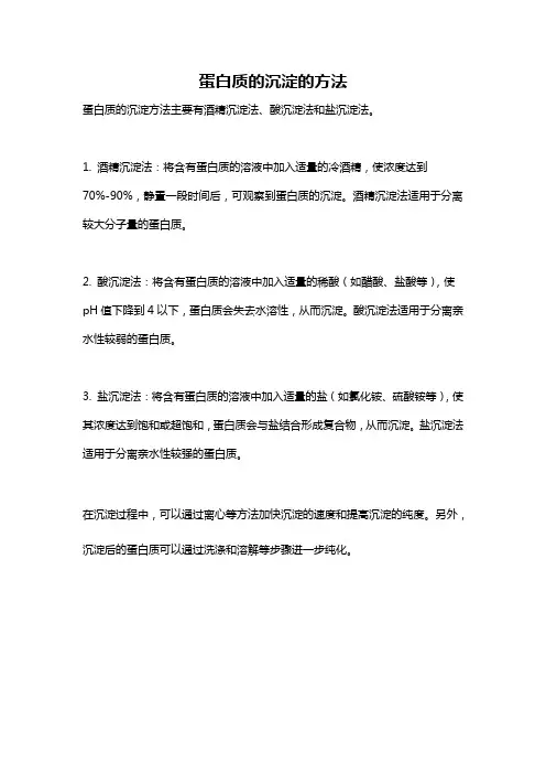
蛋白质的沉淀方法主要有酒精沉淀法、酸沉淀法和盐沉淀法。
1. 酒精沉淀法:将含有蛋白质的溶液中加入适量的冷酒精,使浓度达到
70%-90%,静置一段时间后,可观察到蛋白质的沉淀。
酒精沉淀法适用于分离较大分子量的蛋白质。
2. 酸沉淀法:将含有蛋白质的溶液中加入适量的稀酸(如醋酸、盐酸等),使pH值下降到4以下,蛋白质会失去水溶性,从而沉淀。
酸沉淀法适用于分离亲水性较弱的蛋白质。
3. 盐沉淀法:将含有蛋白质的溶液中加入适量的盐(如氯化铵、硫酸铵等),使其浓度达到饱和或超饱和,蛋白质会与盐结合形成复合物,从而沉淀。
盐沉淀法适用于分离亲水性较强的蛋白质。
在沉淀过程中,可以通过离心等方法加快沉淀的速度和提高沉淀的纯度。
另外,沉淀后的蛋白质可以通过洗涤和溶解等步骤进一步纯化。
四种蛋白纯化方法
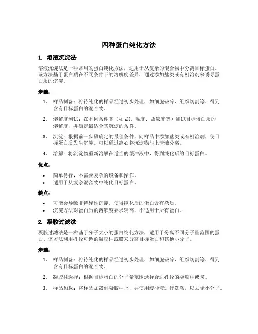
四种蛋白纯化方法1. 溶液沉淀法溶液沉淀法是一种常用的蛋白纯化方法,适用于从复杂的混合物中分离目标蛋白。
该方法基于蛋白质在不同条件下的溶解度差异,通过添加盐类或有机溶剂来诱导蛋白质的沉淀。
步骤:1.样品制备:将待纯化的样品经过初步处理,如细胞破碎、组织切割等,得到含有目标蛋白的混合物。
2.溶解度测试:在不同条件下(如pH、温度、盐浓度等)测试目标蛋白质的溶解度,并确定最适合其沉淀的条件。
3.沉淀:根据前一步骤确定的最佳条件,向样品中添加盐类或有机溶剂,使目标蛋白质发生沉淀。
可以通过离心将沉淀物与上清液分离。
4.溶解:将沉淀物重新溶解在适当的缓冲液中,得到纯化后的目标蛋白。
优点:•简单易行,不需要复杂的设备和操作。
•适用于从复杂混合物中纯化目标蛋白。
缺点:•可能会导致非特异性沉淀,使得纯化后的蛋白含有杂质。
•沉淀方法对蛋白质的溶解度要求较高,不适用于所有蛋白。
2. 凝胶过滤法凝胶过滤法是一种基于分子大小的蛋白纯化方法,适用于分离不同分子量范围的蛋白。
该方法利用孔径可调的凝胶柱或膜来分离目标蛋白和其他小分子。
步骤:1.样品制备:将待纯化的样品经过初步处理,如细胞破碎、组织切割等,得到含有目标蛋白的混合物。
2.凝胶柱选择:根据目标蛋白的分子量范围选择合适孔径的凝胶柱或膜。
3.样品加载:将样品加载到凝胶柱上,并使用缓冲液进行洗涤,以去除小分子。
4.蛋白洗脱:通过改变缓冲液的组成或pH值,使目标蛋白从凝胶柱上洗脱下来。
5.收集纯化蛋白:将洗脱得到的蛋白收集起来,即可得到纯化后的目标蛋白。
优点:•可以根据分子量范围选择合适的凝胶柱,实现高效分离。
•纯化后的蛋白质纯度较高。
缺点:•操作相对复杂,需要一定的专业知识和技术。
•只适用于分子量差异较大的目标蛋白。
3. 亲和层析法亲和层析法是一种基于生物分子间特异性相互作用的蛋白纯化方法,适用于富含目标蛋白的混合物。
该方法利用目标蛋白与特定配体之间的亲和力进行分离和纯化。
蛋白沉淀法
蛋白沉淀法蛋白沉淀法是一种常用的分离蛋白质的方法,其原理是利用化学反应使蛋白质沉淀至底部,从而分离出目标蛋白质。
本文将详细介绍蛋白沉淀法的原理、步骤、优缺点以及应用领域。
一、原理蛋白沉淀法的原理基于化学反应,常用的反应剂包括三氯醋酸(TCA)、硫酸铵(AS)、三硝基苯磺酸(TNBS)等。
其中,TCA法是最常用的方法之一。
TCA与蛋白质反应后,会形成一种不溶于水的复合物,从而使蛋白质沉淀至底部。
TCA法的反应方程式如下:TCA + 蛋白质→ TCA-蛋白复合物二、步骤蛋白沉淀法的步骤通常包括以下几个步骤:1. 样品制备:将待分离的样品加入适量的缓冲液中,使其pH值在7左右。
2. 加入反应剂:将反应剂加入样品中,通常加入的量为样品体积的1/10至1/5。
3. 沉淀:将反应液在4℃下静置30分钟至1小时,使蛋白质充分沉淀至底部。
4. 洗涤:用冷乙醇或冷醚洗涤沉淀,去除残余的反应剂和其他杂质。
5. 脱水:将沉淀放入干燥器中,用低温低压的方式将水分脱除。
6. 重溶:用适量的缓冲液将沉淀重溶,得到目标蛋白质。
三、优缺点1. 优点:蛋白沉淀法操作简单,成本低廉,适用于大规模分离蛋白质。
此外,该方法还可以去除大量的杂质和非蛋白质物质。
2. 缺点:蛋白沉淀法的选择性不够高,可能会将多种蛋白质沉淀至底部。
此外,该方法会对蛋白质的结构和功能产生一定的影响,使得蛋白质的活性降低。
四、应用领域蛋白沉淀法广泛应用于生物学、生化学、医学等领域。
其中,最常见的应用包括:1. 分离纯化蛋白质:蛋白沉淀法可以将目标蛋白质从复杂的混合物中分离出来,得到较为纯净的蛋白质样品。
2. 检测蛋白质含量:蛋白沉淀法可以用于检测样品中蛋白质的含量,并进行定量分析。
3. 蛋白质结构研究:蛋白沉淀法可以用于分离蛋白质的亚单位,从而研究蛋白质的结构和功能。
总之,蛋白沉淀法是一种常用的分离蛋白质的方法,其原理简单,操作方便,适用于大规模分离蛋白质。
但是,由于其选择性不够高,会对蛋白质的结构和功能产生一定的影响,因此在具体应用时需谨慎选择。
蛋白质沉淀
蛋白质沉淀(Protein Precipitation)浓缩方法原理及详细解析在生化制备中,沉淀主要用于浓缩目的,或用于除去留在液相或沉淀在固相中的非必要成分。
在生化制备中常用的有以下几种沉淀方法和沉淀剂:1.盐析法多用于各种蛋白质和酶的分离纯化。
2.有机溶剂沉淀法多用于生物小分子、多糖及核酸产品的分离纯化,有时也用于蛋白质沉淀。
3.等电点沉淀法用于氨基酸、蛋白质及其它两性物质的沉淀。
但此法单独应用较少,多与其它方法结合使用。
4.非离子多聚体沉淀法用于分离生物大分子。
5.生成盐复合物沉淀用于多种化合物,特别是小分子物质的沉淀。
6.热变性及酸碱变性沉淀法用于选择性的除去某些不耐热及在一定PH值下易变性的杂蛋白。
第一节盐析法一般来说,所有固体溶质都可以在溶液中加入中性盐而沉淀析出,这一过程叫盐析。
在生化制备中,许多物质都可以用盐析法进行沉淀分离,如蛋白质、多肽、多糖、核酸等,其中以蛋白质沉淀最为常见,特别是在粗提阶段。
盐析法分为两类,第一类叫Ks分段盐析法,在一定PH和温度下通过改变离子强度实现,用于早期的粗提液;第二种叫Kb分段盐析法,在一定离子强度下通过改变PH和温度来实现,用于后期进一步分离纯化和结晶。
一.影响盐析的若干因素1.蛋白质浓度高浓度蛋白溶液可以节约盐的用量,但许多蛋白质的b 和Ks常数十分接近,若蛋白浓度过高,会发生严重的共沉淀作用;在低浓度蛋白质溶液中盐析,所用的盐量较多,而共沉淀作用比较少,因此需要在两者之间进行适当选择。
用于分步分离提纯时,宁可选择稀一些的蛋白质溶液,多加一点中性盐,使共沉淀作用减至最低限度。
一般认为2.5%-3.0%的蛋白质浓度比较适中。
2.离子强度和类型一般说来,离子强度越大,蛋白质的溶解度越低。
在进行分离的时候,一般从低离子强度到高离子强度顺次进行。
每一组分被盐析出来后,经过过滤或冷冻离心收集,再在溶液中逐渐提高中性盐的饱和度,使另一种蛋白质组分盐析出来。
沉淀蛋白质的常用方法
沉淀蛋白质的常用方法(TCA、乙醇、丙酮沉淀蛋白操作步骤)2010-08-18 15:19TCA-DOCFor precipitation of very low protein concentration1) To one volume of protein solution, add 1/100 vol. of 2% DOC (Na deoxy cholate, detergent).2) Vortex and let sit for 30min at 4oC.3) Add 1/10 of Trichloroacetic acid (TCA) 100% vortex and let sit ON at 4oC (preparation of 100% TCA: 454ml H2O/kg TCA. Maintain in dark bottle at 4oC.Be careful, use gloves!!!).4) Spin 15min 4oC in microfuge at maximum speed (15000g). Carefully discharge supernatant and retain the pellet: dry tube by inversion on tissue paper (pellet may be difficult to see). [OPTION: Wash pellet twice repellet samples 5min at full speed between washes].5) Dry samples under vaccum (speed vac) or dry air. For PAGE-SDS, resuspend samples in a minimal volume of sample buffer. (The presence of some TCA can give a yellow colour as a consequence of the acidification of the sample buffer ; titrate with 1N NaOH or 1M TrisHCl pH8.5 to obtain the normal blue sample buffer colour.)Normal TCATo eliminate TCA soluble interferences and protein concentration1) To a sample of protein solution add Trichloroacetic acid (TCA) 100% to get 13% final concentration. Mix and keep 5min –20oC and then 15min 4oC; or longer time at 4oC without the –20oC step for lower protein concentration. Suggestion: leave ON if the protein concentration is very low.(preparation of 100% TCA: 454ml H2O/kg TCA. Maintain in dark bottle at 4oC.Be careful, use gloves!!!).2) Spin 15min 4oC in microfuge at maximum speed (15000g). Carefully discharge supernatant and retain the pellet: dry tube by inversion on tissue paper (pellet may be difficult to see).3) For PAGE-SDS, resuspend samples in a minimal volume of sample buffer. (The presence of some TCA can give a yellow colour as a consequence of the acidification of the sample buffer ; titrate with 1N NaOH or 1M TrisHCl pH8.5 to obtain the normal blue sample buffer colour.)Acetone PrecipitationTo eliminate acetone soluble interferences and protein concentration1) Add 1 volume of protein solution to 4 volumes of cold acetone. Mix and keep at least 20min –20oC. (Suggestion: leave ON if the protein concentration is very low).2) Spin 15min 4oC in microfuge at maximum speed (15000g). Carefully discharge supernatant and retain the pellet: dry tube by inversion on tissue paper (pellet may be difficult to see).3) Dry samples under vaccum (speed-vac) or dry air to eliminate any acetone residue (smell tubes). For PAGE-SDS, resuspend samples in a minimal volume of sample buffer.Ethanol PrecipitationUseful method to concentrate proteins and removal of Guanidine Hydrochloride before PAGE-SDS1) Add to 1 volume of protein solution 9 volumes of cold Ethanol 100%. Mix and keep at least 10min.at –20oC. (Suggestion: leave ON).2) Spin 15min 4oC in microcentrifuge at maximum speed (15000g). Carefully discharge supernatant and retain the pellet: dry tube by inversion on tissue paper (pellet may be difficult to see).3) Wash pellet with 90% cold ethanol (keep at –20oC). Vortex and repellet samples 5min at full speed.4) Dry samples under vaccum (speed vac) or dry air to eliminate any ethanol residue (smell tubes). For PAGE-SDS, resuspend samples in a minimal volume of sample buffer.TCA-DOC/AcetoneUseful method to concentrate proteins and remove acetone and TCA soluble interferences1. To one volume of protein solution add 2% Na deoxycholate (DOC) to 0.02% final (for 100 μl sample, add 1 μl 2% DOC).2. Mix and keep at room temperature for at least 15 min.3. 100% trichloroacetic acid (TCA) to get 10% final concentration (preparation of 100% TCA: 454ml H2O/kg TCA. Maintain in dark bottle at 4oC.Be careful, use gloves!!!).4. Mix and keep at room temperature for at least 1 hour.5. Spin at 4oC for 10 min, remove supernatant and retain the pellet. Dry tube by inversion on tissue paper.6. Add 200 μl of ice cold acetone to TCA pellet.7. Mix and keep on ice for at least 15 min.8. Spin at 4oC for 10 min in microcentrifuge at maximum speed.9. Remove supernatant as before (5), dry air pellet to eliminate anyacetone residue (smell tubes). For PAGE-SDS, resuspend samples in a minimal volume of sample buffer.10. (The presence of some TCA can give a yellow colour as a consequence of the acidification of the sample buffer ; titrate with 1N NaOH or 1M TrisHCl pH8.5 to obtain the normal blue sample buffer colour.)Acidified Acetone/MethanolUseful method to remove acetone and methanol soluble interferences like SDS before IEF1) Prepare acidified acetone: 120ml acetone + 10μl H Cl (1mM final concentration).2) Prepare precipitation reagent: Mix equal volumes of acidified acetone and methanol and keep at -20oC.3) To one volume of protein solution add 4 volumes of cold precipitation reagent. Mix and keep ON at -20oC.4) Spin 15min 4oC in microfuge at maximum speed (15000g). Carefully discharge supernatant and retain the pellet: dry tube by inversion on tissue paper (pellet may be difficult to see).5) Dry samples under vaccum (speed-vac) or dry air to eliminate any acetone or methanol residue (smell tubes).TCA-Ethanol PrecipitationUseful method to concentrate proteins and removal of Guanidine Hydrochloride before PAGE-SDS1) Dilute 10-25μl samples to 100μl with H2OAdd 100μl of 20% trichloroacetic acid (TCA) and mix (prepa ration of 100% TCA: 454ml H2O/kg TCA. Maintain in dark bottle at 4oC.Be careful, use gloves!!!).2) Leave in ice for 20min. Spin at 4oC for 15 min in microcentrifuge at maximum speed.3) Carefully discharge supernatant and retain the pellet: dry tube by inversion on tissue (pellet may be difficult to see).4) Wash pellet with 100μl ice-cold ethanol, dry and resuspend in sample buffer.5) In case there are traces of GuHCl present, samples should be loaded immediately after boiling for 7 min at 95°C6) (The presence of some TCA can give a yellow colour as a consequence of the acidification of the sample buffer ; titrate with 1N NaOH or 1M TrisHCl pH8.5 to obtain the normal blue sample buffer colour.)PAGE prepTM Protein Clean-up and Enrichment Kit - PIERCEThe PAGE prep? Kit enables removal of many chemicals that interfere with SDS-PAGE analysis: guanidine, ammonium sulfate, other common salts, acids and bases, detergents, dyes, DNA, RNA, and lipids.PIERCE: #26800 - PAGE prepTM Protein Clean-up and Enrichment Kit (pdf)Chloroform Methanol PrecipitationUseful method for Removal of salt and detergents1) To sample of starting volume 100 ul2) Add 400 ul methanol3) Vortex well4) Add 100 ul chloroform5) Vortex6) Add 300 ul H2O7) Vortex8) Spin 1 minute @ 14,0000 g9) Remove top aqueous layer (protein is between layers)10) Add 400 ul methanol11) Vortex12) Spin 2 minutes @ 14,000 g13) Remove as much MeOH as possible without disturbing pellet14) Speed-Vac to dryness15) Bring up in 2X sample buffer for PAGEReference: Wessel, D. and Flugge, U. I. Anal. Biochem. (1984) 138, 141-143哈哈,我做过这个论文哈!1. 配胶缓冲液系统对电泳的影响?在SDS-PAGE不连续电泳中,制胶缓冲液使用的是Tris-HCL缓冲系统,浓缩胶是pH6.7,分离胶pH8.9;而电泳缓冲液使用的Tris-甘氨酸缓冲系统。
沉淀蛋白质的常用方法(TCA、乙醇、丙酮沉淀蛋白操作步骤)
沉淀蛋白质的常用方法(TCA、乙醇、丙酮沉淀蛋白操作步骤)TCA-DOCFor precipitation of very low protein concentration1) To one volume of protein solution, add 1/100 vol. of 2% DOC (Na deoxycholate, detergent).2) Vortex and let sit for 30min at 4oC.3) Add 1/10 of Trichloroacetic acid (TCA) 100% vortex and let sit ON at 4oC (preparation of 100% TCA: 454ml H2O/kg TCA. Maintain in dark bottleat 4oC.Be4) Spin 15min 4oC in microfuge at maximum speed (15000g). Carefully discharge supernatant and retain the pellet: dry tube by inversion on tissue paper (pellet may be difficult to see). [OPTION: Wash pellet twice with one volume of cold acetone (acetone keep at –20oC). Vortex and repellet samples 5min at full speed between washes].5) Dry samples under vaccum (speed vac) or dry air. For PAGE-SDS, resuspend samples in a minimal volume of sample buffer. (The presence of some TCA can give a yellow colour as a consequence of the acidification of the sample buffer ; titrate with 1N NaOH or 1M TrisHCl pH8.5 to obtain the normal blue sample buffer colour.)Normal TCATo eliminate TCA soluble interferences and protein concentration1) To a sample of protein solution add Trichloroacetic acid (TCA) 100% to get 13% final concentration. Mix and keep 5min –20oC and then 15min 4oC; or longer time at 4oC without the –20oC step for lower protein concentration. Suggestion: leave ON if the protein concentration is very low.(preparation of 100% TCA: 454ml H2O/kg TCA. Maintain in dark bottleat 4oC.Be2) Spin 15min 4oC in microfuge at maximum speed (15000g). Carefully discharge supernatant and retain the pellet: dry tube by inversion on tissue paper (pellet may be difficult to see).3) For PAGE-SDS, resuspend samples in a minimal volume of sample buffer. (The presence of some TCA can give a yellow colour as a consequence of the acidification of the sample buffer ; titrate with 1N NaOH or 1M TrisHCl pH8.5 to obtain the normal blue sample buffer colour.)Acetone PrecipitationTo eliminate acetone soluble interferences and protein concentration1) Add to 1 volume of protein solution 4 volumes of cold acetone. Mix and keep at least 20min –20oC. (Suggestion: leave ON if the protein concentration is very low).2) Spin 15min 4oC in microfuge at maximum speed (15000g). Carefully discharge supernatant and retain the pellet: dry tube by inversion on tissue paper (pellet may be difficult to see).3) Dry samples under vaccum (speed-vac) or dry air to eliminate any acetone residue (smell tubes). For PAGE-SDS, resuspend samples in a minimal volume of sample buffer.Ethanol PrecipitationUseful method to concentrate proteins and removal of Guanidine Hydrochloride before PAGE-SDS1) Add to 1 volume of protein solution 9 volumes of cold Ethanol 100%. Mix and keep at least 10min.at –20oC. (Suggestion: leave ON).2) Spin 15min 4oC in microcentrifuge at maximum speed (15000g). Carefully discharge supernatant and retain the pellet: dry tube by inversion on tissue paper (pellet may be difficult to see).3) Wash pellet with 90% cold ethanol (keep at –20oC). Vortex and repellet samples5min at full speed.4) Dry samples under vaccum (speed vac) or dry air to eliminate any ethanol residue (smell tubes). For PAGE-SDS, resuspend samples in a minimal volume of sample buffer.TCA-DOC/AcetoneUseful method to concentrate proteins and remove acetone and TCA soluble interferences1. To one volume of protein solution add 2% Na deoxycholate (DOC) to 0.02% final (for 100 μl sample, add 1 μl 2% DOC).2. Mix and keep at room temperature for at least 15 min.3. 100% trichloroacetic acid (TCA) to get 10% final concentration (preparation of 100% TCA: 454ml H2O/kg TCA. Maintain in dark bottleat 4oC.Be careful, use gloves).4. Mix and keep at room temperature for at least 1 hour.5. Spin at 4oC for 10 min, remove supernatant and retain the pellet. Dry tube by inversion on tissue paper.6. Add 200 μl of ice cold acetone to TCA pellet.7. Mix and keep on ice for at least 15 min.8. Spin at 4oC for 10 min in microcentrifuge at maximum speed.9. Remove supernatant as before (5), dry air pellet to eliminate any acetone residue (smell tubes). For PAGE-SDS, resuspend samples in a minimal volume of sample buffer. 10. (The presence of some TCA can give a yellow colour as a consequence of the acidification of the sample buffer ; titrate with 1N NaOH or 1M TrisHCl pH8.5 to obtain the normal blue sample buffer colour.)Acidified Acetone/MethanolUseful method to remove acetone and methanol soluble interferences like SDS before IEF1) Prepare acidified acetone: 120ml acetone + 10μl HCl (1mM final concentration).2) Prepare precipitation reagent: Mix equal volumes of acidified acetone and methanol and keep at -20oC.3) To one volume of protein solution add 4 volumes of cold precipitation reagent. Mix and keep ON at -20oC.4) Spin 15min 4oC in microfuge at maximum speed (15000g). Carefully discharge supernatant and retain the pellet: dry tube by inversion on tissue paper (pellet may be difficult to see).5) Dry samples under vaccum (speed-vac) or dry air to eliminate any acetone or methanol residue (smell tubes).TCA-Ethanol PrecipitationUseful method to concentrate proteins and removal of Guanidine Hydrochloride before PAGE-SDS1) Dilute 10-25μl samples to 100μl with H2OAdd 100μl of 20% trichloroacetic acid (TCA) and mix (preparation of 100% TCA: 454ml H2O/kg TCA. Maintain in dark bottleat 4oC.Be careful, use gloves).2) Leave in ice for 20min. Spin at 4oC for 15 min in microcentrifuge at maximum speed.3) Carefully discharge supernatant and retain the pellet: dry tube by inversion on tissue (pellet may be difficult to see).4) Wash pellet with 100μl ice-cold ethanol, dry and resuspend in sample buffer.5) In case there are traces of GuHCl present, samples should be loaded immediately after boiling for 7 min at 95°C6) (The presence of some TCA can give a yellow colour as a consequence of the acidification of the sample buffer ; titrate with 1N NaOH or 1M TrisHCl pH8.5 to obtain the normal blue sample buffer colour.)PAGE prep TM Protein Clean-up and Enrichment Kit - PIERCEThe PAGEprep? Kit enables removal of many chemicals that interfere with SDS-PAGE analysis: guanidine, ammonium sulfate, other common salts, acids and bases, detergents, dyes, DNA, RNA, and lipids.PIERCE: #26800 - PAGE prep TM Protein Clean-up and Enrichment Kit (pdf) Chloroform Methanol PrecipitationUseful method for Removal of salt and detergents1) To sample of starting volume 100 ul2) Add 400 ul methanol3) Vortex well4) Add 100 ul chloroform5) Vortex6) Add 300 ul H2O7) Vortex8) Spin 1 minute @ 14,0000 g9) Remove top aqueous layer (protein is between layers)10) Add 400 ul methanol11) Vortex12) Spin 2 minutes @ 14,000 g13) Remove as much MeOH as possible without disturbing pellet14) Speed-Vac to dryness15) Bring up in 2X sample buffer for PAGEReference: Wessel, D. and Flugge, U. I. Anal. Biochem. (1984) 138, 141-143蛋白质浓缩——方法很全1130徐炉李2011-05-28 14:35楼主蛋白质浓缩——方法很全- 丁香园论坛-医学/药学/生命科学论坛蛋白质浓缩方法总结一个简便的方法你可以试试:找一透析袋,底部扎紧,袋口扎一去底的塑料或玻璃试管,将待浓缩的液体从管口灌入透析袋中,将整个装置挂在冰箱中,或者用电风扇吹,液体干后可再继续加入,直至样品浓缩至所需体积。
- 1、下载文档前请自行甄别文档内容的完整性,平台不提供额外的编辑、内容补充、找答案等附加服务。
- 2、"仅部分预览"的文档,不可在线预览部分如存在完整性等问题,可反馈申请退款(可完整预览的文档不适用该条件!)。
- 3、如文档侵犯您的权益,请联系客服反馈,我们会尽快为您处理(人工客服工作时间:9:00-18:30)。
P13三、5 、常用蛋白质沉淀方法有哪些?列举沉淀应用的实例
蛋白质分子凝聚从溶液中析出的现象称为蛋白质沉淀(precipitation),变性蛋白质一般易于沉淀,但也可不变性而使蛋白质沉淀,在一定条件下,变性的蛋白质也可不发生沉淀。
常用蛋白质沉淀的方法有:
(一)盐析(Salting Out)
在蛋白质溶液中加入大量的中性盐以破坏蛋白质的胶体稳定性而使其析出,这种方法称为盐析。
常用的中性盐有硫酸铵、硫酸钠、氯化钠等。
例如用半饱和的硫酸铵来沉淀出血清中的球蛋白,饱和硫酸铵可以使血清中的白蛋白、球蛋白都沉淀出来,盐析沉淀的蛋白质,经透析除盐,仍保证蛋白质的活性。
(二)重金属盐沉淀蛋白质
蛋白质可以与重金属离子如汞、铅、铜、银等结合成盐沉淀。
重金属沉淀的蛋白质常是变性的,但若在低温条件下,并控制重金属离子浓度,也可用于分离制备不变性的蛋白质。
如临床上利用蛋白质能与重金属盐结合的这种性质,抢救误服重金属盐中毒的病人,给病人口服大量蛋白质,然后用催吐剂将结合的重金属盐呕吐出来解毒。
(三)生物碱试剂以及某些酸类沉淀蛋白质
蛋白质又可与生物碱试剂(如苦味酸、钨酸、鞣酸)以及某些酸(如三氯醋酸、过氯酸、硝酸)结合成不溶性的盐沉淀。
如临床血液化学分析时常利用此原理除去血液中的蛋白质,此类沉淀反应也可用于检验尿中蛋白质。
(四)有机溶剂沉淀蛋白质
可与水混合的有机溶剂,如酒精、甲醇、丙酮等,对水的亲和力很大,能破坏蛋白质颗粒的水化膜,在等电点时使蛋白质沉淀。
在常温下,有机溶剂沉淀蛋白质往往引起变性。
例如酒精消毒灭菌就是如此,但若在低温条件下,则变性进行较缓慢,可用于分离制备各种血浆蛋白质。