恶性淋巴瘤疗效评价标准
恶性淋巴瘤疗效评价的修订标准

J OURNAL OF C LINICAL O NCOLOGY恶性淋巴瘤疗效评价的修订标准Bruce D. Cheson, Beate Pfistner, Malik E. Juweid, Randy D. Gascoyne, Lena Specht, Sandra J. Horning, Bertrand Coiffier, Richard I. Fisher, Anton Hagenbeek, Emanuele Zucca, Steven T. Rosen, Sigrid Stroobants, T. Andrew Lister, Richard T. Hoppe, Martin Dreyling, Kensei Tobinai, Julie M. Vose, Joseph M. Connors, Massimo Federico, and Volker Diehl摘 要目的有必要制定规范的疗效标准以用于各个临床试验的解释和对照,以及管理机构对治疗新药的审批。
方法国际工作组疗效标准(Cheson et al, J Clin Oncol 17:1244, 1999)一直被广泛采用,但由于其明显的局限性以及[18F]脱氧葡萄糖正电子发射断层成像(PET )、免疫组化(IHC )与流式细胞学技术的推广应用,需要重新评估该标准。
因此,开展国际性协调议案以进行建议的更新。
结果新指南将PET 、IHC 和流式细胞学技术纳入非霍奇金与霍奇金淋巴瘤的疗效评估。
同时规范了终点的定义。
结论希望这些指南得到各研究组、制药和生物技术公司的广泛采纳,并有助于管理机构对新药和更有效治疗方法的审批,由此改善淋巴瘤患者的预后。
J Clin Oncol 25:579-586. © 2007 by American Society of Clinical Oncology规范的疗效标准为临床试验提供了统一的终点,这使得各研究间具有可比性,有助于明确更有效的治疗方法和管理机构对新药的审批。
肿瘤治疗疗效评价标准
肿瘤治疗疗效评价标准一、肿瘤大小变化1.完全缓解(CR):所有目标病灶消失,无新病灶出现,肿瘤标志物正常。
2.部分缓解(PR):目标病灶体积减小,至少缩小50%,且无新病灶出现,肿瘤标志物正常。
3.稳定(SD):目标病灶体积无变化或缩小不足50%,且无新病灶出现,肿瘤标志物正常。
4.进展(PD):目标病灶体积增大,或出现新病灶,或肿瘤标志物异常。
二、生存期延长生存期是指从开始治疗到死亡的时间。
评价生存期延长的标准可采用总生存期(OS)和无病生存期(DFS)等指标。
OS是指从开始治疗到死亡的时间,DFS是指从开始治疗到疾病复发或进展的时间。
三、症状改善1.完全缓解(CR):所有症状完全消失,且持续时间超过2周。
2.部分缓解(PR):症状明显减轻,且持续时间超过2周。
3.稳定(SD):症状无变化或略有减轻,但未达到PR标准。
4.进展(PD):症状加重或出现新症状。
四、血液学指标血液学指标主要包括血常规、肝肾功能等。
在治疗前后进行血液学检查,以评估患者的一般状况和不良反应情况。
对于某些特定类型的肿瘤,血液学指标也可作为疗效评价的参考。
五、病理学改变对于某些肿瘤,病理学改变是疗效评价的重要指标。
例如,对于淋巴瘤等血液系统肿瘤,病理学改变包括肿瘤细胞坏死、细胞凋亡等反应。
可根据病理学检查的结果进行疗效评价。
六、安全性评估安全性评估是评价抗肿瘤治疗的另一个重要方面。
它包括对治疗过程中出现的所有不良反应进行记录和评估。
不良反应可根据NCI-CTCA E 等标准进行分级和评估。
安全性评估有助于及时发现和治疗不良反应,提高患者的生活质量和生存期。
七、生活质量评估生活质量评估是抗肿瘤治疗疗效评价的另一个重要方面。
它包括对患者的身体状况、心理状况、社会功能等方面进行全面的评估。
生活质量评估有助于了解患者的生活质量状况和抗肿瘤治疗对患者的影响程度,为制定更加全面的治疗方案提供参考。
2019年恶性淋巴瘤疗效评价标准-PPT课件
Use of Positron Emission Tomography for Response Assessment of Lymphoma: Consensus of the Imaging Subcommittee of International Harmonization Project in Lymphoma
• Allowing for comparisons among studies
• Facilitating the identification of more effective therapies
The widely used IWG criteria for response assessment of lymphoma are based predominantly on CT.
It became clear that the International Working Group criteria warranted revision, because of identified limitations and the increased use of :
1. [18F] fluorodeoxyglucose-positron emission tomography (PET), 2. immunohistochemistry (IHC), 3. flow cytometry, 4. molecular biology
“REVISED RESPONSE CRITERIA FOR MALIGNANT LYMPHOMA”
淋巴瘤疗效评价标准

淋巴瘤疗效评价标准
淋巴瘤是一种常见的恶性肿瘤,由淋巴细胞恶性增殖引起。
在淋巴瘤的治疗过程中,评价疗效是非常重要的,可以帮助医生更好地调整治疗方案,提高患者的治疗效果。
淋巴瘤的疗效评价标准是根据患者的临床表现、影像学检查和实验室检查等综合评定的。
下面将介绍淋巴瘤疗效评价的标准及其相关内容。
一、临床表现。
1. 症状改善,包括发热、盗汗、体重减轻、淋巴结肿大等症状的减轻或消失。
2. 肿瘤负荷减轻,淋巴瘤患者的肿瘤负荷可以通过体格检查和淋巴结活检等手段来评价,肿瘤负荷的减轻通常意味着治疗效果好。
二、影像学检查。
1. CT或MRI检查,通过CT或MRI检查可以观察淋巴瘤病灶的大小、数量和分布情况,评估治疗效果。
2. PET-CT检查,PET-CT检查可以更准确地评估淋巴瘤病灶的活动情况,对疗效评价具有重要意义。
三、实验室检查。
1. 血液学检查,包括血常规、肝肾功能、电解质等指标,可以反映淋巴瘤患者的整体健康状况和治疗效果。
2. 淋巴细胞亚群分析,淋巴瘤患者的淋巴细胞亚群分析可以帮助评估免疫功能和疾病活动情况。
综上所述,淋巴瘤的疗效评价需要综合临床表现、影像学检查和实验室检查等多方面的信息。
在评价疗效时,需要注意病情的动态变化,及时调整治疗方案,以
提高治疗效果。
同时,淋巴瘤的疗效评价标准也在不断发展和完善中,希望未来可以有更准确、可靠的评价指标,为淋巴瘤患者提供更好的治疗和管理。
2007年恶性淋巴瘤疗效评价标准电子教案
PET
• False-positive: - Thymic hyperplasia - Infection - Inflammation - Sarcoidosis - Brown fat Other causes of false-positive scans should be ruled out.
Whole-body acquisition using a PET or PET/CT system should encompass at least the region between the base of the skull and themed thigh, and can be acquired in either two- or three-dimensional mode.
Whole-body imaging should begin 50-70 minutes after the administration of FDG.
The reconstructed PET or PET/CT images must be displayed on a computer workstation so that transaxial, sagittal, and coronal images can be viewed simultaneously.
皮肤淋巴瘤疗效评价标准2023

皮肤淋巴瘤是一种罕见的非霍奇金淋巴瘤,通常发生在皮肤和表皮下组织。
由于其症状多样化,治疗方法也多种多样,因此对其疗效的评价标准显得尤为重要。
本文将从多个角度来探讨皮肤淋巴瘤疗效评价标准,并对2023年的相关标准进行深入分析。
1. 临床表现评价皮肤淋巴瘤的临床表现多样,包括皮肤色素沉着、斑块、结节、溃疡等症状。
评价疗效时需要综合考虑病变部位、大小、颜色、形态等因素,以及患者的主观感受,对病变的缓解程度进行评价。
2. 影像学检查评价影像学检查对于评估皮肤淋巴瘤的疗效至关重要。
通常采用超声检查、CT、MRI等技术来观察病变的范围、深度、浸润情况等,从而判断治疗的效果。
3. 病理学检查评价病理学检查是确诊皮肤淋巴瘤的关键。
在评价疗效时,可以通过活检等方式获取组织样本,观察治疗后病变的缓解程度、组织的恢复情况等。
4. 免疫学指标评价免疫学指标在评价皮肤淋巴瘤的疗效中起着重要作用。
对于免疫功能的恢复、炎症因子的水平、肿瘤标志物等进行监测和评价,可以更加客观地了解治疗的效果。
5. 患者生活质量评价治疗的最终目的是改善患者的生活质量。
在评价皮肤淋巴瘤的疗效时,需要考虑患者的生活状态、心理状态、社会功能等方面,通过问卷调查、心理评估等方式进行客观评价。
2023年的皮肤淋巴瘤疗效评价标准应该更加科学、客观、全面。
具体来说,可以考虑以下几个方面的改进:1. 引入新技术随着医学技术的不断进步,可以引入更先进的影像学技术、病理学检查技术、免疫学指标监测技术等,以提高对皮肤淋巴瘤疗效的评价水平。
2. 建立标准化评价体系制定统一的评价指标和评分标准,使得皮肤淋巴瘤的疗效评价更具客观性和标准化,不同医院、不同医生之间的评价结果更具可比性。
3. 结合患者主观感受在评价疗效时,应该更加注重患者的主观感受,比如疼痛程度、舒适度、生活质量等方面,为医生提供更全面的信息,从而更好地指导治疗方案的调整。
4. 加强随访管理对于皮肤淋巴瘤患者,应该建立健全的随访管理制度,对治疗效果进行长期跟踪,从而能更加客观地评价治疗的效果。
2019年恶性淋巴瘤疗效评价标准精品文档
Response duration CR, CRu, PR
Time to next treatment
All patients
Cause-specific death All patients
Definition Death from any cause
Point of Measurement
Entry onto trial
It became clear that the International Working Group criteria warranted revision, because of identified limitations and the increased use of :
1. [18F] fluorodeoxyglucose-positron emission tomography (PET),
A recently developed integrated PET/CT system, which combines a PET camera and CT scanner in a single session, has overcome these drawbacks by providing both anatomical and functional imaging at the same position. PET/CT has become the new standard approach to imaging in the diagnosis and management of many cancer patients.
“REVISED RESPONSE CRITERIA FOR MALIGNANT LYMPHOMA”
淋巴瘤疗效评估标准
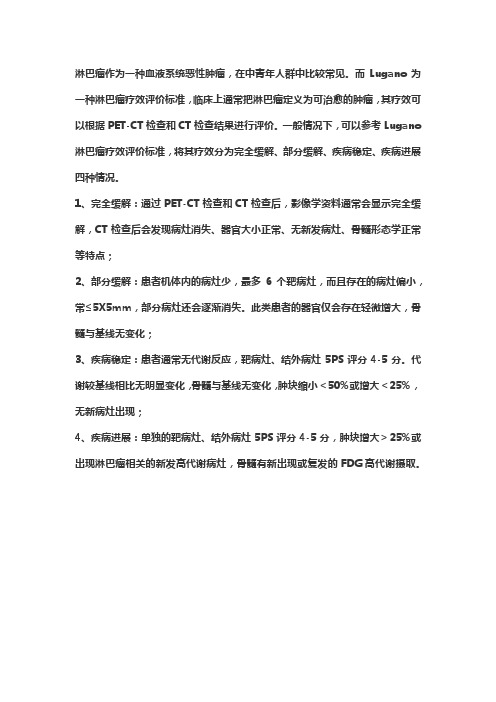
淋巴瘤作为一种血液系统恶性肿瘤,在中青年人群中比较常见。
而Lugano为一种淋巴瘤疗效评价标准,临床上通常把淋巴瘤定义为可治愈的肿瘤,其疗效可以根据PET-CT检查和CT检查结果进行评价。
一般情况下,可以参考Lugano 淋巴瘤疗效评价标准,将其疗效分为完全缓解、部分缓解、疾病稳定、疾病进展四种情况。
1、完全缓解:通过PET-CT检查和CT检查后,影像学资料通常会显示完全缓解,CT检查后会发现病灶消失、器官大小正常、无新发病灶、骨髓形态学正常等特点;
2、部分缓解:患者机体内的病灶少,最多6个靶病灶,而且存在的病灶偏小,常≤5X5mm,部分病灶还会逐渐消失。
此类患者的器官仅会存在轻微增大,骨髓与基线无变化;
3、疾病稳定:患者通常无代谢反应,靶病灶、结外病灶5PS评分4-5分。
代谢较基线相比无明显变化,骨髓与基线无变化,肿块缩小<50%或增大<25%,无新病灶出现;
4、疾病进展:单独的靶病灶、结外病灶5PS评分4-5分,肿块增大>25%或出现淋巴瘤相关的新发高代谢病灶,骨髓有新出现或复发的FDG高代谢摄取。
- 1、下载文档前请自行甄别文档内容的完整性,平台不提供额外的编辑、内容补充、找答案等附加服务。
- 2、"仅部分预览"的文档,不可在线预览部分如存在完整性等问题,可反馈申请退款(可完整预览的文档不适用该条件!)。
- 3、如文档侵犯您的权益,请联系客服反馈,我们会尽快为您处理(人工客服工作时间:9:00-18:30)。
PET
• False-positive: - Thymic hyperplasia - Infection - Inflammation - Sarcoidosis - Brown fat Other causes of false-positive scans should be ruled out.
• Allowing for comparisons among studies
• Facilitating the identification of more effective therapies
The widely used IWG criteria for response assessment of lymphoma are based predominantly on CT.
The Competence Network Malignant Lymphoma convened an International
Harmonization Project at which 5 subcommittees were formed:
• Response Criteria • End Points for Clinical Trials • Imaging • Clinical Features • Pathology/Biology
Use of Positron Emission Tomography for Response Assessment of Lymphoma: Consensus of the Imaging Subcommittee of International Harmonization Project in
Whole-body acquisition using a PET or PET/CT system should encompass at least the region between the base of the skull and themed thigh, and can be acquired in either two- or three-dimensional mode.
• The advantage of PET over conventional imaging techniques, such as TC or RMN, is its ability to distinguish between viable tumor and necrosis or fibrosis in residual mass(es) often present after treatment.
Response Criteria for Lymphoma
Response Category CR CRu
PR
Relapse/ progression
Physical Examination
Normal
Lymph Nodes Lymph Node Masses
Normal
Normal
Bone Marrow Normal
> 75% decrease
Normal
≥50% decrease
≥50% decrease
New or increased
Normal or indeterminate Positive Irrelevant
Irrelevant
Reappearance
Definitions of End Points for Clinical Trials
Normal
Normal
Normal
Indeterminate
Normal
Normal
Normal
Normal
Decrease in liver/spleen Enlarging liver/spleen; new sites
Normal
≥50% decrease ≥ 50% decrease
New or increased
“REVISED RESPONSE CRITERIA FOR MALIGNANT LYMPHOMA”
J Clin Oncol 25:579-586. © 2007 by American Society of Clinical Oncology
Cheson et al, J Clin Oncol 17:1244, 1999
In 1999, an International Working Group (IWG) of clinicians, radiologists, and pathologists with expertise in the evaluation and management of patients with Lymphoma published guidelines for response assessment and outcomes measurement.
Patients should have fasted for at least 4 hours before FDG injection.
Blood glucose level should not exceed 200 mg/dL at the time of FDG injection. If the blood glucose exceeds this level, the FDG-PET study should be rescheduled and an attempt made to control the blood sugar.
Lymphoma
J Clin Oncol 25:571-578. © 2007 by American Society of Clinical Oncology
PET- PET/CT
• PET using [18F]fluorodeoxyglucose (FDG, a radioactive derivative of glucose, is an advanced imaging tool, based on the increased glucose consumption of cancer cells), has emerged as a powerful functional imaging tool for staging, restaging, and response assessment of lymphomas.
It became clear that the International Working Group criteria warranted revision, because of identified limitations and the increased use of :
1. [18F] fluorodeoxyglucose-positron emission tomography (PET),
End Point Overall survival
Response Category
All patients
Event-free survival CR, CRu, PR
Progression-free survival
Disease-free survival
All patients CR, CRu
PET: 1. Increased the number of complete remission (CR)
patients, 2. Eliminated the CRu category 3. Enhanced the ability to discern the difference in
A recently developed integrated PET/CT system, which combines a PET camera and CT scanner in a single session, has overcome these drawbacks by providing both anatomical and functional imaging at the same position. PET/CT has become the new standard approach to imaging in the diagnosis and management of many cancer patients.
• False-negative: - Resolution of the equipment and technique - Variability of FDG avidity among histologic subtypes
Juweid et al. evaluated the impact of integrating PET into the IWG criteria in a retrospective study of 54 patients with diffuse large B-cell NHL who had been treated with an anthracycline-based regimen.
2. immunohistochemistry (IHC), 3. flow cytometry, 4. molecular biology
ห้องสมุดไป่ตู้
“REVISED RESPONSE CRITERIA FOR MALIGNANT LYMPHOMA”
J Clin Oncol 25:579-586. © 2007 by American Society of Clinical Oncology
Standardization of PET and CT Imaging Parameters
Patients undergoing PET imaging should receive an FDG dose of 3.5 to 8 MBq/kg of body weight, with a minimum dose of 185 MBq in adults (5 mCi) and 18.5 MBq (0.5 mCi) in children.
