
恶性淋巴瘤疗效评价标准共27页
Response Criteria for Lymphoma
Response Category CR CRu
PR
Relapse/ progression
Physical Examination
Normal
Lymph Nodes Lymph Node Masses
Normal
Normal
Bone Marrow Normal
Failure or death from any Entry onto trial cause
Disease progression or death from NHL Time to relapse
Time to relapse or progression
Entry onto trial
First documentation of response First documentation of response
It became clear that the International Working Group criteria warranted revision, because of identified limitations and the increased use of :
1. [18F] fluorodeoxyglucose-positron emission tomography (PET),
Cheson et al, J Clin Oncol 17:1244, 2019
In 2019, an International Working Group (IWG) of clinicians, radiologists, and pathologists with expertise in the evaluation and management of patients with Lymphoma published guidelines for response assessment and outcomes measurement.
Lymphoma
J Clin Oncol 25:571-578. © 2019 by American Society of Clinical Oncology
• Allowing for comparisons among studies
• Facilitating the identification of more effective therapies
The widely used IWG criteria for response assessment of lymphoma are based predominantly on CT.
The Competence Network Malignant Lymphoma convened an International
Harmonization Project at which 5 subcommittees were formed:
• Response Criteria • End Points for Clinical Trials • Imaging • Clinical Features • Pathology/Biology
> 75% decrease
Normal
≥50% decrease
≥50% decrease
New or increased
Normal or indeterminate Positive Irrelevant
Irrelevant
Reappearance
Definitions of End Points for Clinical Trials
Response duration CR, CRu, PR
Time to next treatment
All patients
Cause-specific death All patients
Definition Death from any cause
Point of Measurement
Entry onto trial
Normal
Normal
Normal
Indeterminate
Normal
Normal
Normal
Normal
Decrease in liver/spleen Enlarging liver/spleen; new sites
Normal
≥50% decrease ≥ 50% decrease
New or increased
Time when new treatment Entry onto trial is needed
Death related to NHL
Death
Standardized response criteria provide uniform end points for
clinical trials:
Use of Positron Emission Tomography for Response Assessment of Lymphoma: Consensus of the Imaging Subcommittee of International Harmonization Project in
2. immunohistochemistry (IHC), 3. flow cytometry, 4. molecular biology
“REVISED RESPONSE CRITERIA FOR MALIGNANT LYMPHOMA”
J Clin Oncol 25:579-586. © 2019 by American Society of Clinical Oncology
End Point Overall survival
Response Category
All patients
Event-free survival CR, CRu, PR
Progression-free survival
Disease-free survival
All patients CR, CRu
2019年恶性淋巴瘤疗效评价标准-PPT课件
Use of Positron Emission Tomography for Response Assessment of Lymphoma: Consensus of the Imaging Subcommittee of International Harmonization Project in Lymphoma
• Allowing for comparisons among studies
• Facilitating the identification of more effective therapies
The widely used IWG criteria for response assessment of lymphoma are based predominantly on CT.
It became clear that the International Working Group criteria warranted revision, because of identified limitations and the increased use of :
1. [18F] fluorodeoxyglucose-positron emission tomography (PET), 2. immunohistochemistry (IHC), 3. flow cytometry, 4. molecular biology
“REVISED RESPONSE CRITERIA FOR MALIGNANT LYMPHOMA”
淋巴瘤疗效评价标准

淋巴瘤疗效评价标准
淋巴瘤是一种常见的恶性肿瘤,由淋巴细胞恶性增殖引起。
在淋巴瘤的治疗过程中,评价疗效是非常重要的,可以帮助医生更好地调整治疗方案,提高患者的治疗效果。
淋巴瘤的疗效评价标准是根据患者的临床表现、影像学检查和实验室检查等综合评定的。
下面将介绍淋巴瘤疗效评价的标准及其相关内容。
一、临床表现。
1. 症状改善,包括发热、盗汗、体重减轻、淋巴结肿大等症状的减轻或消失。
2. 肿瘤负荷减轻,淋巴瘤患者的肿瘤负荷可以通过体格检查和淋巴结活检等手段来评价,肿瘤负荷的减轻通常意味着治疗效果好。
二、影像学检查。
1. CT或MRI检查,通过CT或MRI检查可以观察淋巴瘤病灶的大小、数量和分布情况,评估治疗效果。
2. PET-CT检查,PET-CT检查可以更准确地评估淋巴瘤病灶的活动情况,对疗效评价具有重要意义。
三、实验室检查。
1. 血液学检查,包括血常规、肝肾功能、电解质等指标,可以反映淋巴瘤患者的整体健康状况和治疗效果。
2. 淋巴细胞亚群分析,淋巴瘤患者的淋巴细胞亚群分析可以帮助评估免疫功能和疾病活动情况。
综上所述,淋巴瘤的疗效评价需要综合临床表现、影像学检查和实验室检查等多方面的信息。
在评价疗效时,需要注意病情的动态变化,及时调整治疗方案,以
提高治疗效果。
同时,淋巴瘤的疗效评价标准也在不断发展和完善中,希望未来可以有更准确、可靠的评价指标,为淋巴瘤患者提供更好的治疗和管理。
淋巴瘤疗效评价标准2014 英文
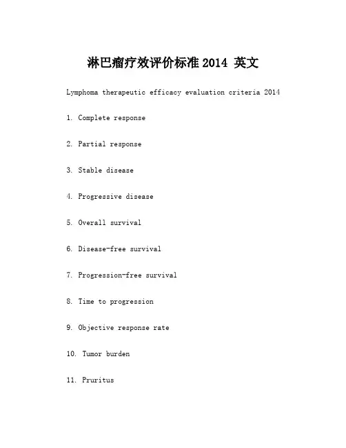
淋巴瘤疗效评价标准2014 英文Lymphoma therapeutic efficacy evaluation criteria 20141. Complete response2. Partial response3. Stable disease4. Progressive disease5. Overall survival6. Disease-free survival7. Progression-free survival8. Time to progression9. Objective response rate10. Tumor burden11. Pruritus12. Fatigue13. Anemia14. Neutropenia15. Thrombocytopenia16. Lymphopenia17. Hypersensitivity reaction18. Infusion-related reaction19. Adverse events20. Quality of life21. Biomarkers22. Tumor markers23. Lymph node involvement24. Extramedullary involvement Example sentences:1. The patient achieved a complete response after undergoing lymphoma therapy.病人通过淋巴瘤治疗后实现了完全缓解。
2. The treatment resulted in a partial response, with a decrease in tumor size.治疗导致部分缓解,肿瘤体积减小。
3. The patient's condition remained stable with no significant changes in tumor size.病人的病情保持稳定,肿瘤体积没有显著变化。
2019年恶性淋巴瘤疗效评价标准精品文档

Response duration CR, CRu, PR
Time to next treatment
All patients
Cause-specific death All patients
Definition Death from any cause
Point of Measurement
Entry onto trial
It became clear that the International Working Group criteria warranted revision, because of identified limitations and the increased use of :
1. [18F] fluorodeoxyglucose-positron emission tomography (PET),
A recently developed integrated PET/CT system, which combines a PET camera and CT scanner in a single session, has overcome these drawbacks by providing both anatomical and functional imaging at the same position. PET/CT has become the new standard approach to imaging in the diagnosis and management of many cancer patients.
“REVISED RESPONSE CRITERIA FOR MALIGNANT LYMPHOMA”
恶性肿瘤的TNM分期与疗效评估共28页

肿瘤增大为25%或更大或 大多数肿瘤增大约25% X-光片或扫描片有肿瘤
(progressive disease)
发现有新的肿瘤或发现有新的肿瘤 Nhomakorabea增加或有新的转移
实体瘤疗效评价的新标准 —RECIST
WHO标准
RECIST(2000年)
• 1985年,Warr等研究发现临床试验疗效评价的偏倚 有5%~10%是因为WHO标准对有些病灶的定义模 糊和肿瘤测量上的误差造成的。
恶性肿瘤的TNM分期与疗效评估
恶性肿瘤的TNM分期与疗效评估
TNM通用定义
• (一) 原发肿瘤(T) • ① Tx —原发肿瘤不能确定; • X 代表未知。 • ② T0 —无原发肿瘤的证据; • 0 代表没有 • ③ Tis—原位癌; • is代表 in situ原位 • ④ T1、T2、T3、T4 —原发肿瘤的体积及/或范围递增,数字越大,肿瘤累及的
• 无病生存期DFS :是指CR后直至复发的时间 或未复发任何原因死亡的时间。
• 无进展生存期PFS:是指从开始对肿瘤进行针 对性治疗直至肿瘤出现继发进展生长的时间。
肿瘤测量的示意图
A B
bidimentional a × b, unidimentional a
WHO提出的癌症疗效评估的共识
可测量的肿瘤 四星期后有确认
残存肿瘤(R)
• RX 无法评估是否存在残余肿瘤 • R0 无残存肿瘤 • R1 镜下可见残存肿瘤 • R2 肉眼可见残存肿瘤
病理学分级(G)
• ① Gx —不能确定肿瘤的分化程度; • ② G1 —高分化; • ③ G2 —中度分化; • ④ G3 —低分化; • ⑤ G4 —未分化。
TNM分期特别说明
霍奇金淋巴瘤疗效评估标准
霍奇金淋巴瘤疗效评估标准
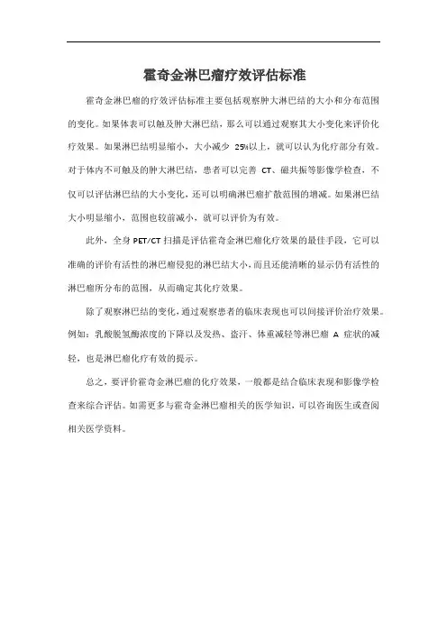
霍奇金淋巴瘤的疗效评估标准主要包括观察肿大淋巴结的大小和分布范围的变化。
如果体表可以触及肿大淋巴结,那么可以通过观察其大小变化来评价化疗效果。
如果淋巴结明显缩小,大小减少25%以上,就可以认为化疗部分有效。
对于体内不可触及的肿大淋巴结,患者可以完善CT、磁共振等影像学检查,不仅可以评估淋巴结的大小变化,还可以明确淋巴瘤扩散范围的增减。
如果淋巴结大小明显缩小,范围也较前减小,就可以评价为有效。
此外,全身PET/CT扫描是评估霍奇金淋巴瘤化疗效果的最佳手段,它可以准确的评价有活性的淋巴瘤侵犯的淋巴结大小,而且还能清晰的显示仍有活性的淋巴瘤所分布的范围,从而确定其化疗效果。
除了观察淋巴结的变化,通过观察患者的临床表现也可以间接评价治疗效果。
例如:乳酸脱氢酶浓度的下降以及发热、盗汗、体重减轻等淋巴瘤A症状的减轻,也是淋巴瘤化疗有效的提示。
总之,要评价霍奇金淋巴瘤的化疗效果,一般都是结合临床表现和影像学检查来综合评估。
如需更多与霍奇金淋巴瘤相关的医学知识,可以咨询医生或查阅相关医学资料。
淋巴瘤疗效评估标准
淋巴瘤作为一种血液系统恶性肿瘤,在中青年人群中比较常见。
而Lugano为一种淋巴瘤疗效评价标准,临床上通常把淋巴瘤定义为可治愈的肿瘤,其疗效可以根据PET-CT检查和CT检查结果进行评价。
一般情况下,可以参考Lugano 淋巴瘤疗效评价标准,将其疗效分为完全缓解、部分缓解、疾病稳定、疾病进展四种情况。
1、完全缓解:通过PET-CT检查和CT检查后,影像学资料通常会显示完全缓解,CT检查后会发现病灶消失、器官大小正常、无新发病灶、骨髓形态学正常等特点;
2、部分缓解:患者机体内的病灶少,最多6个靶病灶,而且存在的病灶偏小,常≤5X5mm,部分病灶还会逐渐消失。
此类患者的器官仅会存在轻微增大,骨髓与基线无变化;
3、疾病稳定:患者通常无代谢反应,靶病灶、结外病灶5PS评分4-5分。
代谢较基线相比无明显变化,骨髓与基线无变化,肿块缩小<50%或增大<25%,无新病灶出现;
4、疾病进展:单独的靶病灶、结外病灶5PS评分4-5分,肿块增大>25%或出现淋巴瘤相关的新发高代谢病灶,骨髓有新出现或复发的FDG高代谢摄取。
恶性淋巴瘤疗效评价标准
J Clin Oncol 25:579-586. © 2007 by American Society of Clinical Oncology
Cheson et al, J Clin Oncol 17:1244, 1999
Response Criteria for Lymphoma
Response Category CR
CRu
Physical Lymph
Examinatio Nodes
Nn ormal
Normal
Normal Normal
Normal Normal
PR
Normal Normal
Relapse/ progression
Failure or death from any cause
Entry onto trial
Disease progression or death Entry onto trial from NHL
Time to relapse
First documentation of response
Time to relapse or progression
Response duration
CR, CRu, PR
Time to next treatment All patients
Cause-specific death All patients
Definition Death from any cause
Point of Measurement Entry onto trial
Use of Positron Emission Tomography for Response Assessment of Lymphoma: Consensus of the Imaging Subcommittee of International Harmonization Project
实体肿瘤疗效评估标准
实体肿瘤临床疗效评价指标基线病灶分类可测量病灶肿瘤性病变:至少有一个不小于(仪器检测)低限的尺寸,必须准确测量;临床检验10 毫米用卡尺测量(不能用卡尺测量的记为不可测量病灶);直径大于10 mm 的病灶用CT 扫描(CT 扫描厚度≤5 毫米);20 毫米用胸部X 光检查恶性淋巴结:当用CT 扫描(CT 扫描厚度≤5 毫米):淋巴结短轴必须达到15 mm 才可将其认为是病理扩大和可测量的。
术前和后续工作中,只测量并跟踪短轴长度。
注:(不同于其它可测量病灶用最长轴作为直径,恶性肿瘤淋巴结测量时用最短轴作为直径)不可测量的病灶:最长直径小于10 毫米的小病灶或短轴为10 毫米到小于15 毫米的病理淋巴结真正不可测的病变:病理学检查确定的脑膜疾病、腹水、胸膜或心包积液、炎症乳腺疾病等(均无法用现有影像学技术测量)。
骨病为不可测量的疾病,软组织成份可采用CT 或MRI 评价,且符合基线时可评价的定义者除外。
位于先前照射区或受到其他局部治疗的部位的肿瘤病灶,通常不被视为可测量的,除非已证明病变仍在继续。
在进行肿瘤评估时,选取病灶进行测量评估,并在之后的随访过程中对其进行跟踪测量,这些病灶我们称之为目标病灶。
除目标病灶外的其它病灶,我们称之为非目标病灶,非目标病灶在肿瘤评价中有其自己独有的价值。
目标病灶:一般来说,所有可测量病灶都应视为基线目标病灶,需记录每个病灶的最长直径(病理学淋巴结应记录短轴)。
基线时所有目标病灶直径(非淋巴结节病灶的最长径,淋巴结节病灶的最短轴)的总和是试验中进行的评价比较的基础。
若两个病灶融合,就测量融合的肿块;若目标病灶分裂,则使用各部分的总和。
应继续记录变小的目标病灶的测量。
目标病灶变的太小而不能测量,如认为病灶已消失则记录为0 mm;反之应记录为默认值5 mm。
淋巴结缩小至<10 mm(正常),仍应记录实际测量结果。
非目标病灶:所有不可测量的疾病均为非目标病灶病灶。
所有未鉴别为目标病灶的可测量病灶也纳入非病灶疾病。
实体肿瘤的疗效评价标准
实体肿瘤的疗效评价标准1.1 版( Response Evaluation Criteria in Solid Tumors RECIST Version 1.1)3. 肿瘤在基线水平的可测量性3.1 定义在基线水平上,肿瘤病灶/淋巴结将按以下定义分为可测量和不可测量两种:3.1.1 可测量病灶肿瘤病灶:至少有一条可以精确测量的径线(记录为最大径),其最小长度如下:●CT扫描10 mm(CT扫描层厚不大于5mm)●临床常规检查仪器10 mm(肿瘤病灶不能用测径仪器准确测量的应记录为不可测量)●胸部X-射线20 mm●恶性淋巴结:病理学增大且可测量,单个淋巴结CT扫描短径须≥15 mm(CT扫描层厚推荐不超过5 mm)。
基线和随访中,仅测量和随访短径。
3.1.2 不可测量病灶所有其他病灶,包括小病灶(最长径<10 mm或者病理淋巴结短径≥10 mm至<15 mm)和无法测量的病灶。
无法测量的病灶包括:脑膜疾病、腹水、胸膜或者心包积液、炎性乳腺癌、皮肤/肺的癌性淋巴管炎、影像学不能确诊和随诊的腹部包块,以及囊性病变。
3.1.3 关于病灶测量的特殊考虑骨病灶、囊性病灶和先前接受过局部治疗的病灶需要特别注明:骨病灶:●骨扫描,PET扫描或者平片不适合于测量骨病灶,但是可用于确认骨病灶的存在或者消失;●溶骨性病灶或者混合性溶骨/成骨病灶有确定的软组织成分,且软组织成分符合上述可测量性定义时,如果这些病灶可用断层影像技术如CT或者MRI进行评价,那么这些病灶可以作为可测量病灶;●成骨病灶属不可测量病灶。
囊性病灶:●符合放射影像学单纯囊肿定义标准的病灶,不应因其为定义上的单纯性囊肿,而认为是恶性病灶,既不属于可测量病灶,也不属于不可测量病灶;●若为囊性转移病灶,且符合上述可测量性定义的,可以作为是可测量病灶。
但如果在同一病人中存在非囊性病灶,应优先选择非囊性病灶作为靶病灶。
局部治疗过的病灶:●位于曾放疗过或经其他局部区域性治疗的部位的病灶,一般作为不可测量病灶,除非该病灶出现明确进展。