壳聚糖+明胶
明胶壳聚糖纳米SiO2复合微胶囊的制备及结构

文章编号:1001-9731(2019)03-03112-06明胶-壳聚糖-纳米SiO2复合微胶囊的制备及结构*侯雪艳1,2,赵文博1,张玉琦1,2,张恩生1,王项项1,王记江1,2,张耀霞1,2(1.延安大学化学与化工学院,陕西延安716000;2.陕西省化学反应工程重点实验室,陕西延安716000)摘 要: 以石蜡和壳聚糖作为囊芯材料,以明胶作为壁材,制备明胶-壳聚糖-纳米SiO2复合微胶囊。
通过油水比、壳聚糖和明胶中分别引入纳米SiO2等因素的调节,研究了其对微胶囊性能、结构的影响。
采用光学显微镜、扫描电子显微镜(SEM)对微胶囊形貌等微观结构进行表征。
结果表明,油水比为3∶1时,制备的微胶囊结构和性能较好,在此基础上分别以不同的方式在壳聚糖和明胶中引入不同粒径的纳米SiO2。
结果表明,未引入纳米SiO2制备的微胶囊呈球形结构,部分为多核微胶囊;当在壳聚糖或明胶中加入SiO2时,微胶囊壁材出现多孔结构,球形度降低,随着SiO2量的增加,微胶囊壳层孔状结构增多,微胶囊的热损失率也随之增大;不同粒径的纳米SiO2无论引入壳聚糖还是明胶中,微胶囊的热损失率都是随着SiO2含量的增加而增大;较大粒径的纳米SiO2引入后微胶囊热损失率小于小粒径纳米SiO2引入后微胶囊的热损失率。
关键词: 明胶;壳聚糖;二氧化硅;微胶囊中图分类号: TB34文献标识码:A DOI:10.3969/j.issn.1001-9731.2019.03.0190 引 言微胶囊技术是将某些组分包裹在成膜物质中,以使包裹物质免受外界环境影响而发生性质的改变,或实现包裹物质的可控释放,该技术已广泛应用于许多领域[1]。
微胶囊的芯材、壁材以及合成方法的选择依赖于微胶囊芯材的组成和微胶囊的具体应用领域[2]。
其中微胶囊壁材的组成及结构对于微胶囊的性质和应用尤为重要[3],目前常用的微胶囊壁材有有机高分子、无机材料、有机高分子与纳米杂化材料[4]。
壳聚糖-明胶海绵治疗气管导管固定器所致口唇皮肤压疮的临床护理

长期 卧床制 动 , 四肢 骨 折后 卧 床牵 引 , 如 骨盆 、 脊柱 受 伤后 需平 卧硬板 甚 至抬 高 床 尾 来 维 持 反 牵 引力 , 种 这 头 低脚高 位及 卧床排 便 , 改变 了排便 的姿 势和 习惯 , 使 患 者难 以适 应 , 以致便 秘 。 因此 , 据 患者 的不 同情 况 , 加 强 心理 护 理 、 根 从 开 展健 康教 育 、 注重饮 食 调 整 , 据 患 者 的不 同情 况 , 根 进
疮 处 , 疮 面干 燥后 取 壳 聚糖 一明胶 海 绵 若 干 分 别 敷 待
于疮面 , 固定 好牙 垫 。根据 创 面渗 液 情 况换 药 。一 般 渗 液少 , 日换 药二 次 , 面 渗 液 多 时 , 6—8 每 创 每 h可 换
药一次。
1 对 象与方 法
1 1 研究 对 象 : 择 2 0 . 选 0 1年 I 至 20 1月 0 6年 8月 在
口唇 皮 肤 压 疮 的 临 床 护 理
章 静 , 孑 建 芳 L
( 江 省 杭 州 市 中 医 院 , 浙 江 杭 州 3 0 0 浙 1 0 7) 关 键 词 : 壳聚糖 明胶 海 绵 ; 压
中图分类号 : R 7 .8 4 3 7
疮 ; 气管导管 固定 器 ; 护
[ ] 姜安丽, 4 石琴. 新编护理学基础[ . M] 北京 i 高等
教 育 出版社 ,9 9 4 I 19 .3 .
ቤተ መጻሕፍቲ ባይዱ
文 章 编 号 :06-63 (07)8- 9 8— 3 10 2 3 20 0 0 9 0
壳 聚 糖 一明 胶 海 绵 治 疗 气 管 导 管 固 定 器 所 致
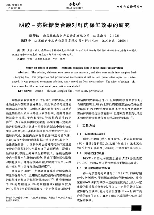
明胶-壳聚糖复合膜对鲜肉保鲜效果的研究

2 1 0 0 4 1
主要 以 明胶 、 壳 聚糖 为 原料 制 成 复 合 保 鲜 膜 , 介 绍天然食 品保 鲜剂的特 性及保 鲜机理 , 并 将 其 配 制 成
膜 液 后 喷 涂 于 鲜 肉表 面 , 研 究其 对 鲜 肉制 品 的 保 鲜 效 果 。
关键词
明胶 一壳聚糖 复合膜
极 易发 生 变 质 、 变 色 和变 味, 导 致 鲜 肉 品 质 的 下
要 略低 于 1 % 的酸 溶性壳 聚糖 , 但 由水 溶性壳 聚糖 处
理 的鲜 肉样 品完 全没 有酸 味 , 且 感观 品质 较好 , 只是
不 如 酸溶性 壳 聚糖保 鲜 肉样 品的货 架期 长 。
降 1 1 。为 了延 长 鲜 肉 的货 架 期 , 必 须 采 用 一 定 的 办
鲜肉 保鲜
S t u dy o n e fe c t o f g e l at i n— —c hi t o s an c ompl e x il f m i n f r e s h me a t pr e s e r v a t i o n Abs t r a c t T he g e l a t i n,c h i t o s a n we r e t a k e n a s r a w ma t e r i a l ,a n d t h e n we r e ma d e i n t o c o mp l e x le f s h
是涂 膜保 鲜法 J 。涂膜保 鲜 法是 将 肉类涂 抹 或 浸 泡
明胶 、 壳聚糖 ( 脱 乙酰 度 8 3 %) 、 转 谷 氨 酰 胺 酶
( T G) 、 甘油( 分 析纯) 、 冰乙酸 ( 分析 纯) 、 无 水 氯 化 钙( 分析 纯 ) 、 碘化钾( 分 析纯 ) 、 三 氯 甲烷 ( 分 析纯 ) 。
壳聚糖-明胶-果胶仿生网络膜对间充质干细胞增殖和矿化的影响

壳聚糖-明胶-果胶仿生网络膜对间充质干细胞增殖和矿化的影响孙红;闫志文;李硕峰;熊艳杰;梁凡;李傲;姚芳莲;车鹏程【摘要】目的:探讨采用壳聚糖、明胶和果胶制备的仿生网络膜对间充质干细胞(MSCs) 增殖和矿化的影响, 评价其用于构建组织工程骨的可行性.方法:采用仿生学方法, 将壳聚糖、明胶和果胶按照一定比例制作成新型仿生网络膜.实验分为对照组(MSCs+常规培养基) 、材料组 (MSCs+网络膜+常规培养基) 和材料+成骨诱导培养基 (OS) 组 (MSCs+网络膜+OS培养基) .倒置相差显微镜下观察细胞形态表现, 扫描电镜 (SEM) 观察细胞生长及细胞外基质分泌情况, MTT法检测细胞增殖情况(MSCs分为阴性对照组和材料组, 分别以空白培养液和含材料的培养液培养) , 茜素红染色检测细胞中钙无机物表达情况, 实时聚合酶链反应 (Real time-PCR) 测定细胞中骨钙素 (OC) mRNA和骨桥蛋白 (OPN) mRNA表达水平.结果:网络膜材料呈半透明薄膜状, 倒置相差显微镜下MSCs为短梭形, 细胞成簇状聚集生长.SEM下观察, 细胞培养第7天可见梭形细胞;培养第14天时细胞数量增多, 以伪足状突起锚定于材料表面;培养第21天时, 细胞聚集并分泌大量细胞外基质.材料组细胞增殖水平与阴性对照组比较差异无统计学意义 (P>0.05) .茜素红染色, 网络膜上细胞被染成橘红色.Real time-PCR法, 材料组和材料+OS组MSCs中OC mRNA在接种后第7和14天时表达水平较低, 第21天时其表达水平明显升高, 达到峰值;OPN mRNA在第7天明显表达, 第14天时表达水平达峰值, 第21天略有下降;与对照组比较, 不同时间点材料组和材料+OS组细胞中OC mRNA和OPN mRNA表达水平明显升高 (P<0.01) , 而材料组与材料+OS组比较差异无统计学意义 (P>0.05) .结论:壳聚糖-明胶-果胶仿生网络膜具有良好的生物相容性, MSC在其表面可正常生长和增殖, 在不加诱导剂的情况下可以诱导MSCs表达矿化相关的基因和蛋白.%Objective::To explore the effects of the biomimetic network membrane prepared by chitosan/gelatin/pectin on the proliferation and mineralization of mesenchymal stem cells (MSCs) , and to evaluate its feasibility of constructing tissue engineering bone.Methods:Chitosan, gelatin and pectin were made into a new biomimetic network membrane in a certain ratio by biomimetics.The experiment was divided into control group (MSCs+conventional medium) , material group (MSCs+network membrane+conventional medium) and material+OS group(MSCs+network membrane+OS medium) .The cell morphology was observed by inverted phase contrast microscope;the growth and secretion of extracellular matrix of the MSCs were observed under scanning electron microscope (SEM) .The proliferation of cells was determined by MTT assay (The MSCs were divided into negative control group and material group, and they were cultivated with blank medium and medium including materials) .The expression of calcium in MSCs was detected by Alizarin Red staining.Real-time polymerase chain reaction (RT-PCR) was used to determine the expression levels of osteocalcin (OC) mRNA and osteopontin (OPN) mRNA in the MSCs.Results:The network membrane was semitransparent thin film.The MSCs were short shuttle and clustered under inverted phase contrast microscope.After cultured for 7d, the MSCs were shuttle;after cultured for 14d, the number of MSCs was increased, with pseudo feet on the membrane;after cultured for21d, the MSCs clustered with a lot of neo-formed extracellular matrix.The MTT results showed that there was no significant difference in the proliferation level of MSCsbetween material group and negative control group (P>0.05) .The Alizarin Red staining results showed that the MSCs in the network membrane were dyed orange red.The RT-PCR results showed that the expression levels of OC mRNA in the MSCs in material group and material+OS group were lower on the 7th and 14th days, but on the 21th day, the expression levels were significantly increased and reached the peak;the expression level of OC mRNA in the MSCs in material group was significantly increased on the 7th day, and the expression level reached the peak on the 14th day, then fell slightly on the 21th day;compared with control group, the expression levels of OC mRNA and OPN mRNA in the cells in material group and material+OS group at different time points were significantly increased (P <0.01) , but there were no significant differences between material group and material+OS group (P>0.05) .Conclusion:Chitosan/gelatin/pectin biomimetic network membrane has good biocompatibility, and MSCs can grow and proliferate well on the membrane.The membrane can induce the MSCs to express mineralization-related genes and proteins without inducers.【期刊名称】《吉林大学学报(医学版)》【年(卷),期】2019(045)001【总页数】7页(P17-22,后插1)【关键词】壳聚糖;明胶;果胶;仿生材料;间充质干细胞;矿化【作者】孙红;闫志文;李硕峰;熊艳杰;梁凡;李傲;姚芳莲;车鹏程【作者单位】华北理工大学基础医学院河北省慢性疾病重点实验室,河北唐山063210;华北理工大学基础医学院河北省慢性疾病重点实验室,河北唐山 063210;华北理工大学基础医学院河北省慢性疾病重点实验室,河北唐山 063210;华北理工大学附属医院病理科,河北唐山 063000;华北理工大学基础医学院河北省慢性疾病重点实验室,河北唐山 063210;华北理工大学基础医学院河北省慢性疾病重点实验室,河北唐山 063210;天津大学化工学院高分子科学与工程系,天津 300072;华北理工大学基础医学院河北省慢性疾病重点实验室,河北唐山 063210【正文语种】中文【中图分类】R318.08一个世纪以来,骨缺损的修复一直是人们深入研究的重要课题,也是热门课题之一,然而,迄今为止临床上对创伤、感染和肿瘤切除后所造成的大范围骨缺损的修复仍未得到有效的解决。
明胶α组分含量对明胶壳聚糖复合膜的影响

明胶α组分含量对明胶-壳聚糖复合膜的性能影响摘要选用明胶α组分含量分别为a%,b%和 c %的A 、B 、C 3 种明胶作为原料,运用溶液共混法制备明胶-壳聚糖复合膜。
研究明胶α组分含量的不同对复合膜力学性能、吸湿性能、溶胀比、孔洞体积分率的影响。
运用X射线衍射和红外光谱分析复合膜的结构。
结果表明:明胶分子量的增加,共混膜的吸湿性、孔洞体积增大;随着共混膜中明胶质量分数的增加,吸湿性和孔洞体积增加,共混膜的溶胀比下降。
明胶与壳聚糖之间存在较强的相互作用,明胶壳聚糖共混可以改变壳聚糖结晶粒的大小,能降低壳聚糖的结晶度。
明胶与壳聚糖之间有良好的生物相容性。
关键词明胶;α组分含量;明胶-壳聚糖复合膜;力学性能;溶胀比;吸湿性能明胶来源广泛,价格低廉,具有良好的生物相容性和可降解性。
但是,它的成膜性、力学性能及抗水性较差。
壳聚糖价廉易得、易于加工,具有良好的生物相容性、可降解性、抗菌防腐性和成膜性等。
明胶-壳聚糖复合膜可用于生物医药、组织工程、食品等。
目前,对增塑及改性明胶-壳聚糖复合膜的性能与应用的研究较多[1~3],但有关未增塑及改性明胶-壳聚糖复合膜的结构与性能的研究很少[4]。
为了开发具有新性能的复合材料,使明胶-壳聚糖复合材料在可食性包装方面得到应用,有必要系统研究明胶-壳聚糖复合物的结构与性能,进一步了解其制备、结构与性能之间的关系,以制备性能特点互补、功能协同增效的绿色包装材料。
本文采用溶液共混法制备了一系列明胶-壳聚糖复合膜,并研究了复合膜的结构和性能1实验部分1.1原材料明胶A,工业级,明胶B和明胶C,照相级,去离子蒸馏水壳聚糖( 脱乙酰度m%),冰醋酸(分析纯)、氢氧化钠(分析纯)、碳酸钾(分析纯)、氯化钠(分析纯)(市售)。
1.2 实验仪器Z 型微机控制电子万能试验机X 型傅立叶变换红外光谱仪Y 型紫外可见分光光度计1.4明胶/壳聚糖复合膜的制备将一定量的明胶α组分含量分别为a%,b%,c%的A,B,C三种明胶溶于去离子水中,然后用40℃的水浴加热,配置成10%的A,B,C三种明胶溶液;将一定量的壳聚糖溶于2%的冰醋酸溶液中配置成2%的壳聚糖溶液。
壳聚糖敷料国内外研究导读

壳聚糖敷料国内外研究导读佚名分享| 收藏壳聚糖是甲壳素脱乙酰基衍生物,具有生物相容性好以及可生物降解等优点;明胶是动物胶原经温和断裂后的产物。
二者均具有良好的成膜性和黏度,具有良好的透气率和吸胀性,这对于膜吸收伤口渗出液和创面保湿非常有利。
用于伤口护理的明胶-壳聚糖敷料的文献报道,较早见诸于由英联邦政府于二十世纪80年代申请的美国专利US4572906。
自90年代末以来,随着组织工程医学的发展,美国、日本、意大利、韩国、中国等国家纷纷对这种生物医学材料予以高度关注。
实验已经证明,明胶-壳聚糖敷料具有良好的柔韧性、吸胀性和透气功能,是烧(创)伤后新鲜创面覆盖敷料的良好选择。
1984年英联邦政府申请的美国专利US4572906揭示了一种用于伤口护理的明胶-壳聚糖敷料,明胶-壳聚糖的重量比为3:1~1:3,还包括0-40%w/w(基于明胶、壳聚糖总重量)的相容的增塑剂山梨糖醇或甘油[1]。
1998年日本产业技术中心Maeda, Takuya等人将明胶和壳聚糖在稀蚁酸溶液中混合,20℃下润胀1小时,在60℃下溶解,然后在10℃玻璃板上干燥72小时。
所得到的明胶-壳聚糖膜用UV(254 nm)照射0、1或6小时(温度23℃,相对湿度50%)。
经历16小时UV照射的明胶-壳聚糖膜在pH 1.8条件下72小时后完全溶解[2]。
2000年韩国Kim, Min Jo等人申请的韩国专利KR2001016482揭示了壳聚糖、明胶和水作为主要成分的软凝胶具有水溶性、生物亲合力、生物降解性、抗菌性,可以吸收伤口分泌物,因此可以被用作伤口护理。
软胶的制备有四个步骤:(1)向明胶中加入水,加入0.1-5%有机酸,加入1-30%壳聚糖,摇晃1小时获得流体溶液;(2)使流体溶液硬化,得到易于成型的凝胶;(3)使凝胶成型为软膜;(4)干燥固化胶膜,得到水溶性固体膜[3]。
2001年意大利U.O Dermatologia对壳聚糖乙醇酸酯凝胶和交联明胶伤口敷料存在下,大鼠组织修复的形态学和免疫组化特性进行了比较和评价。
壳聚糖明胶果胶复合材料的性能研究
5.3.2.2MTT测试
2·0
1.8
1.6
1·4
1.2
81.o
0.8
O·6
0.4
0.2
O.0
012345878
‘Time(d)
图5-8L929细胞在支架上的MTr测试
将L929细胞接种到Cs-Gel支架和Cs.GeI-P支架上,分别于接种后1d、3d、5d和7d用MTr法检测生长在支架上的L929细胞的活性,得到的实验结果如图5-8所示。从图中可以看到,L929细胞在Cs.GeI-P支架上的吸光度值要明显高于Cs—Gel支架和组织培养板,说明细胞在Cs-Gel-P支架上更容易增殖生长。研究发现,果胶的结构和哺乳动物细胞外基质中多糖的结构非常类似,这一特性
Time(hrs)
图5.3:L929细胞在Cs—Gel.P薄膜及组织培养板表面的贴附
5.3.1.2细胞形态
从图5-4中可以看出,当细胞在薄膜上培养两天后,L929细胞的形貌已经基本形成,细胞伸出了伪足,呈多角形或长梭形,贴附在材料表面。培养十天之后,细胞已经长满了材料表面,细胞呈多角形或长梭形,整个材料上的细胞呈铺路石状,生长状态良好。
(a)培养2天,200倍(b)培养2天,400倍
第五章细胞与壳聚糖,明胶,果胶复合材料间的相互作用
(c)培养lO天,200倍(d)培养10天,400倍
图54L929细胞在壳聚糖/明胶,果胶薄膜上生长情况照片,倒置相差显微镜5.3.1.3HE染色观察
(a)培养3天,200倍(b)培养3天,400倍
(c)培养5天,200倍(d)培养7天,200倍
生物支架的含水量是组织工程用生物支架材料的重要指标,特别是用于皮肤组织工程,这是因为吸水量和含水量大的支架材料可以吸收多余的创面渗出液,同时,具有良好的透气作用。
壳聚糖_明胶_APTES改性生物活性玻璃复合支架的制备工艺
文章编号 : 100023851 (2009) 0420047206壳聚糖2明胶/ APTES 改性生物活性玻璃复合支架的制备工艺 力3 1 ,2, 王迎军1 ,2, 陈晓峰1 ,2, 赵营刚1任 ( 1 . 华南理工大学 特种功能材料教育部重点实验室 , 广州 510640 ;2 . 华南理工大学 材料学院 , 广州 510640)摘 要 : 为改善生物活性玻璃与高分子之间的相容性 , 利用 A P T E S 改性生物活性玻璃 ( S B G ) , 通过冷冻干燥法 制备出用于骨和软骨组织工程的壳聚糖2明胶/ A P T E S 改性生物活性玻璃 ( C S 2 G el/ SB G ) 仿生型复合多孔支架 , 并 对其孔隙率 、力学性能和显微形貌进行了表征 ; 探讨了各组分不同含量 、交联剂和冷冻温度对 CS 2G el/ S B G 复合支架孔隙率 、力学性能和显微观结构的影响 。
研究表明 , 当 SB G 和 CS 2 G el 的含量分别为 70 和 40 g 〃L - 1 , 用 EDC 和 N H S 交联 , - 50 ℃急冻 2 h 后 , 又在 - 15 ℃下冷冻 10 h , 最后真空冷冻干燥 , 制备出孔隙分布均匀 、孔隙率达 到 90 %以上 、三维连通的复合多孔支架 。
关键词 : 明胶 ; 壳聚糖 ; 生物活性玻璃 ; 硅烷偶联剂Pr e p ara t i on of the porous sc af f ol d s of chitosan 2gelatin/ APTES modif ied biog la ssR EN L i3 1 ,2, WA N G Yi n gj u n1 ,2, C H EN Xiaof e n g1 ,2, Z H A O Y i n gga n g1(1 . Key L a b o r ato r y of S p e cially Functio n al Mat erial s , S o ut h China U n iver s it y of Tech nolo g y , Mini s t r y of Educatio n , Guangzho u 510640 , China ; 2 . S choo l of Material s S cience a n d Engineering , S o u t h Chi n a U n iver s it y of Tech nolo g y ,Gua n gzho u 510640 , China )Abstract : A new kind of po r o u s co m po s it e used a s b o n e and ca r tilage ti s sue engineeri n g scaffo lds wa s p r ep a r ed wit h chito san , gelatin a nd 3 2 a m inop rop ylt riet ho xysila ne mo dif ied bio gla ss ( SB G ) by t he f reeze 2 dr y ing techniqu e in o r der to imp r o v e t he co m p atibilit y of B G a nd polymer . The po ro sit y , bendi ng st rengt h a nd micro st r uct u re of t h e co mpo sit e scaffo lds were cha ract erized. The eff ect s of t he co ncent ratio n of S B G a nd CS 2 G el , t he cro ss linking ag ent a nd t he sup er 2cooling t emp erat ure o n t he micro st r uct ure of t he co m po sit e scaffolds were inve stigat ed , resp ectively. The re s ult s sho w t h at t h e co m po s it e scaffol d s wit h 3D int erco n nective po r o u s st r u ct u re , high po r o s it y ( 90 %) a n d high mechanical st r engt h co u ld be p r epared un d er 70 g 〃L - 1 of S B G co n cent r atio n and 40 g 〃L - 1 of CS 2 G el co ncent r atio n , t h en cro s slin ked wit h EDC a n d N H S , f rozen at - 50 ℃fo r 2 h a n d - 15 ℃fo r 15 h , and f inally l y op h ilized. K ey w ords : gelatin ; chito s a n ; bio g la s s ; silico n co u p ling agent随着组织工程学和生物材料学的发展 , 对组织 工程支架材料的要求越来越高 , 因此 , 制备出合适 的骨和软骨组织工程支架材料成为解决骨和软骨的缺损 、修复 、重建的瓶颈问题 。
一种壳聚糖复合止血纱布及其制备方法
一种壳聚糖复合止血纱布及其制备方法随着医疗技术的不断进步,止血材料作为医疗领域中的重要一环,其在手术和创伤处理中起着至关重要的作用。
壳聚糖作为一种天然高分子化合物,具有良好的生物相容性和生物降解性,因此被广泛应用于医疗止血领域。
本文将介绍一种壳聚糖复合止血纱布及其制备方法,希望能够为医疗领域的止血材料研究和生产提供参考和借鉴。
一、壳聚糖复合止血纱布的特点1.壳聚糖具有良好的生物相容性和生物降解性,对人体无害,不会引起免疫反应和排斥反应。
2.壳聚糖具有优秀的止血效果,能够迅速吸收创面渗出的血液,形成凝血块,有效止血。
3.壳聚糖复合止血纱布具有良好的柔韧性和透气性,能够与创面完美贴合,不会影响伤口愈合和皮肤呼吸。
二、壳聚糖复合止血纱布的制备方法1.原料准备:准备壳聚糖、明胶和纯棉纱布等原料,将壳聚糖和明胶按一定比例混合并溶解于适量的生理盐水中,然后将纯棉纱布浸泡于混合溶液中。
2.复合处理:将浸泡后的纱布进行复合处理,使壳聚糖和明胶能够均匀地附着在纱布表面,经过干燥处理后形成壳聚糖复合止血纱布。
3.包装存储:将制备好的壳聚糖复合止血纱布进行包装封存,存放在干燥、阴凉的环境中,以确保其质量和性能不受到影响。
三、壳聚糖复合止血纱布的应用前景1.医疗领域:壳聚糖复合止血纱布将成为医疗领域中常用的止血材料,其良好的止血效果和生物相容性将大大提高手术和创伤处理中的止血效率。
2.应急救护:壳聚糖复合止血纱布还可用于应急救护领域,如交通事故、自然灾害等现场急救,能够快速有效地止血,降低伤者的出血量。
3.研究发展:随着生物材料和医疗技术的不断发展,壳聚糖复合止血纱布的制备方法和性能优化将成为医疗领域中的研究热点,为止血材料的开发和应用提供新的思路和方向。
总结:壳聚糖复合止血纱布作为一种新型的止血材料,具有良好的生物相容性和止血效果,其制备方法简单易行,应用前景广阔。
相信随着医疗技术的不断进步和人们对健康需求的不断提高,壳聚糖复合止血纱布必将在医疗领域中发挥重要作用,为患者的康复和健康保驾护航。
壳聚糖 明胶聚合ph
壳聚糖明胶聚合ph
壳聚糖是一种天然高分子多糖,具有生物相容性、生物降解性和无毒性等优点。
它可以在碱性条件下进行水解反应,生成可溶性壳聚糖。
在酸性条件下,壳聚糖可以形成不溶性的聚合物。
壳聚糖具有很好的成膜性和成球性,可以作为药物控释材料、组织工程支架、化妆品添加剂等应用。
明胶是一种动物源性胶体,具有很好的水溶性、成膜性和成球性。
它可以在碱性条件下进行聚合反应,生成可溶性的明胶聚合物。
在酸性条件下,明胶可以形成不溶性的聚合物。
明胶可以作为药物控释材料、组织工程支架、化妆品添加剂等应用。
当壳聚糖和明胶聚合在一起时,它们可以形成一种具有优异性能的复合材料。
这种复合材料具有很好的成膜性、成球性和药物控释性能,可以作为药物载体、组织工程支架、化妆品添加剂等应用。
此外,这种复合材料还具有很好的耐磨性和抗冲击性能,可以用于制备高性能的材料和制品。
- 1、下载文档前请自行甄别文档内容的完整性,平台不提供额外的编辑、内容补充、找答案等附加服务。
- 2、"仅部分预览"的文档,不可在线预览部分如存在完整性等问题,可反馈申请退款(可完整预览的文档不适用该条件!)。
- 3、如文档侵犯您的权益,请联系客服反馈,我们会尽快为您处理(人工客服工作时间:9:00-18:30)。
Applied Surface Science 257 (2011) 2712–2716Contents lists available at ScienceDirectApplied SurfaceSciencej o u r n a l h o m e p a g e :w w w.e l s e v i e r.c o m /l o c a t e /a p s u scSurface derivatization with spacer molecules on glutaraldehyde-activated amino-microplates for covalent immobilization of -glucosidaseYaodong Zhang ∗,Yun Zhang,Juanjuan Jiang,Li Li,Caihong Yu,Tingting HeiKey Laboratory of Applied Surface and Colloid Chemistry (Shaanxi Normal University),Ministry of Education,Key Laboratory of Analytical Chemistry for Life Science of Shaanxi Province,School of Chemistry and Materials Science,Shaanxi Normal University,Changan South Road,Xi’an 710062,Chinaa r t i c l e i n f o Article history:Received 11August 2010Received in revised form 12October 2010Accepted 12October 2010Available online 19 October 2010Keywords:Protein immobilization Microplate -Glucosidase Spacer moleculea b s t r a c tProtein molecules immobilized on a hydrophobic polystyrene microplate by passive adsorption lose their activity and suffer considerable denaturation.In this paper,we report a thorough evaluation of a protocol for enzyme immobilization on a microplate with relatively inexpensive reagents,involving glutaraldehyde coupling and spacer molecules,and employing -glucosidase as a model enzyme.The recommended conditions for the developed method include 2.5%glutaraldehyde to activate the reaction,1%chitosan in an HAc solution to increase the binding capacity,2%bovine serum albumin to block non-specific binding sites,and 0.1M NaBH 4to stabilize Schiff’s base ing this method,the amount of -glucosidase immobilized on amino-microplate was 24-fold with chitosan than without spacer molecules.The procedure is efficient and quite simple,and may thus have potential applications in biosensing and bioreactor systems.© 2010 Elsevier B.V. All rights reserved.1.IntroductionThe microtiter plate has become a common tool in analytical research and clinical diagnostic testing laboratories because it is easy to handle and adaptable to automatic microplate readers.A very common use for this plate is in enzyme-linked immunosorbent assays (ELISAs),which are the bases of most modern medical diag-nostic testing in humans and animals.Biological macromolecules such as proteins are immobilized on the surface of polystyrene microplates through passive adsorption if the surface has not been otherwise treated.Passive adsorption primarily involves multi-ple hydrophobic interactions between the solid phase and the biomolecule,which may interfere with the structure of the lat-ter and lead to conformational changes [1]and alterations in their functions [2].Thus,it is necessary to design a proper surface and rational conjugation for the controlled placement of biomolecules on polystyrene microtiter plates [3].The surfaces of polystyrene can be modified to introduce specific functions.In general,treatments result in the surface incorpora-tion of hydroxyl,carbonyl,and carboxyl functional groups [4,5].Since amine groups can be readily used for the covalent link-ing of bioactive molecules,the introduction of these groups is most often described in the literature [6].The most commonly used treatments for coating or covalently linking amine groups∗Corresponding author.Tel.:+862985303825;fax:+862985307774.E-mail address:ydzhang@ (Y.Zhang).to polystyrene include polymers,such as phenylalanine–lysine [7],nitration–reduction [8],gamma irradiation [9],plasma treat-ments (nitrogen or ammonia plasma)[10],and carbodiimides [11,12].The aminated surface is activated and covalently coupled to the functional groups (primary amines,thiols,and carboxyls)of biomolecules via bifunctional crosslinkers (i.e.,glutaraldehyde and carbodiimide).Glutaraldehyde activation of aminated supports is one of the most popular techniques for immobilizing enzymes [13].The proposed methodology is rather simple and efficient and,in some instances,even allows for the improvement of enzyme stability through multi-point or multi-subunit immobilization [14].Since a microplate consists of a large number of molecules within a small well,the density of the active surface amino groups is a key factor in increasing their binding ability to surfaces and pro-viding higher signals [15].To create a three-dimensional structure that generates sufficient spacing on surfaces and avoids lat-eral steric hindrances between immobilized biomolecules,surface derivation with a spacer molecule has been applied for biomolecu-lar immobilization on the surface of various solid supports [16].The most commonly used spacer molecule includes dendrimers [15,17],polyethyleneimine [18],poly(ethylene glycol)[19,20],chi-tosan [21],and poly(carboxybetaine methacrylate)[22],as well as self-assembled monolayers [23,24].Herein,we demonstrate the suitability of commercially available amine-graft polystyrene microwell plates for enzyme immobilization via a relatively inexpensive glutaraldehyde acti-vation reaction.To improve the enzyme binding efficiency of the amino-microplate,spacer molecules are employed for surface0169-4332/$–see front matter © 2010 Elsevier B.V. All rights reserved.doi:10.1016/j.apsusc.2010.10.050Y.Zhang et al./Applied Surface Science257 (2011) 2712–27162713Fig.1.The scheme of strategy for surface derivatization with spacer molecules on amino-microplate for protein immobilization.Crosslinker:glutaraldehyde,spacer molecule:chitosan.derivatization(Fig.1)and some conditions for immobilization,such as enzyme–support contact time and initial enzyme amount in the attachment solution,among others,are optimized.A compara-tive study between the glutaraldehyde and bis(sulfosuccinimidyl) suberate(BS3)methods is also undertaken.2.Experimental2.1.MaterialsAmine-graft polystyrene microtiter plates were purchased from Corning(96-well,Cat.No.2388).Sodium borohydride and Tween20were purchased from Alfa Aesar.-Glucosidase (E.C.3.2.1.21),4-nitrophenyl-d-glucopyranoside(pNPG),acetyl-cholinesterase(E.C.3.1.1.7),5,5 -dithiobis-(2-nitrobenzoic,acid), acetylthiocholine iodide,BS3,chitosan,glutaraldehyde,bovine serum albumin(BSA),and polyethyleneimine were all purchased from Sigma–Aldrich.2.2.Microplate assay ofˇ-glucosidase activityColor development in microplate wells was determined using a microplate reader(LMR340M,Labexim).A straight line of the average change absorbance versus time for each detected well was constructed.The change in absorbance per unit time per well( abs at405nm/min/well),which represents the quantity of the immo-bilized enzyme,was calculated from the slope of each straight line.-Glucosidase activity was assayed in200L of a0.1M sodium phosphate buffer containing5mM pNPG at pH5.0.After the solu-tion was incubated for15min at room temperature,the absorbance increase was recorded at405nm.A blank containing substrates but no immobilizing enzyme was prepared to control for the nonspe-cific hydrolysis of substrates in the microplate wells.2.3.Protocol for immobilizingˇ-glucosidase using glutaraldehydeThe amino-microplate wells were placed in2.5%glutaraldehyde and0.1M Na phosphate buffer(pH8.0)at room temperature for 15min,after which they were washed thoroughly in more pH8.0 phosphate buffer.The plates were incubated for60min at4◦C in1%chitosan in HAc(1vol.conc.HAc+99vol.H2O),and then washed thoroughly with H2O and0.1M Na phosphate pH8.0buffer.Non-specific binding sites on the plates were blocked by incubation for2h at4◦C for in a solution of2%bovine serum albumin in 0.1M Na phosphate pH8.0buffer.Excess blocking solution was washed away with phosphate buffer.After applying thefirst step of the procedure to the plates once more,they were activated with glutaraldehyde.Afterward,they were incubated in the enzyme solution at4◦C for a minimum of2h,although overnight incuba-tion is preferred.Washing with0.1M Na phosphate pH8.0buffer followed.The plates were incubated for60min at room tempera-ture in0.1M NaBH4in0.1M Na phosphate pH8.0buffer.Finally, the microplate wells were thoroughly and sequentially washed in H2O,0.1M NaCl in0.1M Na phosphate buffer pH8.0containing 0.5%(v/v)Tween20,and0.1M Na phosphate pH8.0buffer.2.4.Immobilization ofˇ-glucosidase using BS3Immobilization was carried out according to the manufacturer’s recommendations.About200L of a1mg/mL solution of BS3in 20mM phosphate buffered saline(pH7.4)solution were added to the detected wells of an amine surface strip plate.After being rins-ing thrice with PBS,the solution was incubated in a-glucosidase solution at4◦C for a minimum of2h,although overnight incuba-tion is preferred.The amount(activity)of-glucosidase in the0.1M Na phosphate pH5.0buffer was the same as in the glutaraldehyde activation method described above.2.5.Storage stabilityAn experiment was conducted to determine the stabilities of the immobilized-glucosidase preparations after storage in a phos-phate buffer(100mM,pH5.0)at4◦C for a predetermined amount of time.The residual activities were then determined as described above.The activity of each preparation was measured in batch oper-ation mode and expressed as a percentage of the residual activity relative to the initial activity.3.Results and discussionThe aminated surface of the polystyrene96-well plates was specifically designed to be used with bifunctional crosslinkers to covalently couple to the functional groups of biomolecules.In this study,-glucosidase was used as a convenient model enzyme to develop the protocol,as shown in Section2.Each of the steps in the procedure is examined individually.3.1.Glutaraldehyde activationGlutaraldehyde activation of aminated supports is one of the most popular techniques for immobilizing enzymes[13].A multi-step procedure for immobilizing proteins and enzymes on nylon has also been developed[25].The precise control of the condi-tions during support activation with glutaraldehyde has enabled the modification of the amino groups of the matrix with one or two glutaraldehyde molecules[14].In this study,amino-microplates were activated with various concentrations of a glutaraldehyde solution and incubated for15min at room temperature in a0.1M phosphate buffer,pH8.0.The glutaraldehyde concentration var-ied from0.5to10%.As can be seen from Fig.2,the amount of -glucosidase bound to the surface in thefinal stage of the proce-dure increased with increasing glutaraldehyde concentrations up to2.5%.At concentrations over2.5%,however,minimal increases in amount of bound-glucosidase were observed.Thus,in consid-eration of toxicity,2.5%(v/v)was chosen as the optimal amount for maximum enzyme immobilization.2714Y.Zhang et al./Applied Surface Science257 (2011) 2712–2716Fig.2.Glutaraldehyde activation of amino-microplate surfaces.Amino-microplates were incubated for 2h at room temperature in 0.1M Na phosphate pH 8.0buffer containing differing amounts of glutaraldehyde.The activated amino-microplates were then passed through the rest of the steps in the protocol to assess the amount of -glucosidase immobilized.n =3.In most cases,Step 2of the glutaraldehyde protocol is fol-lowed by coupling with a spacer molecule,such as chitosan or polyethyleneimine,which then necessitates a second glutaralde-hyde activation (Step 4)to provide sites for enzyme coupling.The obtained plots (not shown)were virtually identical to those shown in Fig.2.Thus,Step 4shows that a concentration of 2.5%glutaralde-hyde and 2h incubation are most favorable for the reaction.The results also suggest that glutaraldehyde levels could be reduced substantially,saving on costs.Reduced toxicity and waste disposal requirements were also observed when longer incubation times were used.For example,0.5%glutaraldehyde incubated for 2h yielded a little over 50%of the maximum enzyme immobi-lization.Prolonging the incubation time,however,increased this amount to almost its maximum level.3.2.Inclusion of spacer moleculesThe amine polystyrene surface was activated with glutaralde-hyde as the bifunctional crosslinker to covalently couple to primary amines on -glucosidase.The results shown in Fig.3indicate that only a small amount of -glucosidase was bound to the plate sur-face.In this study,chitosan or polyethyleneimine wasemployedFig.3.Influence of branched spacer molecules on immobilization efficiency.Acti-vated amino-microplates were incubated with different spacer molecules at various concentrations in Step 2of the immobilization protocol,and then reactivated with glutaraldehyde.The plates were incubated at 4◦C for 15h at pH 8.0in solutions of 0.7U -glucosidase.Chi =chitosan;PEI =polyethyleneimine.n =3.Fig.4.Influence of enzyme amount on the level of immobilized -glucosidase in Step 5of the immobilization protocol.Activated amino-microplates were incubated with 0.5%chitosan,activated with glutaraldehyde,and then incubated at 4◦C for 2or 15h at pH 8.0in -glucosidase solutions varying in activity from 0.1to 1.5U.n =3.( )2h coupling time;(᭹)15h coupling time.as an amplifying spacer arm [18,1,25]to provide more potential binding sites for each NH 2group that was originally generated on the polystyrene surface,and reduce local environmental effects due to both the matrix groups and steric hindrance [26].Surfaces that employed either of the two spacer polymers yielded useful increases in bound activity compared to surfaces that omit the use of a spacer.The use of chitosan resulted in larger proportions of immobilized -glucosidase compared to polyethyleneimine.The use of polyethyleneimine as a spacer molecule,on the other hand,led to larger proportions of immobilized -glucosidase on nylon surfaces activated with glutaraldehyde [25].The activity of immo-bilized -glucosidase was 24-fold higher on surfaces incubated with 1%chitosan compared to that without spacer molecules.These results were confirmed in other experiments wherein the enzyme immobilized was acetylcholinesterase,instead of -glucosidase.3.3.Enzyme couplingImmobilization of proteins on glutaraldehyde activated sup-ports is a very versatile technique.However,several critical variables must be considered when designing either the support or the immobilization conditions.The amino-microplate was passed through the first four steps of the protocol to ensure that the microplate surface was coated with chitosan spacer molecules and activated with glutaraldehyde.It was then incubated at 4◦C for either 2or 15h at pH 8.0in solu-tions of -glucosidase that varied in activity from 0.1to 1.5U.The results in Fig.4show that the 2h plot increased as the amount of -glucosidase increased,while the 15h plot reached a plateau at or increased shortly above 1U.In the case of plates subjected to 15h incubation time,increasing the enzyme activity from 0.7to 1.5U increased the amount of immobilized enzyme only by an additional 22%.As such,the additional costs incurred from longer reaction times are difficult to justify.The 15h plot was higher than the 2h one (Fig.4),showing that more enzymes were bound to the plate surfaces during prolonged incubation.Moreover,incubating the 0.7U -glucosidase solution in the wells for 15h led to levels of immobilization similar to the maximum seen after incubating wells with the 1.5U -glucosidase solution for 2h.This indicates that the amounts of enzyme can be reduced to increase the effi-ciency of enzyme utilization,especially if the immobilized enzyme is not fragile and can tolerate longer incubation times.Fig.5shows the influence of coupling time on the amount of immobilized -Y.Zhang et al./Applied Surface Science257 (2011) 2712–27162715Fig.5.Influence of coupling time on the level of immobilized-glucosidase in Step 5of the immobilization protocol.Activated amino-microplates were incubated with 0.5%chitosan,activated with glutaraldehyde,and then incubated at4◦C in solutions of0.7U-glucosidase with varying coupling times from2to15h.n=3.glucosidase in Step5of the protocol.Here,incubations were carried out at different time periods using0.7U-glucosidase.The results show that a plateau is at or shortly over10h of incubation.3.4.Overall immobilization procedureThis procedure was outlined in Section2.While there is some opportunity at each step of the protocol to increase thefinal amount of bound enzymes,Schiff’s base interactions between aldehydes and amines are typically not stable enough to form irreversible linkages.According to the literature,various reduc-ing reagents,such as sodium cyanoborohydride[27],sodium triacetoxyborohydride[28],Ti(O i-Pr)4/NaBH4[29],Zn(BH4)2/SiO2 [30],and␣-picoline-borane[31]have been developed for this conversion.Reduction with0.1M NaBH4stabilizes Schiff’s base intermediates[32],although it causes a very slight inactivation of bound enzymes on nylon[25].As such,borohydride treatment was introduced to stabilize the linkages of weakly bound enzymes.An amine surface microplate with bifunctional crosslinkers must be blocked using protein blockers capable of interacting with unreacted hydrophobic,ionic,and covalent sites.A number of chemical and biological reagents,such as casein,milk proteins, serum albumins,fish serum,Tween20,and polyethylene glycol, have been used for blocking purposes[33–35].Among the avail-able blocking reagents of pre-activated covalent surfaces(in this case,an amine surface microplate),BSA is the most preferred and commonly used blocking reagent because of its low cost and reduced steric hindrance of specifically binding proteins[36,37]. BSA was used for blocking remaining reactive groups instead of a smaller molecule containing amino groups,e.g.ethanolamine, glycine,ammonium buffers,etc.,because it was also capable of blocking the non-specific hydrophilic and/or hydrophobic adsorp-tion sites on the aminated polystyrene microplate[37,38].BSA is typically used at concentrations of1–3%.Moreover,the inclusion of Tween20to the wash buffer can enhance the removal of non-bound,physically trapped biomolecules[39].Although this work was carried out with a convenient model enzyme(-glucosidase),our work with other proteins and enzymes,such as acetylcholinesterase,showed results entirely consistent with the currentfindings.It appears probable that the developed procedure could yield results close to optimum for any enzyme or protein,including antibodies and inhibitors for bioreac-tor or affinity-based applications as long as they are not inactivated by the reactionparison of different immobilization protocols.Amino-microplates were incubated with0.7or1.1U-glucosidase solution in a0.1M Na phosphate buffer for2h at4◦C.n=3.GA=glutaraldehyde;BS3=bis(sulfosuccinimidyl)suberate.parison of methodsThe developed glutaraldehyde protocol was compared with the BS3protocol prepared according to the manufacturer’s recommen-dation to assess its practicality and reliability.As can be seen from Fig.6,the glutaraldehyde protocol gave values similar to those observed in the BS3protocol obtained from0.7U-glucosidase solutions incubated for2h.The BS3protocol,however,uses an expensive activation reagent.In the1.1U-glucosidase solution, the BS3protocol resulted in only a slightly larger amount of immobilized enzyme.Thus,the developed immobilization method features lower costs and achieve high efficiency immobilization on amino-microplates.3.6.Storage stabilityAn important advantage of immobilized enzymes over free enzymes is their improved stability for repeated use and storage. To investigate the stability of immobilized-glucosidase on the amino-microplate,the ability of the immobilized enzyme to remain active was tested by monitoring the changes in activity after a pre-determined length of time.Fig.7shows that after quickly reducing to nearly85.5%activity after two days,the decrease graduallytem-Fig.7.Storage stability at various storage times.Microplates immobilized with -glucosidase were stored in phosphate buffer(100mM,pH5.0)at4◦C for a prede-termined length of time.A percentage of its residual activity was compared to the initial activity.n=3.2716Y.Zhang et al./Applied Surface Science257 (2011) 2712–2716pered and about80%activity was retained after60days at4◦C in pH 5.0buffer.This indicates that the immobilized enzymes effectively decrease denaturation,as well as obviously increase the endurance and stability of immobilized-glucosidase.4.ConclusionsThe following set of conditions was established for-glucosidase immobilization on aminated polystyrene microplates: wells were activated with2.5%glutaraldehyde for about15min at room temperature,and then incubated in1%chitosan in an HAc solution(1vol.conc.HAc+99vol.H2O)to provide more potential binding sites.Non-specific binding sites were blocked by2%bovine serum albumin.To stabilize Schiff’s base intermediates,reduction with0.1M NaBH4in0.1M Na phosphate pH8.0buffer was included in the steps.The newly designed glutaraldehyde method had higher levels of immobilized protein compared to the conventional glutaralde-hyde method without chitosan as spacer molecules.The amount of-glucosidase immobilized was24-fold with1%chitosan than without spacer molecules.This tremendously increased the signal-to-noise ratio in the activity assays.Adoption of microplate technology for most applications in diagnostic and pharmaceutical screening will be driven,to some extent,by the cost of perform-ing an individual assay.The developed method was designed to use inexpensive reagents for immobilization.Its immobilization efficiency was similar to methods that use very expensive BS3as the activation reagent.Further work is in progress to determine the application of immobilized enzymes for biosensing and high throughput screening of enzyme inhibitors.AcknowledgmentsThis work was supported by the National Natural Science Foun-dation of China(No.30600494)and the Fundamental Research Fund for the Central Universities(No.GK200902010). References[1]J.E.Butler,Methods22(2000)4–23.[2]A.A.Ansari,N.S.Hattikudur,S.R.Joshi,M.A.Medeira,J.Immunol.Methods84(1985)117–124.[3]N.Suzuki,M.S.Quesenberry,J.K.Wang,R.T.Lee,K.Kobayashi,Y.C.Lee,Anal.Biochem.247(1997)412–416.[4]P.V.Narayanan,J.Biomater.Sci.Polym.Ed.6(1994)181–193.[5]J.Kaur,R.C.Boro,N.Wangoo,K.R.Singh,C.R.Suri,Anal.Chim.Acta607(2008)92–99.[6]N.Zammatteo,C.Girardeaux,D.Delforge,J.J.Pireaux,J.Remacle,Anal.Biochem.236(1996)85–94.[7]R.N.Hobbs,D.J.Lea,D.J.Ward,J.Immunol.Methods65(1983)235–243.[8]N.W.Chin,nks,Anal.Biochem.83(1977)709–719.[9]J.M.Varga,P.Fritsch,FASEB J.4(1990)2671–2677.[10]R.M.France,R.D.Short,Polym.Degrad.Stab.45(1994)339–346.[11]A.Niveleau,C.Bruno,E.Drouet,R.Brebant,A.Sergeant,F.Troalen,J.Immunol.Methods182(1995)227–234.[12]A.Niveleau,D.Sage,C.Reynaud,C.Bruno,S.Legastelois,V.Thomas,R.Dante,J.Immunol.Methods159(1993)177–187.[13]F.Lopez-Gallego,L.Betancor,C.Mateo,A.Hidalgo,N.Alonso-Morales,G.Dellamora-Ortiz,J.M.Guisan,R.Fernandez-Lafuente,J.Biotechnol.119(2005) 70–75.[14]L.Betancor,F.López-Gallego,A.Hidalgo,N.Alonso-Morales,G.D.-O.C.Mateo,R.Fernbndez-Lafuente,J.M.Guisbn,Enzyme Microb.Technol.39(2006)877–882.[15]D.Roy,J.W.Kwak,W.J.Maeng,H.Kim,J.W.Park,Langmuir24(2008)14296–14305.[16]F.Rusmini,Z.Zhong,J.Feijen,Biomacromolecules8(2007)1775–1789.[17]R.Benters,C.M.Niemeyer,D.Wohrle,Chembiochem2(2001)686–694.[18]B.P.Wasserman,H.O.Hultin,B.S.Jacobson,Biotechnol.Bioeng.22(1980)271–287.[19]C.Manta,N.Ferraz,L.Betancor,G.Antunes,F.Batista-Viera,J.Carlsson,K.Caldwell,Enzyme Microb.Technol.33(2003)890–898.[20]H.Chen,Y.Chen,H.Sheardown,M.A.Brook,Biomaterials26(2005)7418–7424.[21]P.Ye,Z.K.Xu, A.F.Che,J.Wu,P.Seta,Biomaterials26(2005)6394–6403.[22]Z.Zhang,S.Chen,S.Jiang,Biomacromolecules7(2006)3311–3315.[23]Y.J.Jung,B.J.Hong,W.Zhang,S.J.Tendler,P.M.Williams,S.Allen,J.W.Park,J.Am.Chem.Soc.129(2007)9349–9355.[24]A.Ulman,Chem.Rev.96(1996)1533–1554.[25]F.H.Isgrove,R.J.Williams,G.W.Niven,A.T.Andrews,Enzyme Microb.Technol.28(2001)225–232.[26]L.M.Bonanno,L.A.Delouise,Langmuir23(2007)5817–5823.[27]J.E.Mullersman,J.F.Preston III,Biochem.Cell Biol.69(1991)418–427.[28]A.F.Abdel-Magid,K.G.Carson,B.D.Harris,C.A.Maryanoff,R.D.Shah,.Chem.61(1996)3849–3862.[29]S.Bhattacharyya,.Chem.60(1995)4928–4929.[30]B.C.Ranu,A.Majee,A.Sarkar,.Chem.63(1998)370–373.[31]S.Sato,Tetrahedron60(2004)7899–7906.[32]J.M.Connolly,I.Alferiev,A.Kronsteiner,Z.Lu,R.J.Levy,J.Heart Valve Dis.13(2004)487–493.[33]Z.Peterfi,B.Kocsis,J.Immunoassay21(2000)341–354.[34]M.Steinitz,Anal.Biochem.282(2000)232–238.[35]K.Reimhult,K.Petersson,A.Krozer,Langmuir24(2008)8695–8700.[36]E.Wedege,G.Svenneby,J.Immunol.Methods88(1986)233–237.[37]Y.L.Jeyachandran,J.A.Mielczarski,E.Mielczarski,B.Rai,J.Colloid Interface Sci.341(2010)136–142.[38]Y.L.Jeyachandran,E.Mielczarski,B.Rai,J.A.Mielczarski,Langmuir25(2009)11614–11620.[39]E.Julian,M.Cama,P.Martinez,M.Luquin,J.Immunol.Methods251(2001)21–30.。
