Powerlab 数据采集分析系统
PowerLab系统工作原理、功能及应用 2008.12.12
多导电生理仪的介绍:PowerLab系统工作原理、功能及应用上海中医药大学附属龙华医院科研实验中心王佑华PowerLab生物信号采集处理系统(简称PowerLab系统)是由澳大利亚ADInstruments公司研制开发的计算机数据采集分析系统,可实现多通道生物信号数据的实时采集、记录和分析功能,包括软件和硬件两个部分。
该系统具有多种采样速率和卓越的分辨率,为生命科学的研究提供了强大的功能,己经成为全球使用最为广泛的生物信号采集分析系统之一,在世界各个国家已广泛用于生命科学的研究。
在Pubmed上输入“PowerLab”一词,就可以得到多达7000多个结果。
目前SCI影响因子TOP10的杂志上都时常可以看到应用该系统研究的报道,但多见于心血管、神经科学领域,目前在胃肠系统研究领域的应用有逐渐增多的趋势。
PowerLab系统经过ADInstruments公司近20年来的改良,已经成为全世界在该领域应用最为广泛的多导生理记录仪。
90年代初,PowerLab被部分科学家带进了中国。
1997年ADInstruments在上海成立了办事处,2000年成立了中国公司,2002年公司发布中文软件。
但时至今日,公司竟然还没有推出一份中文操作手册,chart for windows中文软件至今也没有中文操作指南。
ADInstruments公司官方网站上,尽管软件和硬件不断升级换代,但对该系统工作原理、功能及应用始终描述不多。
而中文网站翻译之后又省掉了许多信息,而且很久没有更新,现在基本还是4年前笔者博士期间看到的样子。
这在一定程度上影响了该系统的普及应用。
结合我院实际,本文就PowerLab系统工作原理、功能及应用作一简述。
1.PowerLab系统工作原理、功能应用PowerLab系统通过对生物电(心电、肌电、脑电等)与非生物电信号(压力、温度、pH等)进行采集,然后对采集到的信号进行加工处理,再由USB线将信号传输到计算机系统上的Chart软件,软件最后对采集到的生物电信号进行保存、显示、处理、将处理结果导入Excel 表格、定制实验、数据回放、在线分析、采样过程控制及打印输出结果等多种操作。
用压力-容积环评价大鼠心功能方法的改进及注意事项
用压力-容积环评价大鼠心功能方法的改进及注意事项刘晓雨;谭文【摘要】Objective To investigate the key factors for PV Loop evaluation in rats and to improve this method. To provide examples of cardiac function measurements obtained from normal rats and from rats with cardiac hypertrophy induced by transverse aortic constriction ( TAC ) . To establish a more reliable method for rats heart measurements. Methods Rats underwent left ventricular catheterization through the right carotid artery. Through adjustments of the position of the pressure⁃volume conductance catheter, the optimal PV Loops and a number of cardiac functional parameters were acquired. The key influencing factors, calibration of volume, position of the catheter in the left ventricle ( LV) and suspension of ventilator were assessed. Results 1. The real volume of left ventricle were acquired by injecting appropriate volume of hypertonic saline through jugular vein, which deducted the parallel conductivities of ventricular wall. 2. To get better PV loops, it’ s important to adjust the position of the catheter in the left ventricle until all of the pressure and volume sensors were located in the ventricle as well as out of touch with the ventricular wall. 3. Suspension the ventilator during the test is conducive to stable and reasonable data acquisition. We further assessed the cardiac functions of healthy rats and rats with cardiac hypertrophy with this improved method, which showed better performances. Conclusions This study we have evaluated the influences of calibration of volume,position of the catheter in the left ventricle ( LV) and ventilator on measurements of rats PV loops, and further improved this method. Moreover, we have validated this method with measurements of cardiac functions of normal rats and cardiac hypertrophic rats.%目的:探讨应用压力⁃容积环( pressure⁃volume loop,PV Loop)评价大鼠心功能过程中的影响因素及方法改进原则,分析主动脉弓缩窄(transverse aortic constriction,TAC)诱导的压力负荷对大鼠心脏功能的影响,从而为大鼠健康或疾病状态下的心功能分析提供更为科学全面的方法。
4-苯基丁酸对糖尿病大鼠的心脏保护作用

4-苯基丁酸对糖尿病大鼠的心脏保护作用何玉莲;熊燕;张梅【摘要】目的:观察4-苯基丁酸(4-PBA)对糖尿病大鼠心脏功能的影响并初步探讨其机制。
方法♂Sprague-Dawley ( SD)大鼠随机分成3组:对照组、糖尿病组和糖尿病+4-PBA组。
单次腹腔注射链脲佐菌素(60 mg·kg-1)诱发大鼠糖尿病,在1型糖尿病模型制备成功后第5周开始每天给予大鼠4-PBA(1g·kg-1)灌胃,连续治疗20周。
实验结束后,经右颈总动脉插管检测大鼠大动脉和左心室血流动力学变化,同时收集血清和心脏,比色法检测血糖,血清和心肌组织中超氧化物歧化酶( SOD)和一氧化氮合酶( NOS)活性,丙二醛(MDA)和一氧化氮(NO)含量。
结果4-PBA治疗对糖尿病大鼠血糖、体重和心脏重量无明显影响,但可改善糖尿病大鼠血流动力学,表现为大动脉收缩压和舒张压、心率、左心室收缩压和左心室内压最大上升和下降速率(± dp/dt)的轻度升高,左心室舒张压的轻度降低和舒张时程的缩短(P>0.05),左心室弛缓时间明显缩短(P<0.05)。
4-PBA治疗尚可明显升高糖尿病大鼠血清和心肌组织SOD和NOS活力以及NO含量,并降低MDA含量(P<0.05)。
结论4-PBA治疗对糖尿病大鼠心脏有潜在保护作用,这一作用是通过改善糖尿病大鼠血流动力学和明显缓解其心脏舒张功能障碍而实现,并与其抑制动物体内氧化应激和促进心肌NO生成有关。
%Aim Tostudytheeffectof4-phenlybuty-rate acid(4-PBA) on the heart function of diabetic rats and to explore its underlying mechanism. Methods MaleSprague-Dawley(SD)ratsweredividedintothree groups randomly: ( 1 ) control rats;( 2 ) diabetic rats;(3) 4-PBA treated diabetic rats. Type 1 diabetic mod-el was induced by single intraperitoneal injection of strepotozotocin (60 mg·kg-1 ) . After five weeks dia-betic model was established, diabetic rats weretreated with 4-PBA by P. O at the dose of 1 g·kg-1 per day for 20 weeks. At the end of the experiment, hemody-namics of the left ventricle and main artery were detec-ted through right carotid artery cannulation in rats. The level of blood glucose, the activity of superoxide dis-mutase( SOD) and nitric oxide synthetase ( NOS) and the content of malondialdehyde ( MDA ) and nitric ox-ide( NO) of serum and myocardial tissue in rats were measuredbycolorimetry.Results Thetreatmentof4-PBA had no effect on blood glucose, the weight of body and heart, but slightly increased systolic and diastolic pressure of artery, heart rate, systolic pressure of left ventricular, maximum increase rate and minimum de-crease rate of left ventricular pressure( ± dp/dt) in di-abetic rats. It also slightly decreased diastolic pressure of left ventricular, shortened diastolic duration of left ventricular ( P > 0. 05 ) . Moreover, it significantly shortened relaxation time of left ventricular in diabetic rats (P<0. 05). In addition, the treatment of 4-PBA obviously enhanced the activity of SOD and NOS, NO content, and reduced the content of MDA in serum andheartofdiabeticrats(P<0.05).Conclusion 4-PBA has a potential protective effect on heart function in diabetic rats, which is achieved by ameliorating he-modynamics and significantly improving diastolic dys-function in diabetic rats. The underlying mechanism may be related to the inhibition of oxidative stress and promoting the genesis of NO in myocardial tissue.【期刊名称】《中国药理学通报》【年(卷),期】2014(000)008【总页数】5页(P1137-1141)【关键词】糖尿病;心功能;4-苯基丁酸;血流动力学;氧化应激;一氧化氮【作者】何玉莲;熊燕;张梅【作者单位】广州医科大学药学院蛇毒研究所,广东广州 510082;广州医科大学药学院蛇毒研究所,广东广州 510082;广州医科大学药学院药理学教研室,广东广州 510082【正文语种】中文【中图分类】R-332;R322.11;R331.31;R587.102.2糖尿病患者易并发心血管疾病,其中糖尿病心肌病(diabetic cardiomyopathy,DCM)是患者的主要致死原因之一。
甲泼尼龙琥珀酸钠对窒息型大鼠复苏后早期的心功能影响
甲泼尼龙琥珀酸钠对窒息型大鼠复苏后早期的心功能影响朱旻婕;李响;方唯一【摘要】目的:探讨甲泼尼龙琥珀酸钠(甲强龙)对窒息大鼠模型心肺复苏后早期心功能的影响及机制.方法:45只SD大鼠,雌雄不拘,分为对照组、常规复苏组(常规复苏+肾上腺素10μg/kg)和甲强龙组(常规复苏+肾上腺素10μg/kg+甲强龙1.8mg/kg).对窒息大鼠模型进行心肺复苏,记录平均动脉压(MAP)、左心室收缩压(LVSP)和左心室舒张末压(LVEDP)的变化,ELISA法检测大鼠心脏组织中肾上腺素能α1受体和β1受体水平.结果:在心肺复苏自主循环恢复(ROSC)30 min后,与对照组比较,常规复苏组及甲强龙组大鼠的MAP和LVSP均有明显下降(P均<0.05);在ROSE后0、15 min时,常规复苏组及甲强龙组LVEDP与对照组相比有显著性差异(P均<0.05).甲强龙组MAP在ROSC后60、120 min时,LVSP在ROSC后30、60、120 min时均显著高于常规复苏组(P均<0.05).心肺复苏后,甲强龙组与对照组、常规复苏组比较,心脏组织中肾上腺素能α1受体和β1受体水平均明显升高(P 均<0.05).结论:甲强龙可提高心肺复苏后心脏组织内肾上腺素能α1受体和β1受体含量,有利于心肺复苏后MAP和LVSP的稳定.%Objective:To investigate the effects and mechanism of methylprednisolone on myocardial function at early stage of resuscitation in the rats model of asphyxia. Methods:A totalof 45 health SD rats were randomly divided into the controlgroup,conventional resuscitation group (routine resuscitation + epinephrine 10 μg/kg ) and methylprednisolone group (routiner esuscitation +epinephrine 10 μg/kg + methylprednisolone 1.8mg/kg).Hemodynamic indexes of mean arterial pressure (MAP),left ventricular systolic pressure (LVSP)and left ventricular end-diastolicpressure (LVEDP)were collected to estimate myocardial function.The levels ofα1 andβ1 adrenoreceptors in the cardiac tissue were detected by ELISA. Results:At 30min,60 min and 120 min after recovery of spontaneous circulation (ROSC ), the MAP and LVSP of conventional resuscitation group and methylprednisolone group were significantly decreased compared with the control group (all P<0.05). The LVDEP of conventional resuscitation group and methylprednisolone group were significantly higher than that of the control group at 0 min and 15 min after ROSC (all P<0.05).However,the MAP of methylprednisolone group was significantly higher than that of conventional resuscitation group at 60 min and 120 min after ROSC (both P<0.05 ).The LVSP of methylprednisolone group was significantly higher than that of conventional resuscitation group at30min,60min and 120min after ROSC (all P<0.05).The levels ofα1 andβ1 adrenoreceptors in the cardiac tissue of methylprednisolone group were significantly increased compared with the control group and conventional resuscitation group after cardiopulmonary resuscitation (all P<0.05 ). Conclusion:The methylprednisolone can increase the levels of theα1 andβ1 adrenoreceptors in the cardiac tissue,which is beneficial to the stabilization of MAP and LVSP after cardiopulmonary resuscitation.【期刊名称】《国际心血管病杂志》【年(卷),期】2017(044)001【总页数】4页(P26-29)【关键词】心肺复苏;心功能不全;甲泼尼龙琥珀酸钠【作者】朱旻婕;李响;方唯一【作者单位】200030 上海交通大学附属胸科医院心内科;201199 上海市闵行区中心医院重症监护室;200030 上海交通大学附属胸科医院心内科【正文语种】中文心肺复苏后心功能不全是引起复苏成功后死亡的主要原因之一[1]。
白藜芦醇对大鼠海马CA1区诱发癫痫样放电的影响
白藜芦醇对大鼠海马CA1区诱发癫痫样放电的影响尤竹燕;王斌生;解敏;李珍;王烈成【摘要】Aim To investigate the effects of resvera-trol with different concentrations on evoked epileptiform discharges in rat hippocampal CA1 region in order to provide a theoretical basis for the clinical development of antiepileptic drugs. Methods The epileptiform activity was induced by perfusing GABAA receptor antagonist, bicuculline, in normal hippocampal slices and the temporal lobe epilepsy( TLE ) model of rat was established by injecting kainic acid into the CA3 region of the right hippocampus. The population spike ( PS ) of CA1 pyramidal cell layer was recorded with extracellular recording in hippocampal slices by electrical stimulating Schaffer collaterals in stratum radiatum. Results The recorded PS was of a single peak in normal hippocampal slices. The amplitude of PS recorded in normal hippocampal slices was not significantly affected by perfusing ACSF with low, middle and high dose ( 5 μmol · L-1,15μmol · L-1 and 50 μmol · L-1 ) of resveratrol, respectively( n = 8, P > 0. 05 ). The recorded PS was of 4 ~ 6 peaks like multiple epileptiform discharges evoked by perfusing bicuculline ( 30 μmol· L-1 ). After perfusing low and middle dose of resveratrol, the amplitudes of the first four peaks of PS and the total peak numbers of PS were not significantly affected (n = 8, P > 0. 05). However, after perfusing high dose of resveratrol, the amplitudes of the first four peaks of PS and the total peak numbers of PS were all decreased obviously ( n =8,P <0. 01). The recorded PS was also of 4 ~ 6 peaks likemultiple epileptiform discharges in rat TEL model. After perfusing low and middle dose of resveratrol in rat TLE model, the amplitudes of the first four peaks of PS and the total peak numbers of PS were not significantly affected( n = 6, P>0. 05 ). However, after perfusing high dose of resveratrol, the amplitudes of the first four peaks of PS and the total peak numbers of PS were all decreased obviously( n = 6,P <0. 01). Conclusion High dose of resveratrol can partly inhibit evoked epileptiform discharges in rat hippocampal CA1 region.%目的观察不同浓度白藜芦醇(resveratrol,Res)对癫痫大鼠海马CA1区场电位的影响,为临床研发抗癫痫新药提供理论依据.方法正常大鼠海马脑片灌流γ-氨基丁酸受体拮抗剂bicuculline引起癫痫样活动以及在正常大鼠右侧海马CA3区注射海人藻酸(KA)建立颞叶癫痫(TLE)大鼠模型,应用离体脑片细胞外场电位记录,刺激辐射层Schaffer侧支通路,在海马CA1区锥体细胞层记录群峰电位(PS)的变化.结果正常大鼠海马脑片CA1区锥体细胞记录到的场电位为单个PS,分别灌流含有低、中和高剂量(分别为5、15和50 μmol*L-1)Res的ACSF,PS幅度均无明显变化(n=8,P>0.05).灌流bicuculline(30 μmol*L-1)后,记录到的场电位为多个PS的痫样电位,在此基础上灌流低和中剂量的Res,PS幅度和数目没有明显改变(n=8,P>0.05);而灌流高剂量的Res,前4个PS幅度均有明显降低(n=8,P<0.01),PS数目也有明显减少(n=8,P<0.01).在TLE模型大鼠海马脑片上记录到的场电位也为多个PS的痫样电位,灌流低和中剂量的Res,PS幅度和数目没有明显改变(n=6,P>0.05);灌流高剂量Res,前4个PS幅度均有明显降低(n=6,P<0.01),PS数目也有明显减少(n=6,P<0.01).结论高剂量的Res能部分抑制大鼠海马CA1区诱发癫痫样放电活动.【期刊名称】《中国药理学通报》【年(卷),期】2012(028)002【总页数】6页(P260-265)【关键词】癫痫;海马;白藜芦醇;群峰电位;突触;场电位【作者】尤竹燕;王斌生;解敏;李珍;王烈成【作者单位】安徽医科大学基础医学院生理学教研室,安徽,合肥,230032;浙江大学医学院血液细胞生物学实验室,浙江,杭州,310058;安徽医科大学基础医学院生理学教研室,安徽,合肥,230032;安徽医科大学基础医学院生理学教研室,安徽,合肥,230032;安徽医科大学基础医学院生理学教研室,安徽,合肥,230032【正文语种】中文【中图分类】R-332;R284.1;R322.81;R742.1;R971.6癫痫是由多种原因引起的局部脑区神经元群异常放电所致的行为和功能异常的发作性慢性中枢神经系统疾病。
芦丁片剂药学综合性实验的流程及方法研究
芦丁片剂药学综合性实验的流程及方法研究陈莉;杜红丽;曾仁韬;张叶叶;鲁莹;蔡国君【摘要】目的:以芦丁为模型药物,探讨新药片剂制备及药效学评价的多学科综合性实验流程和方法。
方法采用薄层色谱定性鉴别槐米中的芦丁成分;采用高效液相色谱法测定槐米中芦丁含量及芦丁精制品的含量;采用紫外分光光度法测定芦丁片含量;采用离体大鼠胸主动脉环舒张实验考察芦丁的药效;利用粉末直接压片法制备芦丁片剂。
结果与结论本实验完成了槐米的质量评价,槐米中芦丁的提取纯化、含量测定,芦丁药效学考察及芦丁片处方工艺研究等内容,提高了实验操作技能,实践了芦丁临床前研究的部分内容。
%Objective To imitate the development process of new drugs with rutin as the model and to do multidiscipli-nary experiments of preparation and pharmacodynamics of rutintablets .Methods Thin-layer chromatography was used to i-dentify rutin in Pagodatree flower bud .High-performance liquid chromatography was used for quantitative determination of ru-tin in Pagodatree flower bud and rutin products .The vasodilatation effect of rutin was investigated .The preparation of rutin tablets was completed .Results and Conclusion We completed the identification of Pagodatree flower bud ,extraction andpuri-fication of rutin from Pagodatree flower bud ,the assay of rutin ,the pharmaco-dynamics study and the formulation of rutin tab-lets .The experiments helped the postgraduates to be familiar with the research process of new drugs and to improve their ex-perimental operation skills .【期刊名称】《药学实践杂志》【年(卷),期】2015(000)004【总页数】4页(P359-362)【关键词】多学科综合性实验;芦丁;药效学;片剂【作者】陈莉;杜红丽;曾仁韬;张叶叶;鲁莹;蔡国君【作者单位】第二军医大学药学院,上海200433;第二军医大学药学院,上海200433;第二军医大学药学院,上海200433;第二军医大学药学院,上海200433;第二军医大学药学院,上海200433;第二军医大学药学院,上海200433【正文语种】中文【中图分类】R961;R943芦丁(rutin)又名芸香苷、维生素P,是一种典型的黄酮类化合物,几乎所有的芸香科和石楠科植物中均含有芦丁,尤其是在芸香科的芸香草、豆科植物的槐米中,芦丁含量较为丰富,可用作提取芦丁的原料[1-3]。
Powerlab系统简明操作规程
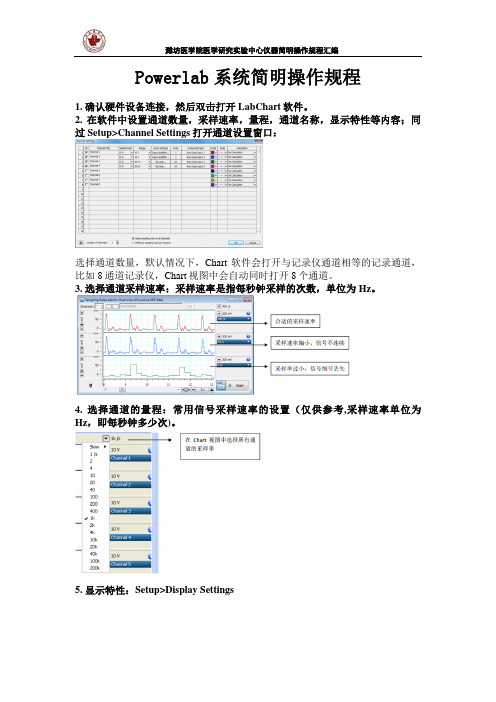
Powerlab系统简明操作规程1. 确认硬件设备连接,然后双击打开LabChart软件。
2. 在软件中设置通道数量,采样速率,量程,通道名称,显示特性等内容;同过Setup>Channel Settings打开通道设置窗口:选择通道数量,默认情况下,Chart软件会打开与记录仪通道相等的记录通道,比如8通道记录仪,Chart视图中会自动同时打开8个通道。
3. 选择通道采样速率:采样速率是指每秒钟采样的次数,单位为 Hz。
4. 选择通道的量程:常用信号采样速率的设置(仅供参考,采样速率单位为Hz,即每秒钟多少次)。
5. 显示特性:Setup>Display SettingsTime format:选择时间的显示格式Frome Start of file:从数据文件打开时计时,一直到记录结束;Frome Start of block:从每个Block 开始时计时,直到本Block 结束,从下个Block 重新计时;Time of Day:以电脑的时钟为准,显示记录数据时的实际时间。
Time as UTC:国际协调时间Always Seconds:以秒为单位计时Display Date:同时在文件的右上方显示日期Black Background:选择Scope 视图的背景色为黑色(关于Scope 视图背景色的设置,在Scope 视图中会有更多种选择)Graticule:选择是否需要背景的网格及网格颜色及线条类型General:选择是否在数据中显示Block 之间的界限,是否显示注释,是否在注释的地方加入竖线,是否在注释的竖线旁边显示注释的具体文本内容。
Sampling:选择采样时,数据是从左到右一直显示还是类似于示波器那样的一屏一屏地显示。
6. 时间轴和信号幅度轴的调整:时间轴(X 轴)的单位默认情况下是秒(S)时间轴的调整:可以通过 Chart 视图右下角的几个工具实现:点击一次,时间轴会压缩一定的倍数,最大的压缩比例为20K:1点击一次,时间轴会扩展一定的倍数,最大扩展比例为 1:1或者直选择水接平轴的压缩比例:Y 轴: Y 轴的坐标显示是由量程决定的,并随量程的变化而变化。
PowerLab生物信号采集系统在医学机能学教学中的应用与体会
一直以来,实验教学中心作为一种具有科学管理、优良设施、先进教学内容的大型现代化生物医学实验教学平台,是培养应用型医学人才的重要基地。
我校病理生理学实验教学中心于2007年被批准为河北省高校实验教学示范中心(建设单位),随后省教育厅逐步加大了建设力度。
为了丰富和创新医学机能学实验教学方法,培养符合现代医疗需要的高素质医学人才,教学中心自2015年开始购入PowerLab 生物信号采集系统,并应用于医学机能学教学。
现回顾总结几年来的使用过程与经验体会,以求进一步扩展PowerLab 生物信号采集系统在医学机能学教学的应用范围,全力提高医学人才培养质量。
1PowerLab 生物信号采集系统简介目前,PowerLab 生物信号采集系统广泛应用于药理学、生物化学、心理学、生理学及病理生理学等学科的教学与科研工作,实现了对实验信号的采集与处理[1]。
PowerLab 生物信号采集系统包括两个重要组成部分及其相关的系统软件,分别是多通道生理信号记录仪、二通道记忆示波器,能够实现多通道生物信号数据的实时采集、记录和分析,能完成所有的经典机能学实验,也能完成创新性、探究性实验,实验操作简洁、方便,实验结果准确、可靠[2]。
此外,PowerLab 生物信号采集系统还包含多通道信号接口等系统硬件,其输出接口通过USB 连接线与计算机主机相接,输入接口则与信号换能器相接,由此形成Power-Lab 生物信号采集系统的工作闭环。
PowerLab 生物信号采集系统在实验教学中心培养应用型人才方面发挥了重要作用。
根据《关于加强医学教育工作提高医学教育质量的若干意见》,夯实应用型人才培养基础,建立新型实验教学管理体制,推进实验教学改革,加强实验技术队伍建设,强化实验教学中心平台功能,将实验教学中心建设成为应用型医学人才的培养基地。
由此,揭开了PowerLab 生物信号采集系统在医学机能学教学中广泛应用的帷幕。
运用诸如PowerLab 生物信号采集系统等教学软硬件设备,建立以学生为主体的新型人才培养模式,推进教学方法改革,并促成基础医学与临床医学教学相融合,提高学生终身学习能力、创新能力、批判性思维能力[3]。