Two Zinc Complexes Supported by in situ Formed Etheric Ligands
基金项目英文翻译

Supported by State Key Development Program of (for) Basic Research of China(项目编号: )
Supported by Science Foundation of Ministry of Education of China
国家教育部博士点专项基金资助
Supported by Doctoral Fund of Ministry of Education of China
国家教育部回国人员科研启动基金资助
国家高技术研究发展计划(863计划)资助
Supported by National High Technology Research and Development Program of China
国家重大科学工程二期工程基金资助
Supported by National Important Project on Science-Phase Ⅱ of NSRL
国家攀登计划—B课题资助
Supported by National Climb—B Plan
国家杰出青年科学基金资助
Supported by National Natural Science Funds for Distinguished Young Scholar
国家科技部基金资助
Supported by State Commission of Science Technology of China(科委)
Supported by Ministry of Science and Technology of China
二联体亲子鉴定 英语

二联体亲子鉴定英语Duplex Parentage Testing: Understanding the Process and Its Applications.Duplex parentage testing, also known as diploid genotyping, is a highly specialized form of genetic testing that aims to establish the biological relationship between two individuals, typically a child and a putative parent. This process involves the analysis of genetic markers present in the DNA of the individuals involved to determine if they share a common genetic heritage. This article will delve into the details of duplex parentage testing, its scientific principles, applications, and ethical considerations.Scientific Principles of Duplex Parentage Testing.Duplex parentage testing relies on the principles of Mendelian inheritance and modern genetic technology. It typically involves the examination of multiple geneticmarkers, such as single nucleotide polymorphisms (SNPs) or short tandem repeats (STRs), which are passed down from parents to their children. These markers are present in specific locations on the DNA, known as loci. By comparing the genetic markers of the putative parent and child, scientists can determine if there is a genetic match, indicating a biological relationship.The process begins with the collection of DNA samples from the individuals involved. These samples can be obtained through a variety of methods, including buccal swabs, blood samples, or even saliva. Once collected, the DNA is isolated and amplified using polymerase chain reaction (PCR) or other similar techniques. This process produces enough DNA material for detailed analysis.Applications of Duplex Parentage Testing.Duplex parentage testing has a wide range of applications in various scenarios. Some of the most common uses include:1. Paternity Testing: This is one of the most common reasons for seeking duplex parentage testing. It involves determining whether a man is the biological father of a child. Paternity testing can be used in cases of disputed paternity, for example, when a child is born out of wedlock or when a man questions his parental status.2. Maternity Testing: Although less common, duplex parentage testing can also be used to establish maternity, or the biological relationship between a woman and her child. This is particularly useful in cases where the biological mother is unknown or disputed.3. Sibling Relationships: Duplex parentage testing can also be used to determine whether two individuals are siblings. This is achieved by comparing the genetic markers of the potential siblings with each other and with those of their putative parents.4. Adoption: In the context of adoption, duplex parentage testing can help to establish the biological relationship between an adoptive parent and child, orbetween siblings who were separated at birth.Ethical Considerations.While duplex parentage testing offers valuable information in resolving questions of biological relationships, it also raises ethical considerations. One of the most significant ethical issues is the privacy and confidentiality of test results. Strict protocols must be followed to ensure that test results are shared only with the authorized parties and that they are not misused or misinterpreted.Another ethical concern is the potential impact of test results on individuals and families. Positive results can confirm biological relationships, but negative results can cause emotional distress and even family breakdowns. It is, therefore, crucial that testing is conducted with the full knowledge and consent of all parties involved, and that they are provided with appropriate support and counseling throughout the process.Conclusion.Duplex parentage testing is a powerful tool that can provide valuable insights into biological relationships. It relies on the principles of Mendelian inheritance and modern genetic technology to analyze genetic markers and establish whether individuals share a common genetic heritage. From paternity testing to adoption and sibling relationships, duplex parentage testing has a wide range of applications. However, it is crucial to approach this testing with caution, respecting the privacy and confidentiality of test results and providing appropriate support and counseling to all parties involved.While duplex parentage testing can offer answers to complex biological questions, it is only one piece of the puzzle. It is essential to remember that genetic relationships are not the sole determinant of family ties. Emotional, cultural, and social factors play an equally important role in defining our sense of family and belonging. In this sense, duplex parentage testing shouldbe seen as a tool to complement other forms of evidence and understanding, rather than a replacement for them.。
新融合大学英语(I)_江西理工大学中国大学mooc课后章节答案期末考试题库2023年

新融合大学英语(I)_江西理工大学中国大学mooc课后章节答案期末考试题库2023年1.What of the following is not true as to the meaning of American Dream?参考答案:All men can embrace a better and richer and fuller life.2.Which of the following is NOT true about Tess?参考答案:She knew more than her parents.3.Which of the following words is not based on the prefix “pre-”?参考答案:Press.4.Before the daughter’s wedding ceremony, Father recalled so many thingsexcept____.参考答案:He can’t wait to dance with his daughter in celebrat ion.5.Which of the following is not the clue of the text?参考答案:Father’s complaint.6.Which of the following statements concerning adverbial clause of time is NOTtrue?参考答案:“While” is used when you are mentioning a time or event in the past and indicating that a situation has continued from then until now.7.Which of the following prefixes does not indicate negation?参考答案:en-8.Which description about the stove is not true?参考答案:It’s as white as snow.9.Which of the following is NOT true about the author?参考答案:Her brother Phillip enjoyed his birthday.10.These crops are extremely ______ to climate change, which has been a trickyproblem for the local farmers.参考答案:vulnerable11.What type of writing does this text fall into?参考答案:A narrative.12.What do you know about Christianity?参考答案:All of the above.13.Which of the following words is an acronym?参考答案:NASA.14.On that unforgettable Thanksgiving, the author received ______ kind deeds.参考答案:Two15.Which of the following statements is not mentioned in the text analysis?参考答案:Her marriage and her husband.16.Now the two older children were very sick with the flu, and the eldest _____bed rest for a week.参考答案:had been prescribed17.Which of the foll owing words is not based on the prefix “over-”?参考答案:Overt.18.The prefix ______ is often used to combine with adjectives and nouns to formnew words indicating that something connects two or more places, things, or groups of people.参考答案:inter-19.By the 6th century BC, athletes began to specialize ______ particular sports,and even began to hire coaches.参考答案:in20.What did students do during the SECOND half?参考答案:Sought help21.What did students do during the FIRST half?参考答案:Explored the natural and social world.22.Which one of the following statements about flashback is not true?参考答案:It narrates the whole story according to a chronological order.23.______ refer to words that consist wholly of two or more free words or roots.参考答案:Compounds24.The day on _______ you start your new job will be very busy.参考答案:which25.I had injured my leg and had to ______ of the race.参考答案:drop out26.Which is not true of the pharmacist’s brother?参考答案:He completed the surgery with charge.27.Which of the following is not one of the formats of exclamative sentences?参考答案:How + adj./adv. + n.28.Which country is the origin place for A.P. course?参考答案:America。
Naming Compounds

Sections (Zumdahl 6th Edition) 2.8-2.9 Outline: The Foundation of Stoichiometry
The Periodic Table helps organize types Binary Compounds: Metal and Non-Metal Binary Compounds: Two Non-Metals Acids (with and without oxygen)
• Binary – only two elements (as ions) combined • Can’t combine two metals (e.g. Cu & Zn –Brass) • The metal ion (cation, or positive) is always named first and the anion second. • A monatomic cation takes its name from the name of the element, e.g. Na+ is called sodium in the names of compounds containing this ion. • The non-metal ion , anion,(or negative ion) is named by taking the first part of the element and adding –ide, e.g. Cl- is chloride.
Different types (Sub-Classes) of Metals
• Alkali metals - (lithium, sodium, potassium, rubidium and cesium) - soft, low melting points, react with water to liberate hydrogen, form 1:1 compounds with chlorine. • Alkaline earths - (beryllium, magnesium, calcium, strontium, barium and radium) - react in a 1:2 ratio with chlorine. (Type I in text) • Transition metals - (e.g. iron, copper, silver, gold, tungsten and cobalt) - structural metals. Multi-valence; (Type II in text) • Actinides and Lantanides: Often multi-valence • Metalloids - (antimony, arsenic, boron, silicon and tellurium) - intermediate between metals and nonmetals. Acts as metals with non-metals; Act as non-metals with metals.
交叉耦合混沌信号源电路建模与设计
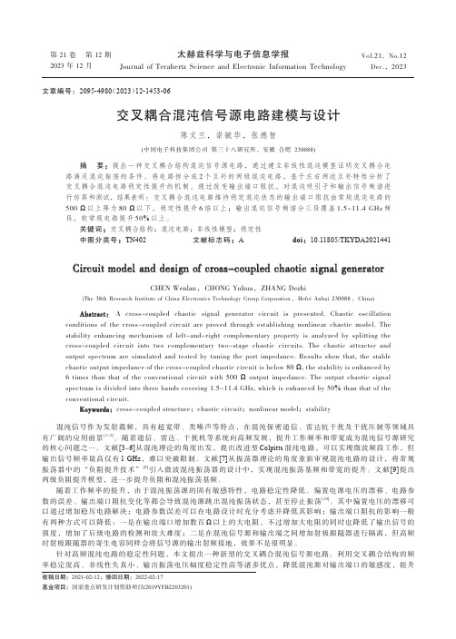
第 21 卷 第 12 期2023 年 12 月Vol.21,No.12Dec.,2023太赫兹科学与电子信息学报Journal of Terahertz Science and Electronic Information Technology交叉耦合混沌信号源电路建模与设计陈文兰,崇毓华,张德智(中国电子科技集团公司第三十八研究所,安徽合肥230088)摘要:提出一种交叉耦合结构混沌信号源电路,通过建立非线性混沌模型证明交叉耦合电路满足混沌振荡的条件。
将电路拆分成2个互补的两级混沌电路,基于左右两边互补特性分析了交叉耦合混沌电路稳定性提升的机制。
通过改变输出端口阻抗,对混沌吸引子和输出信号频谱进行仿真和测试,结果表明:交叉耦合混沌电路维持稳定混沌状态的输出端口阻抗由常规混沌电路的500 Ω以上降为80 Ω以下,稳定性提升6倍以上;输出混沌信号频谱分三段覆盖1.5~11.4 GHz频段,较常规电路提升50%以上。
关键词:交叉耦合结构;混沌电路;非线性模型;稳定性中图分类号:TN402 文献标志码:A doi:10.11805/TKYDA2021441Circuit model and design of cross-coupled chaotic signal generatorCHEN Wenlan,CHONG Yuhua,ZHANG Dezhi(The 38th Research Institute of China Electronics Technology Group Corporation,Hefei Anhui 230088,China)AbstractAbstract::A cross-coupled chaotic signal generator circuit is presented. Chaotic oscillation conditions of the cross-coupled circuit are proved through establishing nonlinear chaotic model. Thestability enhancing mechanism of left-and-right complementary property is analyzed by splitting thecross-coupled circuit into two complementary two-stage chaotic circuits. The chaotic attractor andoutput spectrum are simulated and tested by tuning the port impedance. Results show that, the stablechaotic output impedance of the cross-coupled chaotic circuit is below 80 Ω, the stability is enhanced by6 times than that of the conventional circuit with 500 Ωoutput impedance. The output chaotic signalspectrum is divided into three bands covering 1.5~11.4 GHz, which is enhanced by 50% than that of theconventional circuit.KeywordsKeywords::cross-coupled structure;chaotic circuit;nonlinear model;stability混沌信号作为发射载频,具有超宽带、类噪声等特点,在混沌保密通信、雷达抗干扰及干扰压制等领域具有广阔的应用前景[1-2]。
横坡段桥梁双桩双柱式结构受力特征与解析模型

第 55 卷第 3 期2024 年 3 月中南大学学报(自然科学版)Journal of Central South University (Science and Technology)V ol.55 No.3Mar. 2024横坡段桥梁双桩双柱式结构受力特征与解析模型王佳佳,陈效坤,肖莉丽,陈星潮,陈浩,李枝强(长安大学 公路学院,陕西 西安,710000)摘要:山区桥梁基础不可避免地建立在陡坡上,不稳定斜坡变形或破坏将会对上覆桥跨结构安全造成严重威胁,亟需研究高效、可靠的力学模型。
首先,对横坡段桥梁双桩双柱式结构划分不同特征段,分析其受力特性;其次,考虑桩土相互作用和桩顶变形协调关系并引入边界条件,建立适用于横坡段桥梁双桩双柱式结构内力及位移的简化模型;第三,综合考虑P −Δ效应及盖/系梁对桩柱受力影响,引入相邻特征段满足的连续条件(即位移、转角、剪力及弯矩连续),建立挠曲微分方程并以MATLAB 为平台编制相应计算程序,提出横坡段桥梁双桩双柱式结构基础内力及位移的幂级数解;最后,将模型结果与有限元计算结果对比,验证模型的可靠性。
研究结果表明:本文提出的模型将桥墩与桩基础视为整体,对于多道系梁的结构分析该模型同样适用;模型考虑了横向联系对两桩轴向力和弯矩的分配,可获取横向联系中的横向力;模型不需要假设自由段剪力和迭代计算。
随着剩余下滑力增大,结构各特征段前后桩位移和弯矩明显增加;横向联系对桩基位移及内力进行了重新分配,有较强的约束作用,能够较好地改善前后桩受力与变形情况。
关键词:横坡段桥梁;双桩双柱结构;受力特征;幂级数解;有限元分析中图分类号:U441+.5 文献标志码:A 文章编号:1672-7207(2024)03-1092-15Mechanics characteristics and analytical model of double-pile and double-column structure of bridge on steep transverse slopeWANG Jiajia, CHEN Xiaokun, XIAO Lili, CHEN Xingchao, CHEN Hao, LI Zhiqiang(School of Highway, Chang'an University, Xi'an 710000, China)Abstract: The foundation of bridge in mountain area is inevitably built on steep slope, and the deformation or failure of unstable slope will pose a serious threat to the safety of overlying bridge span, so it is urgent to develop an efficient and reliable mechanical model. Firstly, the double pile and double column structure of the bridge was divided into different characteristic sections, and its mechanical characteristics were analyzed. Secondly,considering the coordination relationship between pile-soil interaction and pile top deformation, and introducing收稿日期: 2023 −07 −10; 修回日期: 2023 −09 −23基金项目(Foundation item):国家自然科学基金资助项目(41907237,41907234);国家重点研发计划项目(2019YFB1600702,2021YFB1600302) (Projects(41907237, 41907234) supported by the National Natural Science Foundation of China; Projects (2019YFB1600702, 2021YFB1600302) supported by the National Key R&D Program of China)通信作者:肖莉丽,博士,副教授,从事公路岩土工程研究;E-mail :**************.cnDOI: 10.11817/j.issn.1672-7207.2024.03.022引用格式: 王佳佳, 陈效坤, 肖莉丽, 等. 横坡段桥梁双桩双柱式结构受力特征与解析模型[J]. 中南大学学报(自然科学版), 2024, 55(3): 1092−1106.Citation: WANG Jiajia, CHEN Xiaokun, XIAO Lili, et al. Mechanics characteristics and analytical model of double-pile and double-column structure of bridge on steep transverse slope[J]. Journal of Central South University(Science and Technology), 2024, 55(3): 1092−1106.第 3 期王佳佳,等:横坡段桥梁双桩双柱式结构受力特征与解析模型boundary conditions, a simplified model for the internal force and displacement of double-pile and double-column structure of bridge on cross slope section was established. Thirdly, considering the influence of P−Δ effect and cap/ tie beam on the force of pile, the continuous conditions (i.e., continuity of displacement, rotation angle, shear force and bending moment) satisfied by adjacent feature sections were introduced, the flexure differential equation was established, and the corresponding calculation program was compiled on the MATLAB platform. The power series solution of the internal force and displacement of the foundation of the double-pile and double-column structure of the bridge on the cross slope section was proposed. Finally, the reliability of the model was verified by comparing the model results with the finite element calculation results. The results show that the proposed model regards pier and pile foundation as a whole, and the model is also applicable to the structural analysis of multi-channel beams.The model takes into account the distribution of axial force and bending moment of the two piles in the transverse connection, and obtains the transverse force in the transverse connection. In addition, the model does not need to assume free section shear forces and iterative calculations. With the increase of residual glide force, the displacement and bending moment of piles in front and back of each characteristic section of the structure increase obviously. The lateral connection redistributes the displacement and internal force of pile foundation and has a strong constraint effect, which can better improve the load and deformation of front and rear piles.Key words: bridge on transverse slope; double pile and double column structure; mechanics characteristic; power series solution; finite element analysis山区高速公路通常采用桥梁来跨越地形障碍,以避免“大填大挖”,减少对生态环境的破坏[1−2]。
Fluorescence in situ hybridiz
2833 Commentary2834Journal of Cell Science 116 (14)2835 Fluorescence in situ hybridizationFig. 2.State of the art in FISH. (A) Many transcription sites (10) detected using barcoded probes can determine the gene expression pattern of each cell (Levsky et al., 2002) (B-D). Detection of single mRNA molecules using double or triple colored probes (Femino et al., 1998). (E) Detection of 24 chromosomes using spectral imaging (Macville et al., 1997).2836re-balance multi-color images composed of bands of varying strengths and to correct for spectral signal overlaps (Castleman, 1998).Difficulties inherent in objective analyses of FISH images have impeded but not stopped the development of automated algorithms for interpretation. Detection of DNA targets with large probes and the use of multi-color fluorescent cytometry algorithms (Galbraith et al., 1991) have allowed the production of automated mechanisms for assisting pathologists (Piper et al., 1994). In addition, the use of diagnostic probe sets and dot-counting approaches have yielded independent platforms capable of making simple diagnostic conclusions (Piper et al., 1990). Although methods have been introduced to analyze and optimize these cell classifiers (Castleman, 1985; Castleman and White, 1980; Castleman and White, 1981), manual cytopathology remains the gold standard for reliable tissue analysis, and automated mechanisms that can yield comparable data are still in development. Nevertheless, the benefits of high-throughput analysis of cell preparations, namely objective, computerized interpretation of cell samples on fixed substrates cannot be understated in the future development of diagnostic medicine. Although morphological analyses remain best suited to human operators, the ability to assay many molecular signatures rapidly in cells is only possible through computer-assisted approaches. Automated processing has recently been extended from the detection of specific DNA loci (Lawrence et al., 1988) and sites of transcription (Lawrence et al., 1989) to the determination of functional cell states by multi-gene transcriptional profiling (Levsky et al., 2002). The ability to assess accurately the transcriptional state of individual cells in situ has begun to influence the way we conceptualize single-cell versus tissue-level gene expression as well as study transcription activation, co-expression, and nuclear structure-function (Levsky and Singer, 2003).Advancing technologyWhereas the initial development of FISH involved expansion of the types of probe and number of detectable targets, the outlook for future development of fluorescence techniques will include expansion of the subjects of investigation. Clinical application of fluorescence imaging will require further advances in mechanization that allow the probes to be delivered, imaged, and analyzed automatically, thereby reducing operator-to-operator variability. Specimen thickness has been a limiting factor in the types of sample that can be analyzed with fluorescence microscopy. Until recently microstructure analysis based on methods such as confocal microscopy and optical coherence tomography was limited to specimens of approximately 1-2 mm thickness. A recent technological improvement known as optical projection tomography has allowed image-reconstruction of specimens up to 15 mm thick, setting the stage for more broad application to biological and clinical specimens (Sharpe et al., 2002). FISH techniques for detecting RNAs have been introduced to living cells (reviewed by Boulon et al., 2002), using either fluorophores that can be ‘un-caged’ in vivo (Politz and Singer, 1999) or probes that fluoresce only when hybridized (Tyagi and Kramer, 1996). Both of these innovative approaches circumvent the high background usually found in scenarios with unbound probe present (such as living cells), to allow the investigator to follow the creation and travels of mRNAs. These approaches are more easily applied to different target molecules than non-hybridization based GFP-fusion protein systems that bind a unique nucleic acid motif (Bertrand et al., 1998). One drawback of live-cell in situ hybridization as opposed to GFP-based assays is that FISH requires mechanical disturbance of the cell to introduce probes. These techniques allow deeper study of live gene expression in a minimally disturbed context, but must be interpreted with consideration of the possible artifacts that may result as physiological ramifications of hybridization. The separation drawn between approaches using fluorescent proteins and FISH should not be considered absolute. In fact, the compatibility of FISH and technologies employing fluorescent fusion proteins promises to allow simultaneous monitoring of proteins and nucleic acids of interest.The use of multi-photon approaches will also expand application of fluorescence imaging. In multi-photon microscopy, a laser source fires short bursts of photons that are focused by the microscope to arrive in pairs or triplets such that they summate to excite the fluorophore of interest. Near-infrared excitation light is used, which penetrates biological specimens more deeply and is far less toxic to live samples than visible light. This scheme has already allowed the application of fluorescence imaging to many living systems, including whole animals. Owing to limitations of our ability to introduce synthetic probes into organisms, most current applications of in vivo fluorescence imaging involve naturally occurring fluorescent molecules or bioluminescence (reviewed by Contag and Bachmann, 2002). Native fluorescent signatures that are present in tissues due to normal physiology or pathophysiological processes can encode important clinical information (reviewed by Konig, 2000). Should a form of organism-friendly probe become a reality, the power to discern many specific nucleic acids could be applied to non-invasive diagnostics, providing an informative adjunct to current methods of medical imaging.Diagnostic FISHThe development of in situ technologies has provided us with a wealth of information regarding the locations and expression patterns of genes in single cells. More complete gene expression profiles of single cells will provide a new level of insight into the correlation of gene expression patterns with particular cellular phenotypes. This will be particularly important in studies of development and disease progression, where complicated, finely demarcated gene expression programs are in play.Surprises are in store, however. The stochastic nature of gene expression revealed by this kind of approach indicates that perhaps our conception of a precisely regulated gene expression pattern is too constrained. Higher levels of tolerance for diversity in cell expression patterns may require a different model (Levsky and Singer, 2003). Computer-interpreted FISH assays are now sufficiently advanced to provide enormous amounts of data from a single cell, and even more from a tissue section. Measurement of expression from 20 genes by scoring for activity of neither, both, or one of the two alleles as mere binary ‘on or off’ signals, yields 320or greater than three billion bits of information per cell. If expression informationJournal of Cell Science 116 (14)2837 Fluorescence in situ hybridizationconcerning 100 genes were to be assayed, the information density would increase to 3100 bits (on the order of 1047). This exponential increase indicates that high-throughput data processing of gene expression information will have to evolve with the technology. The mere enormity of data may reveal insights not dreamt of in our philosophy.The ability to visualize RNA movement in living cells will provide models for how and where specific sequences are expressed and the steps by which transcripts are processed and exported from the nucleus. Our understanding of infectious disease will benefit from elucidation of how retroviruses direct nuclear import, trafficking and packaging for export into infectious particles. We are just beginning to understand the mechanisms by which specific RNAs are localized to subcellular regions of oocytes and some somatic cells for the purposes of asymmetric translation and how this is used to effect permanent structural changes – for instance, in synaptogenesis. When the tools become available for us to visualize multiple gene expression patterns in living cells, we will finally be able to fulfill the promise of FISH technology by building and testing models of molecular transcriptional dynamics within the true native context of the cell.The traditional route to diagnosis has been through morphological evaluation of biopsy specimens and the correlation of this analysis with clinical outcomes. The morphological basis of diagnosis has its limitations. It is well known that tumors that look alike under the microscope, and that appear phenotypically similar, may have radically different clinical courses in real patients. The new field of molecular pathology attempts to obviate the ambiguities of morphology by studying the origins of disease through characterization of genetic mutations and gene products. These investigations promise to provide more reliable biomarker information, founded on recent bioinformatics advances made possible by expression studies using micoarrays and serial analysis of gene expression (SAGE). Transcriptional alterations associated with malignant transformation and markers that correlate with cancer progression are being identified. The mechanism by which these data can be incorporated in the pathologist’s ‘tool box’ is currently being developed.The recently developed tissue microarray technology is an ideal platform for the introduction of high-throughput molecular profiling of tumor specimens at the single cell level. To construct a tissue microarray, small core biopsies are taken from representative areas of paraffin-embedded tumor tissues and assembled in a single block. Microtome sections are taken from the tissue microarray and placed on glass slides for rapid and efficient molecular analyses. In addition to pathological specimens such as tumor tissues, microarrays generally contain corresponding normal tissue and internal controls. The entire group of samples is analyzed simultaneously in one experiment, providing enormous amounts of correlative information about specific biomarkers, in the context of rigorous procedural controls. The next challenge will be to apply multi-gene FISH technology to these samples to correlate putative genes of prognostic value with specific morphological features initially, and then extend studies to samples where the morphology is not sufficiently informative. Certain genes can then be associated with the pre-cancerous state, for instance. Through such developments, one can foresee how molecular pathology could eventually surpass the limitations of morphological pathology. This would allow more judicious use of minimally invasive biopsy techniques that sacrifice retrieved tissue morphology in favor of comfort of the patient. FISH has already colored the way that we visualize and conceptualize genes, chromosomes, transcription and nucleic acid movements. What remains to be seen is how exhaustive molecular analysis of single cells and tissue samples will impact how we identify, diagnose and alter the course of genetic pathology. Over the long term, it is expected that databases correlating gene expression patterns on the single cell level will accumulate as investigators and industries employ the technology of FISH with their favored biomarkers. Ultimately, FISH will be the preferred approach to anticipate the complicated components of gene expression leading to disease.Our work is supported by the Innovative Molecular Analysis Technologies program at the National Cancer Institute, and research and training grants from the NIH.ReferencesArndt-Jovin, D. J., Robert-Nicoud, M., Kaufman, S. J. and Jovin, T. M. (1985). Fluorescence digital imaging microscopy in cell biology. Science 230, 247-256.Baschong, W., Suetterlin, R. and Laeng, R. H.(2001). Control of autofluorescence of archival formaldehyde-fixed, paraffin-embedded tissue in confocal laser scanning microscopy (CLSM). J. Histochem. Cytochem. 49, 1565-1572.Bauman, J. G., Wiegant, J., Borst, P. and van Duijn, P.(1980). A new method for fluorescence microscopical localization of specific DNA sequences by in situ hybridization of fluorochromelabelled RNA. Exp. Cell Res. 128, 485-490.Bertrand, E., Chartrand, P., Schaefer, M., Shenoy, S. M., Singer, R. H. and Long, R. M.(1998). Localization of ASH1 mRNA particles in living yeast. Mol. Cell2, 437-445.Bingham, E. and Hyvarinen, A.(1997). A fast fixed-point algorithm for independent component analysis. Int. J. Neural Syst.10, 1-8.Boulon, S., Basyuk, E., Blanchard, J. M., Bertrand, E. and Verheggen, C. (2002). Intra-nuclear RNA trafficking: insights from live cell imaging. Biochimie 84, 805-813.Brown, J., Horsley, S. W., Jung, C., Saracoglu, K., Janssen, B., Brough, M., Daschner, M., Beedgen, B., Kerkhoffs, G., Eils, R. et al. (2000). Identification of a subtle t(16;19)(p13.3;p13.3) in an infant with multiple congenital abnormalities using a 12-colour multiplex FISH telomere assay, M-TEL. Eur. J. Hum. Genet. 8, 903-910.Carrington, W. A., Lynch, R. M., Moore, E. D., Isenberg, G., Fogarty, K.E. and Fay,F. S.(1995). Superresolution three-dimensional images of fluorescence in cells with minimal light exposure. Science268, 1483-1487. Castleman, K. R.(1985). Estimation of sampling errors. Cytometry6, 276-277.Castleman, K. R.(1998). Concepts in imaging and microscopy: color image processing for microscopy. Biol. Bull. 194, 100-107.Castleman, K. R. and White, B. S.(1980). Optimizing cervical cell classifiers. Anal. Quant. Cytol. 2, 117-122.Castleman, K. R. and White, B. S.(1981). The effect of abnormal cell proportion on specimen classifier performance. Cytometry2, 155-158. Contag, C. H. and Bachmann, M. H.(2002). Advances in in vivo bioluminescence imaging of gene expression. Annu. Rev. Biomed. Eng. 4, 235-260.Dirks, R. W., van Gijlswijk, R. P., Tullis, R. H., Smit, A. B., van Minnen, J., van der Ploeg, M. and Raap, A. K.(1990). Simultaneous detection of different mRNA sequences coding for neuropeptide hormones by double in situ hybridization using FITC- and biotin-labeled oligonucleotides. J. Histochem. Cytochem. 38, 467-473.Dirks, R. W., van Gijlswijk, R. P., Vooijs, M. A., Smit, A. B., Bogerd, J., van Minnen, J., Raap, A. K. and van der Ploeg, M.(1991). 3′-end fluorochromized and haptenized oligonucleotides as in situ hybridization probes for multiple, simultaneous RNA detection. Exp. Cell Res. 194, 310-315.2838Fauth, C. and Speicher, M. R.(2001). Classifying by colors: FISH-based genome analysis. Cytogenet. Cell Genet. 93, 1-10.Femino, A. M., Fay, F. S., Fogarty, K. and Singer, R. H.(1998). Visualization of single RNA transcripts in situ. Science280, 585-590. Forozan, F., Karhu, R., Kononen, J., Kallioniemi, A. and Kallioniemi, O. P.(1997). Genome screening by comparative genomic hybridization. Trends Genet. 13, 405-409.Galbraith, W., Wagner, M. C., Chao, J., Abaza, M., Ernst, L. A., Nederlof, M. A., Hartsock, R. J., Taylor, D. L. and Waggoner, A. S.(1991). Imaging cytometry by multiparameter fluorescence. Cytometry12, 579-596. Gall, J. G. and Pardue, M. L.(1969). Formation and detection of RNA-DNA hybrid molecules in cytological preparations. Proc. Natl. Acad. Sci. USA63, 378-383.Han, M., Gao, X., Su, J. Z. and Nie, S.(2001). Quantum-dot-tagged microbeads for multiplexed optical coding of biomolecules. Nat. Biotechnol. 19, 631-635.Hopman, A. H., Wiegant, J., Raap, A. K., Landegent, J. E., van der Ploeg, M. and van Duijn, P.(1986). Bi-color detection of two target DNAs by non-radioactive in situ hybridization. Histochemistry85, 1-4. Hougaard, D. M., Hansen, H. and Larsson, L. I.(1997). Non-radioactive in situ hybridization for mRNA with emphasis on the use of oligodeoxynucleotide probes. Histochem. Cell Biol. 108, 335-344. Kislauskis, E. H., Li, Z., Singer, R. H. and Taneja, K. L.(1993). Isoform-specific 3′-untranslated sequences sort alpha-cardiac and beta-cytoplasmic actin messenger RNAs to different cytoplasmic compartments. J. Cell Biol. 123, 165-172.Konig, K.(2000). Multiphoton microscopy in life sciences. J. Microsc. 200, 83-104.Landegent, J. E., Jansen in de Wal, N., Dirks, R. W., Baao, F. and van der Ploeg, M.(1987). Use of whole cosmid cloned genomic sequences for chromosomal localization by non-radioactive in situ hybridization. Hum. Genet. 77, 366-370.Langer, P. R., Waldrop, A. A. and Ward, D. C.(1981). Enzymatic synthesis of biotin-labeled polynucleotides: novel nucleic acid affinity probes. Proc. Natl. Acad. Sci. USA78, 6633-6637.Lawrence, J. B. and Singer, R. H.(1986). Intracellular localization of messenger RNAs for cytoskeletal proteins. Cell45, 407-415. Lawrence, J. B., Singer, R. H., Villnave, C. A., Stein, J. L. and Stein, G. S.(1988). Intracellular distribution of histone mRNAs in human fibroblasts studied by in situ hybridization. Proc. Natl. Acad. Sci. USA85, 463-467. Lawrence, J. B., Singer, R. H. and Marselle, L. M.(1989). Highly localized tracks of specific transcripts within interphase nuclei visualized by in situ hybridization. Cell57, 493-502.Levsky, J. M. and Singer, R. H. (2003). Gene expression and the myth of the average cell. Trends Cell Biol. 13, 4-6.Levsky, J. M., Shenoy, S. M., Pezo, R. C. and Singer, R. H.(2002). Single-cell gene expression profiling. Science297, 836-840.Levsky, J. M., Braut, S. A. and Singer, R. H. (2003). Single cell gene expression profiling by multiplexed expression fluorescence in situ hybridization: application to the analysis of cultured cells. In Cell Biology: A Laboratory Handbook(in press) (ed. J. E. Celis): Academic Press. Lichter, P., Cremer, T., Borden, J., Manuelidis, L. and Ward, D. C.(1988). Delineation of individual human chromosomes in metaphase and interphase cells by in situ suppression hybridization using recombinant DNA libraries. Hum. Genet. 80, 224-234.Lichter, P., Joos, S., Bentz, M. and Lampel, S.(2000). Comparative genomic hybridization: uses and limitations. Semin. Hematol. 37, 348-357. Macville, M., Veldman, T., Padilla-Nash, H., Wangsa, D., O’Brien, P., Schrock, E. and Ried, T.(1997). Spectral karyotyping, a 24-colour FISH technique for the identification of chromosomal rearrangements. Histochem. Cell Biol. 108, 299-305.Manuelidis, L., Langer-Safer, P. R. and Ward, D. C.(1982). High-resolution mapping of satellite DNA using biotin-labeled DNA probes. J. Cell Biol. 95, 619-625.Nederlof, P. M., Robinson, D., Abuknesha, R., Wiegant, J., Hopman, A.H., Tanke, H. J. and Raap, A. K.(1989). Three-color fluorescence in situ hybridization for the simultaneous detection of multiple nucleic acid sequences. Cytometry10, 20-27.Nederlof, P. M., van der Flier, S., Wiegant, J., Raap, A. K., Tanke, H. J., Ploem, J. S. and van der Ploeg, M.(1990). Multiple fluorescence in situ hybridization. Cytometry11, 126-131.Nederlof, P. M., van der Flier, S., Raap, A. K. and Tanke, H. J.(1992a). Quantification of inter- and intra-nuclear variation of fluorescence in situ hybridization signals. Cytometry13, 831-838.Nederlof, P. M., van der Flier, S., Vrolijk, J., Tanke, H. J. and Raap, A. K.(1992b). Fluorescence ratio measurements of double-labeled probes for multiple in situ hybridization by digital imaging microscopy. Cytometry13, 839-845.Neumann, M. and Gabel, D.(2002). Simple method for reduction of autofluorescence in fluorescence microscopy. J. Histochem. Cytochem. 50, 437-439.Pachmann, K.(1987). In situ hybridization with fluorochrome-labeled cloned DNA for quantitative determination of the homologous mRNA in individual cells. J. Mol. Cell Immunol. 3, 13-19.Palotie, A., Heiskanen, M., Laan, M. and Horelli-Kuitunen, N.(1996). High-resolution fluorescence in situ hybridization: a new approach in genome mapping. Ann. Med. 28, 101-106.Pinkel, D., Straume, T. and Gray, J. W.(1986). Cytogenetic analysis using quantitative, high-sensitivity, fluorescence hybridization. Proc. Natl. Acad. Sci. USA83, 2934-2938.Piper, J., Fantes, J., Gosden, J. and Ji, L. A.(1990). Automatic detection of fragile X chromosomes using an X centromere probe. Cytometry11, 73-79.Piper, J., Poggensee, M., Hill, W., Jensen, R., Ji, L., Poole, I., Stark, M. and Sudar, D.(1994). Automatic fluorescence metaphase finder speeds translocation scoring in FISH painted chromosomes. Cytometry16, 7-16. Politz, J. C. and Singer, R. H.(1999). In situ reverse transcription for detection of hybridization between oligonucleotides and their intracellular targets. Methods18, 281-285.Puvion-Dutilleul, F. and Puvion, E.(1996). Non-isotopic electron microscope in situ hybridization for studying the functional sub-compartmentalization of the cell nucleus. Histochem. Cell Biol. 106, 59-78. Raap, A. K.(1998). Advances in fluorescence in situ hybridization. Mutat. Res. 400, 287-298.Rudkin, G. T. and Stollar, B. D.(1977). High resolution detection of DNA-RNA hybrids in situ by indirect immunofluorescence. Nature265, 472-473. Schrock, E., du Manoir, S., Veldman, T., Schoell, B., Wienberg, J., Ferguson-Smith, M. A., Ning, Y., Ledbetter, D. H., Bar-Am, I., Soenksen, D. et al. (1996). Multicolor spectral karyotyping of human chromosomes. Science273, 494-497.Sharpe, J., Ahlgren, U., Perry, P., Hill, B., Ross, A., Hecksher-Sorensen, J., Baldock, R. and Davidson, D.(2002). Optical projection tomography as a tool for 3D microscopy and gene expression studies. Science296, 541-545.Singer, R. H. and Ward, D. C.(1982). Actin gene expression visualized in chicken muscle tissue culture by using in situ hybridization with a biotinated nucleotide analog. Proc. Natl. Acad. Sci. USA79, 7331-7335. Speicher, M. R., Gwyn Ballard, S. and Ward, D. C.(1996). Karyotyping human chromosomes by combinatorial multi-fluor FISH. Nat. Genet. 12, 368-375.Tanke, H. J., Florijn, R. J., Wiegant, J., Raap, A. K. and Vrolijk, J.(1995). CCD microscopy and image analysis of cells and chromosomes stained by fluorescence in situ hybridization. Histochem. J. 27, 4-14.Tanke, H. J., Wiegant, J., van Gijlswijk, R. P., Bezrookove, V., Pattenier, H., Heetebrij, R. J., Talman, E. G., Raap, A. K. and Vrolijk, J.(1999). New strategy for multi-colour fluorescence in situ hybridisation: COBRA: COmbined Binary RAtio labelling. Eur. J. Hum. Genet. 7, 2-11.Trask, B. J.(2002). Human Genetics and Disease: Human cytogenetics: 46 chromosomes, 46 years and counting. Nat. Rev. Genet. 3, 769-778. Tyagi, S. and Kramer, F. R.(1996). Molecular beacons: probes that fluoresce upon hybridization. Nat. Biotechnol. 14, 303-308.van der Ploeg, M.(2000). Cytochemical nucleic acid research during the twentieth century. Eur. J. Histochem. 44, 7-42.Wiegant, J., Ried, T., Nederlof, P. M., van der Ploeg, M., Tanke, H. J. and Raap, A. K.(1991). In situ hybridization with fluoresceinated DNA. Nucleic Acids. Res. 19, 3237-3241.Journal of Cell Science 116 (14)。
stiky ends名词解释
stiky ends名词解释Sticky ends (名词解释) :Sticky ends是DNA融合实验中常用的术语,用于描述由于酶切后DNA链断裂形成的具有突出单链DNA序列的末端。
这些具有互补的序列的单链DNA片段能够与其他DNA片段很好地结合,从而形成较稳定的DNA连接,常用于基因工程和重组DNA技术中。
Double-Stranded DNA sticky ends (中文:双链DNA黏性末端)是指由于酶切作用后两个DNA单链的部分被切除而留下的具有互补序列的末端。
这些末端的单链DNA片段具有黏性,可以与其他DNA序列的黏性末端互补结合,形成稳定的DNA连接。
以下是24个双语例句:1. The DNA fragments were ligated together using their sticky ends.DNA片段通过连接其黏性末端进行了连接。
2. Restriction enzymes can produce sticky ends on DNA fragments.限制性酶可以在DNA片段上产生黏性末端。
3. The sticky ends of the DNA can hybridize with complementary sequences.DNA的黏性末端可以与互补序列进行杂交。
4. The sticky ends allow for the formation of recombinant DNA molecules.黏性末端有助于形成重组DNA分子。
5. The DNA fragments were ligated at their sticky ends to create a longer sequence.DNA片段通过其黏性末端连接以创建更长的序列。
6. The sticky ends of the DNA were annealed to form a stable hybrid.DNA的黏性末端被退火形成一个稳定的杂交体。
Chap3_Cell growth 生物反应工程 细胞生长
Unicellular organism: which divide as they grow, increase in biomass, are accompanied in the number of cell present; Mold: the length and number of mycelia increase as the organism grows, so it increases in size and density, but not necessarily in numbers.
Environment (Medium)
Multicomponent
Reaction in solution Acid-base equilibrium
Nutrients Substrate Products
Cell population
Multicomponent Cell-to-cell heterogeneity Multireaction Internal control Adaptability Stochastic
ATP The intracellular ATP concentration (mg ATP/mg cells) is approximately constant for a given organism. Assays:
Luciferin + O2 + ATP
luciferase
Light
When oxygen and luciferin are in excess, total light emission is proportional to total ATP present in sample. And photometers are be used to detect emitted light. This assay is very sensitive, since very low concentration of ATP (10-12g ATP/L) can be measured photometers or scintillation counter.
The Principles of Clinical Cytogenetics说明书
BOOK REVIEWS If you wish to order or require further infor-mation regarding the titles reviewed here, please write to or telephone the BMJ Bookshop,PO Box295,London WC1H9JR. T el020********.Fax020********.Pay-ment can be made by cheque in sterling drawn on a UK bank or by credit card (Mastercard,Visa,or American Express) stating card number,expiry date,and full name.(The price and availability are occa-sionally subject to revision by the Publishers.)The Principles of Clinical Cytogenetics. Editors S L Gersen,M B Keagle.($79.50). T otowa,New Jersey:Humana Press.1999. ISBN0-89603-553-0.The recent considerable expansion in our understanding of human chromosome pa-thology in medicine has been reflected in the production of some excellent textbooks from the“how to”of the encyclopaedic AGT Cytogenetics Manual to the more discursive but invaluable second edition of“Gardner and Sutherland”.This multi-authored book, entitled,The Principles of Clinical Cytogenetics, aims to provide a comprehensive description of the basic concepts of clinical cytogenetics in a single volume.This book is divided into four sections:“Basic Concepts and Background”,“Exam-ining and Analysing Chromosomes”,“Clini-cal Cytogenetics”,and“Beyond Chromo-somes”.Thefirst chapter,“Basic Concepts and Background”,gets o V to a good start with an entertaining history of clinical cytogenetics.This includes an account of Painter’s understandable indecision in the 1920s as to whether to plump for46or48as the diploid chromosome number for man and a reminder that terms like“super-female”were common currency in papers of the late 1950s.“Examining and Analysing Chromo-somes”contains chapters on“Basic Labora-tory Procedures”,“Quality Control and Assurance”,and“Automation”(reflecting one of the editor’s ownfields of interest). Disappointingly,the issue of laboratory safety and its management is dealt with in a single paragraph.Mention is made of approaches to Quality Assurance/Quality Control(QA/QC) outside the United States but only a brief overview is provided.Examination of di V er-ent international approaches to QA/QC in more detail would have been useful.“Clinical Cytogenetics”is the strongest section in the book with comprehensive,well referenced chapters on constitutional,prena-tal,and cancer clinical cytogenetics.The reviewer was disappointed that there was little or no discussion of the role of cytogenetic analysis in the diagnosis of“Breakage”syndromes.Partly because of the rarity of these disorders,this topic would benefit from a review of the di V erent procedures(often involving extremely time consuming meth-ods)used by laboratories.“Beyond Chromo-somes”includes chapters on“Fragile X”,“Imprinting”,“Genetic Counselling”,and on“FISH”.The latter chapter is let down bysome cramped,poor quality black and whitefigures illustrating techniques and the nearuniversal reliance on a commercial companyfor illustrations for the text.Whether this volume realises the editors’ambition to transcend the role of a referencework or a“how to”manual is open toquestion.There is,for instance,little reflec-tion or discussion as to how laboratoriesshould regulate or define the scope of theirworkload(for example,with respect to fragileX testing)in consultation with clinicians.This book is,however,reasonably priced anddoes provide an overview of many of the top-ics which form part of the working knowledgebase of clinical cytogeneticists.JONATHAN J WATERSEye and Face in Syndromes-TheClinical Examination of Eyes and theirSurroundings.Video.Mette Warburg,Ringsbjergvej29,DK-4682Tureby,Denmark.This video has been produced as a trainingaid for ophthalmologists,ophthalmic nurses,orthoptists,genetic counsellors,and nurses.The commentary and content of the videoare the work of Mette Warburg,a respectedDanish geneticist who has published exten-sively in thefield of ophthalmic genetics.Theaim is to outline a system for examination ofthe face and eyes of patients where a syndro-mic diagnosis is a possibility.She explainsthat the video does not describe the embryol-ogy or aetiology of the conditions nor theintraocular signs unless they form an impor-tant part of the syndrome.The introduction details the frequency ofcongenital eye anomalies in dysmorphic syn-dromes before going through an approach toexamination,starting with observation of thechild’s eye contact and use of vision.She thengoes on to discuss examination of the face fol-lowed by the ears,neck,and hands.Possiblefindings are illustrated in turn,using both wellrecognised and rare conditions.The length ofthe video(40minutes)means that only briefdetails can be given about each case and thereare a few technical inaccuracies in thecommentary which may have resulted duringtranslation.The video has been shot as a seriesof still pictures and although this results in theloss of some information,especially when dis-cussing observation of children’s behaviour,the unusual nature of some of the conditionsindicates that they have been collected over aperiod of time,making it di Y cult to producethe video in any other way.From a training point of view,the videoillustrates an interesting set of dysmorphol-ogy cases linked to a system for examination.Most of the cases will be familiar to doctorstraining in clinical genetics and so the videowill probably be most useful to ophthalmolo-gists interested in dysmorphology,who wishto familiarise themselves with some syndro-mic diagnoses and signs to look for in a gen-eral examination.NORA SHANNONNOTICESBritish Human Genetics ConferenceThis conference will be held at the Universityof Y ork on11-13September2000.It willinclude a one day joint symposium“T ech-nologies in Genome Analysis”with theGenetical Society on13September2000.Forfurther information contact the ConferenceO Y ce,British Society for Human Genetics,Clinical Genetics Unit,Birmingham Wom-en’s Hospital,Edgbaston,BirminghamB152TG,UK.T el/fax:01216272634.Email:************.uk Website:http:///bshg7th European Meeting on PsychosocialAspects of Genetics(EMPAG2000)This conference will be held in Manchesteron21-23September2000.Furtherinformation is available on our web site at/directorate/deptstmary/empag2000.htm or from theConference Secretary,Barbara Egan,De-partment of Clinical Genetics,St Mary’sHospital,Hathersage Road,ManchesterM130JH,UK.CORRECTIONSIn the paper by Chotai and Payne(J MedGenet1998;35:472-475)on“A rapid,PCRbased test for di V erential molecular diagnosisof Prader-Willi and Angelman syndromes”,the sequence of primer S2(antisense)waswrong in two places.The correct sequence isas follows with the corrected letters in bold:5'-CCCCTCCTCTA C ACAGCAAT C AT-3'.The authors apologise to any readers whoencountered di Y culties in trying to amplifythe above sequence.In the letter by Webster et al(J Med Genet2000;37:62-63)on“Risk of multisystem dis-ease in isolated ocular angioma(haeman-gioblastoma)”,there was an error in the sec-ond equation where two“|”signs thatdenote conditional probability were omitted.The equation should have been asfollows:J Med Genet2000;37:399399on December 24, 2023 by guest. Protected by copyright./J Med Genet: first published as on 1 May 2000. Downloaded from。
- 1、下载文档前请自行甄别文档内容的完整性,平台不提供额外的编辑、内容补充、找答案等附加服务。
- 2、"仅部分预览"的文档,不可在线预览部分如存在完整性等问题,可反馈申请退款(可完整预览的文档不适用该条件!)。
- 3、如文档侵犯您的权益,请联系客服反馈,我们会尽快为您处理(人工客服工作时间:9:00-18:30)。
2019. 9 V ol. 38, No. 9 Chinese J. Struct. Chem. 1593─1599Two Zinc Complexes Supportedby in situ Formed Etheric Ligands①YANG Le a, b LIU Yong-Lu c ZHANG En-Sheng aCHEN Xiao-Li a GAO Lou-Jun a YANG Hua a②a (Laboratory of New Energy & New Function Materials, Shaanxi Key Laboratoryof Chemical Reaction Engineering, College of Chemistry and ChemicalEngineering, Yan’an University, Sha anxi 716000, China)b (College of Chemistry and Chemical Engineering,Xi’an University of Science and Technology, Xi’an 710054, China)c (College of Chemistry, Chemical Engineering and Materials Science,Soochow University, Suzhou 215123, China)ABSTRACT Two zinc complexes, [Zn(MPIP)Cl2] (1) and [Zn(EPIP)Cl2] (2), where MPIP= 1-(methoxymethyl)-3-(pyridin-2-yl)imidazo[1,5-a]pyridine and EPIP = 1-(ethoxymethyl)-3-(pyri- din-2-yl)imidazo[1,5-a]pyridine, were synthesized under solvothermal conditions. The etheric ligand MPIP was formed in situ via metal ligand reaction between (3-(pyridin-2-yl)imidazo[1,5- a]pyridin-1-yl)methanol (HPIPM) and methanol solvent. Similarly, the ligand EPIP was generated in situ from the reaction of HPIPM with ethanol solvent. The structures of 1and 2were characterized by X-ray single-crystal diffraction analysis. They are four-coordinated mononuclear complexes, and the Zn II ion of 1and 2displays distorted tetrahedral geometry. The photo- luminescent properties of 1 and 2 were investigated. Strong emissions were observed for both 1 and 2, which were ascribed as the intraligand (π-π*) fluorescent transitions. This paper provides a simple and efficient method for the synthesis of etheric compounds.Keywords:zinc complexes, in situ reactions, etheric ligands, photoluminescent propertyDOI: 10.14102/ki.0254-5861.2011-22941 INTRODUCTIONThe coordination compounds supported by imidazo[1,5-a]pyridine and its derivatives continue to receive intense attention due to the potential applications of these complexes on catalysis, lumi- nescent materials and pharmaceuticals[1-12]. Particu- larly, the research of the Zn II complexes based on imidazo[1,5-a]pyridine and its derivatives has attracted sustainable interest owing to the relevance of these complexes to luminescent materials[13-15] and antibacterial reagents[16]. Imidazo[1,5-a]pyri-dine and its derivatives possess two or more coordination donors, and exhibit aromatic nature. These properties render them as superior ligands for the synthesis of Zn II complexes.In this work, we employed (3-(pyridin-2-yl)imi- dazo[1,5-a]pyridin-1-yl)methanol(HPIPM) as ligand to synthesize Zn II complexes. Under solvo- thermal conditions, the reaction of HPIPM and ZnCl2Received 27 December 2018; accepted 28 April 2019 (CCDC 980656 for 1 and 978921 for 2)①Supported by the Natural Science Foundation of Shaanxi Province (2018JM2055, 2018JQ2079, 2018JQ2040)②Corresponding author. E-mail: yanghua_08@1594 YANG L. et al.: Two Zinc Complexes Supported by in situ Formed Etheric Ligands No. 9in methanol gave [Zn(MPIP)Cl2] (1). The etheric ligand MPIP was formed in situ via metal ligand reaction of HPIPM and the solvent MeOH. Treatment of HPIPM with ZnCl2in methanol generated [Zn(EPIP)Cl2] (2). Similarly, the etheric ligand EPIP was afforded in situ via the reaction between HPIPM and EtOH. It is well known that the Williamson method[17]is a commonly used approach for the preparation of eherate compounds. By employing Williamson method, an etherate compound can be produced via a reaction between an alcohol and an alkyl halide at higher temperature in the presence of alkali. Compared with the Williamson method, the present approach is more simple and efficient, giving ethers under a very mild condition. Herein, we report the syntheses, struc- tures and photoluminescent properties of com- pounds 1 and 2.2 EXPERIMENTAL2. 1 Materials and physical measurementsAll manipulations were performed under aerobic and solvothermal conditions using reagents and solvents as received. The C, H and N microanalyses were carried out with an Elementar Vario EL Ⅲelemental analyzer. FT-IR spectra were recorded from KBr pellets in the range of 400~4000 cm-1 ona BRUKER EQUINOX-55 spectrometer.2. 2 Syntheses of the complexes2. 2. 1 Synthesis of [Zn(MPIP)Cl2] (1)A mixture of HPIPM (0.0225 g, 0.10 mmol), ZnCl2(0.0136 g, 0.10 mmol) and MeOH (2 mL) was sealed in a Pyrex-glass tube (8mm × 10mm, 5 mL). The tube was heated at 80 °C for 2 days under autogenous pressure (Scheme 1). Cooling of the resultant solution to room temperature gave yellow crystals. The crystals were collected by filtration, washed with MeOH (2 mL) and dried in air. Yield: 45% based on HPIPM.Elemental Anal. Calcd. for C14H13Cl2N3OZn (375.56): C, 44.77; N, 11.19; H, 3.49%. Found: C, 44.42; N, 11.17; H, 3.35%. Selected IR data for 1(KBr, cm−1): 3150(w), 2922(w), 2878(m), 2818(m), 1601(s), 1488(s), 1443(m), 1376(s), 1273(m), 1113(s), 1017(m), 960(m), 776(m), 730(m), 682(m), 645(w).2. 2. 2 Synthesis of [Zn(EPIP)Cl2] (2)A mixture of HPIPM (0.0225 g, 0.10 mmol), ZnCl2 (0.0136 g, 0.10 mmol) and EtOH (2 mL) was sealed in a Pyrex-glass tube (8mm × 10mm, 5 mL). The tube was heated at 80 °C for 2 days under autogenous pressure (Scheme 1). Cooling of the resultant solution to room temperature gave yellow crystals. The crystals were collected by filtration, washed with EtOH (2 mL) and dried in air. Yield: 45% based on HPIPM. Elemental Anal. Calcd. for C15H15Cl2N3OZn(389.59): C, 46.24; N, 10.79; H, 3.88%. Found: C, 46.04; N, 10.38; H, 3.84%. Selected IR data for 2(KBr, cm−1): 3150(w), 2974(m); 2856(m), 1601(s), 1498(s), 1374(s), 1307(m), 1272(m), 1149(s), 1084(m), 1016(m), 888(m), 774(m), 730(m), 679(m), 644(w).Scheme 1. Syntheses of complexes 1 and 22. 3 Structure determinationThe data collections for 1 and 2 were carried out on a Bruker Smart ApexII diffractometer equipped with a graphite-monochromator utilizing Mo Kαradiation (λ = 0.71073)Å with an ω-2θ scan mode. The structures were solved by direct methods using SHELXS-97 and refined on F2using full-matrix least-squares with the SHELXL program[18, 19]. All2019 V ol. 38 Chinese J. Struct. Chem. 1595non-hydrogen atoms were refined anisotropically. The collected crystal data for the two structures are shown in Table 1, and their selected bond lengths and bond angles are listed in Table 2.Table 1. Crystallographic Data and Structure Refinement Information for 1 and 2 Complex 1 2Empirical formula C14H13Cl2N3OZn C15H15Cl2N3OZn Formula weight (g·mol-1) 375.56 389.59Temperature 296(2) K 296(2) KWavelength 0.71073 Å0.71073 ÅCrystal system Monoclinic MonoclinicSpace group P21/n P21/cUnit cell dimensions a = 8.4421(17) Åa = 9.1491(18) Åb = 10.838(2) Åb = 10.897(2) Åc = 17.277(4) Åc = 17.719(5) Åα = 90° α = 90°β = 100.74(3)° β = 109.57(3)° γ = 90° γ = 90°V olume (Å3) 1553.1(5) 1664.5(6)Z 4 4ρ(g∙cm-3) 1.606 1.555F(000) 760 792Crystal size (mm3) 0.4 ⨯ 0.4 ⨯ 0.1 0.4 ⨯ 0.3 ⨯ 0.2 Theta range for data collection 3.05° to 25.35° 3.01° to 25.00°Limiting indices –9≤h≤10 –10≤h≤8 –12≤k≤13 –12≤k≤9 –17≤l≤20 –20≤l≤21Reflections collected/unique 7266 / 2804 8211 / 2902 Data / restraints / parameters 2804 / 0 / 191 2902 / 1 / 146 GOOF 1.138 1.132R, wR (I > 2σ(I)) R = 0.0765 R = 0.0816 wR = 0.1718 wR= 0.2037R, wR (all data) R= 0.1213 R = 0.1213 wR = 0.2202 wR= 0.2282Largest diff. peak and hole (e·Å-3) 0.637 and –0.860 1.310 and –0.941 Table 2. Selected Bond Lengths (Å) and Bond Angles (°) for 1 and 21596 YANG L. et al.: Two Zinc Complexes Supported by in situ Formed Etheric Ligands No. 93 RESULTS AND DISCUSSION3. 1 Structural description 3. 1. 1 Structure of 1Single-crystal X-ray diffraction analysis indicates that complex 1 crystallizes in the monoclinic space group P 21/n . The molecule of 1 consists of one Zn II ion, one MPIP ligand, and two chloride ions (Fig. 1). The Zn II ion is four-coordinated by two nitrogen atoms from the MPIP ligand and two chloride ions, thus the central metal ion exhibits distorted tetra- hedron geometry. The bond lengths of Zn(1)–N(3), Zn(1)–N(1), Zn(1)–Cl(1) and Zn(1)–Cl(2) are 2.048(6), 2.086(6), 2.193(2) and 2.217(3) Å, respectively. The bond angles of Cl(1)–Zn(1)–Cl(2), Cl(1)–Zn(1)–N(1), N(1)–Zn(1)–N(3) and N(3)– Zn(1)–Cl(2) are 115.11(10), 111.38(19), 80.7(2) and 109.03(18)°, respectively.Two types of π···π stack interactions, namely, theinteraction between pyridine and imidazole of two head-to-tail imidazo[1,5-a]pyridin units, and the π···π stacking interaction between the imidazole of (pyridin-2-yl)imidazo motif and the adjacent pyridine ring, were determined. Two π···π stacking distances are both 3.515 Å. The molecules were linked by these π···π stack interactions to generate a 1-D chain structure (Fig. 2).The most intriguing feature of the complex is the presence of etherate ligand MPIP. Undoubtedly, the ligand was generated in situ by the reaction between the ligand HPIPM and methanol solvent. Solvo- thermal in situ metal ligand reactions have attracted the attention of researchers for many years due to their applications in crystal engineering and organic synthesis [20-26]. However, to the best of our knowledge, the synthesis of ethers by solvothermal in situ metal ligand reactions has never beenreported.Fig. 1.Molecular structure of 1Fig. 2. 1-D Chain structure of 1 created by π···π stacking interactions2019 V ol. 38 Chinese J. Struct. Chem. 15973. 1. 2 Structure of 2Single-crystal X-ray diffraction reveals that complex 2 crystallizes in monoclinic space group P21/c. As can be seen from Fig. 3, the structure of complex 2 is quite similar to that of 1. The Zn II ion is four-coordinated by two nitrogen atoms from the EPIP ligand and two chloride ions, and the central metal ion also displays a distorted tetrahedral geometry. The molecules were linked by π···π stack interactions to generate a 1-D chain structure (Fig. 4). It is worth to mention that the etherate ligand EPIP was also determined in complex 2, which indicated that the reaction between HPIPM and EtOH also occurred, revealing the wide substitute scope of this in situ reaction.Complexes 1and 2join a very small family of coordination complexes supported by imidazo[1,5- a]pyridine and its derivatives[1-12].Although there are three coordination atoms in MPIP and EPIP, they still adopt common two bidentate (N, N) coordination modes in 1 and 2.Fig. 3. Molecular structure of complex 2Fig. 4.1-D Chain structure of 2 created by π···π stack interactions3. 2 Luminescent properties of the ligandand complexes 1 and 2The solid-state luminescent properties of theHPIPM ligand, complexes 1and 2were investiga-ted at room temperature. As shown in Fig. 5, thestrongest emission peak for the HPIPM ligand isobserved at 490 nm upon excitation at 370 nm.When both compounds were excited at 410 nm, theemission maxima are found to be at 520 nm for 1and at 530 nm for2, respectively, which are redshifted relative to the HPIPM ligand. The red-shifted emissions of 1 and 2 may be ascribed as theintraligand (π-π*) fluorescent transitions[27]. Thepronounced fluorescence emissions of 1and 2reveal their potential applications in photolumine-scent materials.1598 YANG L. et al.: Two Zinc Complexes Supported by in situ Formed Etheric Ligands No. 94005006007008000.00.20.40.60.81.0E m i s s i o n (a .u .)Wavelength(nm)HPIPM 10 13Fig. 5. Room-temperature emission spectra for the ligand and complexes 1 and 2 in solid state4 CONCLUSIONTwo Zn II complexes of compositions [Zn- (MPIP)Cl 2] (1) and [Zn(EPIP)Cl 2] (2) (MPIP = 1- (methoxymethyl)-3-(pyridin-2-yl)imidazo[1,5-a]- pyridine, EPIP = 1-(ethoxymethyl)-3-(pyridin-2-yl) imidazo[1,5-a]pyridine) were synthesized and characterized. The etheric ligands MPIP and EPIP were formed in situ from the reactions of (3- (pyridin-2-yl)imidazo[1,5-a]pyridin-1-yl)methanol and corresponding solvents. The photoluminescent properties of 1 and 2 were investigated. The present work gives a simple method for the preparation of etheric compounds under mild conditions.REFERENCES(1)Chen, Y . M.; Li, L.; Chen, Z.; Liu, Y . L.; Hu, H. L.; Chen, W. Q.; Liu, W.; Li, Y . H. Metal-mediated controllable creation of secondary, tertiary, and quaternary carbon centers: a powerful strategy for the synthesis of iron, cobalt, and copper complexes with in situ generated substituted 1-pyridineimidazo[1,5-a]pyridine ligands. Inorg. Chem . 2012, 51, 9705–9713.(2)Zhang, H. F.; Chen, Y . M.; Qin, Y . R.; Li, Y . H.; Li, W. Cu II and Cu I complexes of 1,1΄-(pyridin-2-ylmethylene)-bis[3-(pyridin-2-yl)imidazo [1,5-a]pyridine]: in situ generation of ligand via acetic acid-controlled metal-ligand reactions. Chin. J. Struc. Chem. 2015, 34, 1417–1427.(3)Chen, Y . M.; Li, L.; Cao, Y . Y .; Wu, J.; Gao, Q.; Li, Y . H. Cu II -mediated controllable creation of tertiary and quaternary carbon centers: designed assembly and structures of a new class of copper complexes supported by in situ generated substituted 1-pyridineimidazo[1,5-a]pyridine ligands. CrystEngComm. 2013, 15, 2675–2681.(4) Garino, C.; Ruiu, T.; Salassa, L.; Albertino, A.; V olpi, G .; Gobetto, R.; Hardcastle, K. I. Spectroscopic and computational study on new blue emitting ReL(CO)3Cl complexes containing pyridylimidazo[1,5-a]pyridine ligands. Eur. J. Inorg. Chem . 2008, 2008, 3587–3591.(5) Alvarez, C. M.; Alvarez-Miguel, L.; Garcia-Rodriguez, R.; Martin-Alvarez, J. M.; Miguel, D. 3-(Pyridin-2-yl)imidazo[1,5-a]pyridine (pyridylazaindolizine) as ligand in complexes of transition and main group metals. Eur. J. Inorg. Chem. 2015, 2015, 4921–4934.(6) Roseblade, S. J.; Ros, A.; Monge, D.; Alcarazo, M.; Alvarez, E.; Lassaletta, J. M.; Fernandez, R. Imidazol[1,5-a]pyridine-3-ylidene/thioether mixed C/S ligands and complexes thereof. Organometallics 2007, 26, 2570–2578.(7) Alvarez, C. M.; Alvarez-Miguel, L.; Garcia-Rodriguez, R.; Miguel, D. Complexes with 3-(pyridin-2-yl)imidazo[1,5-a]pyridine ligands by spontaneous dimerization of pyridine-2-carboxaldehyde within the coordination sphere of manganese(II) in a one-pot reaction. Dalton Trans. 2012, 41, 7041–7046.(8) Mukherjee, A.; Dhar, S.; Nethaji, M.; Chakravarty, A. R. Ternary iron(II) complex with an emissive imidazopyridine arm from Schiff base cyclization and its oxidative DNA cleavage activity. Dalton Trans . 2005, 349–353.(9) Salassa, L.; Garino, C.; Albertino, A.; V olpi, G .; Nervi, C.; Gobetto, R.; Hardcastle, K. I. Computational and spectroscopic studies of new rhenium(I) complexes containing pyridylimidazo[1,5-a]pyridine ligands: charge transfer and dual emission by fine-tuning of excited states.2019 V ol. 38 Chinese J. Struct. Chem. 1599 Organometallics2008, 27, 1427–1435.(10)Shibahara, F.; Kitagawa, A.; Yamaguchi, E.; Murai, T. Synthesis of 2-azaindolizines by using an iodine-mediated oxidative desulfurizationpromoted cyclization of N-2-pyridylmethyl thioamides and an investigation of their photophysical properties. Org. Lett. 2006, 8, 5621–5624. (11)Roy, M.; Chakravarthi, B. V. S. K.; Jayabaskaran, C.; Karande, A. A.; Chakravarty, A. R. Impact of metal binding on the antitumor activity andcellular imaging of a metal chelator cationic imidazopyridine derivative. Dalton Trans. 2011, 40, 4855–4864.(12)Bluhm, M. E.; Ciesielski, M. Complexes of Schiff base and intermediates in the copper-catalyzed oxidative heterocyclization by atmosphericoxygen. Inorg. Chem. 2003, 42, 8878–8885.(13)Ardizzoia, G. A.; Brenna, S.; Durini, S.; Therrien, B. Synthesis and characterization of luminescent zinc(II) complexes with a N,N-bidentate1-pyridylimidazo[1,5a]pyridine ligand. Polyhedron2015, 90, 214–220.(14)Tanaka, K.; Kurushima, T.; Iwata, S.; Shimada, S. Fluorescent behavior of 2-(3,4,5,6-tetrafluoro-2-hydroxyphenyl)imidazo[1,2-a]pyridine in thepresence of metal perchlorate. J. Hetero. Chem.2007, 44, 303–307.(15)Ardizzoia, G. A.; Brenna, S.; Durini, S.; Therrien, B.; Veronelli, M. Synthesis, structure, and photophysical properties of blue-emitting zinc(II)complexes with 3-aryl substituted 1-pyridylimidazo[1,5-a]pyridine ligands. Eur. J. Inorg. Chem. 2014, 2014, 4310–4319.(16)Li Kam Wah, H. T. Y.; Bangarigadu-Sanasy, S. Synthesis and antibacterial activity of new Schiff bases derived from 2,3-diaminopyridine and theircopper(II), iron(III), nickel(II) and zinc(II) complexes. Asian J. Chem. 2013, 25, 9221–9225.(17)Sandler, S. R. Organic Functional Group Preparations, VOL 1, 2nd Ed. New York; Academic Press Inc. 1983, 131.(18)Sheldrick, G. M. SHELXS-97, Program for Crystal Structure Solution. University of Göttingen, Germany 1997.(19)Sheldrick, G. M. SHELXL-97, Program for the Refinement of Crystal Structures from Diffraction Data. University of Göttingen, Germany 1997.(20)Chen, X. M.; Tong, M. L. Solvothermal in situ metal/ligand reactions: a new bridge between coordination chemistry and organic syntheticchemistry. Acc. Chem. Res. 2017, 40, 162–170.(21)Li, G. B.; Liu, J. M.; Yu, Z. Q.; Wang, W.; Su, C. Y. Assembly of a 1D coordination polymer through in situ formation of a new ligand by doubleC-C coupling on CHCl3 under solvothermal conditions. Inorg. Chem. 2009, 48, 8659–8661.(22)Xie, X. F.; Chen, S. P.; Xia, Z. Q.; Gao, S. L. Construction of metal-organic frameworks with transition metals based on the3,5-bis(4-pyridyl)-1H-1,2,4triazole ligand. Polyhedron2009, 28, 679–688.(23)Zhong, D. C.; Deng, J. H.; Luo, X. Z.; Lu, T. B.; Wang, K. J. An unprecedented (4,10)-connected porous metal-organic framework containing tworare large secondary building units (SBUs). CrystEngComm. 2012, 14, 1538–1541.(24)Lei, T.; Gao, Q.; Chen, W. Q.; Chen, Y. M.; Liu, W.; Yang. S. M.; Chen, W. W.; Li, W.; Li, Y. H. Formation of CC and CO bonds via solvothermalin situ metal-ligand reaction: synthesis and crystal structures of two novel nickel(II) complexes supported by in situ generated polydentate Schiff base ligands. Inorg. Chem. Commun.2013,30, 92–96.(25)Wei, Q.; Qiao, C. F.; Xia, Z. Q.; Chen, S. P. Efficient in situ synthesis of 3,5-disubstituted-1,2,4-triazoles under microwave-assisted conditions.Synth. Commun. 2013, 43, 3181–3191.(26)Qin, Y. R; Chen, Y. M.; Liu, J. N.; Zhao, J. J.; Gao, D. D.; Li, Y. H.; Liu, W.; Li, W. Cr III and Cu I complexes of2,2΄,2΄΄-(1H-imidazole-2,4,5-triyl)-tripyridine: in situ generation of imidazole ring from the coupling of picolinaldehyde and ammonium acetate.Inorg. Chem. Commun. 2015, 56, 58–61.(27)Chen, Y. M.; Gao, Q.; Liu, Y. L.; Cao, Y. Y.; Gao, D. D.; Liu, J. N.; Zhao, J. J.; Li, Y. H.; Liu, W.; Li, W. Synthesis, crystal structures andluminescent properties of Cd(II) and Zn(II) complexes assembled by 4-aminophenylhydroxamic acid. RSC Adv.2014, 4, 147–153.。