文献1
参考文献的标准格式1
参考文献著录格式标准规范参考文献应引作者亲自阅过的近年的主要文献,一般文章不超过5篇,评述、综述性文章不超过20篇。
未公开发表的资料请勿引用,但可作脚注处理。
参考文献列于文后,正文中也须用上角标标出引用文献的序号。
参考文献用“顺序编码制”,即各篇文献按其在正文中的标注序号依次列出。
参考文献条目著录:个人著者采用姓在前,名在后的著录格式。
作者3人以下全部著录,4人以上只著录前3人,之间加“,”,后加“等” 或“etal”。
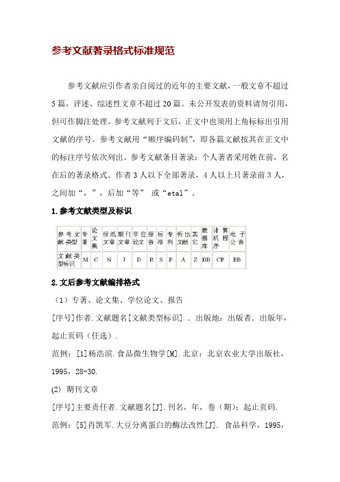
1.参考文献类型及标识2.文后参考文献编排格式(1)专著、论文集、学位论文、报告[序号]作者.文献题名[文献类型标识] .出版地:出版者.出版年,起止页码(任选).范例:[1]杨浩滨.食品微生物学[M].北京:北京农业大学出版社,1995,28-30.(2) 期刊文章[序号]主要责任者.文献题名[J].刊名,年,卷(期):起止页码.范例:[5]肖凯军.大豆分离蛋白的酶法改性[J]. 食品科学,1995,16(9):30-34.范例:[7]OUJP,YOSHIDA O,SOONGT, etal. Recent advance in research on applications energy dissipation systems[J]. Earthquake Eng , 1997,38(3): 358-361.(3) 论文集[序号]作者.文献题名[A].编者.论文题名[C].出版地:出版者,出版年,起止页码.范例:[8]瞿秋白.现代文明的问题与社会主义[A] .罗荣渠.从西化到现代化[C].北京:北京大学出版社,1990,121-133.(4) 报纸文章[序号]作者.文献题名[N].报纸名,出版日期(版次).范例:[10] 胡鞍钢. 中国能够实现粮食自给目标[N].联合早报,1994,10.(5) 国际、国家标准[序号]标准编号,标准名称[S].范例:[11]GB/T 16159-1996 ,汉语拼音正词法基本规则[S]. (6) 专利[序号]专利所有者.专利题名[P].专利国别:专利号,出版日期.范例:[12]姜锡洲. 一种温热外敷药制备方案[P].中国专利:881056073,1989,07,26.(7) 电子文献[序号]作者.电子文献题名[电子文献及载体类型标名].电子文献的出处或可获得地址,发表或更新日期/引用日期(任选).范例:[13]王明亮.关于中国学术期刊标准化数据库系统工程的进展[EB OL]. , 1998,08,16/1998,10,04.(8)各种未定义类型的文献[序号]主要责任者.文献题名.出版地:出版者,出版年.范例:[15]张永禄. 唐代长安词典[Z].西安:陕西人民出版社,1980. 毕业论文参考文献规范格式专著-M;论文集-C;报刊-N;期刊文章-J;学位论文-D;报告-R;专著或论文集中析出的文献-A;标准-S;专利- P;对于不属于上述的文献类型,采用字母“Z”标识。
医学英文文献 (1)
Muscle-selective synaptic disassembly and reorganization in MuSK antibody positive MG miceAnna Rostedt Punga ⁎,1,Shuo Lin,Filippo Oliveri,Sarina Meinen,Markus A.RüeggDepartment of Neurobiology/Pharmacology,Biozentrum,University of Basel,Basel,Switzerlanda b s t r a c ta r t i c l e i n f o Article history:Received 23February 2011Revised 15April 2011Accepted 21April 2011Available online 30April 2011Keywords:Myasthenia Gravis MG MuSKNeuromuscular junction MasseterMuscle atrophy DenervationMuSK antibody seropositive (MuSK+)Myasthenia Gravis (MG)patients present a distinct selective fatigue,and sometimes atrophy,of bulbar,facial and neck muscles.Here,we study the mechanism underlying the focal muscle involvement in mice with MuSK+experimental autoimmune MG (EAMG).8week-old female wildtype C57BL6mice and transgenic mice,which express yellow fluorescence protein (YFP)in their motor neurons,were immunized with the extracellular domain of rat MuSK and compared with control mice.The soleus,EDL,sternomastoid,omohyoid,thoracic paraspinal and masseter muscles were examined for pre-and postsynaptic changes with whole mount immunostaining and confocal microscopy.Neuromuscular junction derangement was quanti fied and compared between muscles and correlated with transcript levels of MuSK and other postsynaptic genes.Correlating with the EAMG disease grade,the postsynaptic acetylcholine receptor (AChR)clusters were severely fragmented with a subsequent reduction also of the presynaptic nerve terminal area.Among the muscles analyzed,the thoracic paraspinal,sternomastoid and masseter muscles were more affected than the leg muscles.The masseter muscle was the most affected,leading to denervation and atrophy and this severity correlated with the lowest levels of MuSK mRNA.On the contrary,the soleus with high MuSK mRNA levels had less postsynaptic perturbation and more terminal nerve sprouting.We propose that low muscle-intrinsic MuSK levels render some muscles,such as the masseter,more vulnerable to the postsynaptic perturbation of MuSK antibodies with subsequent denervation and atrophy.These findings augment our understanding of the sometimes severe,facio-bulbar phenotype of MuSK+MG.©2011Elsevier Inc.All rights reserved.IntroductionAbout 40–70%of acetylcholine receptor (AChR)-antibody seronegative Myasthenia Gravis (MG)patients have antibodies against the muscle tyrosine kinase (MuSK)(Hoch et al.,2001;Sanders et al.,2003).In MuSK-antibody seropositive (MuSK+)MG patients,there is often selective involvement of bulbar-,neck-and facial muscles,as well as muscles that are usually asymptomatic in AChR-antibody seropositive (AChR+)MG,such as the paraspinal muscles (Sanders and Juel,2008).Contrary to conventional AChR+MG patients,the majority of MuSK+patients does not experience symptomatic relief from acetylcholine esterase inhibitors (AChEI)(Evoli et al.,2003)but instead may respond with pronounced nicotinic adverse effects,such as muscle fasciculations and cramps (Punga et al.,2006).Pronounced atrophy of facial muscles has also been described in MuSK+patients,although the concomitant treatment of corticosteroids in most cases has made it dif ficult tojudge whether the MuSK antibodies or the cortisone treatment is the cause of the atrophy (Farrugia et al.,2006).Muscle biopsy studies of the intercostal muscle and biceps brachii muscle from MuSK+patients have shown little AChR loss (Selcen et al.,2004;Shiraishi et al.,2005),however,the neuromuscular junction (NMJ)pathophysiology in the most affected facial or bulbar muscles has not been studied.Nevertheless,MuSK antibodies have been shown to be pathogenic in animals,both after immunization with the extracellular domain of the MuSK protein itself (Jha et al.,2006;Shigemoto et al.,2008;2006;Xu et al.,2006)and after passive transfer of sera from MuSK+MG patients (Cole et al.,2008;ter Beek et al.,2009).MuSK is essential to the process of NMJ formation,maintenance (Wang et al.,2006)and integrity,as perturbations in MuSK protein expression cause pronounced disassembly of the entire NMJ (Hesser et al.,2006;Kong et al.,2004).Other NMJ proteins that are essential for synaptogenesis include Dok-7,a downstream adaptor protein to MuSK (Okada et al.,2006),Lrp4,the co-receptor for neural agrin (Kim et al.,2008;Zhang et al.,2008),rapsyn and the AChR subunits.The effects of MuSK antibodies in-vivo on the gene expression of those synaptic proteins in the facial or bulbar muscles have not yet been established.Here,we hypothesized that low expression levels of MuSK may render some muscles more vulnerable to the effect of MuSK antibodies in the EAMG mouse model.We show that MuSK antibodiesExperimental Neurology 230(2011)207–217⁎Corresponding author at:Institute of Neuroscience,Department of Clinical Neuro-physiology,Uppsala University Hospital,Uppsala,Sweden.Fax:+4618556106.E-mail address:annarostedtpunga@ (A.R.Punga).1Present address:Department of Clinical Neurophysiology,Uppsala University Hospital,Uppsala,Sweden.0014-4886/$–see front matter ©2011Elsevier Inc.All rights reserved.doi:10.1016/j.expneurol.2011.04.018Contents lists available at ScienceDirectExperimental Neurologyj o u r n a l h o me p a g e :w w w.e l s e v i e r.c om /l o c a t e /y e x n rinduce severe fragmentation of the postsynaptic AChR clusters in particular in the masseter and thoracic paraspinal muscles,with less fragmentation in the limb muscles.The severe postsynaptic pertur-bation results in subsequent denervation of musclefibers,not previously described in EAMG or MG.We propose that one underlying mechanism for the severe involvement of the facial masseter muscle, with severely impaired NMJ architecture,atrophy and denervation,is its low intrinsic levels of MuSK.Moreover,muscles respond to the partial denervation caused by MuSK antibodies in two different ways: (1)terminal nerve sprouting in muscles with high intrinsic levels of MuSK(i.e.soleus,sternomastoid)and(2)no nerve sprouting in muscles with low intrinsic MuSK levels(i.e.masseter,omohyoid).MethodsProduction of recombinant rat MuSKpCEP-PU vector containing the His-tagged extracellular domain of recombinant rat MuSK(aa21-491;(Jones et al.,1999))was transfected(Lipofectamine2000;Invitrogen)into HEK293EBNA cells.The overexpressed protein was purified from the cell superna-tant over a Ni-NTA superflow column(Qiagen)and was subsequently dialyzed against PBS.Protein concentration was determined at OD 280nm and purity was ensured by SDS-PAGE.Experimental animalsC57BL6mice and mice expressing yellowfluorescence protein (YFP)in their motor neurons under Thy-1promoter(Feng et al., 2000)were originally supplied from Jackson Laboratories(Bar Harbor, Maine,US).For immunization,8week-old female mice were used.All mice were housed in the Animal Facility of Biozentrum,University of Basel,where they had free access to food and water in a room with controlled temperature and a12hour alternating light–dark cycle.All animal procedures complied with Swiss animal experimental regu-lations(ethical application approval no.2352)and EC Directive 86/609/EEC.ImmunizationThe immunization procedure has been described previously(Jha et al.,2006).Briefly,eleven C57BL6and seven Thy1-YFP female mice aged8weeks were anesthetized(Ketamine:111mg/kg and Xylazine: 22mg/kg)and immunized with10μg of MuSK emulsified in complete Freund's adjuvant(CFA,Difco laboratories,Detroit,Michigan,US) subcutaneously in the hind foot pads,at the base of the tail and dorsolateral on the back.At day28post-injection,immunization was repeated.A3rd immunization was given to mice that did not show any myasthenic weakness after56days.Control mice(8female mice) were immunized with PBS/CFA.Clinical and neurophysiological examinationMuscle weakness was graded every week,as described(Nakayashiki et al.,2000).Briefly,mice were exercised by20consecutive paw grips on a grid and were then placed on an upside-down grid.The time they could hold on to the grid reflected the grade of fatigue and muscle weakness.EAMG grades were as follows:grade0,no weakness;grade1, mild muscle fatigue after exercise;grade2,moderate muscle fatigue; and grade3,severe generalized weakness.Evaluation of the response to AChEIs was performed by i.p.injection of a mix of neostigmine bromide (0.0375mg/kg)and atropine sulfate(0.015mg/kg)in mice with EAMG grades2and3(Berman and Patrick,1980).Repetitive stimulation of the sciatic nerve and recording from the gastrocnemius muscle with monopolar needle electrodes was performed under anesthesia,in mice with EAMG grades2(n=2)and3(n=2),using a Saphire1L EMG machine(Medelec).Decrement was calculated as percent amplitude change between the1st and4th compound motor action potentials evoked by a train of10impulses where10%was considered as pathological.ELISASera were obtained from tail vein blood on day0(preimmune sera)and day35post-immunization.ELISA plates(Nunc MaxiSorp, Fisher Thermo Scientific,Rockford,IL,US)were coated with250ng/ ml of His-labeled rat MuSK(50μl/well),blocked with3%BSA/PBS and then incubated with a sera dilution row(1:3000–1:2,000,000).Pre-immune sera constituted negative and rabbit-anti-MuSK antibody (Scotton et al.,2006)positive controls.After washing,plates were incubated with secondary HRPO-conjugated goat-anti-mouse (1:2000)and goat anti-rabbit antibodies(1:2000;both from Jackson Immuno Research Laboratories,Westgrove,PA,US).HRPO activation by a TMB substrate was terminated with1N HCl after5min. Absorbance was read at450nm.Non-specific binding,determined by incubation of plates with pre-immune serum,was subtracted.The data were displayed as“half maximum MuSK immunoreactivity”,which represents the immunore-activity at a dilution of1:27,000,where the majority of sera obtained50% of maximum absorbance(in the linear range of the absorption at450nm).Western blotWestern blot of masseter muscles was conducted as described (Bentzinger et al.,2008).10μg of protein was resolved on a4–12%Nu-PAGE Bis–Tris gel(Invitrogen,Eugene,OR,US),transferred to nitrocellulose membrane,probed with rat monoclonal anti-NCAM (CD56;1:100;GeneTex)and rabbit polyclonal anti-pan-actin(1:1000; cell signaling)and then recognized with HRPO-conjugated antibodies (1:5000;Jackson Immuno Research Laboratories,Westgrove,PA,US). Quantitative RT-PCR analysisMouse muscle RNA was extracted and purified as previously described(Punga et al.,2011).RT-PCR reactions(triplicates)were carried out with Power SYBR Green PCR Master Mix reagent(Applied Biosystems,Warrington,UK).β-actin was used as endogenous control (Punga et al.,2011;Murphy et al.,2003;Yuzbasioglu et al.,2010).The following primer sets were used:MuSK:5′-GCCTTCAGCGGGACTGAG-3′and5′-GAGGCGTGGTGA-CAGG-3′Lrp4:5′-GGATGGCTGTACGCTGCCTA-3′and5′-TTGCCGTTGTCA-CAGTGGA-3′Dok-7:5′-CTCGGCAGTTACAGGAGGTTG-3′and5′-GCAATGC-CACTGTCAGAGGA-3′A C h Rα1:5′-G C C A T T A A C C C G G A A AG T G A C-3′a n d5′-CCCCGCTCTCCATGAAGTT-3′AChRε:5′-CTGTGAACTTTGCTGAG-3′and5′-GGAGATCAG-GAACTTGGTTG-3′AChRγsubunit:5′-AACGAGACTCGGATGTGGTC-3′and5′-GTCGCACCACTGCATCTCTA-3′Rapsyn:5′-AGGTTGGCAATAAGCTGAGCC-3′and5′-TGCTCTCACT-CAGGCAATGC-3′MuRF-1:5′-ACC TGC TGG TGG AAA ACA-3′and5′-AGG AGC AAG TAG GCA CCT CA-3′β-actin:5′-CAGCTTCTTTGCAGCTCCTT-3′and5′-GCAGCGA-TATCGTCATCCA-3′A C h E:5′-G G G C T C C T A C T T T C T G G T T T A C G-3′a n d5′-GGGCCCGGCTGATGAG-3′208 A.R.Punga et al./Experimental Neurology230(2011)207–217NMJ whole mount analysisAlexa Fluor555-conjugatedα-bungarotoxin(1μg/ml;Invitrogen) was injected into soleus,EDL,sternomastoid,omohyoid,masseter and the thoracic paraspinal muscles as described(Bezakova and Lomo, 2001).At least12images of each muscle per mouse(6MuSK-immunized YFP-transgenic mice and4CFA-immunized control mice) were collected with a confocal laser-scanning microscope(Leica TCS SPE).Laser gain and intensity were equal for all images.Quantification of pre-and postsynaptic area was performed in ImageJ(http://imagej. /ij/index.html).NMJs containing terminal nerve sprouts(processes with YFP expression)were counted in muscles fromfive MuSK+EAMG mice with disease grades1–3.At least275NMJs per muscle were analyzed. The number of postsynapse fragments per NMJ was counted using a fluorescence microscope(Leica DM5000B)in at least50NMJs per muscle deriving fromfive MuSK+EAMG grades1–2and from four control mice(Supplemental Fig.1).Fragmentation was classified as follows:1)normal pretzel-like NMJ;2)slight to moderate fragmen-tation;and3)severe fragmentation or absent postsynapse.The degree of postsynaptic perturbation per muscle was judged based on the percentage of NMJs belonging to each postsynaptic class and was further subdivided into number of postsynapse fragments per NMJ:1–3,4–6,7–9,10–12and more than12.Each NMJ was given the median score for that subgroup.The fragmentation score was obtained by taking the ratio of the score between the EAMG mice and control mice.Statistical analysisIndependent,2-sample t-test was performed for parametric data. For ordinal data(ELISA),the non-parametric test Spearman Rank Correlation was applied.A p-value b0.05was considered significant.Fig.1.(A)Development of EAMG after immunization with recombinant rat MuSK.Progress of clinical EAMG grade at the time points week4,5,7and10.*The mice with EAMG grade 3were sacrificed after week7(hence no new grade3mice week10)and the remaining mice were sacrificed andfinally evaluated at week10.(B)One mouse,representative of the most severely affected MuSK+EAMG mice,withflaccid paralysis and pronounced kyphosis.(C)Repetitive nerve stimulation performed in the same mouse.Stimulation of the sciatic nerve and recording of the gastrocnemius muscle demonstrated a35%decrement between the1st and4th compound motor action potentials at low frequency3Hz stimulation.(D)Correlation of clinical EAMG grade with MuSK antibody titer.Half of maximum MuSK immunoreactivity(1:27,000dilution)in sera from MuSK immunized mice was assessed by Elisa at450nm.R=0.483(Spearman's Rank Correlation);p b0.05.209A.R.Punga et al./Experimental Neurology230(2011)207–217ResultsMuSK+EAMG presents with prominent kyphosis,paralysis and weight lossOut of the18mice immunized with MuSK,thefinal EAMG grade 0was seen in3mice(17%),grade1in8mice(44%),grade2in4mice (22%)and grade3in3mice(17%)(Fig.1A).No difference in disease incidence was found between the groups of C57BL6mice and the YFP-transgenic mice.Based on this,the data were pooled in the current study.The most severe phenotype of EAMG(grade3)includedflaccid paralysis,pronounced kyphosis and weight loss(Fig.1B).In-vivo nerve stimulation at3Hz with recording from the gastrocnemius muscle revealed a decrement of10–40%in the MuSK+EAMG mice (Fig.1C),whereas the control mice had normal neuromuscular transmission(data not shown).MuSK immunoreactivity in sera(day 35)correlated with clinical severity(Fig.1D;Spearman Rank Correlation;R=0.483;p b0.05),although some mice developed measurable MuSK antibodies without showing obvious muscle weakness or fatigue.Bulbar symptoms underlying weight loss in MuSK+EAMG The MuSK immunized mice steadily decreased significantly in body weight after the2nd immunization(Fig.2A),in contrast to the control mice,and thefinal body weight of the MuSK+mice was significantly smaller(p b0.001;Fig.2B).This severe weight loss was slowed but not stopped after introduction of wet food(data not shown).Since the timeline of the weight drop also correlated with development of muscle fatigue,the weight loss was assumed to be indicative of chewing and swallowing difficulties,some of the cardinal symptoms of MuSK+MG.To determine whether loss of muscle weight significantly contributed to the overall lower body weight in the MuSK+EAMG mice,the weight offive different muscles was assessed.The masseter was the only muscle with a significant weight reduction(p b0.01;Fig.2C),implying that this muscle is atrophic and further indicating chewing difficulties.Adverse effects of AChEIs in MuSK+EAMGTo elucidate whether AChEIs have any beneficial effect on fatigue or weakness in MuSK+EAMG,a neostigmine test was performed in the mice with EAMG grade3(n=3).No apparent improvement in weakness at rest or exercise-induced fatigue was seen;instead the mice experienced shivering and constant twitching of the tail,trunk and limbs starting after approximately13min(Supplemental Video1).This effect wore off after40min and was interpreted as nicotinic side effects and neuromuscular hyperactivity,usually seen after an overdose of AChEIs.Thus,this intolerance towards AChEIs in MuSK+EAMG indicates an abnormal sensitivity to acetylcholine.Impairment of NMJs in different musclesBecause muscle groups in the bulbar/facial/back region are selectively involved in the MuSK+EAMG model,we next examined the morphological changes of NMJs in the thoracic paraspinal muscles, masseter,omohyoid and sternomastoid and compared them with those in two limb muscles(EDL and soleus).Typical features of postsynaptic impairment were a fainting of AChRfluorescence,areas lacking AChRs(holes)and disassembly of AChR clusters.To quantify these impairments,we classified NMJs into three classes as illustrated in Fig.3A.All muscles from MuSK+EAMG mice displayeddifferentFig.2.Weight loss in MuSK+EAMG.(A)The course of weight loss in two MuSK+mice with EAMG grade3compared to control(Ctrl CFA)mice(n=8).Initial body weight was comparable and weight loss started after the2nd immunization and weight kept dropping even though wet food was provided ad libitum.(B)Mean body weight was dramatically reduced in the MuSK immunized mice(MuSK+;n=18),compared to the control mice.Results shown as mean±SEM(gram);***p b0.001.(C)Muscles were weighed and compared between control mice(n=8)and MuSK+EAMG mice(n=18).Results displayed as mean muscle weight±SEM(mg).The only muscle which was significantly lighter in the MuSK+mice was the masseter.**p b0.01.210 A.R.Punga et al./Experimental Neurology230(2011)207–217degrees of NMJ impairment (Fig.3B).Moreover,we also observed that the postsynapses were often fragmented into two to three non-continuous fragments (see illustration in Fig.3A),which is in strong contrast to non-interrupted,pretzel-like shape of AChRs in non-immunized mice.This fragmentation was also quanti fied (Supplemental Fig.1)and expressed as “fragmentation score ”.Because fragmentation differs between muscles,this “fragmentation score ”was normalized to the control.As shown in Fig.3C,the fragmentation score varied between a minimum of 2.0in the soleus muscle and a maximum of 3.3in the masseter muscle.The postsynaptic labeling was markedly reduced in the MuSK+EAMG mice compared to control mice (Fig.4A).In the mild to moderately affected mice,AChR cluster area was reduced signi ficantly by 40–50%in all muscles (p b 0.01),except for the soleus,which had a remaining area of 85%(p b 0.05;Fig.4B).In the most severely affected mice,the postsynaptic area was also lost in the soleus muscle and less than 10%of the α-bungarotoxin staining remained in the masseter,sternomastoid and thoracic paraspinal muscles (p b 0.001).In the soleus,the nerve terminal area was unchanged in the severely affected mice,although in the other muscles the presynaptic area was reduced to about 80%in the mild to moderate cases and to 50%in the most severely affected mice (p b 0.01)(Fig.4C).Thus,both the pre-and postsynaptic data suggest that the masseter is the most affected muscle by MuSK antibodies whereas the soleus is the least affected.mRNA transcript expression of NMJ proteins in different muscles in MuSK+EAMGThe mRNA levels of MuSK differ signi ficantly between muscles,with the highest levels in the soleus muscle and lowest levels in the omohyoid muscle (Punga et al.,2011).Consequently,we additionally analyzed MuSK transcript levels in the masseter and these were even lower,with less than 20%of the mRNA levels detected in the soleus muscle (Supplemental Fig.2;p b 0.01).In the MuSK+EAMG mice,MuSK mRNA levels were signi ficantly reduced to 45%of control in the EDL (p b 0.05)and to 25%in the omohyoid muscle (p b 0.001;Fig.5A).Conversely,MuSK mRNA expression was signi ficantly increased in the masseter muscle (p b 0.01)and remained unchanged in the soleus and sternomastoid muscles.The expression of the AChR αsubunit was unchanged (Fig.5B),whereas the AChR εsubunit transcript was downregulated in the soleus and sternomastoid muscles and conversely a trend towards upregulation was seen in the masseter muscle (Fig.5C).The fetal AChR γsubunit mRNA was upregulated up to 1000-fold in the masseter (p b 0.001)and 5-fold in the soleus (p b 0.05;Fig.5D).The transcript levels of rapsyn,Lrp4and Dok-7were not signi ficantly altered (Fig.5E to G).Finally,the transcript levels of acetylcholine esterase (AChE)were signi ficantly down-regulated only in the sternomastoid and omohyoid muscles (p b 0.05;Fig.5H).Fig.3.Postsynaptic fragmentation of NMJs.(A)Examples from each postsynaptic classi fication.AChRs stained for alfa-bungarotoxin.(B)At least 275neuromuscular junctions (NMJs)per muscle were assessed in a total of 5MuSK+EAMG mice with moderate to severe disease.The appearance of NMJ postsynapses were divided into 3classes as indicated.Sol:soleus;EDL:extensor digitorum longus;STM:sternomastoid muscle;omo:omohyoid;mass:masseter.(C)Postsynapses were analyzed in at least 50NMJs per muscle in a total of 5MuSK+EAMG mice with slight to moderate disease grade and in 4control mice.Each NMJ in a certain subclass (for details see Supplemental Fig.1)was given the median score for that subclass.The fragmentation score for each muscle is the ratio in score between the EAMG mice and control mice.211A.R.Punga et al./Experimental Neurology 230(2011)207–217In summary,particularly the massive upregulation of MuSK transcripts and AChR γin conjunction with the overall trend for upregulation of mRNA for the other postsynaptic genes in the masseter muscle most likely re flects the severely disturbed neuro-muscular transmission in this muscle as a result of the MuSK antibodyattack.Fig.4.Pronounced reduction of postsynaptic and presynaptic area in different muscles in MuSK+EAMG.(A)Whole mount staining of single fiber layer bundles from the paraspinal muscle (ps),sternomastoid (STM),masseter (mass),omohyoid (omo),extensor digitorum longus (EDL)and soleus (sol)muscles from one control mouse immunized with CFA/PBS and one mouse with severe MuSK+EAMG.AChRs are visualized by alexa-555-bungarotoxin (red)and the motor nerve terminals by YFP expression (green).The AChRs are almost completely gone from the paraspinal muscles,STM and masseter.Confocal images of 100×magni fication,scale bar is 10μm.Quanti fication of (B)postsynapse (AChR clusters)and (C)presynaptic area in the different muscles;soleus (sol),extensor digitorum longus (EDL),omohyoid (omo),masseter (mass),sternomastoid (STM)and paraspinal muscles (ps).Results are given as %of ctrl area±SEM and at least 12NMJs in each muscle/mouse was measured in mice with EAMG grades 1–2(N =4),in mice with severe grade 3(N =2)and in control mice (N =4).**p b 0.01;#p b 0.001.212 A.R.Punga et al./Experimental Neurology 230(2011)207–217Denervation induced muscle atrophy in MuSK+EAMGThe masseter muscle lost weight and consequently we examined the mRNA levels of the atrophy marker muscle-speci fic RING finger protein 1(MuRF-1)and assessed signs of denervation.For this analysis we included the same muscles as previously examined,with the addition of thoracic paraspinal muscles due to the pronounced kyphosis of the MuSK+EAMG mice.MuRF-1mRNA levels were signi ficantly upregulated in the masseter muscle (p b 0.05;Fig.6A)and on the contrary MuRF-1transcript levels were strongly reduced in the soleus muscle (p b 0.01)in the MuSK+EAMG mice.Further,the protein levels of neural cell adhesion molecule (NCAM),a marker for denervation,were increased in the masseter muscle as detected by Western blot analysis.This is thus additional evidence of denervation in this particular muscle (Fig.6B).Absence of nerve sprouting may contribute to the severe denervation phenotypePresynaptic nerve terminals were visualized by transgenic YFP expression,allowing observation also of NMJs with absent post-synapses.In the masseter,the nerve terminals were still present although many postsynaptic AChRs were partially or completely lost.Intriguingly,no or little nerve sprouting was observed in these cases (Fig.7).This finding raised the possibility that postsynaptic perturbation in this muscle did not elicit any nerve sprouting response.To test this hypothesis,we examined ≥450NMJs in each of the muscles for such sprouting response.The quantitative examination of 4MuSK+EAMG mice revealed terminal nerve sprouting in approximately 20%of NMJs in the soleus and in 15%of endplates in the sternomastoid (Fig.7).On the contrary,theFig.5.mRNA levels of postsynaptic proteins in MuSK+EAMG.C:control mice immunized with CFA/PBS.MG:EAMG mice immunized with MuSK.mRNA levels in the control mice are set to 100%and levels in the EAMG mice are relative to the control levels.n=6control mice;n=6MuSK+EAMG mice.Genes of interest are relative to the house keeping gene β-actin.*p b 0.05;***p b 0.001.213A.R.Punga et al./Experimental Neurology 230(2011)207–217omohyoid,EDL,thoracic paraspinal muscles and masseter,showed terminal sprouting at only a few NMJs.In most samples no sprouting was observed,which differed signi ficantly from the soleus (p b 0.001)and the sternomastoid muscle (p b 0.05).Interestingly,the degree of nerve sprouting in each muscle correlates with their endogenous levels of MuSK,suggesting that MuSK levels regulate the nerve sprouting response and consequently reorganization of NMJs (Punga et al.,2011).DiscussionMuSK+MG patients often present with focal weakness of facial,bulbar,neck and respiratory muscles and sometimes also of paraspinal and upper esophageal muscles (Sanders and Juel,2008).The pathopysiological role of MuSK antibodies has been questioned based on the normal levels of MuSK and AChRs at the NMJ in cross-sections of the intercostal muscle and biceps brachii muscle of MuSK+MG patients (Selcen et al.,2004;Shiraishi et al.,2005).Further studies in mice have now shown that MuSK antibodies deplete MuSK from the NMJ,which results in disassembly of the postsynaptic apparatus and a reduced packing of AChRs (Jha et al.,2006;Shigemoto et al.,2006;Cole et al.,2010).Nevertheless,the exact mechanism of how MuSK antibodies affect muscles has so far not been revealed.Here we report that MuSK antibodies cause a severe pre-and postsynaptic disassembly in the facial,bulbar and paraspinal muscles but only a slight disruption in the limb muscles (i.e.soleus and EDL).The most affected muscle was the masseter and intriguingly this muscle expressed less than 20%of MuSK transcripts than the least affected soleus muscle.Considering that fast-twitch muscle fibers require larger depolarization to initiate contraction compared to slow-twitch fibers,we propose that the superfast twitch pattern of the masseter in combination with its critically low MuSK levels makes this muscle extra vulnerable to the synaptic perturbation following the MuSK antibody attack (Laszewski and Ruff,1985).Hence,over-expression of MuSK in vulnerable muscles could potentially alleviate the effects of the antibody-mediated attack against MuSK.Earlier studies of AChR+EAMG rats have been shown to respond to rapsyn-overexpression in the tibialis anterior muscle with no loss of AChRs and almost normal postsynaptic folds,whereas the NMJs of untreated muscles showed typical AChR loss and morphological damage (Losen et al.,2005).Nevertheless,we did not find any changes in rapsyn mRNA levels across the examined muscles.Additionally,our findings underline the importance of examining the clinically affected muscles in MuSK+MG in order to draw the right conclusions regarding NMJ morphology.In the MuSK+EAMG model,the mice exhibited obvious bulbar and thoracospinal muscle weakness,similar to the mouse model of congenital myasthenic syndrome with MuSK mutation (Chevessier et al.,2008).This implies a common clinical phenotype in mice with impaired MuSK function and corresponds well with the phenotype of human MuSK+MG.In contrast,we have noticed in parallel rounds with immunization of C57BL6mice with AChR from Torpedo californica that the AChR-antibody seropositive mice did not develop signi ficant weight loss,neck muscle weakness or kyphosis and also that their flaccid paralysis was reversible with AChEI treatment (Punga et al.,unpublished observations).The adverse effect of AChEIs in MuSK+EAMG resembles the neuromuscular hyperactivity reported in MuSK+MG patients (Punga et al.,2006).We hypothesize that the underlying reason for the hypersensitivity in the muscles to the additional amount of available ACh at the NMJ is a consequence of (1)the denervation of the muscle fibers with extensive AChR fragmentation and (2)the downregulation of AChE in some muscles (e.g.omohyoid and sternomastoid),which may result in AChEI overdose-like phenotype.The downregulation of AChE mRNA is most probably a direct consequence to the loss of MuSK,since MuSK binds collagen Q,an interaction that is thought to be largely responsible for the synaptic localization of AChE-collagen Q complex at the NMJ (Cartaud et al.,2004).The fragmentation and spatial dispersion of AChRs most likely inhibits the response to the increased ACh at the NMJ induced by AChEIs in the most clinically affected muscles.This study also investigated the possibility of MuSK antibodies to initiate a cascade that results in denervation-induced atrophy.MRI studies of newly diagnosed MuSK+patients have con firmed early muscle atrophy in the temporal,masseter and lingual muscles with fatty replacement (Zouvelou et al.,2009;Farrugia et al.,2006).We observed that,particularly in the masseter and paraspinal muscles,some NMJs were completely depleted of AChRs,and the subsequent loss of synaptic transmission most probably resulted in functional denervation.This explanation is further supported by the fact that pharmacological denervation arises after injection of botulinum toxin A (BotA),which also blocks the cholinergic synaptic transmission,and it is sometimes dif ficult to distinguish this from a surgical denervation due to the similarities in pattern,extent and time course (Drachman and Johnston,1975).Previous studies of passively induced AChR+EAMG in rats have shown an upregulation of the AChR ε,but not the γsubunits;thus suggesting that newly expressed AChRs in the case of antibody mediated AChR loss are of the adult type (Asher et al.,1993).However,our observed massive upregulation of AChR γtranscript in the masseter implies that,except for disturbed neuromuscular transmission,denervation is also ongoing since this usually correlates with the predominant expression of embryonic type receptors and re-expression of the fetal type occurs after experimental denervation (Witzemann et al.,1989).Levels of AChR γmRNA are normally extremely low in all muscles except for the extraocular muscles (Kaminski et al.,1996;MacLennan et al.,1997).Concomitantly with the very prominent increase in transcripts encoding AChR γ,MuSKFig.6.Atrophy-and denervation related markers in MuSK+EAMG mice.(A)mRNA levels of MuRF-1expressed as %of control in relation to β-actin.n=6control mice (C)and n=5MuSK+EAMG mice (MG).*p b 0.05.(B)Western blot of NCAM in the masseter muscle of one MuSK+EAMG mice with disease grade 3(MG)and in one CFA-immunized control mouse (Ctrl).NCAM was detected as 3bands at levels 120,140and 180kD and the loading control β-actin (45kD).An equal amount of protein was loaded.**p b 0.01.214 A.R.Punga et al./Experimental Neurology 230(2011)207–217。
参考文献1

参考文献[1] 姚磊,王静,赵海田.香菇多糖生物活性研究进展[J].中国甜菜糖业,2004,3:27-30.[2] 赵和平,张心团,马振声,等.抗肿瘤真菌药物的研究进展(综述)[J].安徽农业大学学报, 2003,30(4):462-465.[3] 唐省三,朱晓琴.复合真菌多糖的抗肿瘤及免疫增强作用初探[J].基础医学与临床,2004,24(5):599-600.[4] K.Noel Masihi, Katja Madaj, Henny Hintelmann,et al.Down-regulation of tumor necrosis factor-α, moderate reduction of interleukin-1β,but not interleukin-6 or interleukin-10,by glucan immunomodulators curdlan sulfate and lentinan[J].Int J Immunopharmac,1997,19(9/10):463-468.[5] Schepetkin IA, Faulkner C L, Nelson-Overton L K,et al. Macrophage immunomodulatory activity of polysaccharides isolated from Juniperus scopolorum[J]. Int Immunopharmacol, 2005,5(13-14):1783-1799.[6] 龚非力.医学免疫学[M].北京:科学出版社医学出版分社,2004: 171.[7] 陈秀芳,金丽琴,吕建新,等.蝉拟青霉对腹腔及肺泡巨噬细胞的激活作用[ J].中国病理生理杂志, 2002, 18(6): 694-697.[8] 金丽琴,吕建新,袁谦,等.蝉拟青霉对大鼠免疫功能及血液生化指标的影响[J].温州医学院学报, 2001, 31(6): 344-346.[9] 陈秀芳,金丽琴,吕建新,等.蝉拟青霉减轻环磷酰胺所致免疫抑制效应的实验研究[ J].温州医学院学报,2002, 32(6): 351-353.[10] Schepetkin IA,Quinn MT.Botanical polysaccharides:macrophage immunomodulation and therapeuticpotential[J].IntImmunopharma-col,2006,6(3):317.[11] 段博文,李运,刘昕,等.柳茶多糖对小鼠免疫功能的影响[J].中国中医药杂志, 2010, 35(11): 1467-1468.[12] 沈新蛾,龚珊,蒋量红,等.复方虫草精华对小鼠免疫功能的影响.中国血液流变学杂志,2003,13(4):327-330.[13] 张劲松,贾薇,邢增涛,等.灵芝子实体和菌丝体的提取物及其各纯化组分生物活性的比较.菌物学报,2004,23(1):85一92.[14] Kim GY,Lee JY,Lee JO,et al.Partial characterization and im-munostimulatory effect of a novel polysaccharide-protein complex extracted from Phellinuslinteus[J].Biosci Biotechnol Biochem,2006, 70(5):1218.[15] Raveendran N,Sonia R,Reshma R,et al.Immune stimulatingprop-erties of a novelpolysaccharide from the medicinal plant Tinospora cordifolia[J].Int Immunopharmacol,2004,4(13):1645.[16] 董永杰,单铁英,岳峰,等.枸杞多糖对淋巴细胞功能的影响[J].现代中西医结合杂志,2010,19(19):2362.[17] Popov SV,Golovchenko VV,Ovodova RG,et al.Characterisation of the oral adjuvanteffect oflemnan,a pectic polysaccharide ofLemna minor L[J].Vaccine,2006,24(26):5413.[18] WangD,Li X,Xu L,et al.Immunologic synergismwith IL-2 and ef-fects of cCHMIs onmRNA expression of IL-2 and IFN-gamma in chickenperipheral Tlymphocyte[J].Vaccine,2006,24(49-50):7109.[19] 陈西广,张冀伸,李润秋.鬼毛针中水溶性多糖的研究[J].真菌学报,1990,9(2):155-160.[20] 徐秋萍.中药药理学[M].贵阳:贵州科技出版社,1994,10(2):280-331.[21] 朵存莲.浅淡中药在兽医临床的双向调节作用[J].中国畜牧兽医,2008,35(5):115-116.[22] 曲章义,郭彩玲,凌秋,等.香芪口服液增强非特异免疫和体液免疫作用的实验研究[J].中草药,2001,32(10): 908-910.[23] Sun JL,Hu YL,Wang DY,etal.Immunologic enhancement ofcom-pound Chinese herbalmedicinal ingredients and their efficacy comparison with compound Chinese herbal medicines[J].Vaccine,2006,24(13):2343.[24] Kong XF,Hu YL,Yin YL,et al.Chinese herbal ingredients are ef-fective immunestimulators for chickens infected with the Newcastle disease virus[J].PoultSci,2006,85(12):169.[25] WuY,SunH,QinF,etal.Effectofvariousextractsand a polysaccha-ride from theedible mycelia of Cordyceps sinensis on cellular and humoral immune response againstovalbumin in mice[J].Phytother Res,2006,20(8):646.[26] Siegell,LinTL,Gieicher.Theredeell immune system(J).Lancer,1981,2(8246):556.[27] 杨运高,陈先明,傅江南,等.大鼠红细胞免疫功能低下模型的研究[J].中国中医基础医学杂志,2002,8(3):44.[28] 应自忠,张慧,韩志红.壳多糖对荷瘤小鼠红细胞免疫功能的影响[J].中国公共卫生,2000,16(9):831.[29] 廉宜君,谷新利,李炳奇.从中药方剂中提取的复合多糖对小鼠免疫功能的影响[J].畜牧与兽医,2007,39(10):50-52.[30] 单俊杰,王顺春,刘涤,等.黄芪多糖的化学和药理研究进展[J].上海中医药大学学报,2000,14(3):61-65.[31] Cold spring harbor.Signal and gene expression in the immune sys-tem[M].NewYork:Cold spring harbor laboratory,1999(sympo- sium64):71-78.[32] 陆德源.现代免疫学[M].上海:上海科学技术出版社,1993,40(15):65.[33] YangT,Jia M,Meng J,et al.Immunomodulatory activity of polysac-charide isolated from Angelica sinensis[J].Int J Biol Macromol,2006,39(4-5):179.[34] Chen X,Hu ZP,Yang XX,et al.Monitoring of immune responses to a herbal immuno-modulator in patients with advanced colorectal cancer[J].Int Immunopharmacol,2006,6(3):499.[35] Zhao L.Changchun Zhongyiyao Daxue Xuebao 2006;22(3):67-68赵雷.中药多糖类物质的应用研究与现状分析[J].长春中医药大学学报,2006,22(3):67-68.[36] Balachandran P,Pugh ND,Ma G,et al.Toll-like receptor 2-depen-dent activation of monocytes by Spirulina polysaccharide and its immune enhancing action in mice[J].Int Immunopharmacol,2006,6(12):18.[37] 徐军发,侯敢,黄迪南,等.酵母多糖对小鼠腹腔巨噬细胞产生一氧化氮和白细胞介素-1的影响[J].中国生化药物杂志, 2002, 23(2): 67-68.[38] Chen X,Hu ZP,Yang XX,etal.Monitoring of immune responses to a herbal immuno-modulator in patients with advanced colorectal cancer[J].Int Immunopharmacol,2006,6(308):499.[39] LimTS, Na K, Choi E M,etal. Immunomodulating activities of polysaccharides isolated fromPanax ginseng[J]. J Med Food,2004,7(1): 1-6.[40] Kim K A, Choi S K, Choi H S. Corn silk induces nitric oxide synthase in murine macrophages [J]. ExpMol Med, 2004,36(6):545-550.[41]Schepetkin IA, Quinn MT. Botanical polysaccharides: macrophage immunomodulation and therapeutic potential [J]. Int Immunopharmacol, 2006,6(3): 317-333.[42] Kim GY, Oh YH, Park YM. Acidic polysaccharide isolated from Phellinus linteus induces nitric oxide-mediated tumoricidal activity of macrophages through protein tyrosine kinase and protein kinase C[J].Biochem Biophys Res Commun,2003,309(2):339-407.[43]Chanq CL,Liao JC,Kuo L.Macrophage arginase promotes tumor cell growth and suppresses nitric oxide-mediated tumor cytotoxicity[J].Cancer Res,2001,61(3):1100-1106.[44] Soini Y,KalosK,Puhakka A,et al.Expression of inducible nitric ox-ide synthase in healthy pleura and in malignant mesothelioma[J].BrJ Cancer,2000,83(7):880-886. [45] GiYK, Gap SC, SangHL, eta.l Acidic polysaccharide i-solated from Phellinus linteus enhances through the up-regulation of nitric oxide and tumor necrosis factor-αfrom peritoneal macrophages [J]. J Ethnopharmaco,l 2004, 95(1): 69-76.[46] Kun YL,Young JJ. Macrophage activation by polysaccha-ride isolated from Astragalusmembranaceus [J]. Int Im-munopharmaco,l 2005, 5(7-8): 1225-1233.[47] Nose M,Terawaki K,Oguri K,et al.Activation of macrophages by crude polysaccharide fraction obtained from shoots of Glycyrrhiza glabra and hairyroots ofGlycyrrhiza uralensis in vivo[J].Biol Pharm Bull,1998,21(10):1110.[48] 张劲松,李卫军.肿瘤免疫抑制与治疗[J].国外医学:免疫学分册,2001,24(1):50-53.[49] 米蕊芳,张宏冰.雷帕霉素在免疫抑制和肿瘤抑制中的双重作用[J].基础医学与临床.2008,28(12):1329-1331.[50] 张艳,梁华平.黄芪多糖对烧伤小鼠细胞免疫功能的作用及机理探讨[J].中国实验临床免疫学杂志,1995,7(2):41.[51] 石焱,弓小雪,那婕.牛大力多糖对免疫抑制小鼠的免疫调节作用[J].临床军医杂志,2008,36(4):530-532.[52] 许爱华,任莉,郑媛媛,等.银杏外种皮多糖对环磷酰胺诱导的免疫抑制小鼠免疫反应的调节作用[J].中国药理学与毒理学杂志,2008,22(1):69-72.[53] 王惠珍.盐酸左旋咪唑搽剂可安全方便地提高机体免疫力[J].中国社区医师,2007,23(13):16.[54] 董兰凤,刘京生,苗智慧,等.附子多糖对H22和S180荷瘤小鼠的抗肿瘤作用研究[J].中国中医基础医学杂志,2003,9(9):14-17.[55] 刘建东.赤雹果乙醇提取物的抗炎作用的实验研究[J].中国医药指南, 2009, 7(2): 40.[56] XU L,CHEN H,XU H,et al.Anti-tumour and immuno-modulation effects of triptolide-loaded polymeric micelles[J].European Journal of Pharmaceutics and Biopharmaceutics,2008,70(3):741-748.[57] 陈秀芳,金丽琴,吕建新,等.蝉拟青霉减轻环磷酰胺所致免疫抑制效应的实验研究[ J].温州医学院学报,2002, 32(6): 351-353.[58] 邵树军,刘彩玉,刘雄伯,等.牛膝多糖对小鼠免疫功能影响的研究[J].肿瘤防治杂志,2002,9(1):57.[59] 宋春华,金明,陈京,等.保元汤醇提取物不同途径给药对小鼠体重及免疫器官的影[J].中国实验临床免疫学杂志, 1998,10(3):39-41.[60] 于丽萍,邹碧珍,李新玉,等.真菌多糖对小鼠腹腔巨噬细胞免疫功能的影响[J].生物技术,1998,8(2):38-40.[61] 吴小丽,蔡云清,赵岩,等.蒲公英提取物对小鼠免疫功能的调节作用[J].南京医科大学学报:自然科学版,2005,25(3):163-165.[62] 王莉,何赟绵,姚全胜.毛蚶多糖免疫调节作用的实验研究[J].华西药学杂志,2009,24(4):340-342.[63] 张艳,梁华平.黄芪多糖对烧伤小鼠细胞免疫功能的作用及机理探讨[J].中国实验临床免疫学杂志,1995,7(2):41.[64] 吴宪瑞,孔令员、淦洪.黑木耳多糖的医疗保健价值.林业科技,1996,21(3):32-33.[65] 甘勇,吕作丹.阿魏蘑多糖理化性质及免疫活性研究.菌物系统,200l,20(2):228-232.[66] 于敏,沈业寿,梅一德,等.蜜环菌菌索多糖的免疫增强作用研究.生物学杂志,2001,18(4):16-19.[67] 贺新怀,席孝贤.中医药免疫学[M].北京:人民军医出版社,2002,25(9)406-409.[68] 龚非力.医学免疫学[M].北京:科学出版社,2002:77-88.[69] 赵玉珍,陶上乘,王英华,等.小叶黑柴胡药理作用的进一步研究[J].中药材, 1998,21(6):307.[70] 李映丽,吴万兴, 鲍德虎,等 .魔芋甘露聚糖免疫作用的研究[J].陕西林业科技,1999,28(4):68-70.[71] 徐月清,曹志然,李立萍,等.灵芝对小鼠免疫功能调节作用的研究[J].实验动物科学与管理,1999,16(1):18-20.。
文献综述范文1
肺活量测量系统的概述【摘要】自1718年测出肺活量值之后,随着科学技术的不断发展,肺活量检测技术也层出不穷。
肺活量检测对于人体健康评估具有很大的意义,但是如何提高肺活量计的精度、减小装置体积、降低成本,仍是重要的研究课题之一。
本文整理了各种肺活量计的发展历史、制作原理、最新技术的应用及发展前景。
【关键词】机械式肺活量计;电子肺活量计;压差式;节流装置【Abstract】Since 1718 the value of vital capacity was measured,with the development of science and technology, vital capacity detection technology is endless. vital capacity detection is of great significance to human’s health assessment test, but how to improve the accuracy , reduce the volume and the cost, still is one of the most important research projects. This sorted out the history of development, the principle of various spirometer, the application of the latest technologies and development prospects.【Key word】mechanical pneusometer; Electronic lungs;Pressure-Gradient; throttling device1 肺活量计的发展历史肺活量计的发展历史大致可以分为以下二个阶段:机械式肺活量计发展时期(18~19世纪)、电子肺活量计发展时期(20世纪~21世纪)。
中国文献学

1,文献一词最初具有两方面的含义,一是指典籍,二是指贤才。
2,据载,孔子在整理“五经”(即《诗》;《书》;《礼》;《易》;《春秋》)方面贡献很大,也为后世文献整理树立了典范。
3,西汉刘向和刘歆父子在文献学方面的突出贡献是遍校群书,编纂目录学著作《别录》和《七略》。
4,西晋之时,汲郡有著名的文献发掘事件,出土了战国时期的竹简若干,其所得竹简文献一般称汲冢周书。
5,现存最早的雕版印刷文献为唐咸通九年(868)刻印之《金刚经》。
6,文献学上所谓“三通”是指杜佑的《通典》、郑樵的《通志》和马端临的《文献通考》。
7,清代考史三大家为钱大昕、王鸣盛和赵翼,分别著有廿二史考异、《十七史商榷》、廿二史札记等考史名著。
8,金石的“金”指青铜器;金文则指商周时期刻于其上的文字。
9,总集指搜集两种以上文献,按一定理念和体例编校并冠以一个总名的著作。
10,殷墟甲骨文的发现始自1899年,绵延整个20世纪,为中国现代最重要的考古发现之一。
1,1983年颁布的中国国家标准《文献著录总则》规定“文献”为“记录有知识的一切载体”。
《中国大百科全书》则界定“文献”为“记录有知识和信息的一切载体”。
2,秦始皇三十四年的焚书事件对先秦文献的流传产生了很大的破坏。
3,东汉晚期,经学大家郑玄遍注群经,对文献整理作出了重要贡献。
4,明代官方编纂了一部我国古代规模最大的类书,即永乐大典。
5,清代考据学的鼎盛时期为乾嘉时期,有惠栋为代表的吴派,戴震为代表的皖派和汪中、焦循为代表的扬州学派等。
6,近现代学者中,被称为“甲骨四大家”的是罗振玉、王国维、郭沫若和董作宾。
7,我国文献传抄的历史大致经历了由甲骨时代到金石时代简牍时代、缣帛时代、纸质时代再到当今电子时代。
8,古代集部文献的主要类型可分为总集和别集两大类。
9,我国古代目录分类主要有四分法和六分法,《汉书?艺文志》继承《七略》,采用的是六分法,四分法正式确立的标志是《隋书?经籍志》。
10,严复于《天演论》“译例言”中称译事有三难:信、雅、达。
英文文献1
©Smithers Rapra Technology, 2013*Corresponding author: Jiaping Liu, Email : liujiaping@Interfacial Interaction Between Polycarboxylate-based Superplasticizer and Cement Component Minerals1. InTRODUCTIOnPolymer dispersants play a significant role in improving the rheological behaviour of concentrated particle suspensions in many industrial processing s uch a s e .g. p aints, c eramics, cosmetics, and pharmaceutical formulations. Polycarboxylate-based superplasticizers (hereinafter called PCA) with an adsorbing backbone and grafted polyethylene oxide side chains are recognized as the most potential dispersants in concrete technology for their superior performance such as outstanding dispersive capacity and excellent flowability retention 1-4. It is expected that steric hindrance induced by hydrophilic graft side chains of PCA is responsible for a repulsive interparticle force that avoids the formation of agglomerates 5-7.Ordinary Portland cement mainly consists of four minerals, namely Ca 3SiO 5, Ca 2SiO 4, Ca 3Al 2O 6 and Ca 4Al 2Fe 2O 10, which are abbreviated to C 3S, C 2S, C 3A, C 4AF. Gypsum is always added to regulate the reactivity of the aluminate phase (C 3A, C 4AF). Thus, ettringite (AFt) is typically the first-formed product when Portland cement begins to hydrate 8. Due to the complexity of cement composition and the time-dependent process of hydration, the influence of PCA has been increasing investigated on simplified mineral phases rather than entire cement system 9-11. However, the consequence of these results is limited owing to the complex chemical process involved in cement slurry. For instance, pore solution (PS) generally describes the aqueous phase loaded with electrolytes which occurs betweencement, slag or other particles when dispersed in water at specific water-to-solids ratios. The PS of typical cement paste is dominated by various anions (OH -, SO 42-) and cations (K +, Ca 2+, Na +)10. It has been confirmed that sulphate in PS results in a competitive adsorption with PCA on the mineral phase 12. In addition, other ions present in PS have a non-negligible influence on PCA’s conformation in solution which could alter the steric hindrance effect induced by grafted polyethylene oxide side chains 3. All the above factors lead to a distinctive adsorption characteristic. As the model of action of PCA relies on their adsorption on mineral surface, their dispersing power can be altered.In order to investigate the influence of PS on the interfacial interaction between PCA and mineral phase, a synthetic pore solution was used to simulate the condition existing when cement hydration occurs. This work is mainly focused on the surface chemistry of mineral phase in PS and itsInterfacial Interaction Between Polycarboxylate-based Superplasticizer and Cement Component MineralsYinhui Yu, Jiaping liu *, Qianping Ran, Min Qiao, and nanxiao GaoState Key Laboratory of High Performance Civil Engineering Materials, Jiangsu Research Institute of Building Science, Nanjing, 210008, P.R. ChinaReceived: 26 May 2012, Accepted: 3 December 2012SUMMARYThis paper is presented for a better understanding of the interaction between polycarboxylate-based superplasticizer (PCA) and cement component minerals. The impact of PCA on the particle interaction among tricalcium silicate (C 3S), dicalcium silicate (C 2S) and the initial Portland cement hydrates ettringite (AFt) has been systematically examined. The zeta potential was highly dependent on the chemical composition of the solvent. Furthermore, it was demonstrated that adsorption always occurred between the minerals and anionic PCA in spite of their equal charge sign. For silicates (including C 3S and C 2S), the adsorption of high load Ca 2+ formed a positive layer and subsequently adsorbed anionic PCA. For AFt, the direct adsorption onto AFt results from its strong electrostatic interaction. The high affinity of PCA to aluminate phase surface can be attributed to the high reactivity of tricalcium aluminate and its large specific surface.Keywords: Polycarboxylate-based superplasticizer, Mineral phases, Adsorption, Zeta potentialYinhui Yu, Jiaping Liu, Qianping Ran, Min Qiao, and Nanxiao Gaoimpact on the action of PCA. Based on those results, an adsorption distribution of PCA on cement is proposed and the consequences for dispersing are presented.2. MATERIAlS AnD METHODS 2.1 Materials2.1.1 PCAAnionic PCA used in this paper was synthesized by aqueous free radical copolymerization according to a process described in detail in the literature 13. Its general chemical structure is shown in Figure 1. A precise characterization of PCA was performed by size exclusion chromatography (MALLS, Wyatt Technology). 1H NMR spectroscopy (BRUKER DRX-500) was applied to estimate the PCA composition (Figure 2). It showed proton resonance at d = 1.05 ppm (b’), d = 1.2-1.8 ppm (a, a’), d = 1.9-2.5 ppm (b), d = 3.2-3.7 ppm (c, c’, e), d = 4.16 ppm (d), Their characteristic properties are summarized in Table 1. Additionally, a typical IR spectrum of this copolymer displays bands at 3450 cm -1 (O-H), 2880cm -1 (-CH 2), 1730 and 1240 cm -1(-COO-), these bands are characteristic of the desired compounds (Figure 3).2.1.2 Mineral PhaseMineral phases including tricalcium silicate (C 3S), dicalcium silicate (C 2S) and tricalcium aluminate (C 3A) were synthesized by rapid cooling after reacting raw materials at 1500, 1400, 1350 °C, respectively 14. AFt was prepared by precipitation method 10. Mineralogical components of these materials were identified with an X-ray diffractometer, and the results confirmed that no other phases were present. All powders were dried, and then ground to below 80 µm. Particle size distributions presented in Table 2 were determined using laser diffraction (SYMPATEC Inc., Greman).Table 1. Molecular characteristics of PCA Sample Mn/g ×mol -1Mw/g ×mol -1PDI Molar ratio of x to y PCA305001287004.2276.0723.93Mn: number average molecular weight. Mw: weight average molecular weight PDI: Mn/Mw, polydispersity indexTable 2. Percentile volume diameters of mineral phase Material Percentage volume diameter/µmd 10d 50d 90d mC 3S 3.6416.1897.1933.24 C 2S 2.8926.1299.0041.09 C 3A6.9029.2792.1440.06Aft 3.5722.2357.9627.33Note :d 10, d 50 and d 90 represent the particle size below which the volume percentage is 10%, 50% and 90% respectively. d m was the mean particle sizeFigure 1. Chemical composition of the synthesized PCA (x:y=3:1)Figure 2. 1H-nMR spectrum of PCA measured in D 2OInterfacial Interaction Between Polycarboxylate-based Superplasticizer and Cement Component Minerals 2.1.3 Synthetic Pore SolutionThe selected cement from Lafarge wasmixed with deionized water to obtainthe synthetic solution simulating atypical ionic composition of the PSafter 1 h of hydration for a w/c ratioof 2. Table 3 presents the chemicalcomposition of the PS.2.2 Methods2.2.1 Zeta PotentialZeta potential measurement ofthe mineral phase at various PCAconcentrations was performed usingthe ZetaProbe instrument (ColloidalDynamics Inc., USA). This instrumentworks on the basis of the electroacousticmethod and is extensively applied incement systems. Before measuringthe samples, pH-meter (4, 7, and10) and zeta dip probe (KSiW-standard, provided by ColloidalDynamics Inc.) were calibrated. PCAtitration experiments were carried outon suspensions with solid volumefractions around 1%. The syringe unitfor titration was rinsed three times withDI and twice with PCA solution toensure the purity of the titrated solution.Zeta potential is highly sensitive to the chemical composition of the solvent, so two typical solvents, i.e. DI and PS, were used to determine their influence on the zeta potential measurements. To avert the strong background signals caused by the high ionic concentrations solutions, background measurements were also carried out for PS.2.2.2 Adsorption IsothermsThe amount of PCA adsorbed on mineral phase as a function of dosage was determined by a total organic carbon analyzer (TOC), Multi N/C3100 (Analytikjene AG, Germany). 4 g of solution containing various PCA (PCA solutions were prepared in DI and PS, respectively) and 2 g of mineral phase was vigorously agitated for 5 min at 20°C. The liquid phase was removed by centrifugation at 10,000 rpm for 3 min and was stabilized by adding 1 g of 2 mol/L HCl. The differencein concentration before and aftercontacting with mineral phase wasattributed to the adsorption of PCA.2.2.3 Concentrations of Metal IonsConcentrations of metal ionswere directly determined byinductively coupled plasma opticalemission spectroscopy (ICP-OES).3. RESUlTS AnDDISCUSSIOnTo understand the adsorption processbetween anionic PCA and cementcomponent mineral phase, it iscritical to investigate the surfacechemistry of mineral phase in DI andPS, respectively. The presence ofSO42- in PS instantaneously leads to astrong flocculation of C3A, thus, thesuspension could not be measured.Instead, AFt as the main hydrationproduct during the early stage wasselected.3.1 Zeta Potential of Mineralsin DI or PSThe time-dependent zeta potentialof mineral phase dispersed in DI orPS is presented in Figure 4. Over aperiod of 15 minutes, zeta potentialsof all samples increased to either morepositive or negative values both in DIand PS. This behaviour indicates thatthe partial dissolution of minerals isoccurring in an extremely short timeand soon reaches equilibrium.In basic theories, it is assumed that thesurface of silicate phases is negativelycharged. Interestingly, the initial zetapotential determined, whether in DI orPS, shows a slightly positive charge.More specifically, the zeta potentialof C3S ranged from +4.00 mV to+10.67 mV when dissolved in DI Table 3. Chemical composition of synthetic pore solution (mmol/l)Solvent Ca2+a K+a Na+a OH-b SO42-c PS32.7539.15 2.1458.8823.96 a:Directly determined by inductively coupled plasma optical emission spectroscopy (ICP-OES)b:Calculated from the pH value of PSc: Calculated from the charge balanceFigure 3. FTIR spectrum of the synthesized PCAYinhui Yu, Jiaping Liu, Qianping Ran, Min Qiao, and Nanxiao Gaoand PS, respectively. For C 2S, similar values were obtained (+1.51 mV and +12.83 mV). When silicate phases were dissolved in DI or PS, the ionic composition of the Stern layer is most likely to be dominated by cations (mainly Ca 2+). This behaviour could be proved by the fact that the concentrations of Ca 2+ in PS dropped markedly from 32.75 mmol/L (Table 3) to 3.18 mmol/L (Table 4) in the presence of C 3S.In aqueous cement slurries, a huge excess of cations always exists, as a result of dissolution of the cement mineral phase. Depending on the amount of cations present, in the equilibrium state the surface of silicate phases will become much more positive for PS. Previous literature values were measured in very dilute systems (W/C=1500), and thus, no charge inversion was observed for silicate phases. A ctually, similar charge inversion has also been discovered in ground granulated blast furnace slag-water system, and is responsible for the adsorption of anionic PCA 15. On the other hand, charge inversion from positively charged AFt to negative values is attributed to the presence of SO 42- as described by Zingg 10.3.2 Adsorption IsothermsDue to the positively charged layer of adsorbed cations, anionic PCA adsorption onto silicates occurs. PCA adsorption onto silicates in DI or PS was determined. In addition, the adsorption behaviour of PCA onto aluminates (C 3A, C 3A-gypsum mixture and AFt) was studied for a global evaluation of PCA ’s affinity for minerals.Adsorption isotherms obtained on silicates in DI or PS at increasing concentrations of PCA are depicted in Figure 5. The adsorbed amounts of PCA increased with increasing dosage until the point of saturation was reached. The presence of high ionic strength and sulphate resulted in the adsorption onto C 3S declining fromTable 4. Primary cation concentrations (mmol/l) of C 3S dissolve in DI or PS for 30 min Solvent Ca 2+K +Na +DI 4.180.210.30PS3.1839.852.22Figure 4. Time-dependent zeta potentials of the mineral phase dispersed in DI orPSFigure 5. Adsorption isotherms of PCA onto silicate phaseInterfacial Interaction Between Polycarboxylate-based Superplasticizer and Cement Component Minerals0.49 mg/g to 0.36 mg/g. However, the introduction of PS could not reduce the saturation adsorption on C 2S. A possible explanation for this observation is that the adsorption sites of C 2S could be greatly enhanced by the addition of PS.There is a significant difference of PCA adsorption ability between silicates and aluminates. Figure 6 presents the adsorbed PCA onto C 3A or C 3A/gypsum as a function of dosage. No saturation concentration was reached within the dosage of 0 to 0.8 mg/g. The presence of gypsum greatly decreased the adsorbed amount, which could again verify the competitive adsorption between sulphate and PCA. Because C 3A reacts instantaneously with water to form calcium hydroaluminates, when gypsum is added, the first-formed hydrate is AFt. Hence, adsorption ratio of PCA onto hydration products were further determined (Table 5). It becomes obvious from Table 5 that a large amount of PCA favours adsorption onto aluminates.3.3 Impact on Zeta PotentialsTo further investigate the interaction between PCA and the surface of minerals, zeta potentials of aqueous mineral suspensions were measured when the PCA was gradually dosed in. The results are shown in Figure 7. The addition of PCA could hardly modify the zeta potential of silicates, while it could indeed change the potential values toward the isoelectric point, which confirmed that long PEO side chains shift the slip plane of the zeta potential to greater distances away from the AFt surface.3.4 Mechanism of DispersionOnce ordinary Portland cement dissolves in water, flocculation alwaysTable 5. Adsorption ratio of PCA onto various mineral phase in DI or PS (2 mg/g)Sample C 3S+DI C 3S+PS C 2S+DI C 2S+PS C 3AC 3A+GpysumAFt+PS PCA/%24.5182126.561.55745.5Figure 6. Adsorption isotherms of PCA onto C 3A without or with gypsumFigure 7. Zeta potentials of minerals dispersed in DI or PS as a function ofincreasing PCA additionoccurs due to the opposite charge sign of minerals from the point of colloidal science. Previous results assumed that electrostatic repulsion between anionic PCA and negative charged silicates results in adsorption being impossible 11. However, our results indicate that the equal charge sign can still create PCA adsorption with the adsorbing high load Ca 2+Yinhui Yu, Jiaping Liu, Qianping Ran, Min Qiao, and Nanxiao Gaolayer. In addition, it was observed that aluminates always had a higher affinity to PCA. We must note that adsorption (depletion method) was based on the concentration differences before and after contact of powders. On the one hand, PCA intercalation into C 3A hydrates cannot be ignored 16. On the other hand, the larger specific surface of aluminates could lead to a higher adsorption amount. This uneven distribution of PCA in cement mineral systems could prevent the flocculation of fresh cement paste through the steric repulsive effect of PEO side-chains.Figure 8. Schematic illustration of the uneven distribution of PCA in cement mineral systems4. CONCLuSIONZeta potential is controlled by the chemical composition of the solvent. When the silicate phase dissolves in DI or PS, adsorption of high load Ca 2+ forms a positive layer and this subsequently adsorbs anionic PCA. Hence, it is demonstrated that adsorption always occurs between minerals and anionic PCA in spite of their equal charge sign. In this paper, the adsorption behaviour of the minerals and associated changes of zeta potentials are presented. Throughout the experimental data, a schematic illustration of PCA uneven distributionis also given for understanding the mechanism of dispersion.REFERENCES1. Houst Y .F., Bowen P., and Perche F.,Cem. Concr. Res. 38, (2008) 1197.2. Sakai E., Ishida A., and Ohta A., J.Adv. Concr. Technol. 4, (2006) 211.3. Kirby G.H. and Lewis J.A., J. Am.Ceram. Soc. 87, (2004) 1643.4. Sakai E., Ishida A., and Ohta A., J.Adv. Concr. Technol. 40 (2006) 211.5. Uchikawa H., Hanehara S., andSawaki D., Cem. Concr. Res. 27 (1997) 37.6. Chiu-Chun L. and Keng-Ming C., J.Interfacial Interaction Between Polycarboxylate-based Superplasticizer and Cement Component MineralsAppl. Polym. Sci.102, (2006) 3559.7. Nawa T., J. Adv. Concr. Technol.4(2006) 225.8. Taylor H.F.W., Cement Chemistry,Thomas Telford Publishing: London,1997.9. Yoshioka K., Tazawa E.I., and KawaiK., Cem. Concr. Res.32, (2002)1507.10. Z ingg A., Winnefeld F., and HolzerL., J. Colloid. Interface Sci.323,(2008) 301.11. Plank J. and Hirsch C., Cem. Concr.Res.37, (2007) 537.12. P lank J. and Winter Ch., Cem. Concr.Res.38, (2008) 599.13. Qianping R., Ponisseril S., andChangwen Miao., J.ColloidInterface Sci.336, (2009) 624.14. W esselsky A. and Jensen O.M., Cem.Concr. Res.39 (2009) 973.15. H abbaba A. and Plank J., J. Am.Ceram. Soc.95, (2011) 768.16. P lank J., Zhimin D., and Keller H.,Cem. Concr. Res.40, (2010) 45.Copyright of Polymers&Polymer Composites is the property of Rapra Technology and its content may not be copied or emailed to multiple sites or posted to a listserv without the copyright holder's express written permission.However,users may print,download,or email articles for individual use.。
参考文献1
参考文献(一)马列原著和党的文献1.马克思:《1844年经济学——哲学手稿》,人民出版社2000年版。
2.《马克思恩格斯选集》第1、3、4卷,人民出版社1995年版。
3.《马克思恩格斯选集》第1卷,人民出版社1992年版。
4.《马克思恩格斯全集》第3卷,人民出版社2002年版。
5.《列宁全集》第24卷,人民出版社1995年版。
6.《列宁选集》第4卷,人民出版社1995年版。
7.毛泽东:《国民精神总动员的政治方向》,《新中华报》1939年5月10日。
8.《毛泽东选集》第1—4卷1991年版,第5卷1996年版,人民出版社。
9.《建国以来毛泽东文稿》第6册,中央文献出版社1992年版。
10.《刘少奇选集》上卷,人民出版社1982年版。
11.《邓小平文选》第2卷1994年版,第3卷1993年版,人民出版社。
12.江泽民在中国共产党第十五次全国代表大会上的报告《高举邓小平理论伟大旗帜,把建设有中国特色社会主义事业全面推向二十一世纪》13.江泽民在中国共产党第十六次全国代表大会上的报告《全面建设小康社会,开创中国特色社会主义事业新局面》14.中共中央文献研究室编:《江泽民论有中国特色社会主义(专题摘编)》,中央文献出版社2002年版。
15.胡锦涛:《在中国文联第八次全国代表大会、中国作协第七次全国代表大会上的讲话》,《人民日报》2006年11月10日。
16.胡锦涛:《高举中国特色社会主义伟大旗帜为夺取全面建设小康社会新胜利而奋斗》,人民出版社2007年版。
17. 2010年7月,胡锦涛在中共中央政治局就深化我国文化体制改革研究问题进行第二十二次集体学习时的讲话18. 李长春发表十七届六中全会《文化大发展大繁荣决定》说明,人民网2011年10月27日,/xxfb/xwzx/whxw/201110/t20111027-132395.html19.《中共中央关于制定国民经济和社会发展第十一个五年规划的建议》,人民出版社2005年版。
中文文献1
高温过氧化物酶活性的辣根过氧化物酶和血红蛋白的分层锆(ⅳ)C.五.Kumar * 和A.·乔杜里的化学系,康涅狄格大学55 N.伊格尔维尔路,校区,CT 06269 3060,美国。
电子邮件︰ C.V.Kumar@;传真︰860-486-2981;电话︰860-486-3213收到(在美国马萨诸塞州剑桥市)2002 年7 月16 日,接受2002 年8 月16 日首次发布为推进项目在web 上2002 年9 月19 日辣根过氧化物酶和遇见的血红蛋白,当夹在画廊a-Zr(IV) 磷酸,表明过氧化物酶活性在高温(86-90 °C)、率增至 2 —— 3.6 倍率在室温下观察到。
酶是一种高效而特异的生物催化剂,但其在化学反应中的使用是有限的由于其脆弱性,费用,对温度、ph 值或有机solvents.1 酶的敏感性绑定在磷酸锆一(一锆(HPO4) 2·nH2O,一个ZrP)画廊或其它固体可以克服这些局限性,和固体支持提高了酶学性质在特定cases.2 酶绑定到固体经常丢失某些活动的结果这是由于酶结构或构象的变化。
酶是绑定的然而,提高稳定性超过弥补这个loss.3 的酶改善稳定性也可以促进酶活性在较高温度,并耐高温活性的酶是有吸引力的速率加速和驾驶吸热反应。
在这里,我们目前的辣根过氧化物酶(HRP) 和遇见的血红蛋白(Hb) 夹在一ZrP 画廊高温活动第一次报告HRP 固然重要作为有机底物的氧化催化剂,Hb 是廉价模型系统的辣根过氧化物酶过氧化物酶活性。
Hb 是氧气运输protein4 和Hb 广泛研究了氧存储和提取。
虽然Hb 不能作为生物系统中一种酶,其过氧化物酶活性是井documented.5 Hb 可以氧化酚类、胺和芳香hydrocarbons6 过氧化氢存在下的,通过高价的Fe (IV) 氧代中间的的方式类似于HRP.7 的Hb 的过氧化物酶样活性也有人在体内,8 和这可能有重大影响的组织损伤后损伤中风或心脏病发作。
论文的参考文献
论⽂的参考⽂献参考⽂献是在学术研究过程中,对某⼀著作或论⽂的整体的参考或借鉴。
征引过的⽂献在注释中已注明,不再出现于⽂后参考⽂献中。
按照字⾯的意思,参考⽂献是⽂章或著作等写作过程中参考过的⽂献。
论⽂的参考⽂献1 参考⽂献: [1]Ronald WRebore. Human Resources Administration in Education(6th Edition)[M]. Boston:Allyn &Bacon,Pearson Education, Inc.,20xx [2]C Chadwick, P Cappelli. Alternative to Generic Strategy Typologies in Strategic Human Resource Management[M]. Greenwich, Ct: JAI Press, 1999 [3]宋本江胡跃福:⼈才招聘,另请⾼明还是⾃⼰培养[J].中国⼈才,20xx(8) [4]丁敬平:向跨国公司学习什么[J].中国企业家,20xx(11) [5]程桢贾雅军:企业内部⼈才资源的运⽤[J].经济问题,20xx(11) [6]安鸿章:企业⼈才聘任的两种模式[J].中国⼈才,20xx(4) [1]亚瑟W⼩舍曼:乔治W勃兰德·斯科特A斯奈尔著张⽂贤主译:⼈⼒资源管理.东北财经⼤学出版社,20xx [2]魏峰李赕张⽂贤:国内外⼼理契约研究的新进展.管理科学学报,20xx(10) [3]陈加洲:凌⽂辁&⽅俐洛.组织中的⼼理契约,管理科学学报, 20xx,4(2) [4]陈加洲凌⽂辁·⽅俐洛:企业员⼯⼼理契约的结构维度. ⼼理学报, 20xx,35(3) 参考⽂献: [1]王刚编著:《把握好⽤⼈的技巧与管⼈的学问》.地震出版社,20xx年2⽉ [2]斯蒂芬·P·罗宾斯著:《管⼈的真理》.中信出版社,20xx年9⽉ [3]李诚主编:《⼈⼒资源管理的12堂课》.中信出版社,20xx年11⽉ [4]吴志明著:《员⼯招聘与选拔实务⼿册.机械⼯业出版社,20xx年1⽉ [5]赵曙明:《⼈⼒资源管理研究》.中国⼈民⼤学出版社,20xx年 [6]詹姆斯.W.沃克:《⼈⼒资源战略》.中国⼈民⼤学出版社,20xx年1⽉ [7]罗布.⼽菲:《⼈员管理》.中国⼈民⼤学出版社,20xx年01⽉论⽂的参考⽂献2 [1]林红梅.“⽣态⽂明与和谐社会”理论研讨会综述[J].思想理论教育导刊,20xx(5):92-93. [2]杨多贵,周志⽥,陈劭锋.我国⼈与⾃然和谐发展⾯临的挑战及其战略选择[J].上海经济研究,20xx(4):6-12. [3]利奥波德.沙乡年鉴[M].侯⽂惠,译,长春:吉林⼈民出版社, 20xx. [4]卢风,肖葳.应⽤伦理学导论[M].北京:清华⼤学出版社, 20xx. [5]李培超.⾃然的伦理尊严[M].南昌:江西⼈民出版社,20xx:16. [6]林红梅.试论拯救环境危机的多维视⾓[J].⾏政与法,20xx(1):97-99. [7]马克思,恩格斯.马克思恩格斯全集:第25卷[M].北京:⼈民出版社,1971:119. [8]马克思,恩格斯.马克思恩格斯全集:第20卷[M].北京:⼈民出版社,1971:519. [9]何怀宏.⽣态伦理——精神资源与哲学基础[M].保定:河北⼤学出版社,20xx:15.(责任编辑:1011)论⽂的参考⽂献3 学术研究应精确、有据、坦诚、创新和积累。
1文献定义
1文献定义:文献是以文字、图像、公式、声频、视频、代码等手段将信息、知识记录或描绘在一定的物质载体上,并能起到存储和传播信息情报和知识作用的一切载体。
文献包括图书、期刊、科技报告、专利、标准、学位论文、会议文件、政府出版物、样本、微缩资料、视听资料、机读型资料等。
2图书:最早的定义是图书是记录和传播人类知识的工具。
图书文献是人类记录和传播知识或情报的信息载体。
3期刊:期刊,也称(杂志),是指有固定名称、每期版式基本相同、定期或不定期的连续出版物。
它的内容通常是围绕某些学科或某一研究对象的,是由许多短篇编辑而成的。
每期有连续的卷,期号或年月顺序号。
期刊比图书出版周期短,内容新能及时反映最心动科技知识,研究成果和文学艺术的最新作品。
4核心期刊:核心期刊也称重点期刊,这种期刊上的科技情报密度较大,所含的情报数量较多,质量较高;所刊登的论文寿命较长,借阅率和被引用率较,代表着科学技术的发展水平。
5科技期刊:除具有期刊的条件外,主要是报道内容上有学术性,通讯性,消息性,资料性和科普性等五种。
6科技报告:是研究成果的正式报告或关于某项研究的阶段性进展总结报告。
1)AD报告:是美国军事系统的报告,专门收集美国陆海空三军的科研单位,公司企业,大专院校,外国科研机构和国际组织以及美国军事部门翻译的各国的部分文献。
2)PB报告:是政府部门的报告,它的资料来源,除了在第2次世界大战中战败国掠夺之外,主要是从美国政府研究机构;军事科研与情报部门;公司企业及承包单位;高等院校,实验室,研究所,国外研究机构等获得,其内容在六十年前包括基础研究,生产技术,工艺材料,尖端科学技术的探讨等方面。
在60年代以后PB报告的内容已主要是侧重民用工程技术方面,如土木建筑,城市规划,环境保护等,而航空,电子,原子能,军械等文献则收入很少。
3)NASA报告:是国家宇航天局的报告。
它的来源是他的研究中心与实验室,承包公司,企业和大学资助单位等,他收集的内容有科研成果,技术总结报告,专业技术文件,实验报告,数据资料,会议文献,实验成果资料,进展报告,年度报告,技术译文,专题文献目录等4)Doe报告:最早是AEC报告是原子能委员会的报告。
- 1、下载文档前请自行甄别文档内容的完整性,平台不提供额外的编辑、内容补充、找答案等附加服务。
- 2、"仅部分预览"的文档,不可在线预览部分如存在完整性等问题,可反馈申请退款(可完整预览的文档不适用该条件!)。
- 3、如文档侵犯您的权益,请联系客服反馈,我们会尽快为您处理(人工客服工作时间:9:00-18:30)。
方修贵, 池广友.浙江柑橘砂囊悬浮饮料的加工现状及发展方向[J].浙江柑橘,2008,(1):36-39.
任文艳.基于价值链的营运资金管理模式研究——以食品、饮料行业为例[D].山东:中国海洋大学,2013
王化峰.农产品加工价值链的原料供应、产品销售问题研究[J].中国商贸,2010,(14):229-230
陈汝丹.全球价值链视角下我国企业升级的路径研究——以浙江中小制造企业为例[D].浙江:宁波大学,2012
许庆武.我国农产品产业价值链整合研究[J].中国商贸,2011,(15):248-249 杨淑芬.基于核心竞争力的企业价值链整合研究[D].湖北:武汉理工大学,2009毕新娜.质量监督体系的建立在中小型食品企业的应用[J].黑龙江科技信息,2013(19):118
任哲明,孙小羽.浅谈食品企业供应链管理优化[J].中国商贸,2012,(7):171-172
Maillard, petitive quality strategies[M].Hoboken: ISTE Ltd/John Wiley and Sons Inc, 2013.157-160
Michael E. petitive advantage: creating and sustaining superior performance[M].New York: Free Press, 2004.233-237。