超声造影在肝脏良恶性肿瘤鉴别诊断中的价值
超声造影对肝癌的诊断意义
超声造影对肝癌的诊断意义[摘要]目的:为了进一步提高临床对肝细胞肝癌的确诊率,分析实际应用过程中超声造影诊断价值。
方法:按照随机双盲的原则将来我院就诊并通过病理组织确诊的98例肝细胞肝癌患者临床资料作为研究对象,将目前较广泛运用的增强CT作为参照对象,验证超声造影对肝细胞肝癌的诊断意义。
结果:经过统计分析发现,入组患者的病灶经过超声造影和CT增强检查在动脉期都出现增强,其明显强化的表现率分别为100.00%(98/98)、95.92%(94/98),其中CT增强中等强化的表现率为4.08%(4/98),两组检查方法比较差异不具有统计学意义(P>0.05)。
结论:在肝细胞肝癌患者的诊断中,增强CT检查和超声造影在增强模式表现无显著差异,二者可以相互配合来提高临床确诊率。
[关键词]肝细胞肝癌;超声造影;诊断原发性肝癌是我国最常见的恶性肿瘤之一,近年来其发病率有增高趋势,统计表明我国肝癌的年死亡率为20.40/10万,其中城市为19.98/10万,农村为23.59/10万,分别占恶性肿瘤死亡率的第二和第一位.全世界每年新发病的肝癌约45%.在我国大陆,尤以东南沿海区域,高发的趋势更为严峻,发病年龄趋于年轻[1]。
目前临床诊断主要依靠增强CT,典型的肝细胞肝癌表现“早出早归”的增强形式,但是肝细胞肝癌的CT增强部分缺乏特异性[2-4]。
新型超声造影剂及造影软件的不断研制,开发和应用,成为超声领域中最前沿的跨学科研究重点,并且随着分子影像学的迅速发展,靶向性微泡造影剂为影像诊断及治疗学带来革命性的突破[5]。
随着对肝细胞肝癌认识的加深,超声造影技术在鉴别肝脏良恶性肿瘤中也表现出其独有的优势。
回顾性我院超声造影检查诊断肝细胞肝癌的病例,并与增强CT对比,验证超声造影在肝癌诊断中的作用。
1资料与方法1.1一般资料按照随机双盲的原则抽取98例于2013年1月至2015年1月来我院就诊并经病理组织活检确诊的不典型肝癌患者作为研究对象,入组的患者中包括男患者52例,女患者46例;患者年龄(35-80)岁,平均年龄(48.9±6.8)岁;病变大小1.0-17.0cm,平均(5.9±1.8)cm。
实时超声弹性成像技术在肝病灶良恶性病变鉴别中的应用价值
・影像学诊断•doi:10.3969/j.issn.1005-0264.2021.04.021实时超声弹性成像技术在肝病灶良恶性病变鉴别中的应*用价值邹雁冰袁强南方医科大学附属花都医院超声科(广东广州,510800)摘要目的:分析实时超声弹性成像技术在肝病灶良恶性病变鉴别中的应用价值。
方法:选取南方医科大学附属花都医院2016年1月~2019年12月收治的200例肝脏占位性病变患者为研究对象,分析肝良、恶性病灶的实时超声弹性成像评分占比,比较两组患者实时超声弹性结果与超声造影诊断效能(敏感度、特异度、阳性预测值、阴性预测值)的差异。
结果:与恶性患者相比较,肝病灶良性患者实时超声弹性成像评分占比差异有统计学意义(P<0.05);手术结果证实,200例肝脏占位性病变患者中良性136例、恶性64例,两组患者实时超声弹性成像敏感度(94.12%西95.59%)、特异度(93.75%vs95.31%),阳性预测值(96.97%vs 97.74%)、阴性预测值(8&24%”s91.04%),差异无统计学意义(P>0.05)o结论:在肝良、恶性病灶的病变鉴别中实时超声弹性成像技术有取得良好的应用效果,值得于临床诊疗工作中推广使用。
关键词肝病灶;良恶性;实时超声弹性成像技术;超声造影;诊断效能中图分类号R735.7文献标志码AApplication value of real-time ultrasound elastography in identifying benign and malignant lesions of liver lesionsZOU Yan-bing,YUAN Qiang.Department of ultrasound,Huadu hospital,Southern Medical University(Guangzhou Guangdong,510800)China Abstract Objective:To analyze the application value of real-time ultrasound elastography in the identification of benign and malignant liver lesions.Methods:200patients with liver occupying lesions treated in our hospital from January2016to December2019were selected as the research subjects.Real-time ultrasound elastography and contrast-enhanced ultrasound were performed after the patients informed consent,and the surgical results were used as the reference standard.To analyze the difference in the proportion of real-time ultrasound elastography scores between benign and malignant liver lesions,and to compare the difference between real-time ultrasound elasticity results and contrast-enhanced ultrasound diagnostic efficacy(sensitivity,specificity,positive predictive value,and negative predictive value).Results: Compared with malignant patients,the ratio of real-time ultrasound elastography scores in benign patients with liver lesions was statistically significant(P<0.05).Surgical results confirmed that136patients with benign liver lesions and64patients with malignant liver lesions.The real-time ultrasound elastography has a sensitivity of94.12%,specificity of93.75%,a positive predictive value of96.97%,and a negative predictive value of88.24%,compared with95.59%,95.31%,97.74%,and91.04%of contrast-enhanced ultrasound(P>0.05).Con-dusion:Real-time ultrasound elastography can be used to distinguish benign and malignant lesions in liver lesions,and it is worthy of popularization in future clinical diagnosis and treatment.Key Words:liver lesions;benign and malignant;real-time ultrasound elastography;contrast-enhanced ultrasound;diagnostic efficacy 肝病灶是肝脏病变的重要征兆,分为良性病灶与恶性病灶,二者在治疗策略与患者预后方面存在显著差异,准确区分基金项目:广州市花都区科技计划项目(No.18HDWS-058)肝良、恶性病灶性质并予以对症治疗是改善患者预后的重中之重⑴幻。
超声造影与CT及MRI对肝癌的诊断价值探讨
[9]GARGIULOG,VALGIMIGLIM,CAPODANNOD,etal.Stateoftheart:durationofdualantiplatelettherapyafterpercutaneouscoronaryinterventionandcoronarystentimplantation-past,presentandfu tureperspectives[J].EuroIntervention,2017,13(6):717-733.[10]CELIKT,BALTAS,DEMIRM,etal.Predictivevalueofadmissionredcelldistributionwidth-plateletratioforno-reflowphenomenoninacuteSTsegmentelevationmyocardialinfarctionundergo ingprimarypercutaneouscoronaryintervention[J].CardiolJ,2016,23(1):84-92.[11]任艳琴,高胜利,赵凯华,等.降钙素原对急性ST段抬高型心肌梗死急诊PCI术患者不良心血管事件的预测价值[J].临床心血管病杂志,2018,34(4):348-351.[12]方雪娥,顾建芳,廖晓琴,等.急性心肌梗死患者急诊PCI术后住院期间心血管事件的相关因素分析[J].护士进修杂志,2018,33(2):145-148.[13]MAH,LIUY,XIEH,etal.Therenoprotectiveeffectsofsimvastatinandatorvastatininpatientswithacutecoronarysyndromeundergoingpercuta neouscoronaryintervention:anobservationalstudy[J].Medicine(Baltimore),2017,96(32):e7351.[14]肖亚利,王金艳,孟祥茹,等.不同剂量阿托伐他汀对急性冠脉综合征PCI术后血清炎症因子和心血管事件的影响[J].海南医学,2016,27(10):1593-1596.[15]PARKTH.Evaluationofcarotidplaqueusingul trasoundimaging[J].JCardiovascUltrasound,2016,24(2):91-95.收稿日期:2020-03-19(本文编辑:张荣梅)作者简介:曾文杰(1983—),男,江西省丰城县人,学士学位,主治医师,主要从事超声诊断工作。
超声造影在肝脏肿瘤鉴别诊断中的价值

超声造影在肝脏肿瘤鉴别诊断中的价值目的评价超声造影技术在肝脏肿瘤诊断中的价值。
方法应用超声造影技术检查47例肝脏肿瘤患者(良性肿瘤10例10个病灶;恶性肿瘤37例41个病灶)。
观察注射第二代超声造影剂SonoVue后肝脏肿瘤的动态增强表现,并作出造影诊断。
结果肝脏恶性肿瘤的超声造影表现为“快进快出”;肝血管瘤的超声造影表现为“慢进慢出”;肝局灶性结节性增生表现为早期动脉相强化,但持续时间较长;肝硬化结节与肝实质呈同步强化。
结论超声造影能动态显示肝脏肿瘤不同时相的增强情况,对肝脏肿瘤的诊断、鉴别诊断有重要的临床应用价值。
标签:肝肿瘤;超声造影;彩色多普勒【Abstract】objective to evaluate the ultrasonic imaging technique in the diagnosis of liver tumors. Methods 47 patients with liver tumor were examined by ultrasound imaging technology (10 cases of benign tumor of 10 lesions;37 cases of malignant tumors 41 lesions). Observed after injection of the second generation of ultrasound contrast agent SonoVue dynamic enhanced performance of liver cancer,and to make imaging diagnosis. Results liver ultrasound imaging of malignant tumor is “fast into fast out”;Ultrasonic imaging of liver hemangioma is “slow in slow out”;The hepatic focal nodular hyperplasia of the early arterial phase strengthening,but longer duration;Strengthen liver cirrhosis nodules of the liver parenchyma with synchronization. Conclusion dynamic contrast-enhanced ultrasonography can display the liver tumors do not enhanced phase,at the same time for the diagnosis and differential diagnosis of liver tumors has important clinical value.【key words 】liver tumor;Contrast-enhanced ultrasound;Color doppler肝脏肿瘤的早期正确诊断是决定临床治疗方案和患者预后的关键,如何进一步提高肝癌诊断的准确性是影像学科所关注的课题。
原发性肝细胞癌与肝血管瘤在超声下的鉴别诊断研究进展

____________________________________________影像研究与医学应用2021年4月第5卷第8期〔综述]原发性肝细胞癌与肝血管瘤在超声下的鉴别诊断研究进展徐敏(蚌埠医学院〈医学影像学院〉安徽蚌埠233003)【摘要】超声被认为是肝脏良恶性肿瘤检査的首选方法。
由于低回声型肝血管瘤与肝癌在常规的二维超声下进行鉴别诊断存在一定难度,而在很大程度上,超声弹性成像、彩色多普勒成像及超声造影等检查方法能弥补常规二维超声成像的不足。
本文对以上几种方法对肝脏血管瘤和原发性肝细胞癌鉴别诊断的应用价值及研究进展进行综述。
【关键词】原发性肝细胞癌;肝血管瘤;超声成像技术;鉴别诊断【中图分类号】R445.1【文献标识码】A【文章编号】2096-3807(2021)08-0003-02Advances in the differential diagnosis of h epatocellular carcinoma and hepatic hemangioma by ultrasoundXu MinCollege of M edical Imagings Bengbu Medical College,Bengbu,Anhui233003,China【Abstract]Ultrasound is considered to be the first choice for the examination of b enign and malignant liver tumors.It is difficult to differentiate hypoechoic hemangioma from hepatocellular carcinoma by conventional two-dimensional ultrasound.To a large extent, ultrasound elastography,color Doppler imaging and contrast-enhanced ultrasound can make up for the shortcomings of conventional two-dimensional ultrasound.In this paper,the application value and research progress of the above methods in the differential diagnosis of h epatic hemangioma and primary hepatocellular carcinoma are reviewed.【Key words]Primary hepatocellular carcinoma;Hepatic hemangioma;Ultrasound imaging technology;Differential diagnosis消化系统常见恶性肿瘤之一是肝癌,据统计,肝癌的发病率和病死率在我国男性和女性均位列前五位[1],严重威胁患者的生命健康。
超声造影对肝硬化患者良恶性结节的鉴别诊断价值研究
超声造影对肝硬化患者良恶性结节的鉴别诊断价值研究张继红【摘要】目的:探讨超声造影对肝硬化患者良恶性结节的鉴别诊断价值。
方法通过比较54例肝硬化合并小结节患者病理检查结果和超声造影诊断结果是否一致来评价超声造影对肝硬化患者良恶性结节的鉴别诊断价值。
观察两类病灶增强时相、类型及模式,并行定量参数比较研究。
结果纳入患者中,诊断为增生性结节的有27例30个病灶,诊断为小肝癌的有27例共32个病灶,超声造影动脉相高增强,门脉相等或低增强,延迟相低增强较多见;肝硬化增生结节增强表现多样化,以三期等增强最多见,两者各时相增强程度比较差异有统计学意义( P<0.05)。
体重指数、吸烟情况、饮酒情况、一级亲属恶性肿瘤病史对预测肝硬化合并小结节病例中的超声造影鉴别诊断有统计学意义;而年龄、合并高血压、糖尿病等参数用于预测肝硬化合并小结节病例中的超声造影鉴别诊断无统计学意义。
结论超声造影对有肝硬化背景的患者进行增生结节和超声造影的鉴别诊断具有重要价值,值得临床应用推广。
%Objective To study the value of ultrasound contrast in differential diagnosis of benign and malignant nodules in liver cirrhosis patients .Methods Pathologic examination results and ultrasound contrast results of 54 cases of patients with liver cirrhosis complicated with nodules were compared to assess the value of ultrasound contrast in differential diagnosis of benign and malignant nodules in liver cirrhosispatients .Perfusion characteristic and quantitative difference of regenerative nodules and small hepatocellular carcinoma were summarized .Results Among these cases ,27 cases with 30 lesions were diagnosed as regen-erative nodules ,27 cases with 32 lesions werediagnosed as small hapatocellular carcinoma .The majority of small hepatocellular carcinoma showed hyper-enhancement during the arterial phase ,iso-enhancement or hypo-enhancement during the portal phase , hypo-enhancement during the late phase .The enhanced phase and degree of regenerative nodules were diverse ,most of regenera-tive nodules showed iso-enhancement during the 3 phases.The enhanced phase of the 2 groups had significant difference( P<0.05).Weight,smoking,drinking,and first-degree relatives sick history for the differential diagnosis of liver tumor nodules by contrast-enhanced ultrasound had statistical significance .Age,hypertension and diabetes mellitus for the differential diagnosis of liver tumor nodules by contrast-enhanced ultrasound had no statistical significance .Conclusion Ultrasound contrast is a useful in distinguish regenerative nodules and small hepatocellular carcinoma in liver cirrhosis ,and it is worthy of wide application in the clinical practice .【期刊名称】《实用癌症杂志》【年(卷),期】2014(000)009【总页数】3页(P1140-1142)【关键词】肝硬化;超声造影【作者】张继红【作者单位】472000 河南省三门峡市黄河三门峡医院【正文语种】中文【中图分类】R735.7肝硬化合并小结节是目前倍受重视的疾病之一,其原因在于肝细胞癌的患者早期也有此临床症状,而肝细胞癌的治疗效果受发现时间的早晚影响[1]。
超声造影在肝脏良恶性占位病变诊断中的应用价值
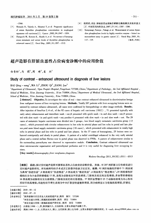
h n e e t p tl n lw y i o tlp a e a c d c n r ea l a d s l p r h s .A at r fa r d a e t f g le h n e n te v r al r r i y o n a p t n o a i c nr u a n a c d i ey e y a ti e l i h r ea l
B IQn —sn MA L Z O i,I N u A ig o g , i, H U Q J G Je A
Dp r etfUt sud eat n laon ,聊 epe o i lT ncu n77 0 C i eatetfP to g ,h n fl t o i l, m o r nP ol" s t ,ogh a 2 20,hn D p r n o ahl y t 2 dA i e H s t s pa H a; m o e f ad pa i Sho o Mei n , inJat g U i rt, in70 0 C i ;Dp r etfM d a lao n ,te2dA ia dH si l col f d i X ' io n nv sy X " 104,hn ea m n ei lUt ud h n fl t o t , ce a o ei a a t o c r s ie pa Sho o Mein , infat gU i rt, in7 00 ,hn . c l o f dc e X  ̄ i oo nv sy X h 10 4 C i i t n ei a
超声造影检查在诊断肝脏疾病中的应用研究

超声造影检查在诊断肝脏疾病中的应用研究徐 足,谷 颖*(贵州医科大学,贵州 贵阳 550004)[摘要]进行超声造影检查是临床上诊断多种疾病的重要方法。
该检查方法能获得人体病变部位清晰的动态图像,从而可为临床医生诊断疾病提供重要的依据。
肝炎、肝癌及肝硬化等都是临床上常见的肝脏疾病。
近年来,超声造影检查在肝脏疾病的诊断和治疗过程中发挥了重要的作用。
本文对超声造影检查的原理及其在诊断肝脏疾病中的应用现状进行研究,以期为临床医生更好地使用该检查方法诊断肝脏疾病提供参考。
[关键词]超声造影检查;肝炎;肝脏良性病变;转移性肝癌;肝硬化[中图分类号]R445 [文献标识码]A [文章编号]2095-7629-(2021)03-0138-02*通讯作者:谷颖超声造影检查又叫声学造影检查。
该检查方法将超声造影剂作为介质,通过向人体内发生病变的部位注射超声造影剂来增强该部位血管的后散射回声,从而提高普通二维超声检查和普通三维超声检查的分辨率、敏感性和特异性[1]。
该检查方法最大的特点是可客观地反映病变组织器官内的血流灌注情况。
本文对超声造影检查的原理及其在诊断肝脏疾病中的应用现状做一综述。
1 超声造影检查的原理及其应用现状1.1 超声造影检查的原理研究发现,与人体软组织对超声的散射回声强度相比,血细胞对超声的散射回声强度更低,仅为前者的千分之一甚至万分之一。
因此,在普通的二维超声图像上,血细胞通常表现为“无回声”。
这使得普通的二维超声检查仅能模糊地识别出心内膜或大血管的边界,无法识别微小血管的边界及大血管内的血流情况[2]。
超声造影剂可使血液的背向反射回声明显增强,从而可使小血管的边界及大血管内的血流情况在二维超声图像上清晰地显现出来。
此外,超声造影剂可随血液的流动在人体内发生病变部位的血管中均匀地分布,故可避免伪像的产生,从而可使二维超声检查的结果具有时效性和可靠性[3]。
1.2 超声造影检查的应用现状自上个世纪60年代末超声造影检查被首次应用于心脏疾病的诊断后,该检查方法已成为临床上诊断先天性心脏病等心脏结构性疾病的优选方法。
- 1、下载文档前请自行甄别文档内容的完整性,平台不提供额外的编辑、内容补充、找答案等附加服务。
- 2、"仅部分预览"的文档,不可在线预览部分如存在完整性等问题,可反馈申请退款(可完整预览的文档不适用该条件!)。
- 3、如文档侵犯您的权益,请联系客服反馈,我们会尽快为您处理(人工客服工作时间:9:00-18:30)。
Ob e t e T eemietev leo n rs—n a cdutao n eda n s f e ina d jci : od tr n a f o t t h n e l s u di t i oi o ng n v h u c a e r nh g s b
蒋 映 丰 ,周 启 昌 ,朱 才 义
( 中南大 学 1 湘雅 医学 院 附属海 口医院超 声科 ,海 口 5 0 0 ; 2 湘雅 二 医院超 声科 ,长 沙 4 0 1) . 72 8 . 10 1
[ 摘要 】 目的 :探讨超声造 影在肝脏 良恶性 肿瘤鉴别诊 断中 的价值 。方法 :采用超声造影技术 对 8 3个肝脏 肿瘤患者 13个病灶进行检查 ,并 比较不 同病变 的肝脏超声造 影增强的特点 。结果 :所有 1 3个肝脏恶性肿瘤 2 0 动脉期或 门脉期均表 现为快速增强 ,9 8个 (5 延 迟期 快速退 出,仅有 5个 (%) 慢退出。其 中 6 9 %) 5 缓 9个肝细胞 癌病灶 中 5 3个 (7 表现为动脉期 整体均匀增 强 ,1 个 (3 由于病灶 中央有 低 回声表 现为不均匀增强 ,6 7 %) 6 2 %) 6 个 (6 表 现迅速快退 ,仅 3 (%) 9 %) 个 4 表现为缓慢退 出。在 3 个转 移性肝癌病灶 中,2 4 4个 (1 表 现为动脉期 7 %) 不 均匀增 强 ,l 个 (9 表 现为动脉 期或 门脉 期 的均 匀增强 ,3 0 2 %) 2个 ( %) 9 4 表现为快 速退 出,2个 (%) 现为 6 表
缓 慢 退 出 。而 另 外 在 2 0个 肝 良性 肿 瘤 中 ,1 个 (0 表 现 为 缓 慢 不 均 匀 增 强 ,1 个 (0 表 现 为 轻 度 增 强 , 8 9 %) 4 7 %)
2 0个 (0 %) 10 表现 为缓慢退 出。结论 :超声 造影有 助于肝脏 良恶性肿瘤 的鉴别 。
atr l h s r otl h sJ n ers 1 2 %) h w dih mo e eu n acme t u r i ae r ae a dt t 6(3 so e o gn o s h n e n t e ap o p ap h e n e b n n a cme tnte e t l ra uigat i h s r otl h s. tl f 6(6 ) oe h n e n nr e r r r l aeo rap aeA t a o 9 % i h c aa d n e ap p o 6
p r lhs. oe e9 s n (5 wa e u uigh tp ae u e et s n ot aeMtgt r 8ei s 9%) s dot r eae hs th s5ei s ap h l o h d nt l b t r l o (% ddn tOfh 9ein f eaoeua crio ,3(7 ) nacdg bl e 5 ) i o. e s s hptcll ac ma5 7 % ehne l ayit t 6l o o l r n o l nh
J etot U i( d c C nSuh n
2 1,71 ht: w wc me. ght: xy. s nt 0 23 () t / w .u d r; t / bx ym. p/ s o p/ x e
超 声 造 影在 肝 脏 良恶 性 肿 瘤 鉴 别 诊 断 中的价 值
Re u t : I he 1 3 lso si p t allrc r i mai r v d q ikyi heat ra ha eo s ls M 0 e i n n he aoc l a acno t u mp o e u c l t reil n p s r
【 键 词 】 超 声 检 查 ;造 影 剂 ;肝脏 肿 瘤 关
DOI1 .9 9 jsn1 7 —3 72 1 .1 1 :03 6 /.s.6 27 4 .0 20 . 0 i 0
Con r s - n a c d u t a o d i h a n i t a t e h n e lr s un t edig oss - n
_ ■ I _ ● ■ ●
beni nc m al gn a l i gnan pa i um or the t ct s
J NGYn f g HOUO ca g Z U C i I A ige Z n hn 2 H a i j , y
.
D pr e oUt s n A lt Ha o H st, i ga c o f d i, e r Su n e i H i u 728 ea m n f la ud f ie i u o il a y Sh l Mein Cn a otU irt a o 500j t t ro , f ad k i p aX n o o ce tl h vs , k y 2D pr et Ut s n S od i ga o i l et lot U irt C ag a 10 C i ) . ea m nol r o d e n Xa y s t, n aSu n ei, hn h 01, h a t f au , c n H pa C r h vs y s 4 1 n
m a g a t e ai t mo s Inn p t i h c u r.
Me o sAt ao aetw t 13oalel i s F L) ne etot sehne t d : t 8 ptn i 2 c re o (L su dr n cn a acd h o lf 3 i s h f lv s n i w r tn u r on (E S ea i t nT e ot sehne a c r ao F L e m a d la u d C U )xm n i .h n a acd hr t i tn fL s rc pr . ts ao c r tn c a ez i o w eo e
