Systematic identification and functional screens of uncharacterized proteins associated
细胞信号转导通路

Chromatin/Epigenetics Resources
Overview of Chromatin / Epigenetics
Chromatin regulation refers to the events affecting chromatin structure and therefore, transcriptional control of gene expression patterns. Epigenetics, specifically, refers to the heritable modifications which result in altered gene expression and are not known to be encoded in DNA. The nucleosome, made up of four histone proteins (H2A, H2B, H3, and H4), is the primary building block of chromatin. Originally thought to function as a static scaffold for DNA packaging, histones have more recently been shown to be dynamic proteins, undergoing multiple types of post-translational modifications (PTMs) and interacting with regulatory proteins to control gene expression. Protein acetylation plays a crucial role in regulating chromatin structure and transcriptional activity. Histone hyperacetylation by histone acetyltransferases (HATs) is associated with transcriptional activation, whereas histone deacetylation by histone deacetylases (HDACs) is associated with transcriptional repression. Hyperacetylation can directly affect chromatin structure by neutralizing the positive charge on histone tails and disrupting nucleosome-nucleosome and nucleosomeDNA interactions. In addition, acetylation creates binding sites for bromodomain-containing chromatin regulatory proteins (histone modification readers). Unlike acetylation, methylation does not alter the charge of arginine and lysine residues and is unlikely to directly modulate nucleosomal interactions required for chromatin folding. Methylated arginine and lysine residues are major determinants for formation of active and inactive regions of the genome. Methylation facilitates binding of chromatin regulatory proteins/histone modification readers that contain various methyl-lysine or methyl-arginine binding domains (PHD, chromo, WD40, Tudor, MBT, Ankyrin repeats, PWWP domains). Recruitment of co-activator and co-repressor proteins is dependent on the specific lysine residue that is modified. The modulation of chromatin structure is an essential component in the regulation of transcriptional activation and repression. One strategy by which chromatin structure can be modulated is through disruption of histone-DNA contacts by ATP-dependent chromatin remodelers, such as the NuRD, Polycomb, and SWI/SNF complexes, which have been shown to regulate gene activation/repression, cell growth, the cell cycle, and differentiation. Chromatin structure is also modulated through other PTMs such as phosphorylation of histone proteins, which affects association with DNA-interacting proteins and has been recently identified to play a role in coordinating other histone modifications. Furthermore, methylation of DNA at cytosine residues in mammalian cells affects chromatin folding and is a heritable, epigenetic modification that is critical for proper regulation of gene silencing, genomic imprinting, and development. Three families of mammalian DNA methyl-transferases have been identified, DNMT1/2/3, that play distinct roles in embryonic stem cells and adult somatic cells. In addition to the core histone proteins, a number of histone variants exist that confer different structural properties to nucleosomes and play a number of specific functions such as DNA repair, proper kinetochore assembly and chromosome segregation during mitosis, and regulation of transcription. Chromatin and epigenetic regulation is crucial for proper programming of the genome during development and under stress conditions, as the misregulation of gene expression can lead to diseased states such as cancer.
大肠杆菌抗氧化应激反应的蛋白质组学分析
大肠杆菌抗氧化应激反应的蛋白质组学分析摘要在细胞代谢过程中,部分电子逃逸出氧化还原系统,以氧分子作为电子受体,产生具有毒害作用的高能量活性氧分子(reactive oxygen species,ROS),包括超氧阴离子、单线态氧、过氧化氢、羟自由基和脂质过氧化的中间产物。
活性氧是一种广谱杀菌剂,是宿主细胞抗菌作用的有效方式。
但细菌在长期的进化过程中必然会发展出多种对策来抵抗宿主所产生的活性氧的杀菌作用。
为检测与此相关的蛋白质,本研究主要通过正常及氧化应激条件下对大肠杆菌k12的培养,差速离心法提取膜蛋白进行SDS-PAGE,挖取具有明显差异的蛋白质条带,经胶内酶切处理,利用基质辅助激光解吸电离飞行时间串联质谱(MALDI-TOF-MS/MS)进行鉴定分析。
结果鉴定到两种具有明显差异的蛋白,麦芽糖孔蛋白、外膜蛋白OmpA表达量明显下降,这两种蛋白主要与小分子物质渗透进入细胞有关,因此可以推测细菌为了适应周围环境的改变关闭膜上小分子通道,减少过氧化氢摄入细胞内,进而减少过氧化氢的毒害作用。
通过本研究,我们可以初步阐明大肠杆菌的氧化应激机制。
关键词:活性氧;氧化应激反应;膜蛋白;MALDI-TOF MS/MS;过氧化氢ABSTRACTDuring the metabolism in the cell, oxygen molecules as an important electron acceptor, Also accompanied by some electronic escape from the redox system, produce some high energy reactive oxygen species which has poisoned, including superoxide anion (O2-), singlet oxygen, hydrogen peroxide, hydroxyl radical, also includes the intermediate products of lipid metabolism. Reactive oxygen species (ROS) is a broad-spectrum fungicide. It is one of the effective way to host cell antimicrobial. However, bacteria in the long-term evolution development of a variety of strategies to resist the host immune bactericidal action. To detect which proteins associated with this system, in this study, mainly through training two group of the pathogenic E. coli, the one Treatment by hydrogen peroxide, the other one as the control group, Broken cells by ultrasound. Extraction the membrane proteins by differential centrifugation, make a SDS-PAGE electrophoresis with the Protein, Digging the significant difference protein band, after gel digestion, then identified by MALDI-TOF-MS/MS. We found two proteins which have significant difference, they are Maltoporin and outer membrane protein A (OmpA). Maltoporin and OmpA are associate to the small molecules penetrate into the cells. Therefore, we can speculate that to adapt to the environment Bacteria Close membrane channels of small molecules to reduce the intake of intracellular hydrogen peroxide, thereby reducing the toxicity of hydrogen peroxide. Through this experiment, we can initially clarify the oxidative stress mechanism of E. coli.Keywords: Reactive oxygen species; Oxidative stress; hydrogen peroxide; Membrane protein; MALDI-TOF-MS/MS目录摘要ABSTRACT第1章细菌抗氧化应激反应的研究进展 (1)1.1 蛋白质组学分析在细菌抗氧化应激研究中的应用 (1)1.1.1 金黄色葡萄球菌的蛋白质组学分析 (1)1.1.2 幽门螺旋杆菌的蛋白质组学分析 (1)1.1.3 枯草芽孢杆菌的蛋白质组学分析 (2)1.1.4 在其他菌株上的蛋白质组学分析 (2)1.2 蛋白质组学分析的概括 (3)第2章材料和方法 (5)2.1 实验流程图 (5)2.2 材料及主要实验仪器 (6)2.3 试剂 (7)2.4 实验过程 (10)2.4.1 大肠杆菌扩大培养 (10)2.4.2 氧化应激处理 (10)2.4.3 细胞破碎 (11)2.4.4 膜蛋白提取 (11)2.4.5 制胶 (11)2.4.6 样品处理 (12)2.4.7 SDS-PAGE分析 (12)2.4.8 MALDI-TOF-MS/MS质谱鉴定 (13)第3章实验结果 (14)第4章讨论 (18)4.1 过氧化氢的浓度 (18)4.2 质谱结果分析 (18)4.3 前景展望 (19)参考文献 (20)致谢 (23)第1章细菌抗氧化应激反应的研究进展1.1蛋白质组学分析在细菌抗氧化应激研究中的应用1.1.1 金黄色葡萄球菌的蛋白质组学分析中性白细胞通过活性氧物质杀伤细菌,借助蛋白质组学方法可以发现不同菌株在氧化应激中蛋白表达差异,以研究其抗氧化机制。
C.parvum全基因组序列

DOI: 10.1126/science.1094786, 441 (2004);304Science et al.Mitchell S. Abrahamsen,Cryptosporidium parvum Complete Genome Sequence of the Apicomplexan, (this information is current as of October 7, 2009 ):The following resources related to this article are available online at/cgi/content/full/304/5669/441version of this article at:including high-resolution figures, can be found in the online Updated information and services,/cgi/content/full/1094786/DC1 can be found at:Supporting Online Material/cgi/content/full/304/5669/441#otherarticles , 9 of which can be accessed for free: cites 25 articles This article 239 article(s) on the ISI Web of Science. cited by This article has been /cgi/content/full/304/5669/441#otherarticles 53 articles hosted by HighWire Press; see: cited by This article has been/cgi/collection/genetics Genetics: subject collections This article appears in the following/about/permissions.dtl in whole or in part can be found at: this article permission to reproduce of this article or about obtaining reprints Information about obtaining registered trademark of AAAS.is a Science 2004 by the American Association for the Advancement of Science; all rights reserved. The title Copyright American Association for the Advancement of Science, 1200 New York Avenue NW, Washington, DC 20005. (print ISSN 0036-8075; online ISSN 1095-9203) is published weekly, except the last week in December, by the Science o n O c t o b e r 7, 2009w w w .s c i e n c e m a g .o r g D o w n l o a d e d f r o m3.R.Jackendoff,Foundations of Language:Brain,Gram-mar,Evolution(Oxford Univ.Press,Oxford,2003).4.Although for Frege(1),reference was established rela-tive to objects in the world,here we follow Jackendoff’s suggestion(3)that this is done relative to objects and the state of affairs as mentally represented.5.S.Zola-Morgan,L.R.Squire,in The Development andNeural Bases of Higher Cognitive Functions(New York Academy of Sciences,New York,1990),pp.434–456.6.N.Chomsky,Reflections on Language(Pantheon,New York,1975).7.J.Katz,Semantic Theory(Harper&Row,New York,1972).8.D.Sperber,D.Wilson,Relevance(Harvard Univ.Press,Cambridge,MA,1986).9.K.I.Forster,in Sentence Processing,W.E.Cooper,C.T.Walker,Eds.(Erlbaum,Hillsdale,NJ,1989),pp.27–85.10.H.H.Clark,Using Language(Cambridge Univ.Press,Cambridge,1996).11.Often word meanings can only be fully determined byinvokingworld knowledg e.For instance,the meaningof “flat”in a“flat road”implies the absence of holes.However,in the expression“aflat tire,”it indicates the presence of a hole.The meaningof“finish”in the phrase “Billfinished the book”implies that Bill completed readingthe book.However,the phrase“the g oatfin-ished the book”can only be interpreted as the goat eatingor destroyingthe book.The examples illustrate that word meaningis often underdetermined and nec-essarily intertwined with general world knowledge.In such cases,it is hard to see how the integration of lexical meaning and general world knowledge could be strictly separated(3,31).12.W.Marslen-Wilson,C.M.Brown,L.K.Tyler,Lang.Cognit.Process.3,1(1988).13.ERPs for30subjects were averaged time-locked to theonset of the critical words,with40items per condition.Sentences were presented word by word on the centerof a computer screen,with a stimulus onset asynchronyof600ms.While subjects were readingthe sentences,their EEG was recorded and amplified with a high-cut-off frequency of70Hz,a time constant of8s,and asamplingfrequency of200Hz.14.Materials and methods are available as supportingmaterial on Science Online.15.M.Kutas,S.A.Hillyard,Science207,203(1980).16.C.Brown,P.Hagoort,J.Cognit.Neurosci.5,34(1993).17.C.M.Brown,P.Hagoort,in Architectures and Mech-anisms for Language Processing,M.W.Crocker,M.Pickering,C.Clifton Jr.,Eds.(Cambridge Univ.Press,Cambridge,1999),pp.213–237.18.F.Varela et al.,Nature Rev.Neurosci.2,229(2001).19.We obtained TFRs of the single-trial EEG data by con-volvingcomplex Morlet wavelets with the EEG data andcomputingthe squared norm for the result of theconvolution.We used wavelets with a7-cycle width,with frequencies ranging from1to70Hz,in1-Hz steps.Power values thus obtained were expressed as a per-centage change relative to the power in a baselineinterval,which was taken from150to0ms before theonset of the critical word.This was done in order tonormalize for individual differences in EEG power anddifferences in baseline power between different fre-quency bands.Two relevant time-frequency compo-nents were identified:(i)a theta component,rangingfrom4to7Hz and from300to800ms after wordonset,and(ii)a gamma component,ranging from35to45Hz and from400to600ms after word onset.20.C.Tallon-Baudry,O.Bertrand,Trends Cognit.Sci.3,151(1999).tner et al.,Nature397,434(1999).22.M.Bastiaansen,P.Hagoort,Cortex39(2003).23.O.Jensen,C.D.Tesche,Eur.J.Neurosci.15,1395(2002).24.Whole brain T2*-weighted echo planar imaging bloodoxygen level–dependent(EPI-BOLD)fMRI data wereacquired with a Siemens Sonata1.5-T magnetic reso-nance scanner with interleaved slice ordering,a volumerepetition time of2.48s,an echo time of40ms,a90°flip angle,31horizontal slices,a64ϫ64slice matrix,and isotropic voxel size of3.5ϫ3.5ϫ3.5mm.For thestructural magnetic resonance image,we used a high-resolution(isotropic voxels of1mm3)T1-weightedmagnetization-prepared rapid gradient-echo pulse se-quence.The fMRI data were preprocessed and analyzedby statistical parametric mappingwith SPM99software(http://www.fi/spm99).25.S.E.Petersen et al.,Nature331,585(1988).26.B.T.Gold,R.L.Buckner,Neuron35,803(2002).27.E.Halgren et al.,J.Psychophysiol.88,1(1994).28.E.Halgren et al.,Neuroimage17,1101(2002).29.M.K.Tanenhaus et al.,Science268,1632(1995).30.J.J.A.van Berkum et al.,J.Cognit.Neurosci.11,657(1999).31.P.A.M.Seuren,Discourse Semantics(Basil Blackwell,Oxford,1985).32.We thank P.Indefrey,P.Fries,P.A.M.Seuren,and M.van Turennout for helpful discussions.Supported bythe Netherlands Organization for Scientific Research,grant no.400-56-384(P.H.).Supporting Online Material/cgi/content/full/1095455/DC1Materials and MethodsFig.S1References and Notes8January2004;accepted9March2004Published online18March2004;10.1126/science.1095455Include this information when citingthis paper.Complete Genome Sequence ofthe Apicomplexan,Cryptosporidium parvumMitchell S.Abrahamsen,1,2*†Thomas J.Templeton,3†Shinichiro Enomoto,1Juan E.Abrahante,1Guan Zhu,4 Cheryl ncto,1Mingqi Deng,1Chang Liu,1‡Giovanni Widmer,5Saul Tzipori,5GregoryA.Buck,6Ping Xu,6 Alan T.Bankier,7Paul H.Dear,7Bernard A.Konfortov,7 Helen F.Spriggs,7Lakshminarayan Iyer,8Vivek Anantharaman,8L.Aravind,8Vivek Kapur2,9The apicomplexan Cryptosporidium parvum is an intestinal parasite that affects healthy humans and animals,and causes an unrelenting infection in immuno-compromised individuals such as AIDS patients.We report the complete ge-nome sequence of C.parvum,type II isolate.Genome analysis identifies ex-tremely streamlined metabolic pathways and a reliance on the host for nu-trients.In contrast to Plasmodium and Toxoplasma,the parasite lacks an api-coplast and its genome,and possesses a degenerate mitochondrion that has lost its genome.Several novel classes of cell-surface and secreted proteins with a potential role in host interactions and pathogenesis were also detected.Elu-cidation of the core metabolism,including enzymes with high similarities to bacterial and plant counterparts,opens new avenues for drug development.Cryptosporidium parvum is a globally impor-tant intracellular pathogen of humans and animals.The duration of infection and patho-genesis of cryptosporidiosis depends on host immune status,ranging from a severe but self-limiting diarrhea in immunocompetent individuals to a life-threatening,prolonged infection in immunocompromised patients.Asubstantial degree of morbidity and mortalityis associated with infections in AIDS pa-tients.Despite intensive efforts over the past20years,there is currently no effective ther-apy for treating or preventing C.parvuminfection in humans.Cryptosporidium belongs to the phylumApicomplexa,whose members share a com-mon apical secretory apparatus mediating lo-comotion and tissue or cellular invasion.Many apicomplexans are of medical or vet-erinary importance,including Plasmodium,Babesia,Toxoplasma,Neosprora,Sarcocys-tis,Cyclospora,and Eimeria.The life cycle ofC.parvum is similar to that of other cyst-forming apicomplexans(e.g.,Eimeria and Tox-oplasma),resulting in the formation of oocysts1Department of Veterinary and Biomedical Science,College of Veterinary Medicine,2Biomedical Genom-ics Center,University of Minnesota,St.Paul,MN55108,USA.3Department of Microbiology and Immu-nology,Weill Medical College and Program in Immu-nology,Weill Graduate School of Medical Sciences ofCornell University,New York,NY10021,USA.4De-partment of Veterinary Pathobiology,College of Vet-erinary Medicine,Texas A&M University,College Sta-tion,TX77843,USA.5Division of Infectious Diseases,Tufts University School of Veterinary Medicine,NorthGrafton,MA01536,USA.6Center for the Study ofBiological Complexity and Department of Microbiol-ogy and Immunology,Virginia Commonwealth Uni-versity,Richmond,VA23198,USA.7MRC Laboratoryof Molecular Biology,Hills Road,Cambridge CB22QH,UK.8National Center for Biotechnology Infor-mation,National Library of Medicine,National Insti-tutes of Health,Bethesda,MD20894,USA.9Depart-ment of Microbiology,University of Minnesota,Min-neapolis,MN55455,USA.*To whom correspondence should be addressed.E-mail:abe@†These authors contributed equally to this work.‡Present address:Bioinformatics Division,Genetic Re-search,GlaxoSmithKline Pharmaceuticals,5MooreDrive,Research Triangle Park,NC27009,USA.R E P O R T S SCIENCE VOL30416APRIL2004441o n O c t o b e r 7 , 2 0 0 9 w w w . s c i e n c e m a g . o r g D o w n l o a d e d f r o mthat are shed in the feces of infected hosts.C.parvum oocysts are highly resistant to environ-mental stresses,including chlorine treatment of community water supplies;hence,the parasite is an important water-and food-borne pathogen (1).The obligate intracellular nature of the par-asite ’s life cycle and the inability to culture the parasite continuously in vitro greatly impair researchers ’ability to obtain purified samples of the different developmental stages.The par-asite cannot be genetically manipulated,and transformation methodologies are currently un-available.To begin to address these limitations,we have obtained the complete C.parvum ge-nome sequence and its predicted protein com-plement.(This whole-genome shotgun project has been deposited at DDBJ/EMBL/GenBank under the project accession AAEE00000000.The version described in this paper is the first version,AAEE01000000.)The random shotgun approach was used to obtain the complete DNA sequence (2)of the Iowa “type II ”isolate of C.parvum .This isolate readily transmits disease among numerous mammals,including humans.The resulting ge-nome sequence has roughly 13ϫgenome cov-erage containing five gaps and 9.1Mb of totalDNA sequence within eight chromosomes.The C.parvum genome is thus quite compact rela-tive to the 23-Mb,14-chromosome genome of Plasmodium falciparum (3);this size difference is predominantly the result of shorter intergenic regions,fewer introns,and a smaller number of genes (Table 1).Comparison of the assembled sequence of chromosome VI to that of the recently published sequence of chromosome VI (4)revealed that our assembly contains an ad-ditional 160kb of sequence and a single gap versus two,with the common sequences dis-playing a 99.993%sequence identity (2).The relative paucity of introns greatly simplified gene predictions and facilitated an-notation (2)of predicted open reading frames (ORFs).These analyses provided an estimate of 3807protein-encoding genes for the C.parvum genome,far fewer than the estimated 5300genes predicted for the Plasmodium genome (3).This difference is primarily due to the absence of an apicoplast and mitochondrial genome,as well as the pres-ence of fewer genes encoding metabolic functions and variant surface proteins,such as the P.falciparum var and rifin molecules (Table 2).An analysis of the encoded pro-tein sequences with the program SEG (5)shows that these protein-encoding genes are not enriched in low-complexity se-quences (34%)to the extent observed in the proteins from Plasmodium (70%).Our sequence analysis indicates that Cryptosporidium ,unlike Plasmodium and Toxoplasma ,lacks both mitochondrion and apicoplast genomes.The overall complete-ness of the genome sequence,together with the fact that similar DNA extraction proce-dures used to isolate total genomic DNA from C.parvum efficiently yielded mito-chondrion and apicoplast genomes from Ei-meria sp.and Toxoplasma (6,7),indicates that the absence of organellar genomes was unlikely to have been the result of method-ological error.These conclusions are con-sistent with the absence of nuclear genes for the DNA replication and translation machinery characteristic of mitochondria and apicoplasts,and with the lack of mito-chondrial or apicoplast targeting signals for tRNA synthetases.A number of putative mitochondrial pro-teins were identified,including components of a mitochondrial protein import apparatus,chaperones,uncoupling proteins,and solute translocators (table S1).However,the ge-nome does not encode any Krebs cycle en-zymes,nor the components constituting the mitochondrial complexes I to IV;this finding indicates that the parasite does not rely on complete oxidation and respiratory chains for synthesizing adenosine triphosphate (ATP).Similar to Plasmodium ,no orthologs for the ␥,␦,or εsubunits or the c subunit of the F 0proton channel were detected (whereas all subunits were found for a V-type ATPase).Cryptosporidium ,like Eimeria (8)and Plas-modium ,possesses a pyridine nucleotide tran-shydrogenase integral membrane protein that may couple reduced nicotinamide adenine dinucleotide (NADH)and reduced nico-tinamide adenine dinucleotide phosphate (NADPH)redox to proton translocation across the inner mitochondrial membrane.Unlike Plasmodium ,the parasite has two copies of the pyridine nucleotide transhydrogenase gene.Also present is a likely mitochondrial membrane –associated,cyanide-resistant alter-native oxidase (AOX )that catalyzes the reduction of molecular oxygen by ubiquinol to produce H 2O,but not superoxide or H 2O 2.Several genes were identified as involved in biogenesis of iron-sulfur [Fe-S]complexes with potential mitochondrial targeting signals (e.g.,nifS,nifU,frataxin,and ferredoxin),supporting the presence of a limited electron flux in the mitochondrial remnant (table S2).Our sequence analysis confirms the absence of a plastid genome (7)and,additionally,the loss of plastid-associated metabolic pathways including the type II fatty acid synthases (FASs)and isoprenoid synthetic enzymes thatTable 1.General features of the C.parvum genome and comparison with other single-celled eukaryotes.Values are derived from respective genome project summaries (3,26–28).ND,not determined.FeatureC.parvum P.falciparum S.pombe S.cerevisiae E.cuniculiSize (Mbp)9.122.912.512.5 2.5(G ϩC)content (%)3019.43638.347No.of genes 38075268492957701997Mean gene length (bp)excluding introns 1795228314261424ND Gene density (bp per gene)23824338252820881256Percent coding75.352.657.570.590Genes with introns (%)553.9435ND Intergenic regions (G ϩC)content %23.913.632.435.145Mean length (bp)5661694952515129RNAsNo.of tRNA genes 454317429944No.of 5S rRNA genes 6330100–2003No.of 5.8S ,18S ,and 28S rRNA units 57200–400100–20022Table parison between predicted C.parvum and P.falciparum proteins.FeatureC.parvum P.falciparum *Common †Total predicted proteins380752681883Mitochondrial targeted/encoded 17(0.45%)246(4.7%)15Apicoplast targeted/encoded 0581(11.0%)0var/rif/stevor ‡0236(4.5%)0Annotated as protease §50(1.3%)31(0.59%)27Annotated as transporter 69(1.8%)34(0.65%)34Assigned EC function ¶167(4.4%)389(7.4%)113Hypothetical proteins925(24.3%)3208(60.9%)126*Values indicated for P.falciparum are as reported (3)with the exception of those for proteins annotated as protease or transporter.†TBLASTN hits (e Ͻ–5)between C.parvum and P.falciparum .‡As reported in (3).§Pre-dicted proteins annotated as “protease or peptidase”for C.parvum (CryptoGenome database,)and P.falciparum (PlasmoDB database,).Predicted proteins annotated as “trans-porter,permease of P-type ATPase”for C.parvum (CryptoGenome)and P.falciparum (PlasmoDB).¶Bidirectional BLAST hit (e Ͻ–15)to orthologs with assigned Enzyme Commission (EC)numbers.Does not include EC assignment numbers for protein kinases or protein phosphatases (due to inconsistent annotation across genomes),or DNA polymerases or RNA polymerases,as a result of issues related to subunit inclusion.(For consistency,46proteins were excluded from the reported P.falciparum values.)R E P O R T S16APRIL 2004VOL 304SCIENCE 442 o n O c t o b e r 7, 2009w w w .s c i e n c e m a g .o r g D o w n l o a d e d f r o mare otherwise localized to the plastid in other apicomplexans.C.parvum fatty acid biosynthe-sis appears to be cytoplasmic,conducted by a large(8252amino acids)modular type I FAS (9)and possibly by another large enzyme that is related to the multidomain bacterial polyketide synthase(10).Comprehensive screening of the C.parvum genome sequence also did not detect orthologs of Plasmodium nuclear-encoded genes that contain apicoplast-targeting and transit sequences(11).C.parvum metabolism is greatly stream-lined relative to that of Plasmodium,and in certain ways it is reminiscent of that of another obligate eukaryotic parasite,the microsporidian Encephalitozoon.The degeneration of the mi-tochondrion and associated metabolic capabili-ties suggests that the parasite largely relies on glycolysis for energy production.The parasite is capable of uptake and catabolism of mono-sugars(e.g.,glucose and fructose)as well as synthesis,storage,and catabolism of polysac-charides such as trehalose and amylopectin. Like many anaerobic organisms,it economizes ATP through the use of pyrophosphate-dependent phosphofructokinases.The conver-sion of pyruvate to acetyl–coenzyme A(CoA) is catalyzed by an atypical pyruvate-NADPH oxidoreductase(Cp PNO)that contains an N-terminal pyruvate–ferredoxin oxidoreductase (PFO)domain fused with a C-terminal NADPH–cytochrome P450reductase domain (CPR).Such a PFO-CPR fusion has previously been observed only in the euglenozoan protist Euglena gracilis(12).Acetyl-CoA can be con-verted to malonyl-CoA,an important precursor for fatty acid and polyketide biosynthesis.Gly-colysis leads to several possible organic end products,including lactate,acetate,and ethanol. The production of acetate from acetyl-CoA may be economically beneficial to the parasite via coupling with ATP production.Ethanol is potentially produced via two in-dependent pathways:(i)from the combination of pyruvate decarboxylase and alcohol dehy-drogenase,or(ii)from acetyl-CoA by means of a bifunctional dehydrogenase(adhE)with ac-etaldehyde and alcohol dehydrogenase activi-ties;adhE first converts acetyl-CoA to acetal-dehyde and then reduces the latter to ethanol. AdhE predominantly occurs in bacteria but has recently been identified in several protozoans, including vertebrate gut parasites such as Enta-moeba and Giardia(13,14).Adjacent to the adhE gene resides a second gene encoding only the AdhE C-terminal Fe-dependent alcohol de-hydrogenase domain.This gene product may form a multisubunit complex with AdhE,or it may function as an alternative alcohol dehydro-genase that is specific to certain growth condi-tions.C.parvum has a glycerol3-phosphate dehydrogenase similar to those of plants,fungi, and the kinetoplastid Trypanosoma,but(unlike trypanosomes)the parasite lacks an ortholog of glycerol kinase and thus this pathway does not yield glycerol production.In addition to themodular fatty acid synthase(Cp FAS1)andpolyketide synthase homolog(Cp PKS1), C.parvum possesses several fatty acyl–CoA syn-thases and a fatty acyl elongase that may partici-pate in fatty acid metabolism.Further,enzymesfor the metabolism of complex lipids(e.g.,glyc-erolipid and inositol phosphate)were identified inthe genome.Fatty acids are apparently not anenergy source,because enzymes of the fatty acidoxidative pathway are absent,with the exceptionof a3-hydroxyacyl-CoA dehydrogenase.C.parvum purine metabolism is greatlysimplified,retaining only an adenosine ki-nase and enzymes catalyzing conversionsof adenosine5Ј-monophosphate(AMP)toinosine,xanthosine,and guanosine5Ј-monophosphates(IMP,XMP,and GMP).Among these enzymes,IMP dehydrogenase(IMPDH)is phylogenetically related toε-proteobacterial IMPDH and is strikinglydifferent from its counterparts in both thehost and other apicomplexans(15).In con-trast to other apicomplexans such as Toxo-plasma gondii and P.falciparum,no geneencoding hypoxanthine-xanthineguaninephosphoribosyltransferase(HXGPRT)is de-tected,in contrast to a previous report on theactivity of this enzyme in C.parvum sporo-zoites(16).The absence of HXGPRT sug-gests that the parasite may rely solely on asingle enzyme system including IMPDH toproduce GMP from AMP.In contrast to otherapicomplexans,the parasite appears to relyon adenosine for purine salvage,a modelsupported by the identification of an adeno-sine transporter.Unlike other apicomplexansand many parasitic protists that can synthe-size pyrimidines de novo,C.parvum relies onpyrimidine salvage and retains the ability forinterconversions among uridine and cytidine5Ј-monophosphates(UMP and CMP),theirdeoxy forms(dUMP and dCMP),and dAMP,as well as their corresponding di-and triphos-phonucleotides.The parasite has also largelyshed the ability to synthesize amino acids denovo,although it retains the ability to convertselect amino acids,and instead appears torely on amino acid uptake from the host bymeans of a set of at least11amino acidtransporters(table S2).Most of the Cryptosporidium core pro-cesses involved in DNA replication,repair,transcription,and translation conform to thebasic eukaryotic blueprint(2).The transcrip-tional apparatus resembles Plasmodium interms of basal transcription machinery.How-ever,a striking numerical difference is seenin the complements of two RNA bindingdomains,Sm and RRM,between P.falcipa-rum(17and71domains,respectively)and C.parvum(9and51domains).This reductionresults in part from the loss of conservedproteins belonging to the spliceosomal ma-chinery,including all genes encoding Smdomain proteins belonging to the U6spliceo-somal particle,which suggests that this par-ticle activity is degenerate or entirely lost.This reduction in spliceosomal machinery isconsistent with the reduced number of pre-dicted introns in Cryptosporidium(5%)rela-tive to Plasmodium(Ͼ50%).In addition,keycomponents of the small RNA–mediatedposttranscriptional gene silencing system aremissing,such as the RNA-dependent RNApolymerase,Argonaute,and Dicer orthologs;hence,RNA interference–related technolo-gies are unlikely to be of much value intargeted disruption of genes in C.parvum.Cryptosporidium invasion of columnarbrush border epithelial cells has been de-scribed as“intracellular,but extracytoplas-mic,”as the parasite resides on the surface ofthe intestinal epithelium but lies underneaththe host cell membrane.This niche may al-low the parasite to evade immune surveil-lance but take advantage of solute transportacross the host microvillus membrane or theextensively convoluted parasitophorous vac-uole.Indeed,Cryptosporidium has numerousgenes(table S2)encoding families of putativesugar transporters(up to9genes)and aminoacid transporters(11genes).This is in starkcontrast to Plasmodium,which has fewersugar transporters and only one putative ami-no acid transporter(GenBank identificationnumber23612372).As a first step toward identification ofmulti–drug-resistant pumps,the genome se-quence was analyzed for all occurrences ofgenes encoding multitransmembrane proteins.Notable are a set of four paralogous proteinsthat belong to the sbmA family(table S2)thatare involved in the transport of peptide antibi-otics in bacteria.A putative ortholog of thePlasmodium chloroquine resistance–linkedgene Pf CRT(17)was also identified,althoughthe parasite does not possess a food vacuole likethe one seen in Plasmodium.Unlike Plasmodium,C.parvum does notpossess extensive subtelomeric clusters of anti-genically variant proteins(exemplified by thelarge families of var and rif/stevor genes)thatare involved in immune evasion.In contrast,more than20genes were identified that encodemucin-like proteins(18,19)having hallmarksof extensive Thr or Ser stretches suggestive ofglycosylation and signal peptide sequences sug-gesting secretion(table S2).One notable exam-ple is an11,700–amino acid protein with anuninterrupted stretch of308Thr residues(cgd3_720).Although large families of secretedproteins analogous to the Plasmodium multi-gene families were not found,several smallermultigene clusters were observed that encodepredicted secreted proteins,with no detectablesimilarity to proteins from other organisms(Fig.1,A and B).Within this group,at leastfour distinct families appear to have emergedthrough gene expansions specific to the Cryp-R E P O R T S SCIENCE VOL30416APRIL2004443o n O c t o b e r 7 , 2 0 0 9 w w w . s c i e n c e m a g . o r g D o w n l o a d e d f r o mtosporidium clade.These families —SKSR,MEDLE,WYLE,FGLN,and GGC —were named after well-conserved sequence motifs (table S2).Reverse transcription polymerase chain reaction (RT-PCR)expression analysis (20)of one cluster,a locus of seven adjacent CpLSP genes (Fig.1B),shows coexpression during the course of in vitro development (Fig.1C).An additional eight genes were identified that encode proteins having a periodic cysteine structure similar to the Cryptosporidium oocyst wall protein;these eight genes are similarly expressed during the onset of oocyst formation and likely participate in the formation of the coccidian rigid oocyst wall in both Cryptospo-ridium and Toxoplasma (21).Whereas the extracellular proteins described above are of apparent apicomplexan or lineage-specific in-vention,Cryptosporidium possesses many genesencodingsecretedproteinshavinglineage-specific multidomain architectures composed of animal-and bacterial-like extracellular adhe-sive domains (fig.S1).Lineage-specific expansions were ob-served for several proteases (table S2),in-cluding an aspartyl protease (six genes),a subtilisin-like protease,a cryptopain-like cys-teine protease (five genes),and a Plas-modium falcilysin-like (insulin degrading enzyme –like)protease (19genes).Nine of the Cryptosporidium falcilysin genes lack the Zn-chelating “HXXEH ”active site motif and are likely to be catalytically inactive copies that may have been reused for specific protein-protein interactions on the cell sur-face.In contrast to the Plasmodium falcilysin,the Cryptosporidium genes possess signal peptide sequences and are likely trafficked to a secretory pathway.The expansion of this family suggests either that the proteins have distinct cleavage specificities or that their diversity may be related to evasion of a host immune response.Completion of the C.parvum genome se-quence has highlighted the lack of conven-tional drug targets currently pursued for the control and treatment of other parasitic protists.On the basis of molecular and bio-chemical studies and drug screening of other apicomplexans,several putative Cryptospo-ridium metabolic pathways or enzymes have been erroneously proposed to be potential drug targets (22),including the apicoplast and its associated metabolic pathways,the shikimate pathway,the mannitol cycle,the electron transport chain,and HXGPRT.Nonetheless,complete genome sequence analysis identifies a number of classic and novel molecular candidates for drug explora-tion,including numerous plant-like and bacterial-like enzymes (tables S3and S4).Although the C.parvum genome lacks HXGPRT,a potent drug target in other api-complexans,it has only the single pathway dependent on IMPDH to convert AMP to GMP.The bacterial-type IMPDH may be a promising target because it differs substan-tially from that of eukaryotic enzymes (15).Because of the lack of de novo biosynthetic capacity for purines,pyrimidines,and amino acids,C.parvum relies solely on scavenge from the host via a series of transporters,which may be exploited for chemotherapy.C.parvum possesses a bacterial-type thymidine kinase,and the role of this enzyme in pyrim-idine metabolism and its drug target candida-cy should be pursued.The presence of an alternative oxidase,likely targeted to the remnant mitochondrion,gives promise to the study of salicylhydroxamic acid (SHAM),as-cofuranone,and their analogs as inhibitors of energy metabolism in the parasite (23).Cryptosporidium possesses at least 15“plant-like ”enzymes that are either absent in or highly divergent from those typically found in mammals (table S3).Within the glycolytic pathway,the plant-like PPi-PFK has been shown to be a potential target in other parasites including T.gondii ,and PEPCL and PGI ap-pear to be plant-type enzymes in C.parvum .Another example is a trehalose-6-phosphate synthase/phosphatase catalyzing trehalose bio-synthesis from glucose-6-phosphate and uridine diphosphate –glucose.Trehalose may serve as a sugar storage source or may function as an antidesiccant,antioxidant,or protein stability agent in oocysts,playing a role similar to that of mannitol in Eimeria oocysts (24).Orthologs of putative Eimeria mannitol synthesis enzymes were not found.However,two oxidoreductases (table S2)were identified in C.parvum ,one of which belongs to the same families as the plant mannose dehydrogenases (25)and the other to the plant cinnamyl alcohol dehydrogenases.In principle,these enzymes could synthesize protective polyol compounds,and the former enzyme could use host-derived mannose to syn-thesize mannitol.References and Notes1.D.G.Korich et al .,Appl.Environ.Microbiol.56,1423(1990).2.See supportingdata on Science Online.3.M.J.Gardner et al .,Nature 419,498(2002).4.A.T.Bankier et al .,Genome Res.13,1787(2003).5.J.C.Wootton,Comput.Chem.18,269(1994).Fig.1.(A )Schematic showing the chromosomal locations of clusters of potentially secreted proteins.Numbers of adjacent genes are indicated in paren-theses.Arrows indicate direc-tion of clusters containinguni-directional genes (encoded on the same strand);squares indi-cate clusters containingg enes encoded on both strands.Non-paralogous genes are indicated by solid gray squares or direc-tional triangles;SKSR (green triangles),FGLN (red trian-gles),and MEDLE (blue trian-gles)indicate three C.parvum –specific families of paralogous genes predominantly located at telomeres.Insl (yellow tri-angles)indicates an insulinase/falcilysin-like paralogous gene family.Cp LSP (white square)indicates the location of a clus-ter of adjacent large secreted proteins (table S2)that are cotranscriptionally regulated.Identified anchored telomeric repeat sequences are indicated by circles.(B )Schematic show-inga select locus containinga cluster of coexpressed large secreted proteins (Cp LSP).Genes and intergenic regions (regions between identified genes)are drawn to scale at the nucleotide level.The length of the intergenic re-gions is indicated above or be-low the locus.(C )Relative ex-pression levels of CpLSP (red lines)and,as a control,C.parvum Hedgehog-type HINT domain gene (blue line)duringin vitro development,as determined by semiquantitative RT-PCR usingg ene-specific primers correspondingto the seven adjacent g enes within the CpLSP locus as shown in (B).Expression levels from three independent time-course experiments are represented as the ratio of the expression of each gene to that of C.parvum 18S rRNA present in each of the infected samples (20).R E P O R T S16APRIL 2004VOL 304SCIENCE 444 o n O c t o b e r 7, 2009w w w .s c i e n c e m a g .o r g D o w n l o a d e d f r o m。
dia质谱和泛素化位点

dia质谱和泛素化位点
Dia质谱(differential ion mobility mass spectrometry)是一种质谱分析技术,在传统质谱基础上增加了离子迁移过程,使得质量/电荷比相同的离子根据其气相中的相对移动率(Drift Time)进行分离。
这项技术可提供比传统质谱更高的离子分辨能力,用于鉴定、分析和定量复杂分子混合物中的化合物。
泛素化位点指的是泛素蛋白连接到其他蛋白质上的特定氨基酸残基位置。
泛素化是一种常见的蛋白质修饰方式,在细胞内起到调控蛋白质降解、信号转导和细胞周期等生物学过程的关键作用。
泛素化位点通常发生在溶酶体相关泛素化系统(UPS)
介导的蛋白质降解过程中。
鉴定和研究泛素化位点可以帮助我们理解泛素相关生物学过程的机制,并发现新的泛素靶点。
一些质谱技术,如质谱鉴定和定量方法,常被用于识别和鉴定泛素化位点,并确定泛素修饰在该位点上的氨基酸残基。
cst 泛素化组学

cst 泛素化组学
CST是Cell Signaling Technology的缩写,是一家专注于生物医学研究的公司。
泛素化组学(Ubiquitinomics)是一种研究细胞中泛素化修饰的方法和技术。
泛素化是一种蛋白质修饰过程,通过连接小泛素蛋白到目标蛋白上,调控其稳定性、功能和亚细胞定位。
泛素化组学基于质谱技术,结合免疫分离和定量方法,可以全面分析细胞中的泛素化修饰,并研究其在细胞信号传导、蛋白质降解、DNA修复等生物学过程中的作用。
通过泛素化组学的研究,可以揭示泛素化修饰在细胞功能和疾病发生中的重要作用,为疾病诊断和治疗提供新的靶点和策略。
CST在泛素化组学领域提供了一系列的抗体和试剂盒,用于检测和研究泛素化修饰相关的蛋白质。
这些工具可以帮助研究人员深入了解泛素化修饰的生物学功能以及其在疾病中的作用机制。
串联亲和层析 protocol
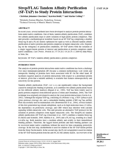
UNIT19.20 Strep/FLAG Tandem Affinity Purification(SF-TAP)to Study Protein InteractionsChristian Johannes Gloeckner,1Karsten Boldt,1,2and Marius Ueffing1,21Helmholtz Zentrum M¨u nchen,Neuherberg,Germany2Technical University of Munich,Munich,GermanyABSTRACTIn recent years,several methods have been developed to analyze protein-protein interac-tions under native conditions.One of them,tandem affinity purification(TAP),combinestwo affinity-purification steps to allow isolation of high-purity protein complexes.Thisunit presents a methodological workflow based on an SF-TAP tag comprising a doubletStrep-tag II and a FLAG moiety optimized for rapid as well as efficient tandem affinitypurification of native proteins and protein complexes in higher eukaryotic cells.Depend-ing on the stringency of purification conditions,SF-TAP allows both the isolation ofa single tagged-fusion protein of interest and purification of protein complexes undernative conditions.Curr.Protoc.Protein Sci.57:19.20.1-19.20.19.C 2009by John Wiley&Sons,Inc.Keywords:SF-TAP r tandem affinity purification r protein complexesINTRODUCTIONThe analysis of protein-protein interactions under native conditions has been a challengeever since immunoprecipitation(IP)became a common methodology.Low yields andnonspecific binding of proteins have been associated with IP.On the other hand,IPfacilitates targeted analysis of protein interactions with respect to a predefined proteinof interest,given that a suitable antibody is available that features monospecificity andselectivity for this protein.Tandem affinity purification(TAP;UNIT19.19)can significantly reduce the backgroundcaused by nonspecific binding of proteins,as it combines two affinity purifications basedon two different affinity matrices(Rigaut et al.,1999).TAP has been widely used topurify protein complexes from different species(Collins and Choudhary,2008).The TAPtechnique was originally developed to analyze the yeast protein interactome(Gavin et al.,2002).Although the original TAP tag,consisting of a Protein A-tag,a TEV(tobacco etchvirus)protease cleavage site,and a calmodulin binding peptide(CBP)tag,has alreadybeen successfully used in mammalian cells(Bouwmeester et al.,2004),several featuresof thisfirst-generation tag remain suboptimal,such as its high molecular mass(21kDa),the dependency on proteolytic cleavage,and CBP,which may interfere with calciumsignaling within eukaryotic cells.This unit presents an alternative TAP protocol for theisolation of protein complexes from higher eukaryotic cells.The Strep/FLAG tandemaffinity purification(SF-TAP)tag(Gloeckner et al.,2007)combines a tandem Strep-tagII(Skerra and Schmidt,2000;Junttila et al.,2005)and a FLAG tag,resulting in a small4.6-kDa tag.Both moieties have a medium affinity and avidity to their immobilizedbinding partners.Therefore,the tagged fusion proteins and their binding partners canbe recovered under native conditions without the need for time-consuming proteolyticcleavage.In thefirst step,desthiobiotin is used for elution of the SF-TAP fusion proteinfrom the Strep-Tactin matrix.In the second step,the FLAG octapeptide is used for elutionof the SF-TAP fusion protein from the anti-FLAG M2affinity matrix.An overview of the Current Protocols in Protein Science19.20.1-19.20.19,August2009Published online August2009in Wiley Interscience().DOI:10.1002/0471140864.ps1920s57Copyright C 2009John Wiley&Sons,Inc.Identification of Protein Interactions19.20.1 Supplement57Strep/FLAGTandem AffinityPurification (SF-TAP)19.20.2Supplement 57Current Protocols in Protein Science A B 1. purification 2. purification binding to Strep-Tactin binding to FLAG matrix elution with desthiobiotin elution with FLAG peptide Key:SF-TAP desthiobiotin FLAG peptide Figure 19.20.1The S trep/FLAG ta n dem affin ity p u rificatio n .(A )N-a n d C-termi n al S F-T AP ta gs (POI,protei n of i n tere s t).(B )Overview of both p u rificatio n s tep s .(1)P u rificatio n by the ta n dem S trep-ta g II moiety:bi n di ng to S trep-T acti n matrix followed by el u tio n with de s thiobioti n .(2)P u rificatio n by the FLAG-ta g moiety:bi n di ng to a n ti-FLAG M2affin ity matrix followed by el u -tio n with FLAG peptide.Abbreviatio ns :s p.,s pecific i n teractor s (s how n a s g ray circle s );n .s p.,n o ns pecific protei ns (co n tami n a n t s ;s how n a s white circle s ).SF-TAP technique and the tag sequence is shown in Figure 19.20.1.The SF-TAP protocol represents an efficient,fast and straightforward purification of protein complexes from mammalian cells within 2hr.This unit describes the full workflow,starting with the cell culture work needed for recombinant expression of the SF-TAP fusion proteins,followed by the SF-TAP protocol (see Basic Protocol 1)and ending with mass spectrometric analysis of the samples (see Basic Protocol 4).Special focus is given to the crucial step of sample preparation for mass spectrometry.For the identification of associated proteins following SF-TAP,the volume of the SF-TAP eluates is reduced by ultrafiltration using centrifugal units with a low molecular weight cut-off or by chloroform/methanol precipitation (see Support Protocol 2).The samples are then directly subjected to proteolytic digestion (see Basic Protocol 2)for analysis on a nano liquid chromatography (LC)–coupled electron sprayIdentification of Protein Interactions 19.20.3Current Protocols in Protein Science Supplement 57Figure 19.20.2Flow chart of a S F-T AP approach i n cl u di ng M S ide n tificatio n of cop u rified pro-tei ns .Thi s figu re co nn ect s all protocol s pre s e n ted i n thi s un it.tandem mass spectrometer.For complex samples,which contain many proteins,an alternative procedure for SDS-PAGE pre-fractionation is provided,including a method for sensitive MS-compatible Coomassie protein staining (see Support Protocol 3)followed by in-gel proteolytic digestion (see Basic Protocol 3).By reducing sample complexity,pre-fractionation helps to increase the number of protein identifications on state-of-the-art LC-coupled tandem mass spectrometers.Representative MS-analysis protocols are provided for an Orbitrap mass spectrometer (Thermo Fisher Scientific),a fast and sensitive system allowing high identification rates from SF-TAP purifications even with low amounts of protein in the sample (see Basic Protocol 4).Finally,a strategy for meta analysis of mass spectrometric data sets using the Scaffold software is provided (see Support Protocol 4).It can generally be used for the analysis of large MS/MS data sets.Figure 19.20.2provides a flowchart of the entire analytical process.Strep/FLAGTandem AffinityPurification (SF-TAP)19.20.4Supplement 57Current Protocols in Protein ScienceBASICPROTOCOL 1STREP/FLAG TANDEM AFFINITY PURIFICATION (SF-TAP)OF PROTEIN COMPLEXES FROM HEK293CELLS A flowchart of the SF-TAP procedure is shown in Figure 19.20.3.Materials HEK293cells (ATCC no.CRL-1573)Complete DMEM containing 10%FBS (APPENDIX 3C )SF-TAP vectors with appropriate insert,and empty control plasmid (see Critical Parameters)Negative control (see annotation to step 3,below)Transfection reagent of choice (see UNIT 5.10)Phosphate-buffered saline (PBS;APPENDIX 2E ),prewarmed Lysis buffer (see recipe)Strep-Tactin Superflow resin (IBA GmbH,cat.no.2-1206-10)Tris-buffered saline (TBS;see recipe)Wash buffer (see recipe)Desthiobiotin elution buffer:dilute 10×buffer E (IBA GmbH,cat.no.2-1000-025)1:10in H 2O (final concentration,2mM desthiobiotin)Anti–FLAG M2agarose (Sigma-Aldrich)FLAG elution buffer (see recipe)14-cm tissue culture plates Cell scraper Millex GP 0.22-μm syringe-driven filter units (Millipore)End-over-end rotator Microspin columns (GE Healthcare,cat.no.27-3565-01)End-over-end rotator Microcon YM-3centrifugal filter devices (Millipore)Additional reagents and equipment for transfection of mammalian cells (UNIT 5.10)Transfect HEK293cells 1.Seed HEK293cells on 14-cm plates at ∼1–2×10⁷cells per dish in complete DMEM medium containing 10%FBS.The amount of cells used for SF-TAP purification can be varied depending on the ex-pression levels of the bait ually,four 14-cm dishes,corresponding to a final amount of ∼4×10⁸HEK293cells,is a good starting point.Strong overexpression of the bait protein usually increases copurification of heat-shock proteins such as HSP70.For in-depth analysis,it is therefore recommended to generate cell lines stably expressing the bait protein.See Support Protocol 1for a stable transfection method.2.Grow cells overnight.3.Transfect cells with the SF-TAP plasmids using a transfection reagent of choice (according to manufacturer’s protocols).HEK293cells can be easily transfected with lipophilic transfection reagents.The trans-fection efficiency is usually >80%.For a typical SF-TAP experiment,1to 4μg plasmid per 14-cm dish is used.Depending on the cell type other transfection reagents may be favorable (also see UNIT 5.10).Although SF-TAP purifications typically exhibit low background caused by nonspecific binding of proteins to the affinity matrix,a suitable negative control should be used in every experiment.Cells transfected with the empty expression vectors may be used in the same amount as for the SF-TAP-tagged bait protein.However,the tag is quite small and expressed at low levels if not fused to a protein.Thus,the untransfected cell line is an acceptable,simple,and inexpensive alternative for a negative control.Identification of Protein Interactions 19.20.5Current Protocols in Protein Science Supplement 571-4 × 10⁸ HEK293 cell s(1-4 co n fl u e n t 14-cm plate s )expre ss i ng S F-TAP f us io n protei nly s i s(15 mi n 4C)vol u mered u ctio nce n trif ug atio n (10 mi n 10,000 × g )a n aly s i sretai n su per n ata n t fi n alel u atei n c u batio n with50 μl/plate S trep-Tacti n matrix (1 hr)el u tio n with200 μl FLAGel u tio n b u ffer(10 mi n )wa s h 3 time s with 500 μl wa s h b u ffer (s pi n 5 s ec, 100 × g )wa s h 3 time s with500 μl wa s h b u ffer(s pi n 5 s ec, 100 × g )el u tio n with 500 μl de s thiobioti n el u tio n b u ffer (10 mi n )i n c u batio n with25 μl/platea n ti-FLAG M2a g aro s e(1 hr)Figure 19.20.3Flow chart for the S F-T AP proced u re.4.Let cells grow for 48hr.If necessary,cells can be starved in DMEM without FBS for 12hr prior to harvesting.Starving might be desirable if cell signaling is to be analyzed,especially prior to differ-ential treatment with growth factors,to eliminate effects of serum growth factors.Lyse cells5.Remove medium from the plates.6.Optional:Rinse cells in warm PBS.Strep/FLAGTandem AffinityPurification (SF-TAP)19.20.6Supplement 57Current Protocols in Protein Science7.Scrape off cells in 1ml lysis buffer per 14-cm plate on ice using a cell scraper,and combine lysates from each experimental condition in a 1.5-ml microcentrifuge tube.8.Lyse cells by incubating 15min on ice with mixing by hand from time to time.9.Pellet cell debris,including nuclei,by centrifuging 10min at 10,000×g ,4◦C.10.Clear lysate supernatant by filtration through a 0.22-μm syringe filter.Perform SF-TAP 11.Wash Strep-Tactin Superflow resin twice,each time with 4resin volumes TBS and once with 4resin volumes lysis buffer.12.Incubate lysates with 50μl per 14-cm plate of settled Strep-Tactin Superflow resin for 1hr at 4◦C (use an end-over-end rotator to keep the resin evenly distributed).Note that a maximum of 200μl settled resin per spin column should not be exceeded.If more than four 14-cm plates (∼4×10⁸HEK293cells)are used,reduce the volume per plate or use additional spin columns in step 13.13.Centrifuge for 30sec at 7000×g ,4◦C,remove the supernatant until 500μl remains,and transfer resin to a microspin column.Snap off bottom closure of the spin column prior to use.The maximum volume of the spin columns is 650μl.Alternatively,centrifugations for wash and elution steps can be performed at room temperature if no cooled centrifuge is available.14.Remove remaining supernatant by centrifugation in the spin column for 5sec at 100×g ,then wash resin three times,each time with 500μl wash buffer (centrifuge 5sec at 100×g each time to remove the supernatant)at 4◦C.Replug spin columns with inverted bottom closure prior to adding the elution buffer in step 15.IMPORTANT NOTE:Do not allow the resin to run dry.Depending on the bait protein,this markedly reduces the yield.15.Add 500μl desthiobiotin elution buffer and gently mix the resin by hand for 10min on ice.16.Remove the plug of the spin column,transfer the column to a new collection tube,and collect the eluate by centrifuging 10sec at 2000×g ,4◦C.If spin columns were closed by the top screw cap during incubation with elution buffer,the cap needs to be removed prior to centrifugation,to allow the pressure to balance out.17.Wash anti–FLAG M2agarose resin three times,each time with 4resin volumes TBS.Suspend resin in TBS and transfer it to microspin columns,then remove the buffer by centrifuging 5sec at 100×g .25μl settled resin per 14-cm plate will be needed.18.Transfer eluate from step 16corresponding to each 14-cm plate to a microspin column containing 25μl settled anti-FLAG M2agarose prepared as in step 17.19.Plug columns,close columns with top screw caps,and incubate for 1hr at 4◦C (on an end-over-end rotator).20.Wash once with 500μl wash buffer,and then twice,each time with 500μl TBS (centrifuge 5sec at 100×g each time to remove the supernatant)at 4◦C.21.For elution,incubate with 4bead volumes (at least 200μl)FLAG elution buffer for 10min,keeping the columns plugged and gently mixing the resin several times.22.After incubation,remove the plugs and top screws of the spin columns,transfer to new collection tubes,and collect the eluate(s)by centrifugation (10sec at 2000×g ).Identification of Protein Interactions 19.20.7Current Protocols in Protein Science Supplement 5723.Depending on downstream method to be used,either precipitate protein (see SupportProtocol 2)or concentrate the eluate by Microcon YM-3centrifugal filter units according to manufacturer’s protocols.SUPPORT PROTOCOL 1GENERATION OF HEK293CLONES STABLY EXPRESSINGSF-TAP-TAGGED PROTEINSIn Basic Protocol 1,SF-TAP-tagged proteins are transiently expressed.However,strong overexpression of the bait protein usually increases copurification of heat-shock proteins such as HSP70.For in-depth analysis,it is therefore recommended to generate cell lines stably expressing the bait protein.This protocol presents a quick method for generating stable HEK293lines.MaterialsHEK293cells (ATCC no.CRL-1573)Complete DMEM containing 10%FBS (APPENDIX 3C )SF-TAP vectors with appropriate insert,and empty control plasmid (see Critical Parameters)Transfection reagent of choice (see UNIT 5.10)Phosphate-buffered saline (PBS;APPENDIX 2E )Complete DMEM medium (APPENDIX 3C )G418(PAA Laboratories, )Freezing solution:90%fetal bovine serum (FBS;Invitrogen)/10%dimethylsulfoxide (DMSO;AR grade)Lysis buffer (see recipe)Blocking reagent:5%(w/v)nonfat dry milk in TBS (see recipe for TBS)containing 0.1%(v/v)Tween 20Anti-FLAG M2antibody (Sigma-Aldrich)10-cm tissue culture dishes12-well and 6-welll tissue culture platesCentrifuge2-ml cryovials (Nunc)Additional reagents and equipment for transfection of mammalian cells (UNIT 5.10),trypsinization and counting of cells (UNIT 5.10),and immunoblotting (UNIT 10.10)Grow and transfect cells1.Grow cells in complete DMEM containing 10%FBS.2.Transfect cells with expression plasmid using a transfection reagent of choice ac-cording to the manufacturer’s protocols.3.Change medium after 6hr.Select cells4.After 48hr,trypsinize and count cells (APPENDIX 3C )and seed them at low density (1×10⁶cells per 10-cm dish)to allow formation of single colonies upon selection.5.Add G418(500to 1000μg/ml)for selection of the SF-TAP expression vectors,which are based on pcDNA3.0and contain a neomycin-resistance gene.6.Grow the cells under G-418selection for 2to 4weeks,changing the medium every second day.7.Collect single colonies with a 200-μl pipet into 12-well plates.8.Keep colonies under G418selection until the cell density is sufficient for expanding them to 6-well dishes (two wells per clone).Strep/FLAGTandem AffinityPurification (SF-TAP)19.20.8Supplement 57Current Protocols in Protein ScienceCryopreserve cells 9.Grow cells to >90%confluency and trypsinize (APPENDIX 3C )one well of each clone for generation of cryostocks.10.Generate cryostocks:a.Wash cells from one well once by adding 3ml PBS,centrifuging 5min at 800×g ,room temperature,and resuspending the pellet in 500μl freezing buffer.b.Transfer resuspended cells to 2-ml cryovials.c.Freeze cells slowly:keep cells for 1hr at −20◦C,then overnight at −80◦C,followed by storage in a liquid nitrogen tank.For cultivation and expansion of confirmed clones,thaw the cryostock at 37◦C,wash cells once with medium,and plate cells onto 10-cm culture dishes.Test for expression of bait protein 11.Lyse one well of each clone in 300μl lysis buffer and test for expression of the bait protein by immunoblotting (UNIT 10.10).SF-TAP proteins can be detected using the anti-FLAG M2antibody (Sigma-Aldrich)at a dilution of 1:1000to 1:5000in blocking reagent.SUPPORTPROTOCOL 2CHLOROFORM/METHANOL PRECIPITATION OF PROTEINS The chloroform/methanol precipitation method described by Wessel and Fl¨u gge (1984)precipitates proteins with high efficiency and yields samples containing low levels of salt contamination.Materials SF-TAP eluate (from Basic Protocol 1)Methanol (AR grade)Chloroform (AR grade)2-ml polypropylene sample tubes 1.Transfer 200μl SF-TAP eluate to a 2-ml sample tube.All steps are performed at ambient temperature.2.Add 0.8ml of methanol,vortex,and centrifuge for 20sec at 9000×g ,room temperature.3.Add 0.2ml chloroform,vortex,and centrifuge for 20sec at 9000×g ,room temperature.4.Add 0.6ml of deionized water,vortex for 5sec,and centrifuge for 1min at 9000×g ,room temperature.5.Carefully remove and discard the upper layer (aqueous phase).The protein precipitate (visible as white flocks)is in the interphase.6.Add 0.6ml of methanol,vortex,and centrifuge for 2min at 16,000×g ,room temperature.7.Carefully remove the supernatant and air dry the pellet.The pellet can be stored for several months at –80◦C.Identification of Protein Interactions 19.20.9Current Protocols in Protein Science Supplement 57BASIC PROTOCOL 2IN-SOLUTION DIGEST OF PROTEINS FOR MASS SPECTROMETRIC ANALYSISThe in-solution digest described here is a quick and efficient method to digest the SF-TAP eluate after protein precipitation (Support Protocol 2).The use of an MS-compatible surfactant helps to solubilize the precipitated proteins.In order to allow the identification of cysteine-containing peptides,random oxidation is prevented,rather than reverted,by applying a DTT/iodoacetamide treatment prior to digestion,leading to a defined-mass adduct.The digested protein sample can then be directly subjected to analysis on an LC-coupled tandem mass spectrometer.MaterialsPrecipitated protein (see Support Protocol 2)50mM ammonium bicarbonate (freshly prepared)RapiGest SF (Waters):prepare 2%(10×)stock solution in deionized water 100mM DTT (prepare from 500mM stock solution;store stock up to 6months at −20◦C)300mM iodoacetamide (prepare fresh)50×(0.5μg/μl)trypsin stock solution (Promega;store at −20◦C)Concentrated (37%)HCl60◦C incubatorPolypropylene inserts (Supelco,cat.no.24722)1to 200μl gel-loader pipet tips (Sorenson Bioscience,/contact.cfm )1.Dissolve the protein pellet in 30μl of 50mM ammonium bicarbonate by extensive vortexing.2.Add 3μl of 10×(2%)RapiGest stock solution (final concentration,0.2%).RapiGest (sodium 3-[(2-methyl-2-undecyl-1,3-dioxolan-4-yl)methoxyl]-1-propanesulfo-nate)is an acid-labile surfactant that helps to solubilize and denature proteins to make them accessible to proteolytic digestion (Yu et al.,2003).3.Add 1μl of 100mM DTT and vortex.4.Incubate 10min at 60◦C.5.Cool the samples to room temperature.6.Add 1μl of 300mM iodoacetamide and vortex.7.Incubate for 30min at room temperature.Samples should be protected from light,since iodoacetamide is light-sensitive.8.Add 2μl trypsin stock solution and vortex.9.Incubate at 37◦C overnight.10.Add 2μl of concentrated (37%)HCl to hydrolyze the RapiGest.For hydrolysis of the RapiGest reagent,the pH must be <2.11.Transfer samples to polypropylene inserts (remove spring).12.Incubate for 30min at room temperature.13.Place inserts in 1.5-ml microcentrifuge tubes and microcentrifuge 10min at 13,000×g ,room temperature.One hydrolysis product of the RapiGest reagent is water-immiscible and can be removed by centrifugation.After centrifugation,it is visible as faint film (oleic phase)on top of theStrep/FLAGTandem Affinity Purification (SF-TAP)19.20.10Supplement 57Current Protocols in Protein Science aqueous sample phase.The other hydrolysis product is an ionic water-soluble component which does not interfere with reversed phase LC or MS analysis.A white pellet might appear.14.Carefully recover the solution between the upper oleic phase and the pellet using gel-loader tips.The sample can now be directly subjected to C18HPLC separation prior to MS/MS-analysis (LC-MS/MS;Basic Protocol 4).Pre-fractionation (Basic Protocol 3)is optional.BASIC PROTOCOL 3PRE-FRACTIONATION VIA SDS-PAGE AND IN-GEL DIGESTION PRIOR TO LC-MS/MS ANALYSIS Pre-fractionation prior to MS analysis increases the number of peptides which can be an-alyzed,and therefore the peptide coverage of identified proteins.This benefit is achieved by overcoming the undersampling problem mainly caused by the limited capacity of the trapping columns used in nano–LC chromatography,or that occurs with high complexity.For these samples,SDS-PAGE pre-fractionation can be used to reduce the complexity.For less complex samples or samples with low protein content,the in-solution digest (Basic Protocol 2)is preferred.Materials Protein sample (e.g.,from Basic Protocol 1or Support Protocol 2)10%NuPAGE gels (Invitrogen)MOPS running buffer (Invitrogen)40%and 100%acetonitrile (AR grade;prepare fresh)5mM DTT (prepare from 500mM stock;store stock up to 6months at −20◦C)25mM iodoacetamide (prepare fresh)Digestion solution:dilute 50×trypsin stock solution (0.5μg/μl,Promega)1:50in 50mM ammonium bicarbonate (freshly prepared)1%and 0.5%(v/v)trifluoroacetic acid (TFA;prepare fresh from 10%v/v stock)50%(v/v)acetonitrile/0.5%(v/v)TFA (prepare fresh)99.5%(v/v)acetonitrile/0.5%(v/v)TFA (prepare fresh)2%(v/v)acetonitrile/0.5%(v/v)TFA Concentration units (e.g.,Microcon from Millipore)Scalpel Polypropylene 96-well microtiter plate:polystyrene material should be avoided since,depending on the product,polymers can be extracted from plastics which produce strong background signals in mass spectrometry 60◦C incubator or heating block Polypropylene 0.5-ml reaction tubes Microtiter plate shaker (e.g.,V ortex mixer equipped with microtiter-plate adaptor)HPLC sample tubes Additional reagents and equipment for SDS-PAGE (UNIT 10.1)and colloidal Coomassie blue staining of gels (Support Protocol 3)Prepare samples 1.Concentrate samples using concentration units (e.g.,Microcon).2.Supplement samples with Laemmli loading buffer (SDS-PAGE loading buffer;UNIT 10.1).A detailed description of the SDS gel electrophoresis and standard buffers can be found in UNIT 10.1or in the protocols supplied with the NuPAGE system.Identification of ProteinInteractions19.20.11Perform electrophoresis and stain gels3.Separate samples on 10%NuPAGE gels according to the manufacturer’s protocols,using MOPS running buffer.4.Stop electrophoresis after the gel front has travelled 1to 2cm.5.Stain gels with colloidal Coomassie blue (see Support Protocol 3).Avoid strong staining of the bands since it increases the time necessary for destaining.6.Excise desired gel pieces with a clean scalpel (three to ten slices,depending on the complexity of the sample).Destain and process gel slices7.Transfer gel pieces into individual wells of a 96-well plate.8.Wash by adding 100μl water to each well and incubating for 30min.9.For destaining:a.Wash twice,each time by incubating the gel slices for 10min in 100μl/well of 40%acetonitrile.b.Wash for 5min in 100μl/well of 100%acetonitrile (if gels are still blue,repeat de-staining).10.Add 100μl of 5mM DTT,then incubate 15min at 60◦C in an incubator or heatingblock.11.Remove DTT solution and cool the plate to room temperature.12.Add 100μl per well of freshly prepared 25mM iodoacetamide,then incubate 30minin the dark.13.Wash twice,each time for 10min with 100μl/well of 40%acetonitrile.14.Wash 5min with 100μl/well of 100%acetonitrile.15.Discard supernatant and air dry (or SpeedVac)the gel pieces to complete dryness.Digest and extract gel slices16.Add 20to 30μl per well of freshly prepared digestion solution (depending on the sizeof the gel plugs).Wrap plates in Parafilm to reduce evaporation during the overnight incubation (or use a humidified incubator in step 17).17.Digest overnight at 37◦C.18.For extraction of the peptides from the gel piece,add 10μl 1%TFA,then shake15min on a V ortex mixer with a microtiter plate adapter.The peptides are extracted in three steps with increasing acetonitrile concentrations (steps 18to 23).19.Transfer liquid (extract 1)to a 0.5-ml polypropylene tube.20.Add 50μl 50%acetonitrile/0.5%TFA to the gel piece and shake 15min on a V ortexmixer with a microtiter plate adapter.21.Remove the liquid (extract 2)and pool extracts 1and 2.22.Add 50μl 99.5%acetonitrile/0.5%TFA to the gel piece,then shake 15min on aV ortex mixer with a microtiter plate adapter.23.Remove the liquid (extract 3)and pool extract 3with 1and 2.Strep/FLAG Tandem AffinityPurification(SF-TAP)19.20.1224.Dry samples to complete dryness in a SpeedVac evaporator.25.Redissolve samples in50μl of2%acetonitrile/0.5%TFA by shaking(e.g.,on aV ortex mixer)for10to15min,then transfer the sample into HPLC sample tubes for LC-MS/MS analysis.SUPPORT PROTOCOL3QUICK MS-COMPATIBLE COLLOIDAL COOMASSIE STAIN OF PROTEINS AFTER SDS-PAGE SEPARATIONThe colloidal Coomassie stain(Kang et al.,2002)represents a fast and sensitive MS-compatible protein staining method.In contrast to the classical staining protocol,no intense and time-consuming destaining is needed to visualize protein bands.Therefore, this method is ideal for a quick staining of the protein bands and provides good orientation on how the gel can be fractionated without splitting predominant bands(see Basic Protocol3).MaterialsElectrophoresed SDS gel containing protein samples of interest(e.g.,from Basic Protocol3)Colloidal Coomassie staining solution(see recipe)Destaining solution:10%(v/v)ethanol/2%(v/v)orthophosphoric acidGel staining trays of appropriate size1.Wash gels twice,each time for10min in deionized water in a staining tray.The SDS must be removed before staining to reduce background signals.2.Incubate gels for10min in colloidal Coomassie staining solution.The incubation steps are kept short for the staining of gels used for pre-fractionation.The staining can be prolonged up to overnight.The maximum staining will be reached after ∼3hr incubation in the staining solution.3.Incubate gels for10min in destaining solution.4.Wash gels twice,each time for10min in deionized water.BASIC PROTOCOL4LC-MS/MS ANALYSIS OF DIGESTED SF-TAP SAMPLESThe following protocol describes MS analysis of digested protein samples on an LC-coupled ESI tandem mass spectrometer.The representative MS-analysis protocol is provided for an Orbitrap mass spectrometer(Thermo Fisher Scientific).The Orbitrap system combines fast data acquisition with high mass accuracy and is therefore ideal for the analysis of SF-TAP samples.Background information on mass spectrometric analysis can be found in UNIT16.11.MaterialsDigested protein sample,either from in-solution digest(Basic Protocol2)or in-gel digest(Basic Protocol3)Nano HPLC loading buffer:0.1%formic acid in HPLC-grade waterNano HPLC buffer A:2%acetonitrile/0.1%formic acid in HPLC-grade waterNano HPLC buffer B:80%acetonitrile/0.1%formic acid in HPLC-grade water HPLC vials(Dionex)Nano HPLC system(UltiMate3000,Dionex)equipped with a trap column (100μm i.d.×2cm,packed with Acclaim PepMap100C18resin,5μm,100◦A;Dionex)and an analytical column(75μm i.d.×15cm,packed with AcclaimPepMap100C18resin,3μm,100◦A;Dionex)Mass spectrometer:Oritrap XL with a nanospray ion source(ThermoFisher Scientific;also see UNIT16.11)。
全转录组测序分析精子发生中RNA结合蛋白质的动态表达
基础医学与临床Basic & Clinical MedicineJune 2021Vol.41 No.62021年6月 第41卷第6期文章编号:1001-6325 ( 2021) 06-0825-06研究论文全转录组测序分析精子发生中RNA 结合蛋白质的动态表达李 凯,邙新雨,邹定峰,李梦真,缪时英,王琳芳,宋 伟**收稿日期:2021-03-29 修回日期:2021・04-16基金项目:国家自然科学基金(31970794,32000586)* 通信作者(corresponding author ) : songwei@ (中国医学科学院基础医学研究所北京协和医学院基础学院生物化学与分子生物学系医学分子生物学国家重点实验室,北京100005)摘要:目的系统解析RNA 结合蛋白质(RBPs)在小鼠精子发生中的动态表达全貌、阶段特异性及协同表达模式,并预测其潜在调控作用。
方法整合6种类型生精细胞的全转录组测序数据,分析精子发生全程差异表达的RBPs ;利用时间序列分析软件(STEM)分析差异表达RBPs 动态表达模式;利用加权基因共表达网络分析(WGCNA)鉴定精子发生中协同表达的RBPs ;通过ClusterProfiler 工具分别对差异表达以及协同表达的RBPs 进行GO 功能富集分析。
结果精子发生中共鉴定519个阶段特异表达的RBPs,并具有7种动态表达模式,其中减数分裂时期的RBPs 占比最高;GO 分析显示RBPs 主要参与mRNA 选择性剪接、加工或翻译过程;WGCNA 分析获得246个共表达RBPs,其中减数分裂时期共表达RBPs 占比最高。
结论RBPs 在精子发生中呈现阶段特异性,并且以协 同表达模式发挥调控作用。
其在精子发生早期阶段参与RNA 加工或剪接等过程,而在后期阶段参与核糖体组装或RNA 翻译等过程。
关键词:RNA 结合蛋白质;转录组;共表达;精子发生中图分类号:Q28文献标志码:ADynamic expression of RNA-bindingproteins in spermatogenesis based on RNA-seqLI Kai, MANG Xin-yu, ZOU Ding-feng, LI Meng-zhen, MIAO Shi-ying, WANG Lin-fang, SONG Wei *(State Key Laboratory of Medical Molecular Biology , Department of Biochemistry and Molecular Biology, Institute ofBasic Medical Sciences Chinese Academy of Medical Sciences , School of Basic Medicine Peking Union Medical College ,Beijing 100005, China)Abstract : Objective To systematically characterize the dynamic expression pattern , stage specificity and co-ex pression pattern of RNA-binding protein ( RBPs) and to predict their potential regulatory role in mouse spermato genesis ・ Methods The whole transcriptome sequencing data of six spermatogenic cells types were integrated for an alyzing the differentially expressed RBPs during spermatogenesis ; STEM was used to analyze the dynamic expressionpattern of RBPs ; WGCNA was used to identify the RBPs co-expressed pattern ; The differentially expressed and co expressed RBPs were analyzed by the ClusterProfiler tool for GO function enrichment analysis. Results A total of 519 stage-specific RBPs were identified during spermatogenesis , and there were 7 dynamic expression patterns , ofwhich RBPs at the meiotic stage accounted for the highest proportion ; GO enrichment analysis showed that RBPs were826基础医学与临床Basic&Clinical Medicine2021.41(6) mainly involved in the selective splicing,processing or translation of mRNA;WGCNA analysis showed that246 RBPs were co-expressed,among which RBPs at the meiotic stage accounted for the highest proportion.Conclusions RBPs exhibit stage specificity and play a regulatory role in spermatogenesis with a coordinated expression mode.It functions mainly in the process of RNA processing or splicing in the early stage of spermatogenesis,and is have significant impact on the process of ribosome assembly or RNA translation in the later stage.Key words:RNA-binding proteins;transcriptome;co-expression;spermatogenesis精子发生是从精原细胞发育成为成熟精子的一个复杂有序的连续细胞分化过程,主要分为3个时期:精原细胞有丝分裂期、精母细胞减数分裂期和精子形成期⑴。
粉尘螨HSP16-1原核表达体系构建与温度应激响应功能鉴定
㊃论㊀著㊃基金项目:国家自然科学基金项目(81471972)作者简介:牛栋玲,女,博士,研究方向:粉尘螨温度应激响应㊂E-mail:niudl2010@ 通信作者:赵亚娥,E-mail:zhaoyae@粉尘螨HSP16-1原核表达体系构建与温度应激响应功能鉴定牛栋玲,赵亚娥,张宛钰,郭宏松,胡丽西安交通大学基础医学院病原生物学与免疫学系,陕西西安710061摘要:目的㊀建立原核表达体系,在蛋白水平确认粉尘螨HSP16-1蛋白的温度应激响应功能㊂方法㊀基于前期RNA-seq 获得的粉尘螨HSP16-1基因序列,设计特异性引物,PCR 扩增克隆测序;利用生物信息学软件,分析理化性质,预测亚细胞定位和三维结构;通过构建pET32a /HSP16-1原核表达质粒,转化大肠杆菌感受态细胞BL21;采用1mmol /L IPTG 在16㊁28和37ħ三个温度诱导HSP16-1重组蛋白表达,分别在2㊁4㊁6和8h 取样进行SDS-PAGE 分析;绘制热胁迫和冷胁迫下重组菌与空载菌生长曲线㊂结果㊀测序获得粉尘螨HSP16-1完整CDS 为462bp,编码153个氨基酸;BLAST 比对显示粉尘螨与近缘物种屋尘螨核苷酸和氨基酸序列相似度分别为84.63%和87.58%;亚细胞定位在细胞核;保守区预测含有α晶体HSP23超家族保守区㊂菌落PCR 及双酶切鉴定pET32a /HSP16-1重组质粒构建成功㊂SDS-PAGE 分析显示,HSP16-1蛋白被成功诱导表达,37ħ诱导6h 是最佳诱导条件㊂细菌生长曲线显示,pET32a /HSP16-1重组菌在热胁迫下的生长均优于pET32a 空载菌,而冷胁迫下则相反,空载菌生长要优于重组菌㊂结论㊀粉尘螨HSP16-1蛋白仅在热胁迫下发挥应激响应功能,而在冷胁迫未表现出相似的功能㊂关键词:粉尘螨;HSP16-1;原核表达;温度应激响应;功能鉴定中图分类号:R384.4㊀㊀文献标识码:A㊀㊀文章编号:1672-2302(2021)02-0064-07DOI 10.3969/j.issn.1672-2302.2021.02.002Construction of prokaryotic expression system of HSP16-1of Dermatophagoides farinae and functional identification of temperature stress responseNIU Dong-ling ZHAO Ya-e ZHANG Wan-yu GUO Hong-song HU LiDepartment of Pathogen Biology and Immunology School of Basic Medical SciencesXi an Jiaotong University Xi an 710061 Shaanxi Province ChinaCorresponding author ZHAO Ya-e E-mail zhaoyae@Abstract Objective ㊀To establish a prokaryotic expression system and confirm the temperature stress response function ofHSP16-1of Dermatophagoides farinae at protein level.Methods ㊀Based on the HSP16-1gene sequence of D.farinae ob-tained by previous RNA-seq specific primers were designed amplified by PCR cloned and sequenced.Bioinformatics soft-ware was used to analyze the physical and chemical properties subcellular location and three-dimensional structure.The pro-karyotic expression of plasmid pET32a /HSP16-1was constructed and transformed into E.coli BL21.The expression ofHSP16-1recombinant protein was induced by 1mmol /L IPTG at 16ħ 28ħand 37ħ and the samples were collected for SDS-PAGE analysis at 2h 4h 6h and 8h respectively.The bacteria growth curves of the recombinant strains pET32a /HSP16-1 and control strains pET32a under heat and cold stress were drawn.Results ㊀The sequencing resultsshowed that the complete coding sequence CDS of D.farinae HSP16-1was 462bp which encoded 153amino acids.BLAST alignment demonstrated that the nucleotide and amino acid sequences of D.farinae HSP16-1were in similarity by 84.63%and 87.58% respectively with the closely related species D.pteronyssinus .Subcell were localized in the nucleusand conservative region prediction revealed conservative region of αcrystallin HSP23superfamily.The recombinant plasmid pET32a /HSP16-1was successfully constructed and identified by bacteria liquid PCR and double restriction enzyme diges-tion.SDS-PAGE analysis showed that HSP16-1protein was successfully expressed and the best induction condition was37ħfor 6h.The bacterial growth curve indicated that the growth of pET32a /HSP16-1recombinant strains was better than that of pET32a control strains under heat stress yet the growth of control strains was better than that of recombinant strains㊃46㊃㊀㊀㊀㊀㊀㊀㊀热带病与寄生虫学㊀2021年4月第19卷第2期㊀㊀J Trop Dis Parasitol Apr.2021 Vol.19 No.2under cold stress.Conclusion㊀HSP16-1protein of D.farinae only generate stress response function under heat stress yet does not show similar function under cold stress.Key words Dermatophagoides farinae HSP16-1 Prokaryotic expression Temperature stress response Functional identifica-tion㊀㊀粉尘螨(Dermatophagoides farinae)是一种广泛存在于人类生活环境中的微小生物,是诱发人类过敏性疾病的重要病原体,可引起过敏性哮喘㊁过敏性鼻炎㊁过敏性皮炎和过敏性休克等变态反应性疾病,严重危害人体健康[1-3]㊂粉尘螨属于变温动物,一年四季都可存活,对外界环境温度具有很强的抵抗力,不仅能耐高温[4-6],还耐低温[7],这给粉尘螨的防制以及螨源性过敏性疾病的预防带来了极大困难㊂然而,粉尘螨抵抗环境温度胁迫的应激响应机制目前尚不清楚㊂㊀㊀本课题组前期通过转录组测序㊁qRT-PCR检测和RNA干扰,确认了热休克蛋白家族(HSPs)中有17种基因参与粉尘螨温度应激响应,起关键应激调节作用的基因有6种,即HSF㊁HSC71㊁HSP70㊁HSP83-1㊁HSP83-2和HSP16-1,且各关键基因调节规律各不相同㊂HSP70㊁HSP83-1㊁HSP83-2和HSP16-1只参与热应激响应,而HSF和HSC71在热应激和冷应激响应中均发挥调节作用[8-10]㊂该研究第一次在mRNA水平确认了公认的生物应激响应基因HSPs参与粉尘螨抵抗温度胁迫的应激响应㊂然而,mRNA水平的改变并不能直接反映蛋白质水平的改变㊂目前虽然能够检索到螨类HSPs原核表达的相关文献[11-13],但都是原核表达体系构建和重组蛋白诱导表达,缺乏蛋白水平的功能验证㊂㊀㊀本研究选择粉尘螨HSP16-1为目的基因,尝试建立原核表达体系,探索重组蛋白表达的最佳诱导条件,通过绘制重组菌和空载菌在热胁迫和冷胁迫下的生长曲线,从蛋白水平验证HSP16-1在温度胁迫下的应激响应功能㊂这些研究结果将为深入探讨粉尘螨HSF㊁HSP70s和HSP90s等重要HSPs在温度应激响应中的作用机制及生物学功能奠定实验基础㊂1㊀材料与方法1.1㊀材料1.1.1㊀螨虫培养与收集㊀实验用粉尘螨来自本实验室人工气候培养箱纯培养,设置温度为(25ʃ1)ħ,湿度为70%~75%㊂振筛分离法获取含螨粉尘[9],采用自制取螨针在显微镜下分离粉尘螨雌性成虫㊂1.1.2㊀主要仪器与试剂㊀智能人工气候培养箱和恒温水浴锅(东南仪器,中国);视频显微镜(麦克奥迪,中国);普通PCR仪(Thermo scientific,美国);实时定量PCR仪(Agilent Mx3005P,美国);超声波细胞破碎仪(上海之信,中国);脱色摇床(其林贝尔,中国);扫描仪(Umax,美国);微量RNA提取试剂盒(Qiagen,美国);逆转录试剂盒㊁高保真酶Prime-STAR HS DNA Polymerase㊁Bam HⅠ㊁XhoⅠ㊁T4连接酶(TaKaRa,中国);E.coli DH5α和BL2(DE3)感受态细胞(天根,中国);胶回收试剂盒(OMEGA,美国);BCA蛋白定量试剂盒(鼎国,中国);SDS-PAGE快速凝胶试剂盒(赫特,中国);预染蛋白Marker(Thermal Fisher,美国);细菌裂解液(行知生物科技,中国);蛋白上样缓冲液(赫特,中国);凝胶成像系统(赛智,中国);电泳仪(伯乐,美国)㊂1.2㊀方法1.2.1㊀粉尘螨总RNA提取与cDNA合成㊀选取本实验室人工气候培养箱纯培养的粉尘螨,振筛分离法获取含螨粉尘,平铺在干净玻片上,静置几分钟后,于显微镜下挑取活雌性成螨约500只,置于15mL无菌无酶离心管,液氮研磨破膜㊂按照微量RNA提取试剂盒说明书提取粉尘螨雌性成螨总RNA,用反转录试剂盒将RNA反转录为cDNA, -20ħ保存备用㊂1.2.2㊀粉尘螨HSP16-1基因克隆㊀根据转录组测序获得的粉尘螨HSP16-1基因的完整开放阅读框(ORF,Genbank:MK896232)设计引物,设计带有正向Bam HⅠ酶切位点的引物:5'-CGGGATCCATGTT-GGCTCTTCCACGAC-3'和反向XhoⅠ酶切位点的引物:5'-CCGCTCGAGTTATTTCTGTTGTTCAACAGCTG-3'(下划线为酶切位点)㊂采用高保真酶PrimeSTAR HS DNA Polymerase对粉尘螨HSP16-1带酶切位点的完整ORF进行PCR扩增㊂反应体系为:5ˑPrime STAR Buffer(Mg2+Plus)10.0μL,dNTP Mix-ture(2.5mmol/L)4.0μL,F引物(10μmol/L) 1.0μL,R引物(10μmol/L)1.0μL,cDNA模板< 200ng,ddH2O补齐至50.0μL,PrimeSTAR HS DNA polymerase(2.5U/μL)0.5μL㊂PCR反应程序:98ħ变性10s,55ħ复性15s,72ħ延伸30s,共30个循环㊂1.5%琼脂糖凝胶电泳检测扩增产物,用胶回㊃56㊃热带病与寄生虫学㊀2021年4月第19卷第2期㊀㊀J Trop Dis Parasitol Apr.2021 Vol.19 No.2㊀㊀㊀㊀㊀㊀㊀收试剂盒回收并纯化目的片段㊂1.2.3㊀pET32a /HSP16-1重组质粒构建与鉴定㊀纯化Bam H Ⅰ和Xho Ⅰ双酶切的HSP16-1ORF 和pET32a 载体,采用T4连接酶连接后,转化E.coli DH5α感受态细胞进行克隆㊂通过Amp(+)LB 固体培养基筛选阳性克隆,随机挑取单菌落进行质粒PCR 鉴定,阳性克隆送苏州金唯智生物科技有限公司进行测序,通过BLAST 比对确认序列及近缘物种㊂1.2.4㊀HSP16-1生物信息学分析㊀用ProtParam软件预测蛋白质组成和理化性质㊂用ExPASy 中的Prot Scale 软件进行氨基酸的疏水性分析㊂蛋白质信号肽用signalP-5.0预测㊂用NCBI Conserved Do-mains Search(https:// /Struc-ture /)预测保守区㊂蛋白亚细胞定位预测使用soft-berry 软件( /berry.phtml?topic =protcom pan &group =programs &subgroup =proloc)㊂使用在线软件SWISS-MODEL (https:// /interactive)对其空间结构进行预测㊂1.2.5㊀重组蛋白的诱导表达和条件优化㊀将测序正确的pET32a /HSP16-1重组质粒转化到E .coli BL21(DE3)感受态细胞中,PCR 电泳初步鉴定阳性克隆㊂提取进一步扩大培养,当OD 600为0.4~0.6时,用1.0mmol /L IPTG 诱导表达,诱导温度选择16㊁28和37ħ[14],诱导时间选择2㊁4㊁6和8h,分别取菌液5mL㊂4ħ10000ˑg 离心10min,弃上清,加入1000μL PBS 重悬洗涤㊂往重悬菌液中加入细菌裂解液150μL,冰上静置30min 后,超声破碎细胞膜,超声功率90W,工作10s 间隙10s,约5~10个循环,直至液体澄清㊂参照BCA 蛋白定量试剂盒说明书进行蛋白浓度测定,绘制BSA 蛋白定量标准曲线,计算样品浓度㊂加入5ˑ上样缓冲液重悬,金属浴煮沸10min 使蛋白样品变性,离心收集上清,经10%SDS-PAGE 电泳检测分析蛋白表达结果,筛选HSP16-1重组蛋白表达的最佳诱导条件㊂1.2.6㊀原核重组表达菌温度胁迫下生长曲线绘制将pET32a 和pET32a /HSP16-1菌株各单克隆3个,在液体LB 培养基中37ħ培养,OD 600为0.4~0.6时,加入IPTG 在最佳条件下诱导蛋白表达,调节各菌液OD 值一致,取1%接种到新鲜培养基,37ħ培养,每隔1h 测定一次各菌液OD 值,并记录数据,绘制诱导后37ħ生长曲线㊂然后分别在41ħ水浴锅和-7ħ冰盐浴处理6㊁12和24h,再次1%接种,37ħ培养,最后绘制热胁迫和冷胁迫下细菌生长曲线㊂1.2.7㊀数据分析㊀采用GraphPad Prism 6.2软件进行数据分析,结果以均数ʃ标准差( x ʃs )表示㊂采用单因素方差分析进行多组间差异分析,两两比较采用LSD-t 检验㊂以P <0.05为差异有统计学意义㊂2㊀结㊀果2.1㊀粉尘螨HSP16-1序列比对与分析㊀粉尘螨HSP16-1经特异性引物PCR 扩增,电泳显示的条带与目的片段大小一致,见图1A;经克隆测序获得462bp 核苷酸序列,比对显示与转录组测序的HSP16-1基因开放阅读框(Genbank:MK896232)完全一致,未发现碱基突变,可编码153个氨基酸,见图1B㊂核苷酸和氨基酸序列经BLAST 比对,覆盖度为100%的分别只比对上一条序列,均为近缘物种屋尘螨α晶体蛋白A 链(XM _027339750,XP _027195551),相似度分别为84.63%和87.58%㊂㊀㊀注:A 为HSP16-1PCR 扩增,M 为DL1000DNA marker,1为HSP16-1ORF;B 为HSP16-1核苷酸氨基酸序列比对,红色字体为预测的保守区;C 为HSP16-1三维结构㊂图1㊀粉尘螨HSP16-1PCR 扩增㊁核苷酸氨基酸比对和三维结构㊃66㊃㊀㊀㊀㊀㊀㊀㊀热带病与寄生虫学㊀2021年4月第19卷第2期㊀㊀J Trop Dis Parasitol Apr.2021 Vol.19 No.2㊀㊀ProtParam分析显示,粉尘螨HSP16-1分子量为17.93kDa,理论等电点为6.83,带负极残基(Asp+ Glu)22个,正极残基(Arg+Lys)22个,总原子数2506,预测分子式为C786H1249N225O237S9㊂在编码的153个氨基酸中,含量最多的为Lys(K)9.2%,其次为Asn(N)8.5%㊁Asp(D)8.5%㊁Gln(Q)8.5%和Val (V)8.5%㊂不稳定系数47.98,为不稳定蛋白;亲水性指数GRAVY-0.808,为疏水性蛋白;脂肪指数74.44㊂蛋白定位综合预测显示HSP16-1定位于细胞核,得分2.5㊂signalP-5.0预测HSP16-1未发现信号肽㊂预测的粉尘螨HSP16-1三维结构如图1C 所示㊂㊀㊀粉尘螨HSP16-1保守区预测,发现氨基酸序列有一个α晶体蛋白HSP23超家族的保守区,共74个氨基酸(在54~127位氨基酸),即NFDVRNFDPN SVQVKLDGNNLQVQAKQEKKADGHYEYREFVRHV TVPQNVQSDQLKCKMDKDGVLRFEAPLKQI;与屋尘螨和梅内欧尘螨相似度高达93%和92%㊂2.2㊀重组质粒构建㊀重组质粒菌液PCR检测显示均有阳性条带,表明转化成功,见图2A㊂挑取阳性单克隆于37ħ恒温振荡过夜培养后提取质粒,双酶切成功得到pET32a和HSP16-1两条带,说明原核表达质粒pET32a/HSP16-1构建成功,见图2B㊂㊀㊀注:A为粉尘螨pET32a/HSP16-1重组质粒菌液PCR,M为DL1000DNA ladder,1-12为pET32a/HSP16-1重组菌单克隆1-12;B为双酶切鉴定,M1为1kb DNA ladder,1为pET32a/HSP16-1重组质粒,2为双酶切pET32a/HSP16-1质粒,M2为DL1000DNA ladder,白色箭头表示双酶切的HSP16-1ORF㊂图2㊀粉尘螨pET32a/HSP16-1重组质粒菌液PCR和双酶切鉴定结果㊀㊀注:A为BSA蛋白标准曲线;B为不同诱导条件HSP16-1重组蛋白表达SDS-PAGE分析,M为蛋白Marker,NC为pET32a空载菌㊂图3㊀HSP16-1重组蛋白浓度测定与不同诱导条件表达SDS-PAGE分析2.3㊀重组蛋白诱导表达㊀BSA蛋白标准曲线为y=0.1063x+0.0195,R2=0.9995,说明线性关系良好,能够准确定量待检蛋白浓度,见图3A㊂HSP16-1重组蛋白可被成功诱导表达,融合蛋白分子量约为35.8kDa,为HSP16-1(17.9kDa)与pET32a标签蛋白(17.9kDa)分子量之和㊂选择16㊁28㊁37ħ三个温度和2㊁4㊁6㊁8h四个诱导时间,SDS-PAGE显示37ħ诱导6h为最佳诱导条件,诱导效果最好,见图㊃76㊃热带病与寄生虫学㊀2021年4月第19卷第2期㊀㊀J Trop Dis Parasitol Apr.2021 Vol.19 No.2㊀㊀㊀㊀㊀㊀㊀3B㊂2.4㊀温度胁迫对HSP16-1重组菌生长的影响㊀细菌生长曲线显示,pET32a/HSP16-1重组菌和pET32a空载菌在37ħ未诱导时生长曲线一致(t=1.580,P=0.1400),见图4A;而在37ħ诱导6h 后,重组菌随着诱导时间的延长,生长好于空载菌,差异有统计学意义(t=5.334,P<0.05),见图4B㊂㊀㊀注:A为37ħ未诱导时生长曲线;B为37ħ诱导后生长曲线;无差异表示两组比较,P>0.05;∗∗∗表示两组比较,P<0.001㊂图4㊀pET32a/HSP16-1重组菌和对照菌未诱导和诱导后细菌生长曲线㊀㊀注:A为41ħ热胁迫6h;B为41ħ热胁迫12h;C为41ħ热胁迫24h;∗∗∗∗表示两组比较,P<0.0001㊂图5㊀pET32a/HSP16-1重组菌和对照菌在41ħ热胁迫生长曲线㊀㊀41ħ分别热胁迫6㊁12和24h,pET32a/HSP16-1重组菌和空载菌生长曲线均明显好于37ħ诱导菌;重组菌生长曲线好于空载菌(t=8.767㊁6.942㊁6.320,P均<0.05);细菌生长曲线与胁迫时间有关,热胁迫时间越长,起初1~3h细菌生长越缓慢,但之后细菌繁殖速度呈快速增长的趋势,见图5㊂㊀㊀在-7ħ分别冷胁迫6㊁12和24h,则结果有所不同㊂冷胁迫后,pET32a/HSP16-1重组菌和空载菌的生长直接受到影响,生长曲线在起初7~8h内低于37ħ诱导菌,之后细菌快速生长,高于37ħ诱导菌;空载菌生长好于重组菌(t=8.874㊁7.158㊁5.550,P均<0.05),见图6㊂㊃86㊃㊀㊀㊀㊀㊀㊀㊀热带病与寄生虫学㊀2021年4月第19卷第2期㊀㊀J Trop Dis Parasitol Apr.2021 Vol.19 No.2㊀㊀注:A为-7ħ冷胁迫6h;B为-7ħ冷胁迫12h;C为-7ħ冷胁迫24h;∗∗∗∗表示两组比较,P<0.0001;∗∗∗表示两组比较,P<0.001㊂图6㊀pET32a/HSP16-1重组菌和对照菌在-7ħ冷胁迫生长曲线3㊀讨㊀论㊀㊀本研究在前期从mRNA水平确认粉尘螨多个HSPs参与温度应激响应的背景下,选择HSP16-1尝试建立原核表达体系,完成了蛋白水平的功能确认㊂选择HSP16-1作为原核表达的目的基因,其原因主要有:相对于HSP70s㊁HSP90s和HSF等多个HSP 基因,HSP16-1基础表达量高(FPKM为3864.36),核苷酸序列较短(462bp),利于PCR扩增测序;相对于HSP16-2在41ħ热胁迫上调3.27倍,HSP16-1上调4.00倍,调节作用强[10]㊂经过生物信息学分析,发现粉尘螨HSP16-1属于HSPs中的α晶体蛋白A链亚家族成员,与HSPs的典型结构特征相一致[15]㊂检索NCBI数据库,目前收录的螨类HSP16s 基因序列仅有4条,涉及屋尘螨(Genbank: XP027195551)㊁梅内欧尘螨(Genbank:OTF73758)㊁地里纤恙螨(Genbank:RWS26778)和二斑叶螨(Genbank:XP015781289),而粉尘螨HSP16基因未被收录㊂我们以前期RNA-seq获得的HSP16-1序列为模板,设计特异性引物,经逆转录㊁PCR扩增和测序,成功获得了粉尘螨HSP16-1全长编码区序列(coding sequence,CDS),全长462bp㊂经序列比对,与RNA-seq获得的ORF模板完全一致,没有碱基突变㊂已投递至GenBank,为今后粉尘螨HSPs基因的研究提供了序列模板㊂㊀㊀蛋白质生物学功能的发挥依赖于正确折叠的三维结构,因此对蛋白质三维结构的预测十分必要㊂常采用计算机模拟来预测三维结构,同源建模是目前比较主流的思路㊂通过对预模拟的蛋白质序列与PDB数据库中的序列进行相似性搜索,根据相似序列的结构来预测,具有预测速度快㊁结果准确度高的优点㊂本研究选择的SWISS-MODEL在线软件基于同源建模方法进行,成功预测出了粉尘螨HSP16-1蛋白质三维结构,为更好地从空间结构的角度研究HSP16-1蛋白的生物学机制奠定了基础㊂㊀㊀要实现蛋白水平的功能研究,第一步是要获得重组蛋白㊂本研究选择大肠杆菌原核表达系统,尝试对粉尘螨HSP16-1进行蛋白表达㊂大肠杆菌表达系统是最早被采用和目前掌握最为成熟的表达系统,它具有遗传背景清楚㊁繁殖快㊁产量高㊁成本低㊁IPTG诱导表达相对简便㊁表达产物容易纯化㊁稳定性好㊁抗污染能力强以及适用范围广等优点,是生产重组蛋白最常用的系统[16-17]㊂载体选择pET32a,是因为pET载体在大肠杆菌原核表达系统中,基础表达水平最低,更利于目标基因的表达㊂研究设计了3个诱导温度和4个时间梯度,最终筛选出粉尘螨HSP16-1最优诱导表达条件为37ħ诱导6h㊂㊀㊀近年有多项通过绘制细菌生长曲线以揭示非生物胁迫对玉米㊁番茄和木薯等植物生长功能影响的研究[18-20]㊂本研究借鉴这一技术,观察HSP16-1蛋白对重组菌和空载菌在热胁迫和冷胁迫下生长曲线变化,验证蛋白功能㊂研究发现,热胁迫时重组菌的生长要明显好于空载菌,可能是HSP16-1蛋白在重组菌中被大量诱导表达,发挥热应激响应功能,使得重组菌对热胁迫的抵抗力增强;而冷胁迫时重组菌的生长相对于空载菌较差,说明HSP16-1蛋白在冷胁迫不具有应激响应功能㊂这与前期HSP16-1(下转第81页)膜血吸虫尾蚴检测结果的比较[J].中国热带医学,2020,20(7):617-620,636.[3]㊀丁兆军.灾区血吸虫病应急处理措施和效果分析[J].现代预防医学,2008,35(18):3638.[4]㊀尹晓梅,王珍丽,汪奇志,等.长江特大洪水溃堤洲滩灾后血吸虫病疫情监测[J].热带病与寄生虫学,2010,8(4):192-194.[5]㊀洪青标,闻礼永,钟波,等.适宜技术:消除血吸虫病进程中的推进器 2015年首届中国血吸虫病防治论坛述评[J].中国血吸虫病防治杂志,2015,27(5):447-450.[6]㊀张世清,汪天平,何家昶,等.三峡水利枢纽工程对长江安徽段血吸虫病流行的影响[J].中华预防医学杂志,2015,49(7): 632-637.[7]㊀孙乐平,梁幼生,戴建荣,等.江苏省血吸虫病监测预警关键技术研究与集成示范Ⅰ监测预警示范点布局与效果[J].中国血吸虫病防治杂志,2015,27(3):221-228.[8]㊀毛守白.血吸虫生物学与血吸虫病的防治[M].北京:人民卫生出版社,1990.[9]㊀吴萍.2006 2012年安徽当涂县血吸虫病疫情分析[J].热带病与寄生虫学,2014,12(2):94-95,100.[10]㊀胡合华,张瑕,张纯彬,等.日本血吸虫病治疗后血清抗体消长观察[J].中国血吸虫病防治杂志,2018,30(1):22-25. [11]㊀高风华,张世清,汪天平,等.2017年安徽省血吸虫病疫情分析[J].热带病与寄生虫学,2018,16(3):125-129,152,封二.[12]㊀高风华,张世清,汪天平,等.2018年安徽省血吸虫病疫情分析[J].热带病与寄生虫学,2020,18(1):41-45,55.收稿日期:2021-01-31㊀本文编辑:张光明(上接第69页)mRNA水平仅在热胁迫上调的结果一致㊂研究还发现冷胁迫细菌恢复生长要滞后于热胁迫,可能与细菌在低温环境下生理代谢和繁殖速度减慢有关㊂热胁迫和冷胁迫24h恢复生长时间要滞后于12h和6h,这可能与长时间温度胁迫,导致部分细菌死亡或者活性减弱有关㊂在温度胁迫后,重组菌和空载菌的生长后期均超过了37ħ诱导菌,究其原因,可能是除了重组表达的HSP16-1对细菌发挥应激响应作用外,细菌自身的应激响应基因也在发挥作用,使得胁迫后细菌的生长反而好于37ħ诱导菌㊂此外,本研究发现重组菌诱导后的细菌生长要低于未诱导,可能是由于IPTG诱导表达的蛋白,对细菌有一定的毒性作用,轻微抑制了细菌的生长[20]㊂㊀㊀本研究成功建立了粉尘螨HSP16-1原核表达方法,揭示了粉尘螨HSP16-1在热胁迫具有应激响应功能,建立的方法为在蛋白水平研究粉尘螨等其他螨虫功能基因提供了技术借鉴㊂参考文献[1]㊀陈广道,吴子谕,刘运可,等.广州地区哮喘及变应性鼻炎和湿疹儿童血清特异性过敏原IgE的对比分析[J].国际医药卫生导报,2020,26(14):2104-2107.[2]㊀钟小青,蔡高涛,沈菲,等.2016-2018年广州地区853例过敏患儿常见过敏原分布及尘螨交叉反应分析[J].中国医学创新,2020,17(23):68-72.[3]㊀Thomas WR,Hales BJ,Smith WA.House dust mite allergens inasthma and allergy[J].Trend Mol Med,2010,16(7):321-328.[4]㊀Tovey ER,Woolcock AJ.Direct exposure of carpets to sunlight cankill all mites[J].J Allergy Clin Immun,1994,93(6):1072-1074.[5]㊀Chang JCS,Arlian LG,Dippold JS,et al.Survival of the HouseDust Mite,Dermatophagoides farinae,at High Temperatures(40–80ʎC)[J].Indoor Air,1998,8(1):34-38.[6]㊀Arlian LG,Morgan MS,Neal JS.Dust mite allergens:ecology anddistribution[J].Curr Allergy Asthm R,2002,2(5):401-411.[7]㊀Paul TC,Sinha RN.Low-temperature Survival of Dermatophagoidesfarinae[J].Environ Ento,1972,1(5):547-549.[8]㊀Yang R,Niu DL,Zhao YE,et al.Function of heat shock protein70in the thermal stress response of Dermatophagoides farinae and es-tablishment of an RNA interference method[J].GENE,2019( 705):82-89.[9]㊀Niu DL,Zhao YE,Gong XJ,et al.Screening of reference genes andquantitative real-time PCR detection and verification in Dermatoph-agoides farinae under temperature stress[J].Exp Parasitol,2019 (206):107754.[10]㊀Niu DL,Zhao YE,Gong XJ,et al.Stress response and silencingverification of heat shock proteins in Dermatophagoides farinaeunder temperature stress[J].Int J Biol Macromol,2020,(144):351-361.[11]㊀冯宏祖,刘映红,何林,等.朱砂叶螨HSP90基因克隆及原核表达[J].昆虫学报,2009,52(8):845-851.[12]㊀刘海娟,洪青,罗晓东,等.粉尘螨Hsp60基因的测序㊁克隆和原核表达[J].中国人兽共患病学报,2015,31(2):154-157.[13]㊀Liao Z,Liu H,Liu J,et al.Heat Shock Protein60:Identificationof an undetected allergen from Dermatophagoides farinae[J].I-ran J Biotechnol,2018,16(3):e1697.[14]㊀严飒,袁红梅.橡胶树MAPK同源蛋白HbNTF4的克隆㊁原核表达及纯化[J].分子植物育种,2020,18(17):5622-5628. [15]㊀Kappe G,Verschuure P,Philipsen RL,et al.Characterization oftwo novel human small heat shock proteins:protein kinase-relatedHspB8and testis-specific HspB9[J].BBA-Gene Struct Expr,2001,1520(1):1-6.[16]㊀Arya R,Sabir JS,Bora RS,et al.Optimization of culture parame-ters and novel strategies to improve protein solubility[J].MethodsMol Biol,2015,1258(26):45-63.[17]㊀Kim S,Jeong H,Kim EY,et al.Genomic and transcriptomic land-scape of Escherichia coli BL21(DE3)[J].Nucleic Acids Res,2017,45(9):5285-5293.[18]㊀陈东滨,王茜茜,孙智仪,等.玉米ZmXTH23的克隆,表达及其对盐胁迫和干旱胁迫的响应[J].农业生物技术学报,2019,27(9):1533-1541.[19]㊀周涛,王娟,王露露,等.番茄转录因子基因SlWRKY16的克隆及原核表达分析[J].园艺学报,2020,47(7):1312-1322.[20]㊀昌燕李,韦运谢.木薯MeCAMTA基因的克隆与原核表达[J].分子植物育种,2020,18(3):744-750.收稿日期:2021-01-15㊀本文编辑:黄殷殷。
- 1、下载文档前请自行甄别文档内容的完整性,平台不提供额外的编辑、内容补充、找答案等附加服务。
- 2、"仅部分预览"的文档,不可在线预览部分如存在完整性等问题,可反馈申请退款(可完整预览的文档不适用该条件!)。
- 3、如文档侵犯您的权益,请联系客服反馈,我们会尽快为您处理(人工客服工作时间:9:00-18:30)。
10.1101/gad.1422006Access the most recent version at doi: 2006 20: 1294-1307Genes Dev. Tracey C. Fleischer, Connie M. Weaver, K. Jill McAfee, et al. proteins associated with eukaryotic ribosomal complexesSystematic identification and functional screens of uncharacterized MaterialSupplemental http://genesdev.cshlp.org/content/suppl/2006/05/15/20.10.1294.DC1.htmlReferences
http://genesdev.cshlp.org/content/20/10/1294.full.html#related-urlsArticle cited in: http://genesdev.cshlp.org/content/20/10/1294.full.html#ref-list-1This article cites 59 articles, 23 of which can be accessed free at:
serviceEmail alerting click heretop right corner of the article orReceive free email alerts when new articles cite this article - sign up in the box at the
http://genesdev.cshlp.org/subscriptions go to: Genes & DevelopmentTo subscribe to Copyright © 2006, Cold Spring Harbor Laboratory Press
Cold Spring Harbor Laboratory Press on July 10, 2012 - Published by genesdev.cshlp.orgDownloaded from Systematicidentificationandfunctionalscreensofuncharacterizedproteinsassociatedwitheukaryoticribosomalcomplexes
TraceyC.Fleischer,ConnieM.Weaver,K.JillMcAfee,JenniferL.Jennings,andAndrewJ.Link1DepartmentofMicrobiologyandImmunology,VanderbiltUniversitySchoolofMedicine,Nashville,Tennessee37232,USA
Translationregulationisacriticalmeansbywhichcellscontrolgrowth,division,andapoptosis.Togainfurtherinsightintotranslationandrelatedprocesses,weperformedmultifacetedmassspectrometry-basedproteomicscreensofyeastribosomalcomplexesanddiscoveredanassociationof77uncharacterizedyeastproteinswithribosomes.ImmunoblottingrevealedanEDTA-dependentcosedimentationwithribosomesinsucrosegradientsfor11candidatetranslation-machinery-associated(TMA)proteins.Tandemaffinitypurificationlinkedonecandidate,LSM12,totheRNAprocessingproteinsPBP1andPBP4.Asecondcandidate,TMA46,interactedwithRBG1,aGTPasethatinteractswithribosomes.Byadaptingtranslationassaystohigh-throughputscreeningmethods,weshowedthatnullyeaststrainsharboringdeletionsforseveraloftheTMAgeneshadalterationsinproteinsynthesisrates(TMA7andTMA19),susceptibilitytodrugsthatinhibittranslation(TMA7),translationfidelity(TMA20),andpolyribosomeprofiles(TMA7,TMA19,andTMA20).TMA20hassignificantsequencehomologywiththeoncogeneMCT-1.ExpressionofhumanMCT-1inthe⌬tma20yeastmutantcomplementedtranslation-relateddefects,stronglyimplyingthatMCT-1functionsintranslation-relatedprocesses.TogetherthesefindingsimplicatetheTMAproteinsand,potentially,theirhumanhomologs,intranslationrelatedprocesses.
[Keywords:Massspectrometry;proteomics;ribosome;Saccharomycescerevisiae;translation]Supplementalmaterialisavailableathttp://www.genesdev.org.ReceivedFebruary17,2006;revisedversionacceptedMarch15,2006.
Proteintranslationisanessentialcellularactivity.Ineukaryoticcells,itisanimportantmechanismforcon-trollinggeneexpression.Regulationofgeneexpressionattheleveloftranslationallowsforarapidresponsetoenvironmentalstimuli,withouttheneedfornewtran-scriptionandnuclearexportofmRNAs.Theimportanceoftranslationalcontrolineukaryoticgeneexpressionisbecomingmoreapparent,withthediscoverythattrans-lationalcontrolplaysaroleinoncogenesis,viralinfec-tion,synapticplasticity,andfragileXsyndrome(forre-views,seePe’eryandMathews2000;Duaetal.2001;Calkovenetal.2002;SchneiderandMohr2003;Hollandetal.2004;KlannandDever2004;RajasekharandHol-land2004;VanderklishandEdelman2005).ThetranslationofmRNAintopolypeptidesiscata-lyzedbytheribosome,alargeriboproteincomplexcom-prisedoftwomajorsubunits:asmaller40Ssubunitandalarger60Ssubunit.Ineukaryotes,alargenumberofinitiationfactorsarerequiredinamultistepprocesstoformthe80Sinitiationcomplexcontainingbothribo-
somalsubunitsplusthemethionylinitiatortRNAbase-pairedwiththeAUGstartcodonofthemRNA(forre-views,seeKozak1999;Hinnebusch2000).Afterinitia-tion,the80Scomplexmatchessuccessivecodonswiththeirrespectiveaminoacylated-tRNAsasthepolypep-tidechainelongates.Eachnewaminoacidformsapep-tidebondwiththepreviouslyrecruitedaminoacid.Elongationcontinuesuntilastopcodonsignalsreleaseofthepolypeptidechainanddissociationoftheribosomesubunits,therebyterminatingtranslation.Thenetworkofproteinsregulatingtranslationiscom-plex,andmanydetailsremaintobediscovered.Forex-ample,∼10%ofcharacterizedyeastgeneshavebeenfoundtoplayrolesinproteinsynthesis(Costanzoetal.2000).Becausetwo-thirdsoftheyeastORFshavenotbeenfunctionallycharacterized(Costanzoetal.2000),asignificantsubsetofthesenovelORFswillprobablyhaverolesinmRNAtranslation.Historically,themajor-ityoftranslationfactorswerepurifiedusingbiochemicalmethodsincludingsucrosegradientsandribosomesaltwashesandidentifiedusingEdmansequencing(Gross-manandMoldave1979).Whilelargelysuccessful,thesemethodswouldnotbeexpectedtoidentifyproteinswithsubtlerolesintranslationorproteinsoflowabundance.