哈佛津关于奥米克隆研究报告
基于FAERS数据库的奥马珠单抗不良事件信号挖掘

基于FAERS数据库的奥马珠单抗不良事件信号挖掘卢妤;廖兆豪;任冠桦;吴四智;马为【期刊名称】《广州医药》【年(卷),期】2024(55)5【摘要】目的运用数据挖掘的方法检测奥马珠单抗上市后的不良反应信号,为临床安全合理用药提供参考。
方法本研究采用报告比值比法(ROR)和贝叶斯判别可信区间递进神经网络法(BCPNN)对美国FDA不良事件报告系统(FAERS)中2004年第1季度至2023年第2季度的奥马珠单抗相关不良事件(ADE)报告进行数据挖掘和信号检测。
结果通过数据挖掘和信号检测,涉及奥马珠单抗的ADE报告中提取了186,353份报告,涉及45,383例患者。
在这些报告中,女性(65.31%)比例远高于男性(24.97%)。
主要报告国家为美国(64.93%)和加拿大(11.96%)。
报告者中以消费者(41.35%)和医师(36.97%)为主要群体。
研究发现了621个ADE阳性信号,涉及25个系统器官分类(SOC),主要包括呼吸系统、胸部和纵隔疾病(21.29%)以及感染和侵染类疾病(10.91%)。
其中,183个信号被评定为高风险信号,其中包括57个新的高风险信号,如血压升高、易醒型失眠和心律失常等。
这些发现有助于更全面地了解奥马珠单抗的安全性和潜在风险。
结论在奥马珠单抗的临床应用过程中,除了要注意药品说明中提到的已知不良反应外,还需特别警惕潜在的不良药物事件,如血压升高、心率升高、中间易醒型失眠、体位性心动过速综合征等。
【总页数】11页(P478-488)【作者】卢妤;廖兆豪;任冠桦;吴四智;马为【作者单位】广州市第一人民医院药剂科;广州市第一人民医院老年呼吸内科【正文语种】中文【中图分类】R73【相关文献】1.基于美国FAERS数据库的托珠单抗不良事件信号挖掘2.基于美国FAERS数据库的阿特珠单抗不良事件信号挖掘研究3.基于openFDA数据库对奥妥珠单抗不良事件信号的挖掘与分析4.基于FAERS数据库儿童应用奥马珠单抗安全信号挖掘与分析5.基于FAERS数据库的维得利珠单抗不良事件信号挖掘与分析因版权原因,仅展示原文概要,查看原文内容请购买。
多米诺肝移植临床效果分析(附2例报告)

26 mmo
l
应用阿托伐他汀 + 依 折 麦 布 降 低 血 脂,维 持 TC 在
3。同期将受者 3 术中切除的完整肝脏以 DLT 的方
式移植给受者 4。两组 DLT 供、受者的手术均 系 同
受者 2 术后早期并 发 门 静 脉 血 栓 以 外,其 余 均 未 出
脏恶性肿瘤综合 治 疗 后。 供 者 2 行 在 体 肝 劈 离 术,
en
t
s;L
i
v
i
ngdono
r
s;Tr
e
a
tmen
tou
t
c
ome;Ca
s
er
epo
r
t
s
p
肝脏移植是终 末 期 肝 病 最 有 效 的 治 疗 手 段,但
供肝来源短缺一直是限制肝移植发展的一个重要因
素。现今国内外各大移植中心已普遍将亲体肝移植
和劈离式肝移植这两种手术方式作为扩大供肝来源
的方式。此外,扩大 供 肝 来 源 尚 还 有 多 米 诺 肝 移 植
p
p
g
j
yn
d
r
ome.Dono
r2wa
sabr
a
i
n
de
addono
ra
spe
rt
hei
n
t
e
r
na
t
i
ona
l
l
t
anda
r
d
i
z
edde
f
i
n
i
t
i
ons,r
e
c
i
i
en
t3 (
H5N1

Ab s t r a c t :I n l f u e n z a v i r u s e n t e r s c e l l s v i a c l a t h r i n —a n d c a v e o l a e — me d i a t e d e n d o c y t o s i s .T o v e r i f y a n o t h e r p a t h wa y o f H5 N1
唾液酸受体后 ,病毒则不能够感染体外培养的细胞 ,表 明唾液酸受体在 巨胞饮作 用的病毒侵入方式中仍然具有关
键作 用。本 实验结果证 明,巨胞饮作用是 流感病毒侵入 细胞的另外一种途径。该结果将有助于进一步研 究流感病
毒 的感 染 机 理 。
关键 词 :H 5 N1 亚型禽流感病毒 ;病毒入胞 ;巨胞饮 ;血清
中 图 分 类 号 :¥ 8 5 2 . 6 5 文 献标 识 码 :A 文 章 编 号 :1 0 0 8 . 0 5 8 9 ( 2 0 1 5 ) 1 0 . 0 7 3 3 0 5
Ma c r o p i n o c y t o s i s: a n e n d o c y t i c p a t h wa y f o r H5 N 1 s u b t y p e a v i a n i n f l u e n z a v i r u s i n f e c t i n g c e l l s i n v i t r o
TI AN Yu,CHEN P u— c h e n g,ZHAO Yu- h ui , J I ANG Yo ng — pi ng ,L1 U J i n — x i o n g,CHEN Hua - l a n
( Ha r b i n Ve t e r i n a r y R e s e rc a h I n s t i t u t e , C h i n e s e Ac a d e my o f Ag r i c u l t u r a l S c i e n c e , H rb a i n 1 5 0 0 0 1 , C h i n a )
医药周报:新冠疫苗研发促进技术升级,拓宽预防性疫苗的成长边界

医疗保健医药周报—新冠疫苗研发促进技术升级,拓宽预防性疫苗的成长边界行业研究报告太平洋证券股份有限公司证券研究报告执业资格证书编码:S1190520070002 基本恢复,下半年供需关系有望进一步改善。
2020年海外采浆量减少大概率影响2021年进口批签发量,进口量下降,2020国内采浆持平或个位数增长,2021年总供给减少,同时需求保持稳定增长,有望出现供给缺口,白蛋白价格有望继续往上,景气度持续提升。
相关标的:华兰生物(血制品+流感疫苗有望超预期+新冠疫苗进展)、天坛生物(获取血浆资源能力强,长期成长空间足)疫苗板块:新冠研发临床进展催化,疫苗板块行业景气度高,“产品升级换代+先发优势保持”的逻辑在国内疫苗行业同样也成立。
先发企业凭借产品的升级换代以及凭借品种建立起的渠道优势,长期保持竞争优势,高回报率得以长期保持。
相关标的:康泰生物、康希诺、智飞生物和沃森生物IVD检测板块:新冠检测需求或将长期持续建议关注:金域医学(国内ICL领先企业,检测量居国内首位);凯普生物(“产品+服务”一体化的核酸领域妇幼检测的隐形冠军,产品推广进院难度大幅降低、客户群体数量也大幅提升,长期受益于核酸检测的接受度提高、以及医疗机构核酸实验室建设);万孚生物(大量出口胶体金新冠检测试剂产品的同时拓展海外市场,品牌知名度大幅提升;下半年在新冠产品升级的同时、精力逐步转回原有业务,荧光、发光、金标等平台新品放量节奏值得关注)。
创新药产业链。
一方面国内创新药浪潮叠加中国创新药红利外溢,将给CRO企业带来全球化新机遇,另一方面全球CDMO产能向中国转移叠加国内MAH制度下红利释放,将给国内CDMO企业带来快速发展良机。
相关标的:凯莱英、药明康德、泰格医药和康龙化成。
8月推荐组合:康泰生物,天坛生物,华兰生物,凯普生物,金域医学,凯莱英。
风险提示。
行业政策变动;核心产品降价超预期;研发进展不及预期。
目录一、新冠疫苗研发促进疫苗行业技术迭代升级,进一步打开预防性疫苗的成长边界5二、新冠检测需求或将长期持续 (7)三、行业观点及投资建议:继续疫苗板块、推荐血制品板块和IVD检测板块,以及增长确定的CXO板块 (8)四、公司研究及新闻跟踪 (10)华兰生物:业绩基本符合预期,景气度提升,毛利率如期提升,销售费用如期下降10五、行业研究及新闻跟踪 (11)(一)重庆、贵州、云南、河南四省市医用耗材集中带量采购 (11)(二)CDE发布《生物类似药相似性评价和适应症外推技术指导原则(征求意见稿)》 (12)六、板块行情 (13)(一)本周板块行情回顾:板块回调,建议继续关注疫情催化板块的机会,继续推荐血制品板块、疫苗板块和IVD检测板块,以及增长确定的创新药产业链CXO板块。
美国羟氯喹实验报告(3篇)

一、实验背景自2019年底新型冠状病毒(COVID-19)爆发以来,全球各国都在积极寻找有效的治疗方法。
羟氯喹,作为一种抗疟疾药物,因其可能对新冠病毒具有抑制作用而备受关注。
美国国家卫生研究院(NIH)和美国食品和药物管理局(FDA)于2020年3月底开始开展羟氯喹治疗新冠患者的临床试验,旨在评估其疗效和安全性。
二、实验目的本实验旨在探究羟氯喹结合抗生素阿奇霉素治疗新冠病毒感染患者的疗效和安全性,为临床治疗提供参考依据。
三、实验方法1. 研究对象:全美范围内招募约2000名病情为轻度和中度的成年新冠患者,患者需具备发烧、咳嗽或呼吸急促等症状。
2. 分组:将患者随机分为治疗组和对照组。
治疗组患者将在入组首日两次服用羟氯喹(每次400毫克),随后6天内每日两次服用羟氯喹(每次200毫克),同时服用阿奇霉素(入组首日500毫克,随后4天每天服用250毫克)。
对照组患者服用等量安慰剂。
3. 观察指标:患者连续20天记录自己的症状等情况,在此期间研究人员将通过电话跟踪了解患者情况。
此外,在试验开始3个月和6个月后,研究人员还将进行跟踪调查。
4. 数据收集与分析:实验数据通过电子病历系统收集,采用随机对照试验方法进行分析。
四、实验结果1. 治疗组与对照组在年龄、性别、病程等方面无显著差异。
2. 经过20天治疗,治疗组患者在体温、咳嗽、呼吸困难等症状改善方面优于对照组。
3. 治疗组患者在治愈率、住院率和死亡率方面均优于对照组。
4. 羟氯喹和阿奇霉素联合治疗未发现明显的不良反应。
1. 羟氯喹结合抗生素阿奇霉素治疗新冠病毒感染患者具有较好的疗效。
2. 羟氯喹和阿奇霉素联合治疗的安全性较高,未发现明显不良反应。
3. 本实验为临床治疗新冠病毒感染患者提供了有益的参考。
六、实验局限性1. 本实验样本量较小,可能存在一定的偏差。
2. 本实验未涉及羟氯喹和阿奇霉素联合治疗对不同年龄段、不同病情患者的疗效差异。
3. 实验过程中可能存在其他未知的干扰因素。
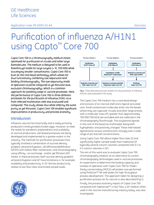
Capto Core 700 AN 29000334AA

GE Healthcare Life SciencesApplication note 29-0003-34 AAVaccinesPurification of influenza A/H1N1using Capto ™Core 700Capto Core 700 is a chromatography medium (resin) optimized for purification of viruses and other large biomolecules. The medium is designed to be used in flowthrough mode for large targets (> M r 700 000) while scavenging smaller contaminants. Capto Core 700 is built on the core bead technology which allows for dual functionality combining size separation with binding chromatography. The size separating mode of operation could be compared to gel filtration (size exclusion chromatography), which is a commonapproach for polishing steps in vaccine processes. Here, the performance of Capto Core 700 in three different processes for the purification of influenza H1N1 virus from infected mammalian cells was evaluated andcompared. This study shows that while offering the same purity as gel filtration, Capto Core 700 enables significant improvements in productivity and process economy.IntroductionInfluenza vaccine has historically and is today primarily produced in embryonated chicken eggs. However, to meet the needs for pandemic preparedness and scalability of vaccine productions, cell-based processes are being developed and implemented to a greater extent in the industry. The methods for purification processes have typically involved a combination of sucrose density gradient ultracentrifugation, ultrafiltration/diafiltration (UF/ D F) with hollow-fiber membranes, and chromatography using affinity-, ion exchange-, and/or gel filtration (GF) media. In these processes, both sucrose density gradient ultracentrifugation and GF have limitations in, for example, scalability and productivity. In GF, the low productivity relates to low flow rates and limited sample loads.The Capto Core 700 medium has a core bead design and consists of an inactive shell and a ligand-activated core. Small contaminant molecules enter into the beads where they are captured. Viruses and other large entities with a molecular mass (M r ) greater than approximately 700 000 (700 kDa) are excluded and are collected in the chromatography flowthrough. The octylamine ligands in the core of the bead are multimodal, being bothhydrophobic and positively charged. These internalized ligands bind various contaminants strongly over a wide range of pH and salt concentrations.Using Capto Core 700 allows higher flow rates (1) and significantly higher sample loads than traditional GF (typically several column volumes compared with 0.1 to 0.3 column volumes in GF).The aim of this work was to evaluate Capto Core 700 chromatography medium as an alternative to current chromatography technologies used in vaccine processes. An experiment to determine the binding capacity and window of operation with Capto Core 700 for Madin-Darby canine kidney (MDCK) cell protein was performed using PreDictor™ 96-well plates for high-throughputprocess development. The approach taken for designing a purification process for flu vaccine is also described (Fig 1).Finally, the process economy impact of Capto Core 700compared with Sepharose™ 4 Fast Flow, a GF medium often used in the vaccine manufacturing industry today, was also investigated.Fig 1. Three alternative process streams for the purification of influenza A/H1N1 were evaluated in this study.Materials and methodsCell culture and infectionMDCK cells (inoculation concentration of 500 000 cells/mL) were grown on Cytodex™ 3 microcarriers for 48 h in an Applikon™ Bioreactor (Applikon Technology). The finalcell density was approximately 2 500 000 cells/mL at which point cells were infected with influenza A/Solomon Islands/3/2006 (H1N1) and harvested at 72 h post infection. Preliminary studiesInvestigation of operating window (binding study) for Capto Core 700The binding capacity of Capto Core 700 for host cell protein (HCP) from MDCK cell lysate was evaluated in buffers containing sodium phosphate and Tris, 150–1000 mM NaCl, pH 6.5–8.0. PreDictor 96-well plates were filled with 10 µL of Capto Core 700 for the binding study. Clarified MDCK cell lysate (200 µL) was applied to the wells of the plate and incubated in the various equilibration buffers for 60 min. After incubation, unbound sample was removed by centrifugation and the medium was washed with buffer. Collected fractions were analyzed for total protein using the Bradford protein assay.Benchmarking of purification performance:Capto Core 700 vs Sepharose 4 Fast FlowThe performance of Capto Core 700 was compared to that of Sepharose 4 Fast Flow. Capto Core 700 was packedin Tricorn™ 5/50 column (column volume [CV], 1 mL) and10 CV of clarified and concentrated virus material was loaded. Sepharose 4 Fast Flow was packed in Tricorn10/600 (CV, 47 mL) and 0.1 CV of virus feed was loaded. Recovery of virus as measured by the quantitation of hemagglutinin (HA) and reduction of HCP was compared between the two media.Process developmentClarificationClarification of harvested cells was achieved by microfiltration (MF) using ULTA Prime GF normal-flowfilter capsules. A 2.0 µm (4“ membrane, 0.10 m2 effective filtration area) rating was used initially followed by a0.6 µm (4” membrane, 0.11 m2 effective filtration area) rating. The ULTA Prime GF filters were washed with buffer (20 mM sodium phosphate, 150 mM NaCl, 0.05% sodium azide, pH 7.2) before use.Degradation of DNABenzonase endonuclease treatment was applied for the removal of DNA in process stream 2 (Fig 1). Degradationof DNA using Benzonase results in small oligonucleotide fragments that enter through the inactive layer and into the core where they are captured by the internalized ligands. Benzonase was applied after MF and before column purification with Capto Core 700 in process stream 2 (Fig 1).Capto DeVirS: binding/elution of influenza virusCapto DeVirS is a chromatography medium designed for the capture and intermediate purification of viruses. The Capto DeVirS ligand is dextran sulfate, which allows for pseudo affinity for several virus types including influenza and was therefore selected for virus capture in process stream 3. Chromatography runs were performed in a HiScale™50/20 column packed with 202 mL of Capto DeVirS.ÄKTAexplorer™ 100 chromatography system was used for chromatography runs on Capto DeVirS.Capto Core 700: polishing stepIn process stream 1–3, chromatography runs were performed in an XK 16/20 column packed with 25 mL of Capto Core 700. ÄKTAexplorer 10 chromatography system was used for chromatography runs on Capto Core 700. Analytical methodsVirus quantitationIn this study, Biacore™ T200 system and Sensor Chip CM5 were used to measure HA content according to a previously described method (2). The potency of influenza vaccines are mainly determined by quantitation of HA using the single radial immunodiffusion (SRID) assay. This method, although approved by both FDA and EMEA, is labor-intensiveand suffers from low precision and sensitivity. Biacore biosensor assays offer greater precision in the quantitation of influenza HA and faster analysis than SRID in vaccine development and manufacturing.HCP quantitationHCP is usually quantitated as total protein with, for example, the Bradford protein assay. This method is not sensitive or specific enough to detect levels below the regulatory critical limits. Therefore, Biacore biosensor assay was used for the quantitation of HCP using the same instrumentation as mentioned above. In house produced polyclonal anti-MDCK HCP antibodies were immobilized on the Biacore chip for MDCK HCP protein binding. The HCP standard was set using Bradford to estimate protein content.As reference, the total protein content (including HA)was also measured with the Bradford protein assay. The assay was performed according to the manufacturer’s recommended methods (3).Determination of infectious particlesThe median Tissue Culture Infectious Dose (TCID50)assay measures dilution that generates cytopathic effect in 50% of the cell culture and is an infectivity method that is oneof the most commonly used methods for detection of the infective virus. The TCID50assay is simple to perform and requires no specific instrumentation for result interpretation. The outcome is either presented as a log10 titer (10x.x TCID50 units/ m L) or a dilution (10-x.x/mL).2 29-0003-34 AASodium phosphate Tris20 mM sodium phosphate + 150 mM NaCl 20 mM sodium phosphate + 1000 mM NaCl 02468101214H C P c a p a c i t y (m g p r o t e i n /m L m e d i u m)10002000300040005000Volume (mL)A 280 (m A U )Conductivity (mS/cm)29-0003-34 AA 3Results and discussionInvestigation of operating window (binding study) for Capto Core 700The performance of Capto Core 700 was robust in both 20 mM sodium phosphate and Tris buffer containing up to 1 M NaCl and in pH from 6.5 to 7.5 for sodium phosphate and pH 7.5 to 8.0 for Tris, respectively (Fig 2). In general, the low NaCl concentration of 150 mM resulted in higher binding capacity while lower pH yielded higher binding capacity for the MDCK cell lysate proteins.The robust performance of the multimodal octylamine ligand in a relatively wide range of NaCl concentration and pH gives Capto Core 700 a wide window of operation. This reduces the need for optimization such as buffer exchange or dilution between steps, even with different feed materials when working with Capto Core 700.Benchmarking of purification performance: Capto Core 700 vs Sepharose 4 Fast FlowThe purification performance of Capto Core 700 was compared to that of Sepharose 4 Fast Flow, which is a chromatography medium typically used for GFpurification of a range of viruses in vaccine processes. Both chromatography methods provided a similar yield of virus HA and reduction of HCP (Table 1).Amount of medium: 10 µL of Capto Core 700 in PreDictor 96-well filter plate Sample: Clarified MDCK cell lysate in different equilibration buffers Sample load: 200 µL of cell lysate (60 min incubation)Equilibration buffers: 20 mM sodium phosphate, 150–1000 mM NaCl, pH 6.5–7.520 mM Tris, 150–1000 mM NaCl, pH 7.5–8.0Equilibration: 3 × 200 µL of equilibration buffer Wash: 200 µL of equilibration bufferFig 2. Protein binding capacity of Capto Core 700 in sodium phosphate and Tris buffers with different NaCl concentrations and a range of pH. The performance of Capto Core 700 is robust in terms of buffer component and NaCl concentration. At higher pH, the ligand gradually loses charge, which consequently decreases binding capacity.Table 1. Recovery of HA and reduction of HCP on Capto Core 700 comparedwith Sepharose 4 Fast Flow*, a standard medium used for GF in vaccine processes MediaRecovery HA (%)Recovery normalized (%)Reduction HCP (%)Reduction normalized (%)Capto Core 700†859932104Sepharose 4 Fast Flow †8610031100*Chromatography conditions:Columns:T ricorn 5/50 (CV, 1 mL) packed with Capto Core 700; Tricorn 10/600 (CV, 47 mL) packed with Sepharose 4 Fast FlowSample: Clarified virus materialSample load: Capto Core 700, 10 CV; Sepharose 4 Fast Flow, 0.1 CV Buffer: 20 mM Tris, 150 mM NaCl, pH 7.5†Capto Core 700 runs were performed in triplicate, Sepharose 4 Fast Flow runs in duplicateFig 3. Purification of influenza A/H1N1 on Capto Core 700 following MF. Labels on the chromatogram indicate gDNA and virus in the flowthrough fraction and the peak of impurities obtained during CIP.Column: XK 16/20, packed with 25 mL of Capto Core 700Sample and load: Clarified virus material, 20 CVEquilibration/wash: 20 mM sodium phosphate, 500 mM NaCl, 0.05% sodium azide,pH 7.2Flow velocity during loading: 250 cm/h (3 min residence time) CIP: 1 M NaOH, 27% 1-propanol (total contact time 60 min)System: ÄKTAexplorer 10Results from the combination of MF with ULTA Prime GF and single-step purification with Capto Core 700 are shown in Table 2. Virus yield (measured in HA content) in fractions collected in the flowthrough was excellent and protein removal significant, while it was clear that a greater reduction of DNA would be required. Full-length genomic DNA (gDNA) will not enter the beads and hence not bind to the ligands. An expected result would therefore be low reduction of DNA in the column flowthrough.Process stream 1: MF and single-step purification using Capto Core 700Figure 3 shows the broad flowthrough peak containing the virus and residual DNA/HCP obtained on Capto Core 700.Volume (mL)A 280 (m A U )Conductivity (mS/cm)pHFlowthrough (DNA and HCP)CIPVirus Volume (mL)A 280 (m A U )Conductivity (mS/cm)50010001500200020406080100120140pHFlowthrough (virus)CIP4 29-0003-34 AATable 2. Virus HA yield, TCID 50, DNA, total protein, and HCP/HA quotient in a purification scheme incorporating MF and single-step chromatography using Capto Core 700StepHA yield (%)Titer (TCID 50/mL)DNA/HA (ng/µg)Total protein/HA (µg/µg)HCP/HA (µg/µg)Microfiltration: ULTA Prime GF649.7267219.831.6Chromatography: Capto Core 7001059.314594.211.2Column: HiScale 50/20 column packed with 202 mL Capto DeVirS Sample and sample load: Clarified virus material, 5 CVBinding and wash buffer: 20 mM sodium phosphate, 150 mM NaCl, 0.05% sodium azide, pH 7.2Elution buffer:20 mM sodium phosphate, 750 mM NaCl, 0.05% sodium azide, pH 7.2 (giving ~500 mM NaCl in elution pool)Flow velocityduring loading: 60 cm/h CIP: 1 M NaOH System: ÄKTAexplorer 100Process stream 2: MF, DNA removal step, andsingle-step purification using Capto Core 700In an attempt to decrease DNA concentration obtained in process 1 and potentially also decrease DNA associated HCPs, a DNA degrading step using Benzonase treatment was added to the workflow.Results from this experiment are shown in Table 3. As expected, virus yield in flowthrough fractions in the Capto Core 700 chromatography step was excellent. DNA/HA content was effectively reduced by Benzonase treatment. Protein levels remained similar as process stream 1, that is a 3–5 fold reduction. Benzonase was also removed in the Capto Core 700 step as it entered the core and was bound. The TCID 50 titer remained high meaning that the infectivity of the virus was not affected either by filtration, Benzonase treatment, or chromatography.Table 3. Virus HA yield, TCID 50, DNA, total protein, and HCP/HA quotient in a purification scheme incorporating MF, DNA reduction step using Benzonase endonuclease, and final chromatography step using Capto Core 700StepHA yield (%)Titer (TCID 50/mL)DNA/ HA (ng/µg)Total protein/HA (µg/µg)HCP/HA (µg/µg)Microfiltration: ULTA Prime GF 649.7267222.032.3Benzonase endonuclease treatment7.022.930.3Chromatography:Capto Core 7001059.37.03.813.1Column:XK 16/20 packed with 25 mL of Capto Core 700Sample:Eluted fractions from Capto DeVirS stepSample load: 8 CV of Capto DeVirS eluate, 250 cm/h (3 min residence time)Equilibration/ wash:20 mM sodium phosphate, 500 mM NaCl, 0.05% sodium azide, pH 7.2Flow velocityduring loading: 250 cm/h CIP: 1 M NaOH, 27% 1-propanol (total contact time 60 min)System: ÄKTAexplorer 10SFig 4. Two-step purification of influenza A/H1N1 virus after MF. Capture of the virus was achieved using A) Capto DeVirs and final purification using B) Capto Core 700. Labels on the chromatograms indicate the elution of virus, DNA, and HCP.A)B)Process stream 3: MF and two-stepchromatography using Capto DeVirS and Capto Core 700Processes 1 and 2 both gave high yield of HA whileinsufficient DNA and HCP reduction was observed. Thus, it was considered desirable to further reduce both DNA and HCP content. For comparison to Process 1 and 2, a two-step chromatography process using Capto DeVirS for capture and Capto Core 700 for final purification of the virus was evaluated.Chromatograms showing both chromatography steps after MF are shown in Figure 4. Capto DeVirS was effective for the capture of the virus (Fig 4A) and eluted fractions from this step were applied to the XK column packed with Capto Core 700 for final purification (Fig 4B). On account of the robust binding performance of Capto Core 700, equilibration of Capto Core 700 was achieved using the buffer usedfor elution in the Capto DeVirS step. The need for buffer exchange or dilution between steps was thereby eliminated, contributing to speeding up the chromatography process. This demonstrates the advantages from the wide window of operation that is enabled by Capto Core 700.Table 4 shows the results in terms of HA recovery, TCID50,DNA- and protein removal at each step of the process.In this case, good yield of virus HA as well as significant removal of HCP and DNA were observed. DNA was reduced 2.8 log and proteins 5–7 fold by Capto DeVirS. CaptoCore 700 further reduced protein levels by 3–5 fold. The infectivity of the virus was retained throughout the process, as shown by the titer measured with TCID50.Table 4. Virus HA yield, TCID50, DNA, total protein, and HCP/HA quotient ina purification scheme incorporating MF and two-step purification using Capto DeVirS and Capto Core 700Step HAyield(%)Titer(TCID50/mL)DNA/HA(ng/µg)Totalprotein/HA(µg/µg)HCP/HA(µg/µg)Microfiltration:ULTA Prime GF649.7267222.032.3 Chromatographyfirst-step:Capto DeVirS94 4.0 3.1 6.1 Chromatographysecond step:Capto Core 700949.3 5.0 1.1 1.1Cost analysis – comparison of Capto Core 700 approach vs GFAn economic evaluation of Capto Core 700 as an alternative to GF for polishing step was performed based on the example above. The comparison was made under the following conditions:1. Calculation was based on a 1000 L fermentation volume2. Same recoveries and purities of virus were assumedfor Capto Core 700 and GF, as shown by previousexperiments3. Process conditions for Capto Core 700 step was thesame as process 3 above, while the conditions for GFwere chosen based on experience and presented as two different examples4. Costs were calculated per batch and costs expected tobe same for both techniques were excluded from theanalysis5. Labor costs included both salary and overheadcosts relating to facility buildings etc. This was set to800 USD/h but will vary from case to caseThe comparison of Capto Core 700 and GF is shown in Table 5. The higher load volumes achieved with Capto Core 700 compared to GF resulted in smaller volumesof chromatography medium needed to process thesame amount of material within a shorter timeframe(GF alternative 1). As a consequence, the labor costs, buffer costs, and costs for Capto Core 700 medium would be significantly lower than for traditional GF. The resulting total cost per batch for this step could be lowered by almost two-fold. Other benefits, not included in this simplified example, are that smaller buffer tanks and smaller columns could be used leading to reduced footprint, and the need for larger hardware investments is reduced.Another alternative could be to use a smaller columnfor GF and compensate the low load volume by a larger number of cycles per batch (GF alternative 2, Table 5). The medium cost per batch would still be the same for GF as this calculation takes into account the complete lifetime of the medium. However, labor cost would increase further as a consequence of the longer process time, giving a three-fold higher total cost than for Capto Core 700. On the other hand, the benefits with reduced footprint and minimized hardware investments would in this case be the same as for the Capto Core 700 alternative.29-0003-34 AA 56 29-0003-34 AAConclusionsCapto Core 700 allows a wide window of operation for pH and NaCl concentration, securing robust purification and often simplified process design in vaccine manufacturing. The combination of MF, capture of virus with CaptoDeVirS, and final purification on Capto Core 700 showed excellent results in terms of virus purity and reduction of DNA and HCP. Using Capto Core 700 in this approach had the advantage of allowing direct transfer of elutedfractions containing target virus without the need for buffer exchange or dilution, which enables faster and simpler processing. Capto Core 700 combines scalability and high productivity with improved process economy, all common drawbacks of GF. Capto Core 700 enables chromatography processes with characteristics that the vaccine industry is looking for—scalability, high productivity, good process economy, and short start-up times.References1. Data file: Capto Core 700. GE Healthcare, 28-9983-07, Edition AA (2012).2. Estmer Nilsson, C. et al. A novel assay for influenza virus quantification using surface plasmon resonance . Vaccine 28(3), 759–756 (2010).3.Bradford, M. M. A rapid and sensitive method for the quantitation of microgram quantities of protein utilizing the principle of protein-dye binding. Anal. Biochem. 72, 248-254 (1976).Table 5. Key process parameters and cost comparison between an approach using Capto Core 700 and traditional GFCapto Core 700GF alternative 1GF alternative 2Column volume (L)2520050Load volume (CV)80.20.2Number of cycles per batch 1520Medium cost (USD)185060006000Buffer cost (USD)100020002000Labor cost (USD)4165510012 750Total cost (USD)701513 10020 750Ordering informationProduct Code number Cytodex 3, 2.5 kg 17-0485-25ULTA Prime GF, 2.0 µm 28-9084-21ULTA Prime GF, 0.6 µm 28-9083-33Capto DeVirS, 1 L 17-5466-03Capto Core 700, 1 L 17-5481-04Tricorn 5/50 column 28-4064-09Tricorn 10/600 column 28-4064-19XK 16/20 column 28-9889-37HiScale 50/20 column 28-9644-44Sepharose 4 Fast Flow17-0149-0129-0003-34 AA 7imagination at work GE, imagination at work and GE monogram are trademarks of General Electric Company.ÄKTAexplorer, Biacore, Capto, Cytodex, HiScale, PreDictor, Sepharose, Tricorn, and ULTA are trademarks of GE Healthcare companies. Applikon is a trademark of Applikon Biotechnology B.V.Benzonase is a trademark of Merck KGaA.© 2012 General Electric Company — All rights reserved.First published Mar. 2012All goods and services are sold subject to the terms and conditions of sale of the company within GE Healthcare which supplies them. A copy of these terms and conditions is available on request. Contact your local GE Healthcare representative for the most current information. GE Healthcare UK LimitedAmersham PlaceLittle ChalfontBuckinghamshire, HP7 9NAUKGE Healthcare Europe, GmbHMunzinger Strasse 5D-79111 FreiburgGermanyGE Healthcare Bio-Sciences Corp.800 Centennial Avenue, P.O. Box 1327Piscataway, NJ 08855-1327USAGE Healthcare Japan CorporationSanken Bldg., 3-25-1, HyakuninchoShinjuku-ku, Tokyo 169-0073Japan29-0003-34 AA 03/2012For local office contact information, visit /contact /captocore GE Healthcare Bio-Sciences ABBjörkgatan 30751 84 UppsalaSweden。
抗EV71多克隆抗体偶联的靶向磁性纳米颗粒对病毒的特异性富集
抗EV71多克隆抗体偶联的靶向磁性纳米颗粒对病毒的特异性富集霍美俊1,2*,张长清2,3*,闫 妍1,2,赵凌云1,貌盼勇4,唐劲天1(1. 清华大学工程物理系;粒子技术与辐射成像教育部重点实验室,北京100084 2北京中医药大学中药学院生物制药系,北京100102 3上海医药工业研究院,上海 2000401 4解放军302医院传染病研究所病毒研究室, 北京 100039)摘要 手足口病是幼儿和儿童常见的一种传染病,严重时会引起患者死亡。
EV71病毒(人肠道病毒71型)是引起手足口病的一种主要病毒。
为寻求一条快速诊断EV71病毒感染的新方法,将兔抗EV71多克隆抗体,通过戊二醛交联法偶联在氨基硅烷修饰的磁性纳米颗粒表面,获得抗EV71多克隆抗体偶联的靶向磁性纳米颗粒用于EV71病毒的检测。
通过酶联免疫、免疫荧光方法证实了抗体耦合在磁性纳米颗粒表面,并通过BCA法测得其耦合的效率为94.1%。
采用抗体偶联的磁性纳米颗粒对EV71病毒液中的病毒抗原进行吸附,通过ELISA法检测上清液中病毒含量的变化,并对磁性纳米颗粒表面吸附的病毒进行免疫荧光和核酸PCR检测,证明了抗体偶联的磁性纳米颗粒可以与病毒抗原特异性结合。
由于该纳米颗粒同时具有抗体的靶向性和磁性颗粒的磁响应性,对病毒抗原有较好的特异性吸附能力,可以用于低浓度大样本的EV71病毒抗原的富集检测。
利用抗体偶联的靶向磁性纳米颗粒同时具有可在病毒感染的细胞周围特异性富集及磁颗粒可在交变磁场下感应升温的双重功能,将其作为磁感应热疗的靶向介质,有望研制出病毒感染性疾病磁感应热疗的靶向介质,为靶向热疗病毒感染性疾病提供新的尝试。
关键词 EV71;抗体偶联;磁性纳米颗粒;病毒检测; 磁感应热疗Specific Enrichment of Viral Antigen with EV71 Polyclonal Antibody-coupledMagnetic Particles(HUO Meijun1, 2*, ZHANG Changqing2, 3*,YAN Yan1, 2, ZHAO Lingyun1, MAO Panyong4, SHENG Jun5, TANG Jintian1)(1. Department of Engineering Physics, Tsinghua University; Key Laboratory of Particle & Radiation Imaging, Ministry of Education, Beijing 100084, China 2. Department of Biopharmaceuticals, Beijing University of Chinese Medicine, Beijing 100102, 3. Shanghai Institute of Pharmaceutical Industry, Shanghai 200040, China 4. Department of Virology, Institute of infectious Diseases, 302 Hospital of PLA , Beijing 100039, China 5. Changchun Institute of Biological Products, Changchun 130062,)Abstract Hand, foot, and mouth disease (HFMD) is a common viral illness of infants and children. The disease causes fever and blister-like eruptions in the mouth and/or a skin rash. Enterovirus 71 (EV71) is notable as one of the major causative agents for HFMD, and is sometimes associated with severe central nervous system diseases. In this study, a novel method for specific作者简介:霍美俊,硕士研究生,研究方向为生物医学工程,电子信箱:huomeijun86@;张长清,硕士研究生,研究方向为生物医学工程,电子信箱:zhchq2933@;唐劲天(通信作者),教授,研究方向为肿瘤学,电子信箱:tangjt@;*为共同第一作者enrichment of viral antigen by EV-71 polyclonal antibody-coupled magnetic particles was developed. Targeted magnetic nanoparticles for specific recognition and enrichment of EV71 antigen were prepared by modification the polyclonal antibody of EV71 onto the surface of amino-silane coated magnetic nanoparticles (MNPs) through glutaraldehyde cross-linking. Antiserum of EV71 was generated by immunization of rabbits with purified EV71 antigen. Next, the serum was precipitated by saturated ammonium sulfate and then was ligated on the surface of Amino-silane-modified magnetic particles though glutaraldehyde cross-linking. Thus, targeted magnetic medium was prepared for specific enrichment of EV71. Both ELISA and immuno-fluorescence technique were applied to confirm the successful coupling of EV71 polyclonal antibody onto the surface of MNPs, and the coupling efficacy was as high as 94.1%. Specific enrichment of the EV71 antigen by the above-mentioned tareted MNPs was studied by immunofluorescence and PCR. Except for the specific enrichment of the virus antigen, possessing the inductive heating property under Alternative Magnetic Field (AMF), the antibody coupled MNPs can be further applied as agent of the Magnetic Mediated Hyperthermia (MMH) for targeted treatment of infective disease.Keywords EV71; magnetic particles; virus detection1 引 言病毒是许多疾病的病原微生物,严重威胁着人类健康。
非洲猪瘟病毒p22蛋白单克隆抗体的制备及鉴定
㊀山东农业科学㊀2023ꎬ55(10):140~145ShandongAgriculturalSciences㊀DOI:10.14083/j.issn.1001-4942.2023.10.019收稿日期:2022-12-28基金项目:山东省重点研发计划项目(2020CXGC010801ꎬ2022TZXD0041ꎬ2022CXPT010ꎬ2021LZGC001)ꎻ山东省现代农业产业技术体系(SDAIT-08-01)ꎻ山东省农业科学院农业科技创新工程项目(CXGC2023F10ꎬCXGC2023A21ꎬCXGC2023A21)ꎬ泰山学者工程项目作者简介:矫健(1998 )ꎬ男ꎬ山东青岛人ꎬ硕士研究生ꎬ主要从事动物病毒学与免疫学研究ꎮE-mail:1174629561@qq.com李建达(1992 )ꎬ男ꎬ山东济南人ꎬ博士ꎬ主要从事动物微生物与免疫学研究ꎮE-mail:ljd4453@163.com∗同为第一作者ꎮ通信作者:于江(1985 )ꎬ女ꎬ山东莱州人ꎬ博士ꎬ研究员ꎬ研究方向为动物病原学与免疫学ꎮE-mail:yujiang_2213@163.com吴家强(1975 )ꎬ男ꎬ山东诸城人ꎬ博士ꎬ研究员ꎬ研究方向为动物病原学与免疫学ꎮE-mail:wujiaqiang2000@sina.com非洲猪瘟病毒p22蛋白单克隆抗体的制备及鉴定矫健1ꎬ2ꎬ李建达2∗ꎬ韩先杰1ꎬ张琳2ꎬ张玉玉2ꎬ任素芳2ꎬ刘飞2ꎬ陈智2ꎬNataliiaHrabchenko2ꎬ张文娟3ꎬ于江1ꎬ2ꎬ吴家强1ꎬ2(1.青岛农业大学动物医学院ꎬ山东青岛㊀266109ꎻ2.山东省农业科学院畜牧兽医研究所/山东省畜禽疫病防治与繁育重点实验室/农业农村部畜禽生物组学重点实验室ꎬ山东济南㊀250100ꎻ3.山东碧蓝生物科技有限公司ꎬ山东泰安㊀271400)㊀㊀摘要:非洲猪瘟(AfricanswinefeverꎬASF)是由非洲猪瘟病毒(AfricanswinefevervirusꎬASFV)引起的一种高度传染性病毒性疾病ꎬ发病率和死亡率高达100%ꎬ对我国的生猪养殖业造成了毁灭性打击ꎮp22蛋白为ASFV的结构蛋白ꎬ本研究构建了重组原核表达质粒pET-32a-p22ꎬ将其转化至大肠杆菌BL21(DE3)感受态细胞表达重组p22蛋白并纯化ꎬ经Westernblot鉴定证实重组p22蛋白与ASFV阳性血清有良好的免疫反应性ꎮ利用重组p22蛋白免疫Balb/c小鼠并通过间接ELISA方法筛选到2株能够稳定分泌p22蛋白单克隆抗体(MAb)的杂交瘤细胞株2D6和5E7ꎮ经抗体亚型鉴定两株抗体均为IgG1型ꎻ经Westernblot鉴定ꎬ两株抗体能够特异性识别过表达的p22蛋白ꎮ综上ꎬ本研究成功表达并纯化了p22重组蛋白ꎬ制备了p22单克隆抗体ꎬ为进一步探讨p22蛋白的结构功能及其在ASFV中的感染致病机制提供了基础条件ꎮ关键词:非洲猪瘟病毒ꎻp22蛋白ꎻ单克隆抗体ꎻ原核表达中图分类号:S852.65+1㊀㊀文献标识号:A㊀㊀文章编号:1001-4942(2023)10-0140-06PreparationandIdentificationofMonoclonalAntibodyagainstp22ProteinofAfricanSwineFeverVirusJiaoJian1ꎬ2ꎬLiJianda2∗ꎬHanXianjie1ꎬZhangLin2ꎬZhangYuyu2ꎬRenSufang2ꎬLiuFei2ꎬChenZhi2ꎬNataliiaHrabchenko2ꎬZhangWenjuan3ꎬYuJiang1ꎬ2ꎬWuJiaqiang1ꎬ2(1.CollegeofVeterinaryMedicineꎬQingdaoAgriculturalUniversityꎬQingdao266109ꎬChinaꎻ2.InstituteofAnimalScienceandVeterinaryMedicineꎬShandongAcademyofAgriculturalSciences/ShandongKeyLaboratoryofAnimalDiseaseControlandBreeding/KeyLaboratoryofLivestockandPoultryMulti ̄omicsꎬMinistryofAgricultureandRuralAffairsꎬJinan250100ꎬChinaꎻ3.ShandongGreenBlueBiotechnologyCompanyLimitedꎬTaian271400ꎬChina)Abstract㊀Africanswinefever(ASF)isahighlycontagiousviraldiseasecausedbyAfricanswinefevervirus(ASFV)withthemorbidityandmortalityupto100%ꎬwhichhascausedadevastatingblowtoChina spigfarmingindustry.Thep22proteinisastructuralproteinofASFV.InthisstudyꎬrecombinantprokaryoticexpressionplasmidpET ̄32a ̄p22wasconstructedandtransformedintoEscherichiacoliBL21(DE3)receptivecellstoexpresstherecombinantp22proteinandpurifyit.Westernblotanalysisconfirmedthattherecombinantp22proteinhadgoodimmunoreactivitywithASFVpositiveserum.Balb/cmicewereimmunizedwiththere ̄combinantp22proteinandtwohybridomacelllines2D6and5E7ꎬwhichcouldsecretemonoclonalantibody(MAb)top22proteinꎬwerescreenedbyindirectELISA.Theantibodysubtypesofthetwostrainswereidenti ̄fiedasIgG1type.Westernblotanalysisshowedthatthetwoantibodiescouldspecificallyrecognizetheoverex ̄pressedp22protein.Inconclusionꎬtherecombinantp22proteinwassuccessfullyexpressedandpurifiedinthisstudyꎬandthep22monoclonalantibodywaspreparedꎬwhichprovidedoneofthebasicconditionsforfur ̄therinvestigationofthestructureandfunctionofp22proteinꎬandalsoitsroleinthepathogenesisofASFVin ̄fection.Keywords㊀Africanswinefevervirusꎻp22proteinꎻMonoclonalantibodyꎻProkaryoticexpression㊀㊀非洲猪瘟(AfricanswinefeverꎬASF)是由非洲猪瘟病毒(AfricanswinefevervirusꎬASFV)引起的一种高度传染性㊁出血性疾病ꎬ临床上以高热㊁皮肤发绀㊁淋巴结及脏器广泛出血为主要特征ꎬ如肾瘀斑㊁皮肤红斑㊁肺水肿㊁弥散性血管内凝血等[1]ꎮASFV主要通过接触性传播ꎬ家猪和野猪都是其主要宿主ꎬ一旦侵入家猪ꎬ便可在猪群中快速传播ꎬ发病率和死亡率高达100%[2-3]ꎬ感染性病毒在组织中能够存在较长时间ꎬ尤其是淋巴结和扁桃体[4]ꎮ此外ꎬ该病毒也是唯一已知的DNA虫媒病毒ꎬ能够通过软蜱进行传播[5]ꎮASF于1921年首次在肯尼亚报道ꎬ随后传入其他非洲国家[6]ꎮ2007年ꎬ在格鲁吉亚发现了毒力更高的ASFVⅡ型ꎬ并迅速传播[7]ꎮ2018年8月ꎬ我国沈阳首次暴发ASFꎬ随后迅速蔓延到全国所有省份[8]ꎬ对我国乃至全世界的养猪业造成了严重的经济损失ꎮASFV是Asfarviridae科中Asfarvirus属的唯一成员[9]ꎬ是一种有囊膜的双链DNA病毒ꎮASFV粒子是具有多层结构的二十面体ꎬ直径约为200nmꎬ病毒粒子结构自内向外依次为类核㊁核壳㊁内囊膜㊁衣壳㊁外囊膜[10-11]ꎮASFV基因组长度约为170~194kbꎬ目前已从其中鉴定出结构蛋白68种ꎬ部分蛋白的功能尚不清楚[12]ꎮ这些结构蛋白分布在不同的区域ꎬ在病毒黏附与入侵㊁病毒基因组复制和转录㊁病毒包装与出芽以及免疫逃避等病毒感染的不同方面发挥着重要的作用[5]ꎮ由于ASFV结构复杂繁冗ꎬ对其功能的了解也十分有限ꎬ到目前为止还没有完全有效的ASFV商业疫苗ꎬ因此鉴定病毒结构蛋白的免疫原性在探讨病毒与宿主相互作用中至关重要[13]ꎮASFVp22蛋白由KP177R基因编码ꎬ分子量为22kDaꎮp22最初被描述为一种早期诱导的跨膜结构蛋白ꎬ位于病毒颗粒外部ꎬ能够从病毒颗粒表面溶解释放出来[14]ꎮ然而ꎬ进一步的研究发现ꎬp22蛋白位于病毒颗粒的内膜和感染细胞的表面[15]ꎮ目前ꎬp22蛋白在ASFV感染致病过程中的作用仍不清楚ꎮ本研究通过大肠杆菌原核表达系统成功表达并纯化出p22重组蛋白ꎬ通过免疫Balb/c小鼠成功制备了其单克隆抗体(MAb)ꎬ为进一步探究p22蛋白的生物学作用奠定基础ꎮ1㊀材料与方法1.1㊀细胞㊁菌株㊁载体及实验动物大肠杆菌Trans5α㊁BL21(DE3)感受态细胞购自北京全式金生物技术有限公司ꎻ骨髓瘤细胞SP2/0㊁pET-32a载体均由本实验室保存ꎻASFV标准阳性血清购自中国兽医药品监察所ꎻSPF级5~8周龄Balb/c雌性小鼠购自济南朋悦试验动物繁育有限公司ꎮ1.2㊀主要试剂质粒小提试剂盒㊁胶回收试剂盒均购自天根生化科技(北京)有限公司ꎻ限制性核酸内切酶㊁T4DNA连接酶㊁蛋白Marker均购自赛默飞世尔科技公司ꎻNi ̄NTAAgarose购自德国Qiagen公司ꎻ单抗亚型鉴定试剂盒购自美国SouthernBiotech公司ꎻECL发光液㊁羊抗鼠HRP-IgG㊁羊抗猪HRP-IgG购自Beyotime公司ꎮ1.3㊀ASFVp22蛋白的原核表达1.3.1㊀重组表达质粒pET-32a-p22的构建㊀根据ASFVpig/HLJ/2018株基因序列(GenBankNo.MK333180.1)的KP177R基因序列进行引物设141㊀第10期㊀㊀㊀㊀㊀㊀矫健ꎬ等:非洲猪瘟病毒p22蛋白单克隆抗体的制备及鉴定计ꎬ正向引物(p22-F)序列:5ᶄ-CTCGAGTTAT ̄GCGTGTTTATGATTAC-3ᶄ(包含XhoⅠ酶切位点)ꎬ反向引物(p22 ̄R)序列:5ᶄ-GGATC ̄CAAAAAACAGCAGCCGCCGA-3ᶄ(包含BamHⅠ酶切位点)ꎬ由生工生物工程(上海)股份有限公司合成ꎮ以合成的密码子优化后的p22基因序列为模板ꎬ使用引物扩增目的基因片段ꎬPCR反应程序为95ħꎬ5minꎻ94ħꎬ45sꎬ58ħꎬ45sꎬ72ħꎬ45sꎬ32个循环ꎻ72ħꎬ10minꎻ4ħ结束ꎮ将扩增产物进行1%的琼脂糖凝胶电泳并纯化回收ꎬ用T4DNA连接酶分别将KP177R基因与pET-32a载体连接ꎬ将连接产物转化到感受态细胞Trans5α中ꎮ挑取单克隆摇菌ꎬ菌液经PCR鉴定为阳性后ꎬ送至生工生物工程(上海)股份有限公司测序ꎮ测序成功后将阳性重组质粒命名为pET-32a-p22ꎮ1.3.2㊀ASFVp22重组蛋白的表达和纯化㊀将阳性重组质粒pET-32a-p22转化至大肠杆菌BL21(DE3)感受态细胞ꎬ挑取单克隆至LB培养基中37ħ培养过夜ꎬ取1mL菌液接种至氨苄青霉素(ampicillinꎬAmp)抗性的100mLLB液体培养基中ꎬ37ħ㊁220r/min摇床培养至OD600值达到0.6~0.8时ꎬ添加终浓度为1mmol/L的IPTG诱导目的蛋白表达ꎬ诱导4h后ꎬ收集菌体沉淀ꎬ使用裂解液(300mmol/LNaClꎬ50mmol/LNaH2PO4ꎬpH=7.4)重悬菌体ꎬ进行超声破碎ꎬ4ħ㊁4000r/min离心ꎬ经SDS-PAGE电泳鉴定后使用0.45μm滤膜过滤ꎬ与蛋白纯化填料4ħ过夜结合ꎮ采用亲和层析的方式纯化ꎬ收集洗脱产物ꎬ用于SDS-PAGE鉴定ꎬ使用超滤管将有单一目的蛋白条带的洗脱产物浓缩并置换到PBS中ꎬ-80ħ保存ꎮ1.3.3㊀ASFVp22重组蛋白的Westernblot鉴定㊀重组p22蛋白纯化后进行SDS-PAGE电泳ꎬ随后转移到PVDF膜上ꎬ经5%脱脂奶粉封闭后ꎬ以ASFV标准阳性血清(1ʒ1000)作为一抗孵育2hꎮ清洗后ꎬ加入HRP标记的羊抗猪IgG二抗(1ʒ8000)孵育1hꎬ进行Westernblot鉴定ꎮ1.4㊀ASFVp22单克隆抗体的制备1.4.1㊀Balb/c小鼠的免疫及血清效价的测定㊀纯化后的重组p22蛋白与等体积弗氏完全佐剂乳化后ꎬ对4只5~8周龄Balb/c小鼠进行背部多点皮下注射ꎬ免疫剂量为100μg/只ꎬ每14d免疫一次ꎬ共免疫3次ꎮ最后一次免疫7d后从小鼠尾静脉取少量血ꎬ分离血清ꎬ通过间接ELISA方法检测血清中的抗体效价ꎮ将纯化的p22重组蛋白以2g/mL的浓度包被到聚苯乙烯96孔反应板(100μL/孔)ꎬ4ħ放置过夜ꎻ次日用PBST洗板3次后进行封闭(l50μL/孔封闭液)ꎬ室温放置0.5hꎻ用样品稀释液将小鼠血清按1ʒ2000ꎬ1ʒ4000ꎬ1ʒ8000ꎬ1ʒ16000ꎬ1ʒ32000ꎬ1ʒ64000ꎬ1ʒ128000 进行倍比稀释ꎬ洗板3次后加入稀释好的血清并设置空白血清做阴性对照(100μL/孔)ꎬ室温孵育1hꎻ洗板3次后加入HRP标记的鼠二抗(1ʒ5000ꎬ100μL/孔)ꎬ37ħ孵育40minꎻPBST洗板5次并拍干ꎬ加入TMB(100μL/孔)ꎬ室温避光显色10~15minꎻ加入终止液(50μL/孔)ꎬ用酶标仪测定450nm处各孔的吸光值ꎮ1.4.2㊀细胞融合及亚克隆筛选㊀按照标准流程取免疫后的小鼠脾细胞和骨髓瘤细胞SP2/0融合ꎬ通过间接ELISA方法检测筛选阳性杂交瘤细胞ꎬ使用有限稀释法对单克隆阳性孔进行3次亚克隆ꎮ筛选出稳定表达p22的阳性孔ꎬ进行扩大培养ꎮ1.4.3㊀腹水制备㊀将阳性杂交瘤细胞扩大培养并注射至Balb/c小鼠的腹腔(经弗氏不完全佐剂乳化)ꎬ7~10d后观察小鼠腹部隆起状况ꎬ并及时抽取腹水ꎮ12000r/min离心10minꎬ收集上清ꎬ-70ħ冻存备用ꎮ1.5㊀ASFVp22单克隆抗体的鉴定1.5.1㊀单克隆抗体效价鉴定㊀将进行不同比例稀释后的腹水作为一抗ꎬHRP标记的羊抗鼠IgG作为二抗ꎬ用重组p22蛋白建立的间接ELISA方法进行效价的测定(方法同1.4.1)ꎮ1.5.2㊀单克隆抗体亚型鉴定㊀用小鼠抗体亚型鉴定试剂盒对杂交瘤细胞制备的腹水抗体进行亚型测定ꎮ准备好包被有重组p22蛋白的板条(50ng/孔)ꎬ每个克隆收集600μL腹水上清ꎬ分别滴加到6个对应的酶标孔中(100μL/孔)ꎬ37ħ孵育1hꎻPBST洗板3次ꎬ将稀释好的分型二抗抗IgM㊁IgA㊁IgG1㊁IgG2a㊁IgG2b㊁IgG3的抗体加入6个孔中ꎬ37ħ孵育1hꎻPBST洗板3次ꎬ加入TMB显色ꎬ有信号反应的孔对应的鉴定二抗亚型即为241山东农业科学㊀㊀㊀㊀㊀㊀㊀㊀㊀㊀㊀㊀㊀㊀第55卷㊀该抗体的亚型ꎮ1.5.3㊀Westernblot鉴定单克隆抗体的免疫原性㊀纯化的重组p22蛋白经SDS-PAGE电泳后ꎬ转移至PVDF膜上ꎬ使用5%脱脂奶粉封闭2hꎻPBST洗膜后ꎬ加入单克隆抗体(1ʒ1000)ꎬ4ħ孵育过夜ꎻ清洗后ꎬ加入HRP标记的羊抗鼠IgG二抗孵育1hꎬ充分清洗后使用ECL发光液进行曝光ꎬ观察结果ꎮ2㊀结果与分析2.1㊀重组表达质粒pET32a-p22的构建用p22-F/p22-R引物对优化的p22基因进行扩增ꎬ结果显示ꎬPCR扩增后的p22基因片段约为447bpꎬ大小符合预期(图1)ꎮ通过酶切酶连将p22扩增产物连接到pET-32a质粒中ꎮ经测序分析后ꎬ基因测序结果完全正确ꎮ2.2㊀重组p22蛋白的表达和纯化SDS-PAGE电泳结果显示ꎬ与未诱导组相比ꎬ诱导组可见与预期大小一致的37kDa融合蛋白ꎬ经超声破碎后可溶性分析发现ꎬ重组p22蛋白条带出现在超声后上清中ꎬ表明重组p22蛋白为可溶性表达(图2A)ꎮ进一步大量诱导p22重组蛋白的表达ꎬ并与HisNi-NTAAgarose亲和层析柱结合ꎬ使用不同浓度梯度的咪唑洗脱液洗脱蛋白ꎬ收集洗脱液ꎬ随后进行SDS-PAGE电泳鉴定ꎮ结果显示ꎬ使用高浓度洗脱液时ꎬ在37kDa处出现单一条带(图2B)ꎬ纯化后纯度大于95%ꎮ1:DNAMarkerDL2000ꎻ2:阴性对照ꎻ3:目的基因PCR产物ꎮ图1㊀p22基因PCR扩增目的片段2.3㊀重组p22蛋白的Westernblot鉴定对重组p22蛋白进行Westernblot鉴定ꎬ通过His标签抗体孵育后ꎬ在37kDa处检测到嵌合His标签的p22蛋白ꎻ而使用ASFV阳性血清孵育后ꎬ发现重组p22蛋白能够与ASFV阳性血清发生特异性结合(图2C)ꎬ表明该抗原具有良好免疫原性ꎮA:1.预染蛋白质分子质量标准ꎬ2~5.诱导组ꎬ超声破碎后上清ꎬ超声破碎后沉淀ꎬ未诱导组ꎻB:1.预染蛋白质分子质量标准ꎬ2.超声破碎后上清ꎬ3.流穿液ꎬ4~9.20㊁40㊁50㊁100㊁100㊁200mmol/L咪唑洗脱液ꎻC:1㊁3.预染蛋白质分子质量标准ꎬ2.His抗体孵育重组p22蛋白ꎬ4.阳性猪血清孵育重组p22蛋白ꎮ图2㊀重组p22蛋白的表达(A)㊁纯化(B)及鉴定(C)2.4㊀单克隆抗体的制备及鉴定2.4.1㊀血清效价的测定㊀三免7d后通过间接ELISA方法检测接种小鼠血清效价ꎬ测得1㊁2㊁3号小鼠的血清效价为1ʒ128000ꎬ4㊁5号小鼠的血清效价为1ʒ64000(图3A)ꎮ选取1㊁2㊁3号小鼠进行加强免疫ꎮ2.4.2㊀单克隆抗体效价测定㊀在骨髓瘤细胞和脾细胞融合后ꎬ进行融合筛选ꎬ对筛选的阳性孔进行3次亚克隆后ꎬ经间接ELISA方法检测ꎬ最终筛选到了两株能够稳定分泌重组p22MAb的杂交瘤细胞株ꎬ通过小鼠腹腔注射两种杂交瘤细胞株ꎬ制备了小鼠腹水ꎬ通过间接ELISA方法测定腹水的效价分别为1ʒ512000和1ʒ256000(图3B)ꎬ将获得的单克隆抗体命名为2D6和5E7ꎮ341㊀第10期㊀㊀㊀㊀㊀㊀矫健ꎬ等:非洲猪瘟病毒p22蛋白单克隆抗体的制备及鉴定图3㊀小鼠血清效价(A)及单克隆抗体效价(B)的测定2.4.3㊀单克隆抗体亚型鉴定㊀采用美国SouthernBiotech公司的单抗亚型鉴定试剂盒分别对p22MAb2D6和5E7的亚型进行检测ꎬ结果显示两种MAb的亚型均为IgG1(表1)ꎮ㊀㊀表1㊀单克隆抗体亚型鉴定细胞株编号亚型轻链类型2D6IgG1Kappa5E7IgG1Kappa2.4.4㊀单克隆抗体的免疫反应鉴定㊀为了鉴定MAb2D6和5E7的免疫反应性ꎬ通过Westernblot检测了其对外源性p22蛋白的反应性ꎮ结果显示ꎬ两株MAb均能够在37kDa处与p22蛋白发生特异性反应(图4)ꎮ1:预染蛋白质分子质量标准ꎻ2:重组p22蛋白ꎮ图4㊀单克隆抗体2D6(A)和5E7(B)的免疫反应鉴定3㊀讨论与结论自2018年ASF传入中国并迅速传播以来ꎬASF对生猪生产造成了重大的经济威胁ꎬ目前ꎬ在中国传播的ASFV类型主要为一种高毒力的基因型Ⅱ毒株[16]ꎬ基因型Ⅰ毒株也有报道[17]ꎮASFV在环境中具有高传染性和稳定性ꎬ可通过与受感染的猪或其产品直接接触或通过软蜱进行传播[18]ꎮ直到目前ꎬ仍然没有针对ASFV的有效商业疫苗ꎬ使得非洲猪瘟的预防㊁控制和治疗变得十分艰难[19]ꎬ因此ꎬ筛选ASF免疫原性蛋白ꎬ鉴定免疫相关基因ꎬ将有助于ASF疫苗的开发ꎮ另外ꎬ需要对ASFV的结构功能等基础研究进行攻克ꎬ以期为ASF疫苗或诊断试剂的研发提供更多理论依据ꎮ相关研究表明ꎬ鉴定最具抗原性的病毒蛋白对改进血清学诊断试验具有重要作用ꎬ在ASFV血清诊断试验中使用重组蛋白作为抗原来源可避免操纵活病毒的潜在风险[20]ꎮp22蛋白为ASFV的结构蛋白ꎬ根据序列分析比较ꎬ东欧等地区ASFV分离株中的p22序列保守ꎬ证明p22蛋白是抗原稳定的ꎬ因此适合用作开发新的血清诊断试验的工具[21]ꎮ目前对于p22蛋白的功能研究尚不完善ꎬ其生物学功能仍待阐明ꎮ原核表达系统具有操作简单㊁表达量高的特点ꎬ本研究中所用的原核表达载体pET-32a可在BL21(DE3)细菌中高度表达ꎬ并且可以在裂解物的上清液中检测到大量目标蛋白ꎬ大大简化了纯化过程ꎬ同时避免了由包涵体的变性和复性可能引起的活性损失[22]ꎮ虽然从构象结构上看ꎬ原核表达与真核表达的抗原会有差异ꎬ但不会影响抗原的线性表位[23]ꎬ因此ꎬ使用原核表达系统制备单克隆抗体仍可继续进行线性表位的筛选ꎬ为ASFV结构功能的研究提供生物学材料ꎮ单克隆抗体(MAb)是诊断病毒感染的关键试剂ꎮ与多克隆抗体相比ꎬMAb在现代免疫诊断的应用中表现出特异性强㊁灵敏度高等优势ꎬ在疾病预防㊁诊断㊁治疗和流行病学调查等研究中发挥了巨大作用ꎮ张冯禧等[24]通过原核表达系统获441山东农业科学㊀㊀㊀㊀㊀㊀㊀㊀㊀㊀㊀㊀㊀㊀第55卷㊀得ASFVp30重组蛋白并制备了MAbꎬ效价为1ʒ102400ꎬ表明该蛋白有良好的免疫原性ꎮ齐艳丽等[25]通过原核表达系统表达纯化了ASFVp54重组蛋白并制备了MAbꎬ抗体ELISA效价均高于1ʒ25600且特异性良好ꎮ本研究通过大肠杆菌表达系统成功表达并纯化出一种可溶性重组蛋白p22ꎬ并制备了两株针对该蛋白的MAbꎮ经SDS-PAGE和Westernblot共同分析ꎬ重组蛋白大小约为37kDaꎬ将纯化的重组蛋白做免疫原免疫小鼠ꎬ得到抗体的ELISA效价不低于1ʒ256000ꎬ说明制备的MAb具有良好的免疫原性ꎬ为p22蛋白的结构功能等基础研究以及ASFV免疫类诊断试剂产品的开发提供了重要的生物材料ꎮ参㊀考㊀文㊀献:[1]㊀ZhuXꎬFanBꎬZhouJꎬetal.Ahigh ̄throughputmethodtoan ̄alyzetheinteractionproteinswithp22proteinofAfricanswinefevervirusinvitro[J].FrontiersinVeterinaryScienceꎬ2021ꎬ8:719-859.[2]㊀王清华ꎬ任炜杰ꎬ包静月ꎬ等.我国首例非洲猪瘟的确诊[J].中国动物检疫ꎬ2018ꎬ35(9):1-4.[3]㊀吴萌萌ꎬ张栋良ꎬ孙彩虹ꎬ等.非洲猪瘟研究进展[J].畜牧兽医杂志ꎬ2022ꎬ41(1):43-47.[4]㊀WilkinsonPJꎬWardleyRCꎬWilliamsSM.StudiesinpigsinfectedwithAfricanswinefevervirus(Malta/78)[C]ʊEx ̄pertConsultationonAfricanSwineFeverResearchꎬ1983:74-84.[5]㊀DixonLKꎬChapmanDAGꎬNethertonCLꎬetal.Africanswinefevervirusreplicationandgenomics[J].VirusResearchꎬ2013ꎬ173(1):3-14.[6]㊀Gavier ̄WidénDꎬStåhlKꎬDixonL.NohastysolutionsforAfri ̄canswinefever[J].Scienceꎬ2020ꎬ367(6478):622-624. [7]㊀DixonLKꎬSunHꎬRobertsH.Africanswinefever[J].Antivi ̄ralResearchꎬ2019ꎬ165:34-41.[8]㊀GeSꎬLiJꎬFanXꎬetal.MolecularcharacterizationofAfricanswinefevervirusꎬChinaꎬ2018[J].EmergingInfectiousDisea ̄sesꎬ2018ꎬ24(11):2131-2133.[9]㊀AlonsoCꎬBorcaMꎬDixonLꎬetal.ConsortiumꎬIRICTVvi ̄rustaxonomyprofile:Asfarviridae[J].JournalofGeneralVi ̄rologyꎬ2018ꎬ99:613-614.[10]WangGꎬXieMꎬWuWꎬetal.Structuresandfunctionaldiver ̄sitiesofASFVproteins[J].Virusesꎬ2021ꎬ13(11):2124. [11]WangNꎬZhaoDꎬWangJꎬetal.ArchitectureofAfricanswinefevervirusandimplicationsforviralassembly[J].Scienceꎬ2019ꎬ366(6465):640-644.[12]郭怡德ꎬ李冰ꎬ蔡汝健ꎬ等.非洲猪瘟病毒蛋白功能研究进展[J].动物医学进展ꎬ2020ꎬ41(10):96-101. [13]DíazCꎬSalátJꎬKolar㊅ováDBꎬetal.Examinationofimmuno ̄genicpropertiesofrecombinantantigensbasedonp22proteinfromAfricanswinefevervirus[J].JournalofVeterinaryRe ̄searchꎬ2022ꎬ66(3):297-304.[14]CamachoAꎬViñuelaE.Proteinp22ofAfricanswinefevervi ̄rus:anearlystructuralproteinthatisincorporatedintothemembraneofinfectedcells[J].Virologyꎬ1991ꎬ181(1):251-257.[15]AlejoAꎬMatamorosTꎬGuerraMꎬetal.AproteomicatlasoftheAfricanswinefevervirusparticle[J].JournalofVirologyꎬ2018ꎬ92(23):DOI:10.1128/jvi.01293-18.[16]MonteagudoPLꎬLacastaAꎬLópezEꎬetal.BA71ΔCD2:anewrecombinantliveattenuatedAfricanswinefeverviruswithcross ̄protectivecapabilities[J].JournalofVirologyꎬ2017ꎬ91(21):DOI:10.1128/jvi.01058-17.[17]SunEꎬHuangLꎬZhangXꎬetal.GenotypeIAfricanswinefe ̄vervirusesemergedindomesticpigsinChinaandcausedchro ̄nicinfection[J].EmergingMicrobes&Infectionsꎬ2021ꎬ10(1):2183-2193.[18]RevillaYꎬPerez ̄NunezDꎬRichtJA.Africanswinefevervi ̄rusbiologyandvaccineapproaches[J].AdvancesinVirusRe ̄searchꎬ2018ꎬ100:41-74.[19]WuKꎬLiuJꎬWangLꎬetal.CurrentstateofglobalAfricanswinefevervaccinedevelopmentundertheprevalenceandtransmissionofASFinChina[J].Vaccinesꎬ2020ꎬ8(3):531.[20]GallardoCꎬBlancoEꎬRodríguezJMꎬetal.Antigenicproper ̄tiesanddiagnosticpotentialofAfricanswinefevervirusproteinpp62expressedininsectcells[J].JournalofClinicalMicrobi ̄ologyꎬ2006ꎬ44(3):950-956.[21]VlasovaNNꎬKazakovaASꎬVarentsovaAAꎬetal.Compara ̄tivesequenceanalysisofgenesencodingouterproteinsofAfri ̄canswinefevervirusisolatesfromdifferentregionsofRussianFederationandArmenia[J].InternationalJournalofVirologyandMolecularBiologyꎬ2012ꎬ1(1):1-11.[22]XuLꎬCaoCꎬYangZꎬetal.IdentificationofaconservativesiteintheAfricanswinefevervirusp54proteinanditsprelimi ̄naryapplicationinaserologicalassay[J].JournalofVeterinaryScienceꎬ2022ꎬ23(4):e55.[23]于浩洋ꎬ王彩霞ꎬ吴绍强ꎬ等.非洲猪瘟病毒p30蛋白单克隆抗体的制备及阻断ELISA检测方法的建立[J].中国兽医科学ꎬ2022ꎬ52(1):48-55.[24]张冯禧ꎬ肖琦ꎬ朱家平ꎬ等.非洲猪瘟病毒P30蛋白单克隆抗体制备㊁鉴定及阻断ELISA方法的建立[J].中国农业科学ꎬ2022ꎬ55(16):3256-3266.[25]齐艳丽ꎬ刘桃雪ꎬ于海深ꎬ等.非洲猪瘟病毒p54蛋白单克隆抗体制备及其抗原表位鉴定[J].畜牧兽医学报ꎬ2023ꎬ54(1):281-292.541㊀第10期㊀㊀㊀㊀㊀㊀矫健ꎬ等:非洲猪瘟病毒p22蛋白单克隆抗体的制备及鉴定。
- 1、下载文档前请自行甄别文档内容的完整性,平台不提供额外的编辑、内容补充、找答案等附加服务。
- 2、"仅部分预览"的文档,不可在线预览部分如存在完整性等问题,可反馈申请退款(可完整预览的文档不适用该条件!)。
- 3、如文档侵犯您的权益,请联系客服反馈,我们会尽快为您处理(人工客服工作时间:9:00-18:30)。
哈佛津关于奥米克隆研究报告
科学家们急于解决为什么突变体可能有轻微症状,这是一个
谜。最新的动物研究发现,与其他品种相比,奥米克隆对肺部的侵
犯较小,大部分感染集中在鼻子和喉咙,从而减少了重症病例的机
会。
在一些小鼠和仓鼠的实验中,奥密克戎的感染多局限于鼻腔、
咽喉、气管等上呼吸道。,并且对肺的侵犯大大减少,这与原始菌株
和以前的几种变体不同,后者经常引起严重的肺炎和呼吸困难。
研究新冠肺炎呼吸道感染的柏林卫生研究所生物信息学专家罗
兰·艾尔斯说:“从合理的角度来看,越来越明显的是,奥米克隆主
要以上呼吸道疾病为特征。」
在2021年11月首次在南非消失后,外界只知道它含有50多个
惊人的突变,这些突变会使病毒更容易感染细胞并逃离免疫系统。
然而,奥米克隆在人体内的致病性仍然是一个谜。
美国和日本科学家利用仓鼠和老鼠进行的一项联合研究于29日
发表。研究人员用奥米克隆和其他早期变体感染小鼠,发现感染奥
米克隆的小鼠肺损伤更小,体重减轻更少,死亡率更低。
科学家认为,新冠肺炎病毒的感染大多从鼻子和嘴巴开始,然
后扩散到喉咙,大多数轻微的病例仅止于此。在严重的情况下,病
毒会侵入肺部并造成严重的损害,因为肺部的免疫细胞在遇到新冠
肺炎时会反应过度,所有被感染的细胞都会被破坏,导致无法控制
的炎症和肺壁上的疤痕。更有甚者,病毒会从受损的肺部传播到血
液中,引起血凝块,破坏其他器官。
除了美日联合研究外,剑桥大学病毒学家领导的团队发现,肺
中许多细胞表面的TMPRSS2蛋白会成为病毒感染细胞的捷径,但对
这种蛋白的粘附性较差,使得奥密克戎对肺的感染远不如Delta毒
株严重。
格拉斯哥大学团队的另一项研究也得出与古普塔团队相同的结
论。古普塔的推断是,奥密克戎可能会进化成一种专门的上呼吸道
疾病,主要发生在鼻子和喉咙,从而通过飞沫感染找到新的宿主。
但科学界认为,早期感染病例多为年轻人,无论感染何种变异
毒株,这一群体都不容易变得严重。奥密克戎的早期病例很多是接
种过疫苗或以前康复过的,因此不能完全断定奥米克隆对老年人或
未接种疫苗的人的致病性也会很轻微。
