The topic is Biointerfacing polymeric microcapsules for In vivo near
pH响应型苯硼酸酯连接嵌段聚合物的合成及药物控释
pH响应型苯硼酸酯连接嵌段聚合物的合成及药物控释何文涛;刘燕;施萍;彭立聪;祁芊芊;袁建超【摘要】以含苯硼酸酯(PBE)的聚乙二醇单甲醚(mPEG)大分子(mPEG-PBE-O)H)为引发剂,引发ε-己内酯(ε-CL)开环聚合,制备了以硼酸酯结构连接的pH敏感两亲性聚合物(mPEG-PBE-PCL).然后,使该聚合物在水相环境中自组装形成“核-壳”结构纳米胶束,并将阿霉素(DOX)负载在胶束内核中,形成载药胶束(DOX@mPEG-PBE-PCL).通过核磁共振氢谱(1H-NMR)、红外光谱(FT-IR)和凝胶渗透色谱(GPC)对聚合物结构进行了表征,通过透射电镜(TEM)和动态光散射(DLS)等对胶束的形貌和粒径进行了表征,通过紫外吸收光谱分析了胶束载药量和载药效率,并对胶束的pH敏感释药性能与体外细胞毒性进行了验证.结果表明:聚合物自组装形成粒径约127nm的球形胶束,对DOX具有较高的负载能力;聚合物具有良好的pH响应性和生物相容性,DOX@mPEG-PBE-PCL能在肿瘤细胞弱酸性环境中释放DOX,有效递送至细胞核;与游离的DOX· HC1相比,DOX@mPEG-PBE-PCL对鼠源黑色素瘤B16F10细胞具有相近的抗肿瘤活性.【期刊名称】《功能高分子学报》【年(卷),期】2019(032)003【总页数】9页(P336-344)【关键词】苯硼酸酯键;纳米胶束;DOX;pH响应;药物递送【作者】何文涛;刘燕;施萍;彭立聪;祁芊芊;袁建超【作者单位】西北师范大学化学化工学院,甘肃省高分子材料重点实验室,兰州730070;河西学院医学院,甘肃张掖734000;河西学院医学院,甘肃张掖734000;兰州大学基础医学院病理生理学研究所,兰州730000;西北师范大学化学化工学院,甘肃省高分子材料重点实验室,兰州730070;西北师范大学化学化工学院,甘肃省高分子材料重点实验室,兰州730070;西北师范大学化学化工学院,甘肃省高分子材料重点实验室,兰州730070;西北师范大学化学化工学院,甘肃省高分子材料重点实验室,兰州730070【正文语种】中文【中图分类】O632化疗是癌症最常见的治疗方法之一[1],其中阿霉素(DOX)作为一种最有效的抗肿瘤化疗药物,可以抑制脱氧核糖核酸(DNA)和核糖核酸(RNA)的合成广泛应用于临床[2]。
两相体系中微生物转化法制备依西美坦

扫描隧道显微镜
国 家 数 自 理 hv 然 学 科 e 部 学 实 基 验 金 物 委 理 }员 讲 会 习 班 I=I + I
Definition: STM Induced Luminescence (STML) Beyond imaging and manipulation Elastic Tunneling Process:
Theoretical calculations
国 Introduction to the Background Brief 家 数 自 理 然 学 科 部 学 Experimental Techniques 实 基 验 金 物 委 Status of STM Induced Luminescence 员 理 讲 会 习 班
_
hv
原子分辨 STM
+
e–
ET
e–h
单光子计数 Optical Detectors
h+
recombination
HT
空间、能量、时间 高分辨
+
能量转化动力学
纳米元件、生物分子识别?
国分子尺度调控研究的多功能复合技术 家 数 自 理 扫描探针技术 伏安曲线测量 光学技术 然 学 科 部 学 实 基 验 金 物 委 + “Z” (X,Y) (X,Y) 理 (X,Y) + I-V 员 讲 + dI/dV 会 (X,Y) 习 (X,Y) 班 I/dV + d
Interface parameter control for organic electronics (e.g., OLED, η↑) Spectral imaging of single defects or impurities in electronic devices Radiative decay engineering for bio-molecular (DNA) mapping Nanoscale light source for optical spectroscopy and imaging Single-molecule electroluminescence and single-photon sources
两性离子-阴离子双交联P(AAm-co-AAc-co-SBMA-co-AMPS)

第 4 期第 183-191 页材料工程Vol.52Apr. 2024Journal of Materials EngineeringNo.4pp.183-191第 52 卷2024 年 4 月两性离子-阴离子双交联P(AAm-co-AAc-co-SBMA-co-AMPS)/Fe3+水凝胶的摩擦学性能研究Tribological properties of zwitterionic-anionicdual-crosslinked P(AAm-co-AAc-co-SBMA-co-AMPS)/Fe3+ hydrogel李子恒1,王斌斌1,尤德强1,李卫1,王小健1,2*(1 暨南大学先进耐磨蚀及功能材料研究院,广州 510632;2 暨南大学韶关研究院,广东韶关 512029)LI Ziheng1,WANG Binbin1,YOU Deqiang1,LI Wei1,WANG Xiaojian1,2*(1 Advanced Wear & Corrosion Resistant and Functional Materials,Jinan University,Guangzhou 510632,China;2 Shaoguan ResearchInstitute of Jinan University,Shaoguan 512029,Guangdong,China)摘要:水凝胶是一种理想的软骨修复材料,但目前很难有人工材料能实现软骨的超低摩擦因数。
使用两性离子单体[2-(甲基丙烯酰氧基)乙基]二甲基-(3-磺丙基)(SBMA)和阴离子单体2-丙烯酰氨基-2-甲基丙磺酸(AMPS)合成一种两性离子-阴离子双交联P(AAm-co-AAc-co-SBMA-co-AMPS)/Fe3+水凝胶。
在水和PBS中进行摩擦学测试,以评估两性离子和阴离子基团对摩擦因数(CoF)的影响。
结果表明:SBMA和AMPS引入的物理交联点可以提高水凝胶的抗压强度,在水中实现了较低CoF(0.04);此外,在PBS中观察到CoF进一步降低至0.015,CoF的降低是由于水凝胶在PBS中浸泡产生的高度水合上层所造成的。
超薄二维Au@BiVO_(4)纳米片光催化降解HPAM

超薄二维Au@BiVO 4纳米片光催化降解HPAM董泽坤1,李辉2,3,姚诗余4,刘轶1,鲁新2,刘汉军2(1.吉林大学化学学院,吉林长春130000;2.中国石油集团川庆钻探工程有限公司安全环保质量监督检测研究院,四川广汉618000;3.西华师范大学环境科学与工程学院,四川南充637009;4.吉林大学物理学院,吉林长春130000)[摘要]针对钻井废液中有机物难降解的难题,首先利用两相法制备了单斜晶相的超薄二维BiVO 4纳米片,然后利用光还原法在BiVO 4表面沉积Au 纳米粒子,制得超薄二维Au@BiVO 4纳米片,用于光催化降解聚丙烯酰胺(HPAM )。
考察了催化剂的制备条件、催化剂投加量、HPAM 初始浓度对光催化降解性能的影响,探究了其光催化降解HPAM 的机理。
结果表明,当n (Au )∶n (BiVO 4)为1∶100时,所得到的产品对HPAM 的光催化降解效果最好,其降解效果主要依靠催化剂在光照条件下产生·OH 来实现。
[关键词]两相法;Au@BiVO 4;光催化降解;聚丙烯酰胺[中图分类号]X703.1[文献标识码]A[文章编号]1005-829X (2021)09-0074-07Ultrathin two ⁃dimensional Au@BiVO 4nanosheetsfor photocatalytic degradation of polyacrylamideDong Zekun 1,Li Hui 2,3,Yao Shiyu 4,Liu Yi 1,Lu Xin 2,Liu Hanjun 2(1.College of Chemistry ,Jilin University ,Changchun 130000,China ;PC CCDE SafetyEnvironment Quality Surveillance &Inspection Research Institute ,Guanghan 618000,China ;3.College of Environmental Science and Engineering ,Southwest Normal University ,Nanchong 637009,China ;4.College of Physics ,Jilin University ,Changchun 130000,China )Abstract :Aiming the problem of organic compound hard degradation in drilling waste liquid ,two ⁃dimensional ultra ⁃thin BiVO 4nanosheets with monoclinic crystal phase were firstly prepared via a simple two ⁃phase method ,followedby the deposition of Au nanoparticles under the irradiation.The prepared nanosheets was used to photocatalytic degra ⁃dation of polyacrylamide (HPAM ).The effects of the preparation conditions ,the dosage of the photocatalyst and the initial concentration of the hydrolyzed polyacrylamide (HPAM )on the performance of the photocatalytic degradation efficiency were studied.The mechanism of photocatalytic degradation of HPAM was investigated as well.The results showed that Au@BiVO 4nanosheets exhibited the highest photocatalytic efficiency for HPAM degradation when the molar ratio of Au to BiVO 4was 1∶100.And the photocatalytic degradation mechanism is mainly depended on the ·OH.Key words :two ⁃phase method ;Au@BiVO 4;photocatalytic degradation ;polyacrylamide [基金项目]国家自然科学基金项目(51803070);川庆钻探工程公司资助项目(CQ2020B-41-1-7)油气勘探过程中产生的液态废弃物主要来源于钻井过程中报废的水基与油基钻井液等〔1-2〕。
Manuscript
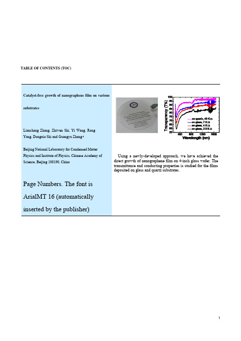
Using a newly-developed approach, we have achieved the direct growth of nanographene film on 4-inch glass wafer. The transmittance and conducting properties is studied for the films deposited on glass and quartz substrates.
KEYWORDS
nanographene, catalyst‐free, PECVD, transparent and conductive film
———————————— Address correspondence to Guangyu Zhang, email: gyzhang@ 2
TABLE OF CONTENTS (TOC)
Catalyst-free growth of nanographene film on various Insert your TOC graphics here. substrates
Lianchang Zhang, Zhiwen Shi, Yi Wang, Rong Yang, Dongxia Shi and Guangyu Zhang Beijing National Laboratory for Condensed Matter Physics and Institute of Physics, Chinese Academy of Science, Beijing 100190, China
Graphene has attracted much attention owing to its unique structures [1, 2], superior properties [1, 3‐5], and promising device applications [1, 6, 7]. Among many studies on graphene, growth of graphene has been intensively explored recently as it provides potential routes for scaled‐up applications. Several techniques have been developed: the epitaxial growth deals [8‐14] with quite expensive single crystalline substrates, thus has limited applications; chemical vapor deposition (CVD) approach on various catalytic metal films [15‐18] is low‐cost, while it needs catalyst films and post transfer [19] or additional catalyst removal techniques [20] are necessary for putting the graphene on a nonspecific substrate. Here we report for the first time a catalyst free graphene growth approach. Without using any catalyst, nanocrystalline graphene with a size about tens of nanometers were directly grown on various substrates at relatively low temperature of 550 ºC. The sp2 bonded honey‐comb carbon lattice structure of the as‐grown materials were confirmed by Raman spectroscopy, X‐ray photoemission spectroscopy (XPS) and scanning tunneling microscopy (STM). Conductivity and transparency properties of the nanographene film were also studied. This simple, low‐cost , and scalable growth approach is compatible to semiconductor processing technologies and might have potential applications for thin film resistor, gas sensor, electrode materials, transparent and conductive films, and so on. The graphene growth was carried out in a remote plasma enhanced chemical vapor deposition (r‐PECVD) system at a substrate temperature of ~550 ºC using pure methane as the precursor (see more details in supporting information). No catalyst was needed during the growth and the growth temperature, i.e. 550 C, is much lower than the CVD graphene growth temperature (900–1000 C,) [15‐18], thus enabling the growth of graphene on substrates like glass. Atomic force microscope (AFM) images of the as‐grown nanographene on SiO2 substrates with different growth durations are shown in figure 1. At the early growth stage, nanographene is nucleated everywhere on the SiO2 surface (Fig. 1a). Those larger graphene islands are ~1.2‐1.5 nm thick, which
3D电极的介电泳力与惯性力的粒子连续分选仿真
2022年第41卷第3期 传感器与微系统(TransducerandMicrosystemTechnologies)DOI:10.13873/J.1000—9787(2022)03—0043—043D电极的介电泳力与惯性力的粒子连续分选仿真李晓红1,2,张斌珍1,段俊萍1,王佳云1,屈 增1,冀苗苗1(1.中北大学仪器科学与动态测试教育部重点实验室,山西太原030051;2.太原工业学院电子工程系,山西太原030008)摘 要:细胞分选在生物医学中起着重要的作用,而其中介电泳分选由于其无需生物标记,对粒子损伤小等优势得到了广泛的应用。
本文设计了一种结合三维(3D)电极的介电泳力和收缩—扩张结构的惯性力的微流控芯片,通过COMSOLMultiphysics仿真软件对流体的流速分布、电场分布及粒子的运动轨迹进行仿真分析。
仿真结果表明:三维电极相较于传统的平面电极,能够提供粒子垂直运动方向上的非均匀电场,更有助于实现粒子的高效率分选。
此外,当粒子随流体运动时,不同的流道收缩—膨胀比分选效果不同,当收缩—膨胀比为361时,分选效果会更好。
通过仿真证明了所设计结构的有效性,确定了芯片尺寸,为后续的粒子连续高通量高效率分选,提供了重要的参考价值。
关键词:微流控芯片;惯性力分选;介电泳力分选;三维电极中图分类号:TP212.3 文献标识码:A 文章编号:1000—9787(2022)03—0043—04Particlecontinuousseparationsimulationbasedon3DelectrodecoupledwithdielectricelectrophoresisforceandinertiaforceLIXiaohong1,2,ZHANGBinzhen1,DUANJunping1,WANGJiayun1,QUZeng1,JIMiaomiao1(1.KeyLaboratoryofInstrumentationScienceandDynamicMeasurement,MinistryofEducation,NorthUniversityofChina,Taiyuan030051,China;2.DepartmentofElectronicEngineering,TaiyuanInstituteofTechnology,Taiyuan030008,China)Abstract:Cellseparationplaysanimportantroleinbiomedicalapplications.Dielectrophoresisiswidelyusedduetoitsadvantagesofnorequirementforbiologicalmarkersandnodamagecausedtoparticles.Amicrofluidicchipcombinedwiththeelectrophoreticforceofthe3Delectrodeandtheinertiaforceofthecontract expansionstructureisdesigned.COMSOLMutiphysicssimulationsoftwareisusedtoanalyzetheflowfield,theelectrodefieldandparticletrajectory.Thesimulationresultsshowthatcomparedwithtraditionalplanarelectrodes,the3Delectrodescanprovidenon uniformelectricfieldinverticaldirection,whichismoreusefultorealizeefficientparticleseparation.Inaddition,whentheparticlesmovewiththefluid,theseparationresultsaredifferentwithdifferentcontraction expansionratio.Thesortingeffectisbetterwithwhenthecontraction expansionratiois361.Thedesignedstructureisprovedeffectivethroughthesimulations,andthesizeofthemicro deviceisconfirmed.Thisstructureprovidesanimportantreferencevalueforcontinuoushighthroughputandhighefficiencyseparationofparticles.Keywords:microfluidicschip;inertialforceseparation;electrophoresisseparation;3Delectrodes0 引 言随着微流控芯片的快速发展,与传统细胞分离技术如离心和过滤等相比,由于其样品消耗少,制备成本低,灵敏度高等潜在优势,得到了人们广泛的关注。
北大考研-化学与分子工程学院研究生导师简介- 梁德海
Liu, Y.; Li, Z. C.; Liang, D. H. Behaviors of liposomes in a thermo-responsive poly(N-isopropylacrylamide) hydrogel Soft Matter , 2012, 8, 4517-4523 Abstract pdf 46 Lei, T.; Cheng, C. Y.; Guo, Z. H.; Zheng, C.; Zhou, Y.; Liang, D. H.; Pei, J. Supramolecular polymeric nanowires: preparation and orthogonal modification of their photophysical propertiesJ. Mater. Chem. , 2012, 22, 4306-4311 Abstract pdf 45
Chen, Y.; Ke, F. Y. M.; Liang, D. H. Phase Separation in Mixtures of Ionic Liquids and Water ChemPhysChem, 2012,13, 160-167Abstract pdf 48
Dehai Liang, Xuyan Yang, Lin Niu, Yuqiong Xia, Yun Liu, Cui Zheng, Jihan Zhou, CuiCui Su, Jianbo Sun, a method for preparation of DNA/RNA complex, Application Number or Patent Number: 201110150739.7(under review) 论文: 2012 52
核酸适配体
a b s t r a c t
Herein, we have successfully built up connections between nanoparticles and nanoclusters, and further constructed a surface-enhanced fluorescence (SEF) strategy based on the two types of nanomaterials for selectively assaying carcinoembryonic antigen (CEA). Specifically, silver nanoclusters provided the original fluorescence signal, while gold nanoparticles modified with DNA served as the fluorescence enhancer simultaneously. On the basis of this proposed nano-system, the two nanomaterials were linked by CEA–aptamer, thus facilitating SEF occurring. Nevertheless, more competitive interactions between CEA and CEA–aptamer emerged once CEA added, leading to SEF failed and their fluorescence decreased. Significantly, this creative method was further applied to detect CEA, and showed the linear relationship between the fluorescence intensity and CEA concentrations in the range of 0.01–1 ng mL À 1 with a detection limit of 3 pg mL À 1 at a signal-to-noise ratio of 3, demonstrating its sensitivity and promising towards multiple applications. On the whole, this approach we established may broaden potential ways of combining nanoparticles and nanoclusters for detecting trace targets in bioanalytical fields. & 2014 Elsevier B.V. All rights reserved.
PDMS基摩擦纳米发电机膜内掺杂
第58卷第4期 2021年4月撳鈉电子故术Micronanoelectronic TechnologyVol. 58 No. 4April 2021DOI:10. 13250/j. cnki. wndz. 2021. 04. 005PDMS基摩擦纳米发电机膜内掺杂张宁,何剑,丑修建(中北大学仪器科学与动态测试教育部重点实验室,太原 030051)摘要:基于新的力电转换机理,摩擦纳米发电机(T E N G)将自然界中微弱机械能转化为电能,并为小型用电设备供电成为可能,而柔性材料的应用更为低功耗设备的便携性及适应环境能 力提供了保证。
基于聚二甲基硅氧烷(P D M S)材料制备出柔性T E N G,并研究膜内掺杂工艺对 T E N G输出性能的影响规律。
通过系统测试可知,使用碳黑材料作为填充物可以有效提升T E N G输出性能,其输出电压峰峰值达到1 000 V左右,输出电流峰峰值达到12 juA左右,输出 功率提高到33 m W,且T E N G等效阻抗明显降低。
提出的膜内掺杂方法可有效提升柔性T E N G 供电能力,进一步推动T E N G的实用化进程。
关键词:摩擦纳米发电机(T E N G);聚二甲基硅氧烷(P D M S);柔性材料;膜内掺杂工艺;碳黑材料 中图分类号:T M919;TB381 文献标识码:A文章编号:1671-4776 (2021) 04-0309-07Intramembrane Doping of PDMS-Based Triboelectric NanogeneratorZhang Ning,H e Jian,Chou Xiujian(K e y Laboratory o f Instrum entation Science and D ynam ic M easurement o f M in istry o f E ducation,North U niversity o f C hina, Taiyuan030051* C hina)Abstract:Based on the new power-to-electricity conversion mechanism, the triboelectric nanogenerator (T E N G)converts the weak mechanical energy in nature into electrical energy and powers small electrical equipment. The application of flexible materials provides guarantee for portability and environmental adaptability of low-power equipment. A flexible T E N G was fabricated based on polydimethylsiloxane (P D M S) materials, and the influence rule of doping process for intramembrane on the output performance of the T E N G was studied. Through the system test, i t can be found that the carbon black material as a f i l l e r can effectively improve the output performance of the T E N G.The peak-to-peak value of the output voltage reaches about 1 0()()V,the peak-to-peak value of the output current reaches about 12 p A,the output power increases to33 m W,and the equivalent impedance of the T E N G obviously decreases. The proposed intramembrane doping method can effectively improve the power supply capacity of the flexible T E N G and further promote the practical process of the T E N G.Key words:triboelectric nanogenerator (T E N G); polydimethylsiloxane (P D M S);flexible material; intramembrane doping process; carbon black materialEEACC:8460;0585收稿日期:2020-10-17基金项目:国家自然科学基金资助项目(51705476, 51975542)通倍作者:丑修建•E-mai丨:*******************.cn309撳M电子故术()引百物联网时代背景下,随着工艺水平的不断提高,电子器件及智能可穿戴设备得到进一步的发展,不仅要求这些器件具有强大的功能,而且人们 对智能可穿戴设备的便携性和舒适性的要求更高,人和机械设备“一体化”是不断追求的目标。
- 1、下载文档前请自行甄别文档内容的完整性,平台不提供额外的编辑、内容补充、找答案等附加服务。
- 2、"仅部分预览"的文档,不可在线预览部分如存在完整性等问题,可反馈申请退款(可完整预览的文档不适用该条件!)。
- 3、如文档侵犯您的权益,请联系客服反馈,我们会尽快为您处理(人工客服工作时间:9:00-18:30)。
The topic is Biointerfacing polymeric microcapsules for In vivo near-infrared light-triggered drug release.Written by Shao Jingxin The First part:Abstract To seek safe and effective water-soluble drug carrier, the group present a novel drug delivery system based on biointerfaceing hollow polymeric microcapsules for effectively encapsulating water-soluble antitumor drug and gold nanorods (GNRs) fuctionalization for triggeredly releasing therapeutic drugs on-demand using low power near-infrared (NIR) radiation. The surface of polymeric microcapsules is covered with fluidic lipid bilayers to decrease thepermeability of the wall of polymeric capsules. In vivo antitumor tests demonstrate that this microcapsule has the effective ability of inhibiting tumor growth and preventing metastases. Realtime in vivo fluorescence imaging results confirm that capsules can be excreted gradually from the animal body which in turn demostrates the biocompatibility and biodegradation of these biointerfacing GNRs-microcapsules. This intelligent system provides a novel anticancer platform with the advantages of controled release, biological friendliness and credible biosafety. The Second part:introduction A novel NIR-responsive microcapsule with controlled release function by embedding gold nanorod (GNRs) into a wall of microcapsules was prepared as illustrated in Fig. 1A. Microcapsules are synthesized by Lb L assembly of biocompatible polysaccharide chitosan and alginate on the surface of silica microparticles following by the removal of sacrifice templates. Usually, small molecular hydrophilic drugs are difficult to be encapsulated since the larger pore sizes of these microcapsules. Thus, forming fluidic natural lipid bilayers on the surface of microcapsules through vesicle fusion method can effectively seal hydrophilic anti-cancer drugs. Furthermore, GNRs embedded in the wall of capsules were used to control drug release by transforming absorbed light energy into heat and thus disturbing the integrity of both lipid membrane and the wall of capsules. In order to evaluate the anti-tumor therapy of this controlled release system, in vivo test was conducted as shown in Fig. 1B. Drug loading GNRs-capsules were intratumoral injected into tumor-bearing mouse, followed by NIR irradiation. In vitro and in vivo experiments demonstrate the high efficient antitumor activity of this controlled release system. The third part:Results and Discussion 1.Fabrication and characterization of GNRs-microcapsules Fig. 2 (A) SEM image of GNRs. The inset is TEM image (B) SEM image of GNRs-microcapsules. The insert is the TEM image of GNRs-microcapsule. (C) UV-Vis-NIRextinction spectra of GNRs and GNRs-capsules. (D) Theoretical stimulation of temperature distribution of GNRs irradiated with a laser power .Inset image is corresponding TEM image of GNRs distribution on the capsule with higher magnification. From A and B,It can be demonstrated that GNRs were successfully assembled into the wall of microcapsules. NIR light (650-900 nm) damages skin and tissue minimally and became a hot topic for anticancer therapy .As-prepared GNRs has maximum absorption wavelength (836 nm) in near-infrared region Justas(Fig. 2C). GNRs were selected as a component of polyelectrolyte multilayer films for gathering NIR light responsive property.For FIG2DIt shows that laser-induced temperature increment localized in a small area around gold nanorod (nanometer size), which is significantly smaller than the cells (micrometer size), thus the photothermal effect towards cells can be ignored. 2. Encapsulating DOX into GNRs-microcapsules (A) CLSM Image of lipid modified microcapsules. (B) CLSM image of DOX loading microcapsules。The fluorescent intensity profile shows that biointerfacing GNRs-microcapsules were successfully filled with DOX. (C) Release profiles of DOX in GNRs-microcapsules and in lipid modified microcapsules at 37 ℃with or without NIR laser illumination. The release of lipid bilayers modified capsule was remarkably slow,herefore, lipid bilayers significantly reduce the permeability of the wall of microcapsules.After exposed to the NIR laser, 80% of DOX from lipid modified capsules was release. It directly proved that the release of encapsulated drug can be remotely controlled by NIR laser. 3. NIR controlled release of DOX from GNRs-microcapsules in vitro Fig. 4 Controlled release of anticancer drug DOX from GNRs-microcapsules by means of the NIR laser and characterized by TP-CLSM. (A) MCF-7 cells incubated with GNRs-microcapsules filled with DOX under NIR irradiation(B) MCF-7 cells incubated with GNRs-microcapsules without DOX loading under NIR irradiation.(C) MCF-7 cells incubated with DOX loaded microcapsules under the same laser irradiation condition. (D) MCF-7 cells directly irradiated with NIR laser For A group,encapsulated DOX was instantaneously released and cellbegun to necrosis by DOX effect subsequently.For B,C and D group,No obvious cell necrosis was observed.The above results conclude that NIR light responsive GNRs-capsules can be opened without killing tumor cells, laser only have the effect of drug controlled release under this condition. 4. Biodistribution of GNRs-microcapsules in vivo Fig. 5 (A) In vivo fluorescence images of the 4T1 tumor bearing nude mouse after treatment with Cy7 labelled GNRs-microcapsules. (B) In vivo fluorescence image of the 4T1 tumor bearing nude mousewith PBS injection.(C)Biodistribution of GNRs-microcapsules in the organ and tumor tissues harvested 24 h after intratumoral injection. As shown in Fig. 5A, at 1h post-injection, it is obvious that the capsules were mainly distributed around the tumor site. As shown in Fig. 5c, the content of Au