FALSE POSITIVES IN FUNCTIONAL NEARINFRARED TOPOGRAPHY
大学英语新四级试卷分析

Summarize key ideas and arguments in your own words to ensure understanding
04
Identify and analyze important evidence or examples provided in the text
Listening comprehension
Difficulty analysis
This section mainly tests the candidate's understanding and application ability of listening materials. Listening materials may include lectures, interviews, news reports, etc., covering a wide range of topics and fields.
03 Analysis of Reading Section
Quick reading
01
02
03
04
05
Quick reading: This type of reading aids to skim the text quickly to get the main idea or gift It helps the reader to identify the topic, structure, and key points of the text
Response strategy: Candidates should learn to grasp key information and themes, and pay attention to the turning points and logical relationships of the dialogue. At the same time, it is necessary to improve listening concentration and avoid anxiety caused by a large amount of information.
A Micropower Analog-Digital Heart Rate Detector Chip-微功耗模拟数字心率检测器芯片

to the person's chest and a portable monitor unit. The detector unit measures the electrocardiogram (ECG) signal, detects the heart beat from the QRS complex and sends a code to the monitor unit, which displays a continuous heart rate figure. The monitor unit is also able to store heart rate data, which can subsequently be transferred to a computer for further processing. The system is optimized for reliable monitoring of the heart rate itself, and the actual waveform of the ECG signal is of no interest, as no ECG analysis is performed. The instrument is mainly used by top athletes and sportsmen who want to optimize their training, but it can also be used elsewhere, e.g., in medical applications where continuous heart rate recording is needed. As a standard CMOS process was preferred over the dedicated low-voltage processes, and silicon foundries did not have suitable micropower cells available for standard processes, and as the use of general-purpose cells would have led to an intolerable use of silicon area and current consumption, a full-custom approach was required in the design of the chip. The methods and structures used are described in this paper.
briefings in functional genomics oxford -回复

briefings in functional genomics oxford -回复“Functional Genomics in Oxford: Unleashing the Potential of Genome Research”Introduction:Functional genomics is a rapidly evolving field of study that aims to understand the functions and interactions of genes in order to unravel the mysteries of life. The University of Oxford, with its esteemed reputation in scientific research, plays a pivotal role in advancing the frontiers of functional genomics. In this article, we will delve into the exciting world of functional genomics at Oxford, exploring the key focus areas, cutting-edge techniques, and significant contributions made by researchers in thisever-expanding field.1. Understanding Functional Genomics:Functional genomics encompasses the study of how the genome regulates biological processes and influences the phenotype of an organism. At Oxford, researchers employ various approaches, including computational biology, next-generation sequencing, andhigh-throughput screening, to enhance our understanding of gene function.2. Key Focus Areas at Oxford:a. Disease Research: Advances in functional genomics have paved the way for a deeper understanding of the genetic basis of diseases. Oxford researchers employ functional genomics techniques to unravel the complex mechanisms underlying diseases such as cancer, cardiovascular disorders, and neurodegenerative conditions, with the ultimate goal of developing targeted therapeutics.b. Epigenomics: The study of epigenetic modifications, such as DNA methylation and histone modifications, is a vibrant area of research at Oxford's functional genomics laboratories. By elucidating the role of epigenetics in gene expression and disease development, researchers are discovering novel therapeutic targets and potential biomarkers for early diagnosis.c. Gene Regulation: Oxford's functional genomics researchers investigate the intricate web of gene regulation mechanisms, including transcription factors, non-coding RNA, and chromatinstructure. The elucidation of these mechanisms enhances our knowledge of gene expression control, providing insights into normal development and disease progression.d. Functional Annotation of Genomes: Identifying the functions of genes encoded within a genome is a fundamental aim of functional genomics. Oxford researchers apply computational and experimental approaches to annotate gene functions, deciphering the roles of genes in various biological processes and shedding light on the evolutionary significance of gene function divergence.3. Cutting-Edge Techniques at Oxford:a. Next-Generation Sequencing (NGS): NGS technologies have revolutionized functional genomics research at Oxford. These high-throughput sequencing techniques allow for the characterization of entire genomes, transcriptomes, and epigenomes in a cost-effective and time-efficient manner. Researchers use NGS to unravel gene expression profiles, detect genetic variants, and investigate epigenetic alterations associated with diseases.b. CRISPR-Cas9 Genome Editing: Oxford researchers spearhead breakthroughs in CRISPR-Cas9-mediated genome editing, enabling precise manipulation of the genome to study gene function. This technique has expanded the possibilities of functional genomics research, offering unprecedented opportunities to elucidate the role of specific genes in disease mechanisms and therapeutic interventions.c. Functional Screens: High-throughput functional screens allow Oxford researchers to systematically identify genes involved in specific biological processes or diseases. These screens involve large-scale genetic perturbations, such as RNA interference (RNAi) or CRISPR knockout libraries, coupled with phenotypic analyses. By identifying genes essential for specific cellular functions, functional screens contribute to our understanding of gene function and potential therapeutic targets.4. Significant Contributions by Oxford Researchers:a. The Cancer Genome Atlas (TCGA): Oxford researchers were instrumental in the international collaboration that led to the creation of TCGA, a comprehensive catalog of genomic alterationsin various cancer types. TCGA has provided crucial insights into the genetic basis of cancer, paving the way for personalized medicine approaches and targeted therapies.b. ENCODE Project: As part of the ENCODE Project, Oxford researchers contributed to the functional annotation of the human genome. This project aimed to identify all functional elements within the genome, shedding light on gene regulation, non-coding RNA, and the three-dimensional architecture of the genome.c. Single-Cell Genomics: Oxford researchers have made significant contributions to the emerging field of single-cell genomics. By studying individual cells, researchers can unravel cellular heterogeneity, identify rare cell types, and investigate gene expression dynamics at unprecedented resolution. These insights have the potential to revolutionize our understanding of development, diseases, and therapeutic interventions.Conclusion:Functional genomics research at the University of Oxford continues to push the boundaries of our understanding of gene function andits impact on health and disease. Through their focused research areas, cutting-edge techniques, and noteworthy contributions, Oxford researchers play a crucial role in unraveling the mysteries of the genome. As the field of functional genomics continues to evolve, Oxford will undoubtedly remain at the forefront of groundbreaking discoveries with far-reaching implications for human health.。
the number of positive predictions

the number of positive predictionsThe Number of Positive Predictions: Its Importance and ApplicationsIn the realm of statistics and machine learning, the number of positive predictions refers to the count of instances where a model correctly identifies a positive outcome. This metric is crucial in evaluating the performance of a predictive model, especially in scenarios where positive outcomes are of particular interest or have significant implications.The importance of positive predictions cannot be overstated. In medical diagnostics, for instance, a high number of positive predictions ensures that patients with diseases are accurately identified, enabling timely treatment and improved outcomes. In marketing, positive predictions might refer to the successful prediction of customer behavior, such as purchases or responses to advertisements. In such cases, a high number of positive predictions can lead to increased sales and revenue.To achieve a high number of positive predictions, it is essential to have a robust and accurate predictive model. This involves the careful selection of relevant features, the use of appropriate algorithms, and the continuous refinement of models through training and testing. Additionally, it is crucial to have a balanced dataset that represents the real-world distribution of positive and negative outcomes to avoid biases in predictions.The applications of positive predictions are diverse and span across multiple industries. In finance, for instance, positive predictions might involve the accurate identification of profitable investment opportunities. In the field of security, they might refer to the successful detection of malicious activities or threats. In human resources, positive predictions might involve the accurate identification of top candidates for a job position based on their past performance and skills.In conclusion, the number of positive predictions is a critical metric inevaluating the performance of predictive models. It not only provides insights into the model's accuracy but also helps in making informed decisions in various scenarios where positive outcomes are of particular interest. By investing in robust predictive models and continuously improving their accuracy, we can ensure a higher number of positive predictions, leading to better outcomes and increased efficiency across various industries.。
正负相关英语

正负相关英语In the realm of language learning, the concepts of positive and negative correlation play a pivotal role in understanding the relationship between language elements and proficiency. A positive correlation indicates that as one variable increases, so does the other, and vice versa. Conversely, a negative correlation implies that an increase in one variable leads to a decrease in the other.Positive Correlation in Language Learning。
When discussing positive correlation in language learning, we often refer to the relationship between exposure and acquisition. The more exposure a learner has to the target language, the greater their proficiency tends to be. This can be seen in immersive environments where learners are constantly surrounded by the language, necessitating its use for daily interactions. For instance, vocabulary acquisition is typically positively correlated with reading; the more a person reads, the broader their vocabulary becomes.Another aspect of positive correlation is the relationship between practice and fluency. Regular speaking practice helps learners become more fluent, as it allows them to use language structures spontaneously and with increasing ease. This is supported by the theory of comprehensible output, which suggests that the act of producing language contributes to language development.Negative Correlation in Language Learning。
spm使用培训课件05JustinMultipletesting

• extrinsic smoothness
– resampling during preprocessing – matched filter theorem
deliberate additional smoothing to increase SNR
Multiple tests
hhhh
tt
hh
t
h
contrast of estimated parameters
t=
variance estimate
What is the problem?
Multiple tests
hhhh
tt
hh
t
h
contrast of estimated parameters
t=
variance estimate
Inference at a single voxel
h
Decision: H0 , H1: zero/non-zero activation
t h
contrast of estimated parameters
t=
variance estimate
Inference at a single voxel
small volume correction (SVC)
Computing EC wrt. search volume and threshold
E(u) () ||1/2 (u 2 -1) exp(-u 2/2) / (2)2
–
Search region R3
(GossypiumhirsutumL.)

T ITLEIdentification of a RAPD marker linked to a male fertility restoration gene in cotton (Gossypium hirsutum L.)Tien-Hung Lan1, Charles G. Cook2 & Andrew H. Paterson1, *1Department of Soil and Crop Sciences, Texas A&M University, College Station, TX 77843-24742USDA-ARS, Weslaco, TX 78596(*authorforcorrespondence,**********************.edu,fax:409-845-0456)K EY WORDSbulked segregant analysis, cytoplasmic male sterility, near-isogenic line, linkage mappingA BSTRACTOne RAPD marker, 6 cM away from a gene which restore male-fertility of a male-sterile cytoplasm was found in upland cotton (Gossypium hirsutum L.). This marker was discovered after screening 400 decamers to identify DNA polymorphism between near-isogenic lines and subsequently verified by bulked segregant analysis in an F2 population of 89 individuals derived from a cross between a cytoplasmic-male-sterile line and a restorer line. The RAPD marker was sub-cloned, sequenced, and mapped to a cotton high density RFLP map. The evaluation and utilization of this RAPD for tagging and ultimately cloning the fertility-restoring gene is discussed.I NTRODUCTIONCytoplasmic male sterility (CMS) is a maternally inherited trait conferring the inability to produce functional pollen because of interaction between cytoplasmic and nuclear genes. Since CMS does not affect female fertility, male sterile plants are able to set seeds as long as viable pollen are provided. The presence of certain nuclear genes, Rf (Restoring fertility), can effectively suppress the male-sterile cytoplasm and restore pollen fertility. The application of CMS/Rf system has proved to be an effective means to produce commercial F1 hybrid seed for many crops (Williams 1992). Cotton is predominantly a self-pollinated crop, however, cotton breeders have long been trying to breed F1 hybrid cotton, to harness F1 heterosis for many desirable traits such as high seedling-vigor, earliness, superior fiber quality and yield (Davis 1978), but also because F1 hybrid seeds could generate huge revenue for the seed industry (Anonymous 1985). Therefore, attempts to produce F1 hybrid cotton on a commercial scale have never stopped (Anonymous 1987). Before the introduction of the CMS/Rf system into Upland cotton, the only way to generate commercial hybrid cotton was through hand emasculation and crossing, which was economical only in China and India where labor cost are low. In US where labor costs are high, hand-crossing made the price of hybrid cotton prohibitive.The first CMS line of commercial cotton was introduced by crossing an upland cotton, G. hirsutum, as male parent, to a wild species, G. harknessii (Meyer 1973). A Rf line was also developed by transferring a nuclear restorer gene from G. harknessii into G. hirsutum simultaneously (Meyer 1975). The F1 hybrid population generated by crossing the CMS line to the Rf line showed a wide range of male fertility expression. Several CMS/Rf lines were then developed through the backcross method (Weaver and Weaver 1977). Later, a second Rf which expresses incomplete dominance, was also identified in G.barbadense (Sheetz and Weaver 1980). Identification of molecular markers closely linked to the nuclear Rf genes could help breeders to distinguish male-sterile and fertile plants prior to pollen shed. Here we report the identification of molecular markers closely-linked to the nuclear Rf gene under male-sterile cytoplasm in commercial cotton.M ATERIAL AND M ETHODA pair of cotton near-isogenic lines, HAF277 and DELCOT277 (kindly provided by R. Bridge, USDA-ARS, Stoneville, MS; Meyer 1975; Weaver and Weaver 1977; Sheetz and Weaver 1980), carrying the Rf gene and CMS respectively were used for initial RAPD-PCR screening. A single primer was used in each PCR reaction, and the PCR products were resolved in 1.6 % agarose gels using established techniques (Williams et al. 1990). An F2 segregating population comprised of 89 individuals from a cross of a CMS line, A2, and a Rf line, B418, were used for bulked segregant analysis (Giovannoni et al. 1991, Michelmore et al. 1991).Linkage analysis was performed using MapMaker (Lander et al. 1987), Macintosh version 2.0 (kindly provided by S. Tingey, DuPont), using the Kosambi centiMorgan function. A threshold of LOD 3.0 was used to test linkage.ResultRAPD screeningAfter screening 400 random decamers obtained from the University of British Columbia, 15 positive RAPDs were identified, which represent 3.75 % of the tested primers . These positive RAPDs were verified twice, on fresh DNA extractions, to avoid false-positives. We applied bulked segregant analysis (Giovannoni et al. 1991; Michelmore et al. 1991) for further exploration of the 15 positive RAPD markers identified in the near-isogenic lines. From the A2 x B418 F2 population, we chose 19 individuals that were clearly fertile, and 19 individuals that were clearly sterile to construct the DNA pools, and to avoid individuals that were of doubtful phenotype. To increase the stringency of our test, two pairs of synthetic DNA pools were constructed for each phenotype (fertile and sterile). One pair of pools contained 9 individuals each in the CMS and Rf pool, the other contained 10 individuals each. The pooled DNA was then used as template for RAPD-PCR analysis. Among the fifteen identified markers, two RAPD markers, R6952 (sequence CGGTTTCGTA ) and R6861 (sequence CGTGACAGGA), putatively distinguished between male-sterile and male-fertile pools.Two RAPD markers were then subjected to linkage analysis using the 38 F2 plants used in constructing the synthetic DNA pools. Four replicas were used, and each amplified band was scored as a dominant locus. Linkage analysis show R6861 was 24.6 cM from the Rf gene, and R6592 was 2.3 cM away.SequencingThe marker R6592 was eluted from the agarose gel, and cloned into the EcoRV site of dTTP-end-filled pBluescript(KS), then sequenced from both ends using the T3/T7 primers on a Applied Biosystem 373 DNA Sequencing System. From the end sequences of R6592, the correct RAPD primer sequence was identified (Fig. 1), and the sequence ofR6952 was deposited into Genbank with accession number AF094829 and AF094830. A blast search of R6592 in Genbank did not found any corresponding sequence at 95% confidence level.Southern AnalysisGenomic DNA of A4 and B418 was digested by BamH1, Cfo1, EcoR1, EcoRV, HindIII and XbaI, gel-separated, and blotted to Hybond N+ membrane as described (Reinisch et al. 1994). Gel-isolated R6592 and R6861 were P32 labeled and applied to the blots. However, a smear pattern was observed and no restriction-enzyme-polymorphism could be identified between A4 and B418, suggesting that both R6592 and R6861 contain repetitive sequence. Thus, R6592 was further digested with a mixture of Acc1, Cfo1, Hinf1, HindIII and EcoR1, and sub-cloned into pBluescript to try to remove the interfering repetitive sequence. One clone, R6592a14, identified a HindIII polymorphism between A4 and B418, though the background on the p32-exposed film was still pretty high. R6592a14 was then used to genotype the 89 individuals of A4 x B418 F2 population for the HindIII polymorphism. Linkage analysis revealed the distance between Rf gene and R6592 to be 6 cM (Fig 2a), not significantly different from the earlier estimate based on 38 individuals. Further, we tried to map R6592a14 in a cotton high-density linkage map based on a cross of Gossypium hirsutum and Gossypium barbadense (Reinisch et al. 1994). R6592a14 did not detect HindIII polymorphism in this population, but one of six genomic restriction fragments did detect EcoRV polymorphism. R6592a14 mapped to a linkage group that was tentatively identified as chromosome 20 (Reinisch et al. 1994), between markers pAR959 and pAR3-41 (Fig. 2b).DiscussionSince the near-isogenic lines we used have been backcrossed for at least 8 generations, the introgressed region should be less than 14.1 cM (Hanson 1959; using the average cotton chromosome length of 200 cM from Reinisch et al. 1994). This is consistent with the distance of R6592a14 to the Rf locus. The larger distance between R6861 and Rfwas unexpected, but R6861 was not extensively verified, so the estimated map distance is based on a small number of individuals.For the purpose of marker-assisted selection, the best scenario was to find tightly linked markers on both side of the target gene to reduce the risk of mis-genotyping due to single recombination events between the marker and the target gene. More markers linked to the Rf gene would be desirable and could be found either by targeted RAPD or AFLP screens, or in the course of further enrichment of the cotton map. The CMS/Rf is a potentially cost-effective way to produce F1 hybrid seeds. Rf genes have been successfully mapped in rice and common bean by RAPD/bulk segregant analysis (He et al. 1995; Zhang et al. 1997), and we provide a marker diagnostic of this important phenotype in cotton. In addition to its utilization in marker-assisted selection, this marker may serve as a starting point for positional cloning of the Rf gene.A CKNOWLEDGMENTWe thank Mark D. Burow for technical assistance. Aspects of the work described here were supported by USDA 91-37300-6570, to AHP, and Texas Higher Education Coordinating Board Award 999902-148 to AHP and Rod A. Wing.Fig. 1Partial DNA sequence of clone R6592. Primer sequences are underlined.Fig. 2a. Linkage of Rf gene and marker R6592 in G. hirsutum A2 x B418 F2 population.b. Marker R6592 mapped to cotton chromosome 20 in G. hirsutum (race palmeri) x G. barbadense “k101” primary mapping population (Reinisch et al., 1994).ReferenceAnonymous, 1985 Seed Money. Forbes 136: 219-210.Anonymous, 1987 Finger-Picking Good: Hybrid Cotton. The Economist 303: 91. Davis, D. D., 1978 Hybrid cotton: specific problems and potentials. Adv Agron 30: 129-157.Giovannoni, J., R. Wing, M. Ganal and S. Tanksley, 1991 Isolation of molecular markers from specific chromosomal intervals using DNA pools from existing mapping populations. Nucl Acids Res 19: 6553-6558.Hanson, W., 1959 Early generation analysis of lengths of heterozygous chromosome segments around a locus held heterozygous with backcrossing or selfing. Genetics 44: 833-837.He, S., Z. H. Yu, C. E. Vallejos and S. A. Mackenzie 1995 Pollen fertility restoration by nuclear gene Fr in CMS common bean: An Fr linkage map and the mode of Fr action. Theor Appl Genet 90: 1056-1062.Lander, E., P. Green, J. Abrahamson, A. Barlow, M. Daly et al., 1987 MAPMAKER: An interactive computer package for constructing primary genetic linkage maps of experimental and natural populations. Genomics 1: 174-181.Meyer, V., 1975 Male sterility from Gossypium harknessii. J Hered 66: 23-27.Meyer, V. G., 1973 Fertility restorer genes for cytoplasmic male-sterility from Gossypium harknessii. Beltwide Cotton Prod. Res. Conf. Proc., p65.Michelmore, R., I. Paran and R. Kesseli, 1991 Identification of markers linked to disease-resistance genes by bulked segregant analysis: a rapid method to detect markers in specific genomic regions by using segregating populations. Proc. Natl. Acad. Sci. USA 88: 9828-9832.Reinisch, A., J.-M. Dong, C. Brubaker, D. Stelly, J. Wendel et al., 1994 A detailed RFLP map of cotton (Gossypium hirsutum x G. barbadense): Chromosome organization and evolution in a disomic polyploid genome. Genetics 138: 829-847.Sheetz, R. H., and J. B. Weaver, 1980 Inheritance of a fertility enhancer factor from Pima cotton when transferred into Upland cotton with Gossypium harknessii Brandegee. Crop Science 20: 272-275.Weaver, D., and J. Weaver, 1977 Inheritance of pollen fertility restoration in cytoplasmic male-sterile upland cotton. Crop Sci 17: 497-499.Williams, J. G. K., A. R. Kabelik, K. J. Livak, J. A. Rafalski and S. V. Tingey, 1990 DNA polymorphisms amplified by arbitrary primers are useful as genetic markers. Nucl. Acids Res. 18: 6531-6535.Williams, M. E. L. C. S., III, 1992 Molecular Biology Of Cytoplasmic Male Sterility, pp. 23-51 in Plant Breeding Reviews, edited by J. Janick. John Wiley and Sons, Inc., New York.Zhang, G., T. S. Bharaj, Y. Lu, S. S. Virmani and N. Huang, 1997 Mapping of the Rf-3 nuclear fertility-restoring gene for WA cytoplasmic male sterility in rice using RAPD and RFLP markers. Theor Appl Genet 94: 27-33.。
微软高级威胁分析系统(Advanced Threat Analytics,简称ATA)说明书
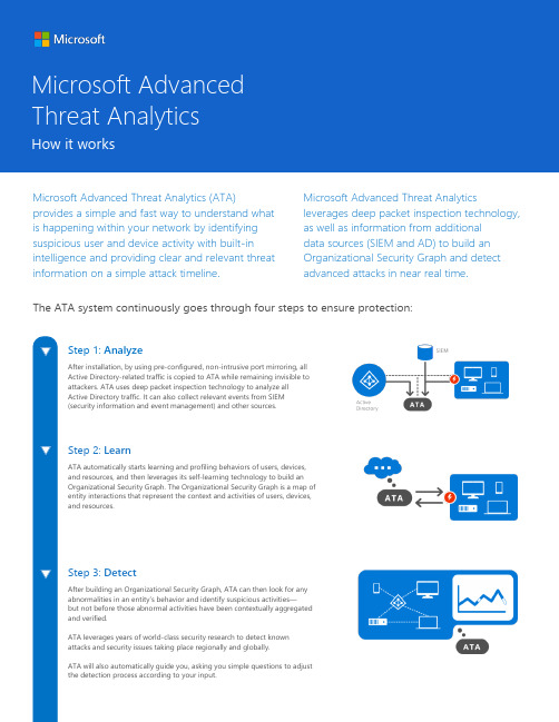
Microsoft Advanced Threat Analytics (ATA)provides a simple and fast way to understand whatis happening within your network by identifyingsuspicious user and device activity with built-inintelligence and providing clear and relevant threatinformation on a simple attack timeline.Microsoft Advanced Threat Analytics leverages deep packet inspection technology, as well as information from additional data sources (SIEM and AD) to build an Organizational Security Graph and detect advanced attacks in near real time. The ATA system continuously goes through four steps to ensure protection:How it works Step 3: Detect After building an Organizational Security Graph, ATA can then look for any abnormalities in an entity’s behavior and identify suspicious activities — but not before those abnormal activities have been contextually aggregated and verified. ATA leverages years of world-class security research to detect known attacks and security issues taking place regionally and globally.ATA will also automatically guide you, asking you simple questions to adjustthe detection process according to your input.Step 1: Analyze After installation, by using pre-configured, non-intrusive port mirroring, allActive Directory-related traffic is copied to ATA while remaining invisible to attackers. ATA uses deep packet inspection technology to analyze allActive Directory traffic. It can also collect relevant events from SIEM (security information and event management) and other sources.Step 2: Learn ATA automatically starts learning and profiling behaviors of users, devices, and resources, and then leverages its self-learning technology to build an Organizational Security Graph. The Organizational Security Graph is a map of entity interactions that represent the context and activities of users, devices, and resources.Microsoft Advanced Threat AnalyticsStep 4: Alert While the hope is that this stage is rarely reached, ATA is there to alert you of abnormal and suspicious activities. To further increase accuracy and save you time and resources, ATA doesn’t only compare the entity’s behavior to its own, but also to the behavior of other entities in its interaction path before issuing an alert. This means that the number of false positives are dramatically reduced, freeing you up to focus on the real threats. At this point, it is important for reports to be clear, functional, and actionable in the information presented. The simple attack timeline is similar to a social media feed on a web interface and surfaces events in an easy-to-understand way.© 2015 Microsoft Corporation. All rights reserved. This document is provided "as-is." Information and views expressed in this document, including URL and other Internet Web site references, may change without notice. You bear the risk of using it. This document does not provide you with any legal rights to any intellectual property in any Microsoft product. You may copy and use this document for your internal, reference purposes. You may modify this document for your internal, reference purposes.。
- 1、下载文档前请自行甄别文档内容的完整性,平台不提供额外的编辑、内容补充、找答案等附加服务。
- 2、"仅部分预览"的文档,不可在线预览部分如存在完整性等问题,可反馈申请退款(可完整预览的文档不适用该条件!)。
- 3、如文档侵犯您的权益,请联系客服反馈,我们会尽快为您处理(人工客服工作时间:9:00-18:30)。
FALSE POSITIVES IN FUNCTIONAL NEAR-INFRARED TOPOGRAPHY Ilias Tachtsidis1, Terence S. Leung1, Anchal Chopra1, Peck H. Koh1, Caroline B. Reid1, and Clare E. Elwell1Abstract:Functional cranial near-infrared spectroscopy (NIRS) has been widely used to investigate the haemodynamic changes which occur in response to functional activation. The technique exploits the different absorption spectra of oxy- and deoxy-haemoglobin ([HbO2] [HHb]) in the near-infrared region to measure the changes in oxygenation and haemodynamics in the cortical tissue. The aim of this study was to use an optical topography system to produce topographic maps of the haemodynamic response of both frontal cortex (FC) and motor cortex (MC) during anagram solving while simultaneously monitoring the systemic physiology (mean blood pressure, heart rate, scalp flux). A total of 22 young healthy adults were studied. The activation paradigm comprised of 4-, 6- and 8- letter anagrams. 12 channels of the optical topography system were positioned over the FC and 12 channels over the MC. During the task 12 subjects demonstrated a significant change in at least one systemic variable (p≤0.05). Statistical analysis of task-related changes in [HbO2] and [HHb], based on a Student’s t-test was insufficient to distinguish between cortical haemodynamic activation and systemic interference. This lead to false positive haemodynamic maps of activation. It is therefore necessary to use statistical testing that incorporates the systemic changes that occur during brain activation.1. INTRODUCTIONWhen analysing cerebral haemodynamic activation data using functional neuroimaging the task-specific activation observed is due to the existence of a close coupling between regional changes in neuronal activation, brain tissue metabolism and regional changes in cerebral blood flow (CBF). Cranial functional near-infrared spectroscopy (NIRS) has been widely used to investigate the haemodynamic changes, which occur in response to functional activation of specific regions of the cerebral cortex. The technique exploits the different absorption spectra of oxy-haemoglobin (HbO2) and 1 Department of Medical Physics and Bioengineering, Malet Place Engineering Building, University College London, Gower Street, London WC1E 6BT, UK2 I. TACHTSIDIS ET AL. deoxy-haemoglobin (HHb) in the near-infrared region to measure the changes in oxygenation and haemodynamics in the brain cortical tissue. In order for this response to be monitored unambiguously it is important that the haemodynamic task-related activity is occurring on top of an unchanged global systemic and brain resting state.We have previously reported that significant changes in mean blood pressure (MBP) and heart rate (HR) occur during anagram activation tasks and observed that NIRS haemodynamic changes were in some volunteers significantly correlated with changes in these systemic variables.1 Most recently,2 we reported that during a frontal lobe anagram activation task, task-related haemodynamic changes were observed both over the frontal cortex (activated region) and motor cortex (control region). The task-related changes were correlated with increases in MBP and scalp blood flow (flux) measured with laser Doppler. This implies the possibility of some systemic “global interference” in our NIRS measured data. It is possible that the anagram task elicits an emotional response, which produces changes in blood pressure that are likely to cause passive changes in the scalp blood flow. These changes can produce small task-related, but non cortical alterations in the [HbO2] and [HHb] signals as measured by cranial NIRS.Over the last decade or so, many studies have been published describing the use of the optical topography (OT) technique to map functional brain activation.3-5 By making simultaneous NIRS measurements at multiple brain sites, one can produce spatial maps of the haemoglobin concentration changes that correspond to specific regions of the cerebral cortex. OT can therefore potentially discriminate between regional activated cortical areas and global haemodynamic changes.The aim of this study is to investigate the functional haemodynamic changes during frontal lobe anagram activation using optical topography both over the activated and control area while continuously monitoring systemic and scalp blood flow changes.2. MATERIAL AND METHODSThis study was approved by the UCL Research Ethics Committee. We studied 22 young healthy subjects with English as their first language (15 male, 7 female, median age 22 years, range 20-39).NIRS measurements were conducted with the ETG-100 Optical Topography System (Hitachi Medical Co., Japan) using two 12-channel arrays. Each optode array consisted of 5 source optodes (each delivering light at 780 and 830 nm) and 4 detector optodes. The source-detector interoptode spacing was 30mm and data were acquired at 10Hz. The optodes were placed over the subject’s left frontal cortex and positioned according to the international 10-20 system of electrode placement such that channels 1-12 were centred approximately over the frontopolar region (Fp) and channels 13-24 were centred approximately over the left primary motor cortex (C3). A schematic illustration of optode placement is show in Figure 1.A Portapres® system (TNO Institute of Applied Physics) was used to continuously and non-invasively measure MBP and HR from the finger. A laser Doppler probe (FloLab, Moore Instruments) was placed over the forehead to monitor the changes in scalp blood flow (flux).FALSE POSITIVES IN FUNCTIONAL NEAR-INFRARED TOPOGRAPHY 3locations of corresponding measuring positions/channels. One array was centred on the frontopolar region (Fp), the other on the left motor cortex (C3).All the volunteers were positioned in a comfortable sitting position. Data wererecorded during two minutes of the subject at rest (baseline), followed by 45 seconds of the subject solving 4-letter anagrams (9 anagrams, 5 seconds per anagram), 30 seconds rest, 45 seconds of solving 6-letter anagrams (5 anagrams, 9 seconds per anagram), 30seconds of rest, 45 seconds of solving 8-letter anagram (5 anagrams, 9 seconds per anagram) and 30 seconds of rest. Each anagram-solving period was repeated a total of four times, with the study ending after a 2-minute rest period (total study time 19 minutes). In this study solving an anagram was defined as producing one coherent word using only the letters from another word (e.g. icon – coin). Subjects were encouraged to solve as many anagrams as possible and were instructed to say possible solutions out loud (without moving).All optical data were subjected to an identical processing procedure using the functional Optical Signal Analysis program 6 (fOSA, University College London, UK) to convert the relative changes in light intensities to concentration changes in haemoglobin (HbO 2, HHb and their sum, HbT) using a differential pathlength factor correction of 6.26. All the signals including MBP, HR and flux, were then decimated from 10Hz to 1Hz and low pass filtered at 0.08Hz. The data were filtered using a 5th order low pass Butterworth digital filter in forward backward directions to avoid introducing a phase delay. The last pre-processing stage, prior to statistical analysis was to de-trend the time-course to remove both drift introduced by the system and any slowly changing unrelated physiological signals. A first-order linear baseline was drawn as the reference and then subsequently subtracted from the activation signal.The response to stimulation was calculated for each subject as the difference between the average of 10 seconds worth of baseline data at the end of the rest period, and the average of 10 seconds of data commencing 15 seconds after the onset of the 4, 6 or 8 letter anagram solving periods respectively. A ‘Student’s t-test’ was used to assess the significance of these responses (the threshold of significance was set at p ≤0.05 from baseline). For the optical topographic data we then calculated the cumulative total number of channels across subjects in which we observed activation. We define activation as a statistical significant increase in [HbO 2], a statistical significant decrease or no change in [HHb] and a statistical significant increase in [HbT]. Systemic interference was measured by using the Pearson correlation model to calculate correlations between the systemic variables and changes in [HbO 2] and [HHb] in all of the OT channels.30 mmSource Detector4 I. TACHTSIDIS ET AL.3. RESULTSA summary of the activation data for the whole group is shown in Figure 2. Each paradigm is shown separately and data are normalised to the number of valid channels. Across paradigms similar activation response was observed in both frontal cortex and motor cortex. Channels in which the highest number of subjects showed activation were channel 23 (55.56%) for the 4-letter task, channel 1 (52.94%) for the 6-letter task, and channels 6 and 21 (33.33%) for the 8-letter task. Taking into account all of the tasks, an average of 30% of the subjects showed activation (range 25-35%) on the frontal cortex and 27% (range 17-37%) on the motor cortex.Analysis of the systemic variables show that at least 50% of the subjects demonstrated a change in at least one systemic variable. Table 1 shows the mean changes in each systemic variable for those subjects that showed a significant change.Correlation analysis of the NIRS and systemic data shows a large variability across different OT channels and across subjects. Figure 3 shows the results of the correlation analysis between MBP and the NIRS data, across all channels for (a) subject 3 who showed generally high correlations (r>0.5), and (b) subject 18 who showed generally low correlations (r<0.5). Both subjects showed significant changes in systemic variables during the anagram tasks and both subjects had channels that showed activation. This trend was observed across subjects. The correlation between the systemic data and the NIRS data from the frontal cortex channels show no difference from the correlation between the systemic data and the NIRS data from the motor cortex channels.Table 1. Group changes from rest to activation are presented as mean ± standard deviation for those subjects that demonstrated a significant change.Systemic Variables 4-letter task 6-letter task 8-letter task∆[MBP] (mmHg) (n=11) 6.9±2.7 (n=12) 6.3±4.9 (n=12) 6.9±1.9∆[HR] (beats/min) (n=5) 4.3±4.9 (n=6) -0.4±6 (n=6) 2.4±4.3∆[Flux] (%) (n=4) 14.3±31.1 (n=3) 20.3±10.3 (n=1) -17.84. DISCUSSIONIn this study we used an optical topography system to investigate the changes in [HbO2] and [HHb] during anagram solving over the frontal lobe (activated area) and motor cortex (control area) while simultaneously monitoring systemic variables. We used a Student’s t-test to define significant changes in [HbO2], [HHb] and [HbT] for each OT channel and for each subject during the different anagram solving tasks and used these data to define where and when activation was detected. The same analysis was performed on the systemic variables. We observed a large variability in activated OT channels across subjects. The OT results failed to define specific regional areas of activation. 50% of subjects showed a significant change in at least one systemic variable. These systemic changes appear in some subjects to correlate with the observed functional changes in [HbO2] and [HHb] across the OT channels. Figure 4 shows an example of changes in [HbO2] and [HHb] from an OT channel over the frontal cortex and an OT channel over the motor cortex with the simultaneously recorded changes in MBP and scalp flux.FALSE POSITIVES IN FUNCTIONAL NEAR-INFRARED TOPOGRAPHY 5Clearly systemic interference during the anagram task can lead to false positives in defining activated OT channels.Figure 2. Group analysis shows the percentage of subjects that demonstrated activation in specific channels during the three different anagram solving paradigms.Frontal CortexMotor Cortex4-Letters6-Letters8-Letters0 %50 %100 %117.65%217.65%327.78%433.33%544.44%633.33%741.18%844.44%938.89%1050.00%1141.18%1235.29%1329.41%1435.29%1527.78%1641.18%1727.78%1838.89%1933.33%2050.00%2133.33%2227.78%2355.56%2444.44%117.65%217.65%316.67%416.67%527.78%633.33%717.65%822.22%922.22%1022.22%1117.65%1211.76%1323.53%1423.53%1522.22%1617.65%1722.22%1827.78%1922.22%2027.78%2116.67%2216.67%2333.33%2427.78%1329.41%1423.53%1522.22%1629.41%1722.22%1827.78%1922.22%2011.11%2133.33%225.56%2322.22%245.56%152.94%247.06%338.89%450.00%527.78%633.33%729.41%827.78%922.22%1033.33%1135.29%1229.41%Scale of number of subjects showing activation6 I. TACHTSIDIS ET AL.Figure 3. Individual correlation coefficients between MBP and ∆[HbO 2] and MBP and ∆[HHb] across all channels for (a) subject 3 and (b) subject 18.In this study we used the classical approach to define significant changes in haemoglobin concentrations by employing a “Student’s t-test”. This approach compares two different states of the brain, i.e. “rest” versus “activation”. The “rest” period is usually defined as a baseline period before the stimulus onset and the “activation” period is defined as the period 10-20 seconds after the onset of the stimulus. By keeping the rest and activation periods constant across subjects one can investigate the functional response to specific tasks. Whilst a simplistic approach of this kind helps to provide a quick assessment of the haemodynamic response to the task it does not consider any spatial coherence in the OT data. It also assumes that the measured changes in haemoglobin concentrations are due solely to the neuronal activation, and that there are no tasks-related systemic effects. We have shown that this latter assumption is not true for all subjects performing an anagram solving task. One can include a priori information regarding systemic changes and can de-correlate the physiological noise (cardiac, respiratory and vasomotion related fluctuations) from the evoked haemodynamic response, by using techniques such as Principal Component Analysis,7 Independent Component Analysis,8 and more recently Statistical Parametric Mapping (SPM).6 SPM has been widely used for the analysis of functional activation data from other neuroimaging modalities such as the BOLD response in fMRI studies.9 SPM uses massChannel Number rr-1.0-0.8-0.6-0.4-0.20.00.20.40.60.81.0Channel NumberMBP and ∆[HbO 2] MBP and ∆[HHb]-1.0-0.8-0.6-0.4-0.20.00.20.40.60.81.0123456789101112131415161718192021222324FALSE POSITIVES IN FUNCTIONAL NEAR-INFRARED TOPOGRAPHY 7univariate approach to modelling the spatiotemporal neuroimaging data by assigning a statistic value to every brain voxel. It enables the construction of spatial statistical processes to test hypotheses about regional specific effects in the brain. Unlike the classical approach mentioned earlier, where the two different time courses compared, SPM employs a modelling approach for each brain voxel. In our study all of the explanatory variables (HbO 2, MBP, HR and flux) were treated as regressors in the linear model. To treat the variability of haemodynamic responses arising from different events between different brain voxels, SPM allows the modelling of latency and dispersion derivatives as additional regressors to its canonical response function. The associated parameter estimates are the coefficients for each of the regressors that best model the observed response for the voxel in question (here a voxel is defined as an OT channel). To account for the spatial coherence of the functional data, SPM provides the necessary family-wise correction based on the theory of Gaussian random field to resolve the multiple comparison problem.Figure 4.data from channel 15 (motor cortex); (c) MBP and (d) scalp flux.As an example of this method we have used fOSA-SPM software 6 to analyse NIRSand systemic data from one subject collected during the 6-letter anagram solving task. Using the “Student’s t-test” analysis, this subject demonstrated activation across all OT channels. Figure 5 shows the results of the SPM analysis on the same subject’s data. These are presented as an SPM t-result for the HbO 2 signal over all channels and show a spatial localisation of the haemodynamic response. Unlike the “Student’s t-test” approach which compares the difference between two specific physiological states, SPM offers a more rigorous approach to analysing functional OT data by taking into account the globalTime (seconds) Time (seconds) Time (seconds) Time (seconds) C o n c e n t r a t i o n s (µM ) C o n c e n t r a t i o n s (µM )M e a n B l o o d P r e s s u r e (m m H g )S c a l p F l u x (a .u .) (a) (b) (c) (d)∆[HHb]∆[HHb] ∆[HbO 2] ∆[HbO 2] 8-Letters8 I. TACHTSIDIS ET AL. systemic effects by means of fitting a haemodynamic response function and performing spatial correlations across all channels.Figure 5.significant t-values.In conclusion, when analysing OT data for evidence of functional activation theeffect of task-related changes in systemic variables should be taken into account. SPM may be a useful tool for analysing simultaneously measured multi-channel OT NIRS data and systemic variables.5. ACKNOWLEDGMENTSThe authors would like to acknowledge the EPSRC (Grant No EP/D060982/1).6. REFERENCES1. I. Tachtsidis, T.S. Leung, L. Devoto, D.T. Delpy, and C.E. Elwell, Measurement of frontal lobe functionalactivation and related systemic effects: a near-infrared spectroscopy investigation, Adv. Exp. Med. Biol. InPress (2008).2. I. Tachtsidis, T.S. Leung, M.M. Tisdall, D. Presheena, M. Smith, D.T. Delpy, and C.E. Elwell,Investigation of frontal cortex, motor cortex and systemic haemodynamic changes during anagram solving,Adv. Exp. Med. Biol. In Press (2008).3. Y. Hoshi, B. H. Tsou, V. A. Billock, M. Tanosaki, Y. Iguchi, M. Shimada, T. Shinba, Y. Yamada, and I.Oda, Spatiotemporal characteristics of hemodynamic changes in the human lateral prefrontal cortexduring working memory tasks, NeuroImage20(3), 1493-1504 (2003).4. B. Chance, S. Nioka, S. Sadi, and C. Li, Oxygenation and blood concentration changes in human subjectprefrontal activation by anagram solutions, Adv. Exp. Med. Biol.510, 397-401 (2003).5. R.P. Kennan, D. Kim, A. Maki, H. Koizumi, and R.T. Constable, Non-invasive assessment of languagelateralization by transcranial near infrared optical topography and functional MRI, Hum. Brain Mapp.16(3), 183-189 (2002).6. P.H. Koh, D.E. Glaser, G. Flandin, S. Kiebel, B. Butterworth, A. Maki, D.T. Delpy, and C.E. Elwell,Functional optical signal analysis (fOSA): a software tool for NIRS data processing incorporating statistical parametric mapping (SPM), JBO In Press (2007).7. X. Zhang, V. Toronov, and A. Webb, Simultaneous integrated diffuse optical tomography and functionalmagnetic resonance imaging of the human brain, Opt. Express13(14), 5513-5521 (2005).8. I. Schiessl, M. Stetter, J.E.W. Mayhew, N. McLoughlin, J.S. Lund, and K. Obermayer, Blind signalseparation from optical imaging recordings with extended spatial decorrelation, IEEE Transactions onBiomedical Engineering, 47(5), 573-577 (2000).9. K.J. Friston, A.P. Holmes, J.B. Poline, P.J. Grasby, S.C. Williams, R.S. Frackowiak, and R. Turner,Analysis of fMRI time-series revisited, NeuroImage2(1), 45-53 (1995).。