X-Ray Spectral Variability Signatures of Flares in BL Lac Objects
X-ray Spectrometry

X-ray SpectrometryImre Szalo´kiInstitute of Experimental Physics,University of Debrecen,Bem te´r18/a,H-4026Debrecen,HungaryJa´nos Osa´nHungarian Academy of Sciences,KFKI Atomic Energy Research Institute,P.O.Box.49,H-1525Budapest,Hungary Rene´E.Van Grieken*Department of Chemistry,University of Antwerp,B-2610Antwerp,BelgiumReview ContentsOverview4069 Detection4071 Instrumentation and X-ray Optics4074 Quantification and Fundamental Data4078 Tomography,Holography,and X-ray Scattering4081 Total Reflection X-ray Fluorescence Analysis4083 Electron Probe Microanalysis4084 Particle-Induced X-ray Emission4086 X-ray Absorption Spectrometry4087 Applications4089 Sample Preparation4089ED-XRF4089Micro-XRF4089TXRF4090EPMA4090PIXE4091XAS4092Standards4093 Literature Cited4093OVERVIEWIn this review,we focus on the most significant and essential progress in X-ray spectrometry(XRS),published in the period 2004-2005,covering the developments and improvements in the performance of detection and instrumentation of X-ray techniques and X-ray optics,new quantification models in X-ray spectra and data evaluation,calculation and experimental determination of fundamental atomic data,tomography and holography methods for2D or3D imaging of microstructures,electron probe micro-analysis(EPMA),total reflection X-ray fluorescence(TXRF), particle-induced X-ray emission(PIXE)analysis,and X-ray absorp-tion spectrometry(XAS).Finally,different applications in each subfield of XRS are shown.This review involves only a selected minority of the published papers and we try to look over the current trends in this analytical field with a critically selected citing of papers,to support the research activity of the community of X-ray scientists.Since our last review,an international group of scientists published two overviews on XRS(A1,A2)in the Journal of Analytical Atomic Spectrometry,covering the period2003-2004 in the main field of this analytical spectroscopic method and instruments.These review articles involve all the important sections of XRS in nine chapters:reviews,instrumentation, spectrum analysis,matrix correction and calibration,X-ray optics and microfluorescence,synchrotron radiation,TXRF,portable and mobile X-ray fluorescence(XRF),and on-line XRF and applications. In the present review,we follow another way for classification of the information published in the literature in the2004-2005 period:detection,instrumentation(except of detectors)and optics,quantification models and related fundamental data,to-mography,and holography methods as the main tool of X-ray imaging,TXRF,EPMA,PIXE,XAS,and a final chapter that reviews the application of these methods in geology,environmen-tal research,industry,biology.and medicine.Current trends in pixel-type detectors were reviewed by Wermes(A3)showing their necessity in X-ray imaging devices,radiography,autoradiography, protein crystallography,and X-ray astrophysics research.The fundamental aims of R&D activity in the field of detectors are to arrange less material in the detector bulk,to build high-speed readout electronics,and to construct large radiation-tolerant detectors.The author describes the working principle and structure of different categories of pixilated detectors:hybrid pixel detectors when the sensor module and the electronic chips are connected by short bumps,diamond detectors and their applica-tions for protein crystallography and radiography.The author describes the dream of the detector developer:a fully monolithic pixilated detector where both the sensor bulk and the electronic circuits are in one entity and these devices can be produced by commercially available technology,and he outlines that this will not be possible in the future due to the lack of proper technology.In the past decade,the greatest hit in XRS was probably the cryogenically cooled quantum detectors such as superconductive tunnel junction(STJ),transition edge sensors(TESs),and micro-calorimeters.These provide excellent energy resolution for a wide range of X-ray energies,from the optical range up to several kiloelectronvolts.Kurakado published(A4)a short characteriza-tion about nonequilibrium superconductivity and STJ detectors. The tutorial paper introduces the reader into the main features, such as high count rate capability,stability against temperature fluctuation,ultra-high-energy resolution,and the working principle of the STJs.The most common Nb/Al/AlOx/Al/Nb structure has excellent working parameters and very high stability against temperature variation from50to500mK.A100×100µm2AlAnal.Chem.2006,78,406910.1021/ac060688j CCC:$33.50©2006American Chemical Society Analytical Chemistry,Vol.78,No.12,June15,20064069 Published on Web00/00/0000STJ located on a Si3N4membrane,covered by a1.3-µm-thick Pb absorber,was described;this device had an energy resolution of 12.4eV and the noise was4eV at5.9-keV X-ray nkosz et al.(A5)reviewed quantitative X-ray microanalysis for biological and glass material samples in order to verify the fundamental parameter method on the basis of the application of standard samples.The principal aim of their investigation was to determine the analytical capability of a microbeam(X-ray beam diameter of ∼30µm)XRF spectrometer equipped with capillary optics.They considered all the possible sources of uncertainty of the whole analysis:operating stability of the detector and X-ray source, sample movement,and errors originating from the spectral deconvolution calculations.The sample thickness was estimated on the basis of the measurement of the elastic and inelastic scattered intensities of the source radiation,and it was applied for the matrix correction as well.The authors demonstrate their analytical considerations and calculations by some examples using the synchrotron microbeam XRF technique for quantitative analysis of human brain tissue samples with both monoenergetic and polychromatic excitation modes.The accuracy for glass standard samples was found between4and40%relative. Janulewicz et al.(A6)reviewed the state-of-the-art of the tabletop type X-ray lasers(XRLs)and described the working principle of these lasers and their characteristic spectral output properties, from a practical point of view.They concluded that the XRL sources are competitive in comparison with the third-generation synchrotron radiation(SR)facilities:brilliance of the beam and time-resolved measurements.The author details the operating processes of two types of XRFs based on the quantum pumping mechanisms in generating the excitation states:(i)recombination and(ii)collisional-type XRLs.The size of the XRLs is the central problem in development research;it can be decreased in the case of a hybrid-type XRL,when the pumping medium is gas plasma in a capillary tube in which the inverse population is generated by a short picosecond optical laser beam,entering into the axial direction of the capillary.Most observed objects in the sky naturally emit X-ray radiation that transports information on the atomic-level processes occurring in the source objects,on the motion of the bulk material,etc.,or the transmission of X-rays through absorption can be studied.Therefore,the detectors of X-ray telescopes are the central devices in the sky observatories, such as Chandra and XMM-Newton.Porter reviewed(A7)the low-temperature detectors developed for space missions such as X-ray cameras and dispersive X-ray spectrometers.The Astro-E2 launched in2005was the first mission that contained a low-temperature microcalorimeter-based observatory,and three more low-temperature detector-based observatories are being developed (NeXT,Constellation-X,ZEUS).Due to the high-level ionization of the individual atoms in different sky objects,very complex X-ray emission spectra must be detected resolving the satellite lines. The newest spectrometers that suit this requirement are high-energy resolution microcalorimeters.The other essential group of X-ray detectors are the CCD cameras;these have moderate spectral sensitivity and therefore they have to be used with selective absorbing filters for energy-sensitive X-ray imaging.In the future observatories,new detector types are needed,which have∼1000pixels with a high-energy resolution below4eV at 6-keV X-ray energy,in a miniature,monolithic arraying form,having three-dimension wiring layers,cryogenic multiplexing schemes,complex and flexible electronics,and finally an as low as possible mass.SR is widely used for all fields of XRS for the characterization of materials for basic research and for practical aims.In view of the lack of a detailed description of this analytical technique for the nonsynchrotron-expert forensic community,Kempson and co-workers(A8)published a review paper on application of synchro-tron radiation in forensic trace analysis.The authors pointed out the benefits of SR and discuss XRF techniques,described the tomographic method,X-ray diffraction,and scattering applications, and finally,they highlighted the X-ray absorption near edge structure(XANES)and extended X-ray absorption fine-structure (EXAFS)methods and their significant contribution to the characterization of materials.They outlined the unique advanta-geous spectroscopic characters of the synchrotron beams:high brightness,energy tenability,nearly100%of polarization and coherence,and possibility of time-resolved measurements;finally they showed some examples demonstrating the benefit of ap-plication of the SR in this practical field.A general tutorial paper was published by Van der Veen and Pfeiffer(A9)about the properties of coherent hard X-ray beams and their use in structural analysis of solid materials.Because of a fully coherent X-ray beam is provided by SR,it is suitable for such experiments where the interference of the X-ray beam gives information on the inner structure of the investigated object material.In the near-forward scattering direction,the differences in the phases between waves traversing different parts of the object enable imaging the object structure into a phase contrast. In larger scattering angles the interference provides coherent diffractive imaging without lenses and it is called holography without reference beam.The authors give a simple mathematical description of the investigated effects:transverse coherence, longitudinal coherence,phase contrast,X-ray photon correlation spectroscopy,diffracting imaging,and waveguide analysis on the basis of the Fraunhoffer diffraction pattern.In this last review period,an increasing number of papers were published on the1D and2D waveguides due to the strong needs of a microsized X-ray beam,which has a high flux density,for the analysis of submicrometer-sized objects.Egorov and Egorov published a tutorial on state-of-art of X-ray waveguides(A10),with the general description of the operating principles and the spectroscopic characteristic properties and possible applications.X-ray microanalytical methods are more widely applied in archaeometry and in cultural heritage conservation research, especially nondestructive micro-X-ray analytical methods.Selected presentations of the7th International Conference on Nondestruc-tive Testing and Microanalysis for the Diagnosis and Conservation of the Cultural and Environmental Heritage,dealing with different types of instrumental analysis of materials and artifacts of cultural-historical value,were published in Cultural Heritage Conservation and Environmental Impact Assessment by Non-Destructive Testing and Micro-Analysis(A11).The publication gives a survey of the possible solutions for analytical problems in the cultural heritage sector,using portable energy-dispersive XRF spectrometers, TXRF,microfocus X-ray tomography,confocal micro-XRF,and micro-XAS.4070Analytical Chemistry,Vol.78,No.12,June15,2006The in-field analysis of hazardous material and environ-mental samples obviously requires portable XRF spectrometers. Melquiades and Appoloni published a review article(A12)about the basic methodology aspects of laboratory and portable X-ray boratory measurements can be slow,sometimes laborious,and of course more expensive;however,they are more precise in comparison with portable XRF spectrometers.The use of portable spectrometers allows rapid mapping and ranking of contaminated sites,and a large number of semiquantitative data can be generated on site in nearly real time.The authors overview the possible detector types that are suitable for the portable technique and concluded that the Peltier cooled detectors(Si-PIN,CdZnTe,Si drift)are the most appropriate devices because no cryogenic cooling is required.The most significant applications and portable designs were summarized in a separate table involving the characteristic parameters:excitation source,re-quired sample preparation technique,possible matrixes for sample bulk,and minimum detection levels for different elements.The authors give some examples for applications published in the literature,and they discuss the main steps of in-field analysis such as preconcentration techniques and data evaluation methods. Finally,in this introductory section,we mention an article about the direct analysis of biological samples applying TXRF analysis, published Marco and Herna´ndez-Caraballo(A13).They claim that, for the purpose of direct analysis of biological origin samples,the most suitable XRS method is TXRF due to the limited matrix effect and the multielement character.TXRF is used mostly after a sample digestion procedure;however,in some cases,the biologi-cal wet samples can be analyzed without preparation due to the low matrix interference.This technique has advantageous proper-ties such as shorter analysis time,low reagent consumption,and simplified analysis procedure.DETECTIONCryogenic energy-dispersive(ED)detectors have a superior energy resolution due to the very low operating temperature,that reduces the thermal noise,and to the appearance of low excitation states of electron energies.The working principle is based on the fact that one absorbed X-ray photon(the energy is∼1keV) generates an∼100times higher number of charge carriers in the superconductive-type detectors than in conventional Si sensors. This effect leads to the excellent2-4-eV energy resolution in the X-ray energy range of several kiloelectronvolts,while the best energy resolution of the conventional Si(Li)detectors is∼120eV at5.9-keV energy.The microcalorimeters are principally very sensitive solid-state thermometers changing their temperature through the absorption of X-ray energy quanta.The construction of the STJs is based on two superconductive electrodes separated by a tunneling barrier.The tunneling current between the electrodes depends on the absorbed X-ray energy in the barrier layer,and this changing of current can be applied as a signal of detected X-ray quanta.Metallic magnetic calorimeters(MMC)for high-resolution XRS were developed by Fleischmann and co-workers(B1);they show an fwhm of3.4eV for X-ray energies up to6.5keV.This type of detector is based on a paramagnetic sensor placed in a magnetic field and in contact with a metallic absorber. The magnetization of the sensor indicates the temperature of the absorber calorimeter.The spectroscopic capability of this cryo-genic MMC detector was demonstrated by55Fe spectra with separation of the Mn-K R1and Mn-K R2peaks.A German physics research group(B2)developed a prototype of a new construction of an STJ detector having two thin electrode layers separated by a thin tunnel barrier,and on this layer a lead absorber is layered. Due to the high absorption properties of the Pb layer,the quantum efficiency of the STJ detector increased considerably from1%up to50%of X-ray energy of6keV.This structure helps reduce the doubling of the peaks.The achieved energy resolution of the detector was10.8eV at an energy of5.9keV,at a working temperature of70mK.The authors are going to install this type of detector into a new high-resolution cryogenic spectrometer used in an electron microscope.The microcalorimeters offer the most impressive energy resolution,2-3eV,in the energy range of2-6 keV(B3).However,their detectable count rate is low,∼500 counts/s.In contrast with these operating properties the STJs have a little bit less resolution capability,which is∼3-12eV in the energy range of2-6keV,but their maximum count rate should be10times higher than in the case of the microcalorimeters.The authors developed a new STJ with structure of Nb-Al-AlO x-Al-Nb for high count rate detection of synchrotron radiation. They produced an impressive count rate at10000counts/s with an energy resolution between7and15eV fwmh.Their device was able to detect100000counts/s as well,but then the energy resolution deteriorated to43eV.The first industrially applied spectrometer was reviewed by Hollerith et al.(B4),who used an Ir/Au TES in a scanning electron microscopy(SEM).The superconductor layer of the detector is 400×400mm2with a250×250mm2Au absorber with a thickness of500nm.Due to the500times smaller detector area of this TES compared to the conventional Si(Li)detectors,the solid angle has to be decreased by mounting a polycapillary lens in front of the Ir/Au ing this X-ray optics,the detectable count rate was improved by a factor of5at the Si-K R energy.Three American authors(B5)outlined the general and specific properties of the microcalorimeters and their applicability to SEM analysis.They called the attention to the following facts:(i)the spectral resolution of the microcalorimeters is better than the best alternative detection technology in wavelength-dispersive spec-trometry(WDS),(ii)this energy-dispersive property is signifi-cantly better than what Si(Li)technology can provide,(iii)the detection efficiency is between Si(Li)and WDS modes,(iv)the disadvantageous detection characters of this cryogenic detector are the limited count rate and the limited geometrical efficiency capabilities compared to WDS and Si(Li)detection.These nonideal working characteristics can be avoided by the development of appropriate microcalorimeter array detector system that should be the ideal detector for EPMA of low atomic number elements. Bechstein and co-workers(B6)characterized a cryogenic super-conductive tunnel junction detector set that consists of Nb/Al/ AlO x/Al/Nb layers segmented in four individual structures having an area between70×70µm2and200×200µm2.The aim of their investigation was to clarify the dependence of the detection efficiency and the energy resolution properties on the impinging X-ray energy(up to1500eV)and the count rate and its influence on the detector response function.The X-ray microbeam had a 5-µm diameter,and this size allowed scanning the beam vertically and irradiating different parts of the detector surface.The authorsAnalytical Chemistry,Vol.78,No.12,June15,20064071found an unexpected degradation of the energy resolution in a wide edge zone.The laterally resolved measurements provide a better understanding of the physical processes in STJs and promote design of an improved detector layout.A Japanese research group(B7)developed a new design for a set of STJ detectors,capable of providing multispectra of X-rays for purposes of X-ray computer tomography in the energy range below1keV. The typical size of each individual STJ on the multipixel chip is ∼100×100µm2.An energy resolution of41eV fwhm was observed at5.9-keV energy,and that value is three times better than the theoretical limit of conventional Si detectors.The authors carried out experiments with their STJ chip in the5-25-keV energy range and concluded that this type of cryogenic detector is a promising candidate for3-D X-ray absorption and fluorescence CT imaging.Bruijn et al.reported(B8)a new development of a cryogenic5×5matrix-shaped array of microcalorimeters using Ti/Au TES with Cu/Bi absorber and Si3N4cooling connector.The array was tested with irradiation of5.9-keV monoenergetic X-ray, and the response spectra had a6-7eV fwhm.An Italian research group reviewed(B9)a similar experiment with TES that consists of a300×400µm2thin and25-µm-thick polycrystalline Sn foil acting as an energy absorber.The TES surface was protected against possible chemical damaging during the photolithographic procedure.The TES microcalorimeter was tested at radiation beamline GILDA of the European Synchrotron Radiation Facility (ESRF),detecting X-ray fluorescence spectra emitted by a Renais-sance gold luster on ceramic;during this analysis,the energy resolution of the TES was found to be70eV between6-and9-keV X-ray energy.On the basis of their experimental results,the authors concluded that the TES calorimeters are very applicable for the synchrotron based X-ray fluorescence analysis.A special monolithic X-ray detector based on the connection of an SDD and a CsI(Tl)scintillation device was reviewed by Marisaldi et al. (B10).In this coupled detector,the SDD worked as a direct X-ray sensor for photons that interacted in the Si body of the SDD and in parallel as a photodetector for photons generated in the scintillation crystal.The source of the electronic signal is dis-criminated on the basis of the pulse shape evaluation and that process yields the correct determination of the deposited photon energy in the complex detector for both X-ray andγ-ray energy ranges.The authors systematically tested the dependence of the detector efficiency on the operating temperature and found that, at10°C,the energy efficiency is nearly100%for the8-200keV of energy range.They concluded that this type of combined detector would be of great interest in X-andγ-ray detection in astrophysics due to the advantageous spectroscopic capabilities, i.e.,the high detection efficiency from low-energy X-rays up to several hundred kiloelectronvolts.Goulon et al.published(B11) the spectroscopic properties of their advanced detector systems, especially SDD arrays,used for XRF spectrometry at the ID12 beamline in ESRF in Grenoble in the last15years.An improved energy resolution was achieved during this period:82eV at Si-K R and126eV at Fe-K R,by means of mathematical spectral deconvolution.Now,the SDD has become a commercial detector in varying sizes and shapes,in large array form,including200-400detection channels for different applications of conventional and specific X-ray emission analysis such as XRF tomography and holography or EXAFS analysis.They discussed the use of SDD as ED detectors,the modes of minimization of the electronic readout noise,and the first result of a35-element SDD array consisting of7×7individual cylindrical SDDs.A special annular SDD,developed for PIXE analysis,was tested and reviewed(B12). PIXE always suffers from the small solid angle of detection; therefore,this new Peltier-cooled SDD was designed with annular shape in order to maximize the detection angle.The area of the detector is60mm2,and the working distance is only1mm.This commercial ED detector is able to collect at high rate of1 Mcounts/s with an energy resolution better than200eV at6.4-keV X-ray energy.In comparison with conventional Si(Li)detec-tors,the solid angle and the count rate capability are both larger by2orders of magnitude.A novel SDD with integrated FET was published by Lechner and co-workers(B13);it has a new chip layout that allows constructing the readout anode in a smaller size than earlier designed,which reduced the anode capacitance of120fF instead of200-250fF in the case of commercial SDDs. This improved detector structure and properties yielded a better energy resolution:147eV at5.9-keV X-ray energy at-10°C detector temperature.On the basis of these results,the authors proposed a new SDD device with increased effective area in order to achieve higher geometrical efficiency.Eggert and colleagues (B14)also studied the improvement possibilities of SDDs with enlarged active detector surface area,from5to10mm2;that required more intensive cooling from-15°C down to-20°C, reducing the leakage current that causes degradation of the energy resolution.On the basis of their experimental results,they concluded that instead of enlargement of the active area of the SDD detector a group of segmented detectors with a small(5 mm2)sensitive surface is more reasonable to apply.On the other hand,in cases when a low count rate is available,a large sensitive area is more reasonable.The research group of Sokolov reviewed their results(B15)of systematic study about Si(Li),Si PIN,and CdZnTe X-ray detectors,cooled by Peltier effect devices in order to develop a portable ED X-ray spectrometer.They found that the spectroscopic characters of the Peltier-cooled Si(Li)detector was similar to those of detector cooled with liquid nitrogen,and it was selected as the most suitable type of detector for XRF analysis.They showed in this article illustrative spectra from the studied semiconductor detectors using55Fe and241Am radioactive sources.Streli and co-workers(B16)published their experimental comparison of Si(Li)detectors and SDD equipped both with a thin polymer window in order to detect low X-ray energies from 200eV.With the Si(Li),it was possible to detect down to the C K R and the limit for SDD was O.The detection limit for this latter element was found to be36ng in the case of SDD and4ng for a Si(Li)detector,which shows the better applicability of the Si(Li) detector for XRF analysis of low-Z elements.Their results suggest that the SDDs should be a promising candidate as a future detection device for low atomic X-ray radiation especially at the case of a high X-ray flux.In ref B17,Wright et al.emphasized that there is a strong demand for more efficient,more radiation-tolerant sensors in X-andγ-ray physics,SR applications and medical imaging.For these detection tasks,an excellent solution is offered by the3-D detector architecture in which the electrodes traverse the detector bulk,neglecting the limitation of the distance between electrodes by the thickness of the wafer.This3-D structure allows faster charge collection and a very low depletion4072Analytical Chemistry,Vol.78,No.12,June15,2006voltage of1-20kV;on the other hand,the planar pixel detectors need∼80V.The authors performed a simulation model calcula-tion for the charge collection in these two types of detectors and found that the current pulse was produced in∼5ns,and in case of a pixilated planar detector,this value was found to be∼80ns. The3-D Medipix1sensor was created on high-resistivity n-type Si bulk,the electrodes were formed by photochemical etching, and the p-type electrodes were etched and doped with B by diffusion.Finally,the electrode pores were metallized with a Ti layer and Au and Al.The energy resolution of the gaseous ED detectors is more and more comparable to that of the semiconductor detectors;in addition,the structure of this low-cost detector is not complex and constructing it is much easier than for high-purity Ge or Si(Li) detectors.Beyond that they can have large area window for the entering X-rays and they have less sophisticated operating requirements than solid-state detectors,up to25-keV X-ray energy.A Portuguese research group(B18)improved their xenon-filled gas proportional-scintillation counter,applying a polyimide window with6-mm diameter with∼30%transmission at250eV for soft X-ray energies below3keV.The pressure was set as800Torr, and the gas was continuously purified by convection,using nonevaporable getters allowing the same operating gas during several months.The gaseous detector is also one of the most suitable devices for two-dimensional X-ray imaging because this type of detector can be constructed with a large sensitive area.A European research group at ELETTRA(B19)reviewed a two-dimensional X-ray imaging detector based on a single-photon counter that has a gas electron multiplier(GEM)inner structure. The GEM detector was applied for detecting X-rays having energy of8keV,and it was capable of time-resolving spectral data down to millisecond time.The sensitive area of the detector was56×56mm,divided into seven xy cells;the voltage of the drift cathode was set at4000V.On the basis of the electronic signals,each cells of the GEM can be subdivided into virtual pixels(ViP)having a sensitive spatial resolution of∼100µm(fwhm).This detection property depends significantly on the speed of the applied electronics and the gas gain,which was∼104.The authors concluded on the basis of their experience that the ViP detector offers a high quantum efficiency up to25-keV X-ray energy using 2×10⁵Pa pressure of Xe mixture gas,and the time resolution, which determines the special resolution,can be improved down to a few hundred nanoseconds.A special application of large-area gaseous detector was shown by the research group of Despre´s (B20)by construction of a new radiographic scanning system. The detector was built by two orthogonally oriented gas microstrip detectors used for scanning a human body with a speed of15 cm/s.Both detectors consisted of1764channels,and the individual pixel sizes in the obtained X-ray image were0.254×0.254mm2.Due to the favorable detection properties,such as short signal evaluation and large sensitive area,this newly constructed device is a future candidate for low-dose radiography,since the entrance dose rate at the irradiated human skins was0.1mSv while at the case of conventional X-ray films this value is close to1.45mSv.A review article was published by Shekhtman(B21)describing the newest design of micropattern detectors and gap-resistive plate chambers and the general operating effects and processes,the design structure,and their influence on the properties of the gaseous detectors.The efficiency through the absorption strongly correlated with the atomic number of the gas material(Ar,Kr, Xe)and its pressure.The article overviewed the most important application areas of the gaseous detectors:(i)wire chambers for medical radiography applications and for SR,and(ii)micropattern gas detectors and gas electron multiplier-based detectors for high count rate synchrotron experiments.Newbury(B22)tested and applied a silicon multicathode drift detector for X-ray spectrometry on a SEM investigating spectros-copy parameters such as the output count rate,energy resolution, and peak stability in the ED spectra.He demonstrated the capability of this multichannel SDD for recording an X-ray spectrum image of an Al-Ni alloy containing4wt%Fe as well in a128×128pixel size with220-kHz output rate.The X-ray spectrum image was generated by fixing the excitation electron beam,and the SDD array was readout,and in this way the overall mapping time was found to be185s while100Mb information was paring with a Si(Li)detector having a50mm2 area,he found that the array SDD has a better resolution,namely, 134eV fwhm at6.4keV(the resolution of Si(Li)was140-145 eV),and the SDD can achieve a shorter peaking time for a given resolution.The main advantages of the silicon drift detectors(SDD)are the high count rate capability,the possibility of a large effective detection area,and the relatively high working temperature(-10°C to room temperature).The main idea is that the signal charges generated in the bulk crystal are transported to the electrodes in a controlled way,mainly parallel to the large surface of the detector.Gatti and Rehak(B23)reviewed the principal properties of the SDD,discussed different applications in charged particle physics and XRS,and gave a short description about their physical structure and the physical processes in the detector bulk,e.g., for55Fe spectra recorded with a ring-shaped SDD at-10°C,in which spectra the two Mn lines were clearly resolved.The working capabilities of two standard Si PIN and conven-tional Si(Li)detectors were studied by Kump and co-workers (B24)using55Fe and109Cd annular radioactive sources and the same measuring geometrical configuration set up.The0.3-mm-thick PIN diode had a7-mm2active area,and the energy resolution at count rates∼1000counts/s was found to be195eV,while the parameters for the Si(Li)detector were30mm2,and fwhm of175 eV(and this value is worse than the average energy resolution of commercial Si(Li)detectors).The authors compared the analytical capabilities of these detectors by quantitative analysis of SRM2710 soil standard sample,a similar dependence of sensitivities versus atomic number was found up to Z)30,and over this value,much less sensitivity was obtained with the PIN diode due to the lower detection efficiency.A new set of large area HPGe detector was reviewed in ref B25for the purpose of PIXE analysis.The detector set is built in annular form of eight individual detectors,and each item had a 100-mm2effective area;this provides high quantum efficiency.The detector chambers are mounted vertically,and each subdetector acts as an individual detector;however,they use a common main amplifier.The fwhm of the set of detectors was found to be between144and168.The authors outlined the unique spectro-scopic properties of this set,which can be used for detection ofAnalytical Chemistry,Vol.78,No.12,June15,20064073。
x射线吸收光谱 样品 基底材料

X射线吸收光谱(X-ray absorption spectroscopy,XAS)是一种用于研究材料结构和化学状态的有力技术。
它通过测定材料对X射线的吸收情况,可以提供有关材料内部原子的信息,包括电子态、原子之间的相互作用以及晶格结构等。
在XAS技术中,样品的基底材料对实验结果有着重要的影响,不同的基底材料可能会对样品的反射、吸收等性质产生显著影响。
本文将就X射线吸收光谱中样品基底材料的选择和影响进行讨论。
1. 基底材料的选择在进行X射线吸收光谱实验时,选择合适的基底材料对于获得准确的实验数据至关重要。
一般来说,基底材料需要具备以下特点:(1)化学稳定性:基底材料需要在实验条件下具有良好的化学稳定性,不会与样品发生化学反应,从而影响实验结果的准确性。
(2)透射性:基底材料需要具有较好的透射性,能够充分地透射X射线,使得样品的吸收信号能够得到准确测量。
(3)机械稳定性:基底材料还需要具有良好的机械稳定性,能够确保在实验过程中不会发生形变或破损。
在实际应用中,常用的基底材料包括石英、硅、玻璃等。
这些材料具有良好的化学稳定性和透射性,并且相对容易加工成薄膜样品,适用于XAS实验的要求。
2. 基底材料的影响基底材料的选择不仅仅影响到实验过程中的操作和信号测量,还会对实验结果产生一定的影响。
基底材料对X射线吸收光谱的影响主要包括以下几个方面:(1)背景信号:基底材料本身也会对X射线的透射和吸收产生影响,因此在测量实验时需要对基底材料的背景信号进行准确的修正,以确保获得准确的样品吸收信号。
(2)光能衍射:一些基底材料具有一定的光能衍射性质,会导致X射线的散射,从而影响实验信号的测量。
(3)化学相容性:部分样品可能与特定的基底材料相容性较差,会导致样品在基底上的固定困难或者发生化学反应,从而影响实验结果的稳定性。
对于不同的样品和实验要求,需要针对性地选择合适的基底材料,并针对基底材料可能产生的影响进行充分的考虑和修正,从而得到可靠的实验结果。
X射线衍射分析法 X-Ray diffraction Analysis

Principle
When a single pure crystal cannot be obtained, X-ray powder diffraction can be used instead.
It can still yield important information about the crystalline structure, such as crystal size, purity and texture, but the data set may not be as complete as X-ray crystallography.
晶面间距与晶胞参数之间存在确定的关系,因此 布拉格方程能由衍射方向确定晶胞的形状和大小 。
使用单色X射线与晶体粉末或多晶样品进行衍 射分析称为X射线粉末衍射法或X射线多晶衍射法 。
由瑞士人Debye和Scherrer在1916年首先提出的 。翌年,美国人Hull也独立提出了这一方法。粉 末衍射法的样品可以是粉末或各种形式的多晶聚 集体,可使用的样品面很宽。
1.33322×10²Pa,下同。
产生X射线的设备
X射线管示意图
Principle
The two main types of XRD are X-ray crystallography and Xray powder diffraction.
X射线萤光光谱仪XRF检验规范标准
AG-0801-M006-F2Rev.:A1AG-0801-M006-F3 Rev.:A1目錄Content一、目的Purpose二、適用範圍Scope三、樣品檢測條件及方法Test condition & method for sample四、零件及產品測試原則Testing Rule五、測試方法Test Method六、注意事項Attention item七、Attachment1.XRF測試零件及產品類別之有害物質限值表限值表EHS threshold value of ponent type for XRF test2.拆解治工具管理流程The management process for disassembly tools of the ponent.3. XRF治工具季驗證紀錄表(AT-0801-M427-F1)The record of disassembly tools in quarter inspection by XRF (AT-0801-M427-F1).目的:Purpose本規範在建立進料之XRF檢驗標準,以確保品質符合既定之標準。
The purpose of this program is to establish the specification of the XRF inspection for ining materials to assure the quality of materials and ply with specified criteria.一、適用範圍:Scope適用於各大類零件之檢驗。
Apply to all types of materials.二、樣品檢測條件及方法:Test condition & method1.分析方法分為檢量線法及FP法,說明如下:Test methods can be separated to the method of calibration curve and FP method.1.1.檢量線法:對應於XRF之分析條件為〞Cd,Pb, Hg,Br,Cr.bcc〞,適用之材質為塑料、紙張、木材及Mg,Al,Si較輕元素)為主材質之樣品。
trixell 探测器参数
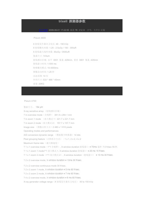
trixell 探测器参数医疗器械2009-06-01 17:22:55 阅读78 评论0 字号:大中小订阅 Pixium 4600X射线发生器名义电压: 40 - 150 kVpX射线曝光剂量: 1.25 - 2.5uGy / 150 - 300uRX射线最大线性剂量: 30uGy / 3500uR象素尺寸: 143um射线感应区域: 水平3001 象素, 429mm, 垂直3001 象素, 429mm射线窗口时间: 1-500 ms特殊曝光模式: 10-4000ms图像读出时间: 1.25秒动态范围: 14位外形尺寸: 533 * 488 * 45mm重量: 20KGPixium 4700像素尺寸:154 μmX-ray sensitive array:(射线感应区域)in overview mode(全视野)381.9 x 294.1 mmin zoom 1 mode (放大模式1)221.7 x 221.7 mmin zoom 2 mode(放大模式2)157.7 x 157.7 mmImage size (图像点阵大小)2 480 x 1 910 pixelsOperating modes and performancesA/D conversion dynamic range (模拟数字转换器)14 bitsPixel grouping feature(点阵组合方式): 1 x 1, 2 x 2, 4 x 2Maximum frame rate:(最大帧速度)1 x 1 overview mode(1*1全视野), X-window duration射线窗口≤ 70ms毫秒7.5 fr/sec帧/秒.1 x 1 zoom 1 mode1*1放大模式1, X-window duration射线窗口≤ 25 ms 15 fr/sec.1 x 1 zoom2 mode(1*1放大模式2), X-window duration(射线窗口)≤ 10 ms 30 fr/sec.2 x 2 overview mode, X-window duration ≤ 13ms 30 fr/sec.2 x 2 overview continuous mode 30 fr/sec.2 x 2 zoom 1 mode, X-window duration ≤ 5 ms 60 fr/sec.2 x 2 zoom 2 mode, X-window duration ≤ 7 ms 60 fr/se c.4 x 2 overview mode, X-window duration ≤ 8 ms 60 fr/sec.X-ray generator voltage range(X射线发生器名义电压)40 to 150 kVpDose range (剂量范围)5 to 4 500 nGy/frMaximum linear dose(最大线性剂量)45 μGy/frSensitivity:(灵敏度)highest gain(最大增益)( 2 x 2 mode(2*2模式)) 6.41 LSB/nGy typ.(6.41SB/nGy标准值) lowest gain (最小增益)0.14 LSB/nGy typ.(0.14SB/nGy标准值)Signal / Electronic noise:(信噪比)@ 5 nGy/fr in highest gain (1) 14 dB min.(在5nGy/fr,最大增益14DB)@ 1μGy/fr in lowest gain (1) 54 dB min.(在1nGy/fr,最小增益54DB)MTF @ 1 lp/mm, RQA5 (2) 60 % min.(MTF值,1线对/mm,60%)MTF @ 2 lp/mm, RQA5 (2) 30 % min.(MTF值,2线对/mm,30%)DQE @ 0 lp/mm, 1μGy/fr,RQA5 (2) 73 % typ.Residual signal (lag & memory effect) after 10 sec. exposure* at 30 fr/sec.:after 1 sec. ≤ 1.1 %after 10 sec. ≤ 0.25 %* Residual signal values in mode 2 gain 7 with 30 fr/sec.Electrical interfaces电源Single DC input voltage 24 V直流24VElectrical power (功率)75 WMechanical characteristicsOverall dimensions(外形尺寸)478 x 366 x 85 mm max.Weight 20 kg typ.(1) 1nGy = 0.115 μR @ RQA(2) RQA5 = 70 kVp, filtration = 2.5 + 21 mm aluminium。
x射线荧光光谱法 英文

x射线荧光光谱法英文X-Ray Fluorescence Spectrometry (XRF)。
X-ray fluorescence spectrometry (XRF) is an analytical technique used to determine the elemental composition of materials by measuring the X-rays emitted by the material when it is exposed to a high-energy X-ray beam. This method is widely used in various fields, including geology, environmental science, forensic science, archaeology, and materials science.Principle of Operation.XRF is based on the principle that when a material is irradiated with high-energy X-rays, electrons in the atoms of the material are excited and ejected from their orbits. The resulting vacancies are filled by electrons from higher energy levels, releasing X-rays with energiescharacteristic of the elements present in the material.The energy of the emitted X-rays is specific to each element, and the intensity of the X-rays is proportional to the concentration of the element in the material. By measuring the energies and intensities of the emitted X-rays, it is possible to identify and quantify the elements present in the sample.Instrumentation.A typical XRF spectrometer consists of the following components:X-ray source: Generates high-energy X-rays that bombard the sample.Sample chamber: Holds the sample to be analyzed.Detector: Converts X-rays into electrical signals.Multichannel analyzer (MCA): Digitizes and analyzes the electrical signals from the detector.Types of XRF Spectrometers.There are several types of XRF spectrometers, each with its own advantages and limitations:Energy-dispersive XRF (EDXRF): Uses a solid-state detector to measure the energies of the emitted X-rays. EDXRF is relatively inexpensive and easy to operate, but it has lower energy resolution compared to other types of XRF spectrometers.Wavelength-dispersive XRF (WDXRF): Uses a crystal monochromator to separate the emitted X-rays by wavelength. WDXRF offers higher energy resolution than EDXRF, but it is more complex, expensive, and time-consuming to operate.Total reflection XRF (TXRF): Utilizes total reflection conditions to enhance the sensitivity for analyzing trace elements in liquids. TXRF is highly sensitive, but it requires sample preparation and is not suitable for solid samples.Applications of XRF.XRF is a versatile analytical technique with a wide range of applications:Geochemistry: Determining the elemental composition of rocks, minerals, and soils.Environmental science: Monitoring pollutants in air, water, and soil.Forensic science: Analyzing trace evidence, such as gunshot residue and paint chips.Archaeology: Studying the composition of artifacts and ancient materials.Materials science: Characterizing the elemental composition of metals, alloys, and other materials.Advantages of XRF.Nondestructive: Does not damage the sample being analyzed.Multi-elemental: Can identify and quantify multiple elements simultaneously.Rapid: Provides real-time analysis results.Sensitive: Can detect elements at trace levels.Versatile: Can be applied to various sample types, including solids, liquids, and powders.Limitations of XRF.Limited sensitivity: Cannot detect elements present in very low concentrations.Matrix effects: The presence of other elements in the sample can affect the accuracy of the analysis.Sample preparation: May require sample preparation,such as grinding or homogenization.Cost: XRF spectrometers can be expensive, especially WDXRF systems.Conclusion.X-Ray Fluorescence Spectrometry is a powerful analytical technique that provides valuable information about the elemental composition of materials. It is widely used in various fields and offers advantages such as non-destructiveness, multi-elemental analysis, and rapid results. However, it has limitations in sensitivity and potential matrix effects, which should be considered when selecting this technique for specific applications.。
x荧光光谱法

x荧光光谱法X荧光光谱法(X-ray fluorescent spectroscopy,XRF)是现代分析科学中常用的一种无损表面分析技术。
它通过测量物质被激发后放射出的X射线能谱图,从而确定样品中各种基本元素的相对含量和结构信息。
X荧光光谱法具有高灵敏度、高分辨率、广泛适用性等优点,在材料科学、地球科学、环境科学、矿业勘探等领域有着广泛的应用。
本文将详细介绍X荧光光谱法的原理、仪器设备以及应用领域。
一、X荧光光谱法的原理1.1 X射线的产生和相互作用X射线是电磁波谱中波长最短的一种辐射。
X射线的产生主要有两种途径:一种是由高能电子通过急剧的减速过程产生的,称为广义X射线;另一种是由高能粒子与物质相互作用而产生的,如β粒子与重原子核相互作用产生的射线,称为硬X射线。
当高能电子与物质相互作用时,会发生三种主要的相互作用过程:电离作用、激发作用和散射作用。
这些相互作用过程对物质的特性有很大的影响。
其中,电离作用是指电子与物质原子中的电子发生碰撞,导致电子被打出原子,产生电离现象。
激发作用是指电子与物质原子中的内层电子发生碰撞,使内层电子被激发到高能级,然后返回基态时放出能量。
散射作用是指电子与物质原子中的电子发生弹性碰撞,改变方向后出射。
1.2 X荧光光谱法的原理X荧光光谱法是利用物质受激发后放射出的X射线能谱图来分析样品中的成分和结构信息。
当X射线照射到物质上时,物质原子的内层电子可以被激发到高能级,然后返回基态时会放出能量。
这些能量的大小和原子的电子能级差有关,不同元素的电子能级差是不同的。
当物质被X射线照射时,其中的原子会被激发,激发后返回基态时放出的能量就形成了一系列特定的X射线能谱线。
这些能谱线对应着不同元素的电子能级差,因此可以通过测量物质放射出的X射线能谱图来确定样品中各种基本元素的相对含量和结构信息。
1.3 X荧光光谱法的仪器设备X荧光光谱法主要的仪器设备有X射线发生器、样品支架、能谱仪和数据处理系统。
x-ray稳定性测试步骤(精)
对FISCHERSCOPE X-RAY测厚仪进行测量稳定性测试的步骤
(基于WinFTM 软件)
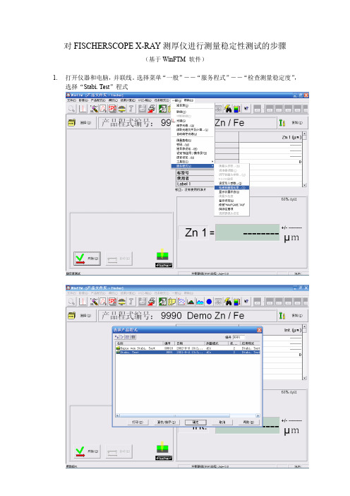
1.打开仪器和电脑,并联线。
选择菜单“一般”――“服务程式”――“检查测量稳定度”,
选择“Stabi. Test”程式
2.选择“产品程式”――“复制”,复制一个“Stabi. Test”程式,为了便于筛选,将复制
的程式取名为日期,例如“Stabi. Test 20070101”。
按“复制”后关闭窗口。
3.打开仪器和电脑,并联线。
选择菜单“一般”――“服务程式”――“检查测量稳定度”,
选择“Stabi. Test 20070101”程式。
4.选择“调校”――“归一化”,按“是”确认。
5.选择“产品程式”――“连续测量”,进行测试。
6.放入Ag基准,连续测量24或48小时后,停止测量。
选择“产品程式”――“复制文
件由/到”,在“产品程式>>>文件”卡中,在“复制那个产品程式?”中选择刚才进行稳定性测试的程式,例如“Stabi. Test 20070101”;在“产品程式复制到那里?”中选择目标目录,例如C盘,D盘等。
7.把复制出来的文件,如“Stabi. Test 20070101.sv1”,通过电子邮件传送到FISCHER公
司,以便进行分析。
- 1、下载文档前请自行甄别文档内容的完整性,平台不提供额外的编辑、内容补充、找答案等附加服务。
- 2、"仅部分预览"的文档,不可在线预览部分如存在完整性等问题,可反馈申请退款(可完整预览的文档不适用该条件!)。
- 3、如文档侵犯您的权益,请联系客服反馈,我们会尽快为您处理(人工客服工作时间:9:00-18:30)。
Chandra Fellow Current address: Department of Physics and Astronomy; Ohio University; Athens, OH 45701
–3– understanding of the relevant radiation mechanisms responsible for the high-energy emission of blazars and the underlying particle acceleration mechanisms. The low-energy component of blazar SEDs is well understood as synchrotron emission from ultrarelativistic electrons in a relativistic jet directed at a small angle with respect to the line of sight. In the framework of leptonic models (for a review of the alternative class of hadronic jet models, see, e.g., Rachen (2000)), high-energy emission will result from Compton scattering of lower-frequency photons off the relativistic electrons. Possible target photon fields for Compton scattering are the synchrotron photons produced within the jet (the SSC process; Marscher & Gear (1985); Maraschi, Ghisellini, & Celotti (1992); Bloom & Marscher (1996)), or external photons (the EC process). Sources of external seed photons include the UV – soft X-ray emission from the disk — either entering the jet directly (Dermer, Schlickeiser, & Mastichiadis 1992; Dermer & Schlickeiser 1993) or after reprocessing in the broad line region (BLR) or other circumnuclear material (Sikora, Begelman, & Rees 1994; Blandford & Levinson 1995; Dermer, Sturner, & Schlickeiser 1997) —, jet synchrotron radiation reflected at the BLR (Ghisellini & Madau 1996; Bednarek 1998; B¨ ottcher & Dermer 1998), or the infrared emission from circumnuclear dust (Bla˙ zejowski et al. 2000; Arbeiter, Pohl, & Schlickeiser 2002). According to the now well-established AGN unification scheme (Urry & Padovani 1995), blazars can be unified with other classes of AGN, in particular radio galaxies, through orientation effects. However, Sambruna, Maraschi, & Urry (1996) have pointed out that such orientation effects can not explain the differences between different blazar sub-classes. Instead, it has been suggested that the sequence of spectral properties of blazars from HBLs via LBLs to FSRQs can be interpreted in terms of an increasing total power input into nonthermal electrons in the jet, accompanied by an increasing contribution of external photons to the seed photon field for Compton upscattering (Madejski 1998; Ghisellini et al. 1998). It has been suggested that this may be related to an evolutionary effect due to the gradual depletion of the circumnuclear material being accreted onto the central black hole (D’Elia & Cavaliere 2000; Cavaliere & D’Elia 2002; B¨ ottcher & Dermer 2002). Detailed modeling of blazars in the different sub-classes (FSRQs, LBLs and HBLs) seems to confirm this conjecture (for a recent review, see, e.g., B¨ ottcher (2002)). As mentioned earlier, blazars tend to exhibit rapid flux and spectral variability. The variability is most dramatic and occurs on the shortest time scales at the high-energy ends of the two nonthermal spectral components of their broadband SEDs. Particularly interesting variability patterns could be observed at X-ray energies for those blazars whose X-ray emission is dominated by synchrotron emission. Observational studies of X-ray variability in blazars have so far focused on HBLs and, in particular, on the attempt to identify clear pat-
Department of Physics and Astronomy, Rice University, 6100 Main Street, Houston, TX 77005-1892 mboett@ and James Chiang NASA Goddard Space Flight Center, Code 661, Greenbelt, MD 20771 Joint Center for Astrophysics and Physics Department, University of Maryland, Baltimore, MD 21250 jchiang@ ABSTRACT We are presenting a detailed parameter study of the time-dependent electron injection and kinematics and the self-consistent radiation transport in jets of intermediate and low-frequency peaked BL Lac objects. Using a time-dependent, combined synchrotron-self-Compton and external-Compton jet model, we study the influence of variations of several essential model parameters, such as the electron injection compactness, the relative contribution of synchrotron to external soft photons to the soft photon compactness, the electron-injection spectral index, and the details of the time profiles of the electron injection episodes giving rise to flaring activity. In the analysis of our results, we focus on the expected X-ray spectral variability signatures in a region of parameter space particularly well suited to reproduce the broadband spectral energy distributions of intermediate and low-frequency peaked BL Lac objects. We demonstrate that SSCand external-Compton dominated models for the γ -ray emission from blazars are producing significantly different signatures in the X-ray variability, in particular in the soft X-ray light curves and the spectral hysteresis at soft X-ray energies, which can be used as a powerful diagnostic to unveil the nature of the high-energy emission from BL Lac objects.