动态光散射基本原理及其在纳米科技中的应用——Zeta电位测量
粒径及zeta电位实验原理
粒径及zeta电位实验原理一、引言粒径及zeta电位是纳米颗粒表征中最常用的两种参数,它们能够反映纳米颗粒的大小和表面电荷特性,对于纳米材料的研究具有重要的意义。
本文将详细介绍粒径及zeta电位实验原理。
二、粒径实验原理1.动态光散射法(DLS)动态光散射法(Dynamic Light Scattering, DLS)是一种常用的测量纳米颗粒粒径分布的方法。
其基本原理是利用激光散射现象,通过测量散射光强度随时间变化的自相关函数来计算出颗粒在溶液中的有效半径分布。
该方法适用于测量直径在几纳米到几微米之间的颗粒。
2.静态光散射法(SLS)静态光散射法(Static Light Scattering, SLS)是一种通过测量溶液中颗粒对入射激光束进行非弹性散射而得到样品分子量和分子大小信息的技术。
该方法适用于大分子或聚合物等高分子材料。
三、zeta电位实验原理1.电泳光散射法(ELS)电泳光散射法(Electrophoretic Light Scattering, ELS)是一种通过测量颗粒在外加电场下的运动速度和其所受到的电荷数目来计算出颗粒表面电荷特性的方法。
该方法适用于测量直径在几纳米到几微米之间的颗粒。
2.激光多角度光散射法(MALS)激光多角度光散射法(Multi-Angle Light Scattering, MALS)是一种通过测量样品对入射激光束进行非弹性散射而得到样品分子量和分子大小信息的技术。
该方法适用于大分子或聚合物等高分子材料。
四、实验操作流程1.样品制备:将需要测试的样品溶解在适当溶剂中,并进行必要的稀释。
2.仪器预热:打开仪器并进行预热,保证仪器处于稳定状态。
3.参数设置:根据实验要求,设置相应的参数,如激光波长、探测角度等。
4.测试操作:将样品注入测试池中,并启动测试程序。
5.数据处理:根据实验结果进行数据处理和分析,如计算粒径分布、zeta电位等参数。
五、实验注意事项1.样品制备应注意避免气泡的产生和溶剂的挥发。
zate电位仪原理方面的资料

动态光散射基本原理及其在纳米科技中的应用——Zeta电位测量前言:Zeta电位是纳米材料的一种重要表征参数。
现代仪器可以通过简便的手段快速准确地测得。
大致原理为:通过电化学原理将Zeta电位的测量转化成带电粒子淌度的测量,而粒子淌度的测量测是通过动态光散射,运用波的多普勒效应测得。
1.Zeta电位与双电层(图1)粒子表面存在的净电荷,影响粒子界面周围区域的离子分布,导致接近表面抗衡离子(与粒子电。
荷相反的离子)浓度增加。
于是,每个粒子周围均存在双电层。
围绕粒子的液体层存在两部分:一是内层区,称为Stern层,其中离子与粒子紧紧地结合在一起;另一个是外层分散区,其中离子不那么紧密的与粒子相吸附。
在分散层内,有一个抽象边界,在边界内的离子和粒子形成稳定实体。
当粒子运动时(如由于重力),在此边界内的离子随着粒子运动,但此边界外的离子不随着粒子运动。
这个边界称为流体力学剪切层或滑动面(slippingplane)。
在这个边界上存在的电位即称为Zeta电位。
2.Zeta电位与胶体的稳定性(DLVO理论)在1940年代Derjaguin, Landau, Verway与Overbeek 提出了描述胶体稳定的理论,认为胶体体系的稳定性是当颗粒相互接近时它们之间的双电层互斥力与范德瓦尔互吸力的净结果。
此理论提出当颗粒接近时颗粒之间的能量障碍来自于互斥力,当颗粒有足够的能量克服此障碍时,互吸力将使颗粒进一步接近并不可逆的粘在一起。
(图2) Zeta电位可用来作为胶体体系稳定性的指示:如果颗粒带有很多负的或正的电荷,也就是说很高的Zeta电位,它们会相互排斥,从而达到整个体系的稳定性;如果颗粒带有很少负的或正的电荷,也就是说它的Zeta电位很低,它们会相互吸引,从而达到整个体系的不稳定性。
一般来说, Zeta电位愈高,颗粒的分散体系愈稳定,水相中颗粒分散稳定性的分界线一般认为在+30mV或-30mV,如果所有颗粒都带有高于+30mV或低于-30mV的zeta电位,则该分散体系应该比较稳定3.影响Zeta电位的因素分散体系的Zeta电位可因下列因素而变化:A. pH 的变化B. 溶液电导率的变化C. 某种特殊添加剂的浓度,如表面活性剂,高分子测量一个颗粒的zeta势能作为上述变量的变化可了解产品的稳定性,反过来也可决定生成絮凝的最佳条件。
zeta电位测定原理

zeta电位测定原理
Zeta 电位是指胶体分散系中分散粒子表面的电位,它是表征胶体分散系稳定性的重要指标。
Zeta 电位的测定是通过在胶体分散系中引入电极,测量电极间的电势差得出的。
在测量 Zeta 电位时,需要将胶体分散系中的分散粒子引入到电解液中,然后将电极放置在电解液中,施加外部电场,使分散粒子做电泳运动。
通过测量电极间的电势差,可以计算出 Zeta 电位。
目前,测量 Zeta 电位的方法主要有电泳法、电渗法、流动电位法和超声波法,其中电泳法应用最广。
Zeta 电位的数值与胶态分散的稳定性相关,分子或分散粒子越小,Zeta 电位越高,体系越稳定,即溶解或分散可以抵抗聚集。
动态光散射基本原理及其在纳米科技中的应用——Zeta电位测量

【专题】动态光散射基本原理及其在纳米科技中的应用——Zeta电位测量--------------------------------------------------------------------------------作者: 骑着蜗牛追火箭收录日期: 2009-11-28 发布日期: 2009-11-28动态光散射基本原理及其在纳米科技中的应用——Zeta电位测量前言:Zeta电位是纳米材料的一种重要表征参数。
现代仪器可以通过简便的手段快速准确地测得。
大致原理为:通过电化学原理将Zeta电位的测量转化成带电粒子淌度的测量,而粒子淌度的测量测是通过动态光散射,运用波的多普勒效应测得。
1.Zeta电位与双电层(图1)粒子表面存在的净电荷,影响粒子界面周围区域的离子分布,导致接近表面抗衡离子(与粒子电。
荷相反的离子)浓度增加。
于是,每个粒子周围均存在双电层。
围绕粒子的液体层存在两部分:一是内层区,称为Stern层,其中离子与粒子紧紧地结合在一起;另一个是外层分散区,其中离子不那么紧密的与粒子相吸附。
在分散层内,有一个抽象边界,在边界内的离子和粒子形成稳定实体。
当粒子运动时(如由于重力),在此边界内的离子随着粒子运动,但此边界外的离子不随着粒子运动。
这个边界称为流体力学剪切层或滑动面(slippingplane)。
在这个边界上存在的电位即称为Zeta电位。
ZETA电位是一个表征分散体系稳定性的重要指标。
由于带电微粒吸引分散系中带相反电荷的粒子,离颗粒表面近的离子被强烈束缚着,而那些距离较远的离子形成一个松散的电子云,电子云的内外电位差就叫Zeta电位。
也称电动电位(只有当胶粒在介质中运动时才会表现出来),实际上就是扩散层内的电位差。
ξ电位较高时,粒子能保持一定距离消弱和抵消了范德华引力从而提高了颗粒悬浮系统的稳定性。
反之,当ξ电位较低时,粒子间的斥力减小并逐步靠近,进入范德华引力范围内,粒子就会互相吸引、团聚。
马尔文纳米粒度 zeta
马尔文纳米粒度 zeta
马尔文纳米粒度和zeta电位是常用的表征纳米颗粒物理性质的指标。
本文将详细介绍马尔文纳米粒度和zeta电位的定义、测量原理和应用。
马尔文纳米粒度(Malvern NanoSizer)是由英国马尔文仪器公司开发的一种用于粒子的大小分析的仪器。
马尔文纳米粒度采用动态光散射(Dynamic Light Scattering,DLS)技术,可以测量纳米级别的颗粒的尺寸分布。
马尔文纳米粒度利用激光散射原理测量颗粒在液体中的动态行为,根据光散射强度的强弱和时间的变化,分析颗粒的大小分布和聚集状态。
马尔文纳米粒度可以测量的颗粒直径范围一般在1nm~1μm之间。
1、研究颗粒尺寸、分散性、聚集状态
2、研究颗粒表面性质
3、研究颗粒在不同媒质中的动态行为
4、测量颗粒的分布系数和均匀度
二、zeta电位
zeta电位是一种表征颗粒在溶液中带电状态的参数。
zeta电位的值可以反映颗粒表面的电荷,是影响颗粒相互作用的重要物理参数。
zeta电位采用激光多角度散射(Laser Doppler Anemometry,LDA)技术,利用测量颗粒在电场中的运动和电荷情况,计算出颗粒表面的电荷密度和zeta电位。
1、评价颗粒的稳定性
2、研究离子强度对颗粒间相互作用的影响
3、研究表面改性对颗粒表面电荷的影响
4、评价纳米材料的表面性能和稳定性
综上所述,马尔文纳米粒度和zeta电位是纳米颗粒表征的重要参数。
通过这两项指标的测量,可以了解颗粒的尺寸分布、表面电荷和稳定性等物理性质,有助于研究纳米颗粒的合成、应用和性质优化。
动态光散射基本原理及其在纳米科技中的应用——粒径测量
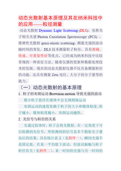
动态光散射基本原理及其在纳米科技中的应用——粒径测量动态光散射Dynamic Light Scattering (DLS),也称光子相关光谱Photon Correlation Spectroscopy (PCS) ,准弹性光散射quasi-elastic scattering,测量光强的波动随时间的变化。
DLS技术测量粒子粒径,具有准确、快速、可重复性好等优点,已经成为纳米科技中比较常规的一种表征方法。
随着仪器的更新和数据处理技术的发展,现在的动态光散射仪器不仅具备测量粒径的功能,还具有测量Zeta电位、大分子的分子量等的能力。
(一)动态光散射的基本原理1. 粒子的布朗运动Brownian motion导致光强的波动微小粒子悬浮在液体中会无规则地运动布朗运动的速度依赖于粒子的大小和媒体粘度,粒子越小,媒体粘度越小,布朗运动越快。
2. 光信号与粒径的关系光通过胶体时,粒子会将光散射,在一定角度下可以检测到光信号,所检测到的信号是多个散射光子叠加后的结果,具有统计意义(见附件一)。
瞬间光强不是固定值,在某一平均值下波动,但波动振幅与粒子粒径有关(见附件二)。
某一时间的光强与另一时间的光强相比,在极短时间内,可以认识是相同的,我们可以认为相关度为1,在稍长时间后,光强相似度下降,时间无穷长时,光强完全与之前的不同,认为相关度为0(此原理见附件三)。
根据光学理论可得出光强相关议程(见附件四)。
之前提到,正在做布朗运动的粒子速度,与粒径(粒子大小)相关(Stokes - Einstein 方程)。
大颗粒运动缓慢,小粒子运动快速。
如果测量大颗粒,那么由于它们运动缓慢,散射光斑的强度也将缓慢波动。
类似地,如果测量小粒子,那么由于它们运动快速,散射光斑的密度也将快速波动。
附件五显示了大颗粒和小粒子的相关关系函数。
可以看到,相关关系函数衰减的速度与粒径相关,小粒子的衰减速度大大快于大颗粒的。
最后通过光强波动变化和光强相关函数计算出粒径及其分布(见附件六)。
动态光散射基本原理及其在纳米科技中的应用——粒径测量
动态光散射基本原理及其在纳米科技中的应用——粒径测量(总5页)-CAL-FENGHAI.-(YICAI)-Company One1-CAL-本页仅作为文档封面,使用请直接删除动态光散射基本原理及其在纳米科技中的应用——粒径测量动态光散射Dynamic Light Scattering(DLS),也称光子相关光谱Photon Correlation Spectroscopy (PCS) ,准弹性光散射quasi-elastic scattering,测量光强的波动随时间的变化。
DLS技术测量粒子粒径,具有准确、快速、可重复性好等优点,已经成为纳米科技中比较常规的一种表征方法。
随着仪器的更新和数据处理技术的发展,现在的动态光散射仪器不仅具备测量粒径的功能,还具有测量Zeta电位、大分子的分子量等的能力。
(一)动态光散射的基本原理1. 粒子的布朗运动Brownian motion导致光强的波动微小粒子悬浮在液体中会无规则地运动布朗运动的速度依赖于粒子的大小和媒体粘度,粒子越小,媒体粘度越小,布朗运动越快。
2. 光信号与粒径的关系光通过胶体时,粒子会将光散射,在一定角度下可以检测到光信号,所检测到的信号是多个散射光子叠加后的结果,具有统计意义(见附件一)。
瞬间光强不是固定值,在某一平均值下波动,但波动振幅与粒子粒径有关(见附件二)。
某一时间的光强与另一时间的光强相比,在极短时间内,可以认识是相同的,我们可以认为相关度为1,在稍长时间后,光强相似度下降,时间无穷长时,光强完全与之前的不同,认为相关度为0(此原理见附件三)。
根据光学理论可得出光强相关议程(见附件四)。
之前提到,正在做布朗运动的粒子速度,与粒径(粒子大小)相关(Stokes - Einstein方程)。
大颗粒运动缓慢,小粒子运动快速。
如果测量大颗粒,那么由于它们运动缓慢,散射光斑的强度也将缓慢波动。
类似地,如果测量小粒子,那么由于它们运动快速,散射光斑的密度也将快速波动。
zeta电位仪原理
zeta电位仪原理
Zeta电位仪是一种常用于表征颗粒表面电荷特性的仪器,它基于电动力学原理和光学散射技术。
其测量原理如下:
在液体中悬浮的颗粒表面通常带有电荷,形成一个电化学双层结构。
当这些带电的颗粒被置于外加电场中时,电场会作用于双层结构,通过测量颗粒的周围电场分布来计算颗粒的电势。
Zeta电位仪测量过程中,首先将样品悬浮液注入电池,然后通过两个电极施加一定电场强度。
接下来,使用激光器产生的激光束照射样品液中的颗粒,在颗粒与激光束相互作用下,产生光散射。
在光学系统中,通过探测器收集和分析散射光的强度和角度信息。
通过测量散射光的强度和角度,可以得到颗粒的大小和分布情况。
根据电势和电场的关系,结合颗粒的大小和分布数据,可以计算出颗粒表面的电场分布,进而得到颗粒表面的Zeta电位。
Zeta电位作为表征颗粒表面电荷特性的重要参数,在颗粒的稳定性、沉降速率、表面吸附等方面具有重要意义。
因此,Zeta 电位仪在颗粒表面电荷特性研究、液固界面性质表征等领域得到了广泛应用。
动态光散射基本原理及其在纳米科技中的应用——粒径测量
动态光散射基本原理及其在纳米科技中的应用——粒径测量动态光散射Dynamic Light Scattering (DLS),也称光子相关光谱Photon Correlation Spectroscopy (PCS) ,准弹性光散射quasi-elastic scattering,测量光强的波动随时间的变化。
DLS技术测量粒子粒径,具有准确、快速、可重复性好等优点,已经成为纳米科技中比较常规的一种表征方法。
随着仪器的更新和数据处理技术的发展,现在的动态光散射仪器不仅具备测量粒径的功能,还具有测量Zeta电位、大分子的分子量等的能力。
(一)动态光散射的基本原理1. 粒子的布朗运动Brownian motion导致光强的波动微小粒子悬浮在液体中会无规则地运动布朗运动的速度依赖于粒子的大小和媒体粘度,粒子越小,媒体粘度越小,布朗运动越快。
2. 光信号与粒径的关系光通过胶体时,粒子会将光散射,在一定角度下可以检测到光信号,所检测到的信号是多个散射光子叠加后的结果,具有统计意义(见附件一)。
瞬间光强不是固定值,在某一平均值下波动,但波动振幅与粒子粒径有关(见附件二)。
某一时间的光强与另一时间的光强相比,在极短时间内,可以认识是相同的,我们可以认为相关度为1,在稍长时间后,光强相似度下降,时间无穷长时,光强完全与之前的不同,认为相关度为0(此原理见附件三)。
根据光学理论可得出光强相关议程(见附件四)。
之前提到,正在做布朗运动的粒子速度,与粒径(粒子大小)相关(Stokes - Einstein方程)。
大颗粒运动缓慢,小粒子运动快速。
如果测量大颗粒,那么由于它们运动缓慢,散射光斑的强度也将缓慢波动。
类似地,如果测量小粒子,那么由于它们运动快速,散射光斑的密度也将快速波动。
附件五显示了大颗粒和小粒子的相关关系函数。
可以看到,相关关系函数衰减的速度与粒径相关,小粒子的衰减速度大大快于大颗粒的。
最后通过光强波动变化和光强相关函数计算出粒径及其分布(见附件六)。
- 1、下载文档前请自行甄别文档内容的完整性,平台不提供额外的编辑、内容补充、找答案等附加服务。
- 2、"仅部分预览"的文档,不可在线预览部分如存在完整性等问题,可反馈申请退款(可完整预览的文档不适用该条件!)。
- 3、如文档侵犯您的权益,请联系客服反馈,我们会尽快为您处理(人工客服工作时间:9:00-18:30)。
【专题】动态光散射基本原理及其在纳米科技中的应用——Zeta电位测量--------------------------------------------------------------------------------作者: 骑着蜗牛追火箭收录日期: 2009-11-28 发布日期: 2009-11-28动态光散射基本原理及其在纳米科技中的应用——Zeta电位测量前言:Zeta电位是纳米材料的一种重要表征参数。
现代仪器可以通过简便的手段快速准确地测得。
大致原理为:通过电化学原理将Zeta电位的测量转化成带电粒子淌度的测量,而粒子淌度的测量测是通过动态光散射,运用波的多普勒效应测得。
1.Zeta电位与双电层(图1)粒子表面存在的净电荷,影响粒子界面周围区域的离子分布,导致接近表面抗衡离子(与粒子电。
荷相反的离子)浓度增加。
于是,每个粒子周围均存在双电层。
围绕粒子的液体层存在两部分:一是内层区,称为Stern层,其中离子与粒子紧紧地结合在一起;另一个是外层分散区,其中离子不那么紧密的与粒子相吸附。
在分散层内,有一个抽象边界,在边界内的离子和粒子形成稳定实体。
当粒子运动时(如由于重力),在此边界内的离子随着粒子运动,但此边界外的离子不随着粒子运动。
这个边界称为流体力学剪切层或滑动面(slippingplane)。
在这个边界上存在的电位即称为Zeta电位。
ZETA电位是一个表征分散体系稳定性的重要指标。
由于带电微粒吸引分散系中带相反电荷的粒子,离颗粒表面近的离子被强烈束缚着,而那些距离较远的离子形成一个松散的电子云,电子云的内外电位差就叫Zeta电位。
也称电动电位(只有当胶粒在介质中运动时才会表现出来),实际上就是扩散层内的电位差。
ξ电位较高时,粒子能保持一定距离消弱和抵消了范德华引力从而提高了颗粒悬浮系统的稳定性。
反之,当ξ电位较低时,粒子间的斥力减小并逐步靠近,进入范德华引力范围内,粒子就会互相吸引、团聚。
ξ电位与液体递质内的粒子质量分数有关,改变液体的pH值、增加体系的盐含量都会引起双电层压缩,改变粒子的ξ电位,降低颗粒间的静电排斥作用,从而影响颗粒悬浮系统的稳定性。
2.Zeta电位与胶体的稳定性(DLVO理论)在1940年代Derjaguin, Landau, Verway与Overbeek 提出了描述胶体稳定的理论,认为胶体体系的稳定性是当颗粒相互接近时它们之间的双电层互斥力与范德瓦尔互吸力的净结果。
此理论提出当颗粒接近时颗粒之间的能量障碍来自于互斥力,当颗粒有足够的能量克服此障碍时,互吸力将使颗粒进一步接近并不可逆的粘在一起。
(图2)Zeta电位可用来作为胶体体系稳定性的指示:如果颗粒带有很多负的或正的电荷,也就是说很高的Zeta电位,它们会相互排斥,从而达到整个体系的稳定性;如果颗粒带有很少负的或正的电荷,也就是说它的Zeta电位很低,它们会相互吸引,从而达到整个体系的不稳定性。
一般来说, Zeta电位愈高,颗粒的分散体系愈稳定,水相中颗粒分散稳定性的分界线一般认为在+30mV或-30mV,如果所有颗粒都带有高于+30mV或低于-30mV的zeta电位,则该分散体系应该比较稳定3.影响Zeta电位的因素分散体系的Zeta电位可因下列因素而变化:A. pH 的变化B. 溶液电导率的变化C. 某种特殊添加剂的浓度,如表面活性剂,高分子测量一个颗粒的zeta势能作为上述变量的变化可了解产品的稳定性,反过来也可决定生成絮凝的最佳条件。
3.1 Zeta电位与pH(图3)影响zeta电位最重要的因素是pH,当谈论zeta电位时,不指明pH根本一点意义都没有。
假定在悬浮液中有一个带负电的颗粒;假如往这一悬浮液中加入碱性物质,颗粒会得到更多的负电;假如往这一悬浮液中加入酸性物质,在一定程度时,颗粒的电荷将会被中和;进一步加入酸,颗粒将会带更多的正电。
Zeta电位对pH作图在低pH将是正的,在高pH将是负的,这中间一定有一点会通过零zeta电位,这一点称为等电点,是相当重要的一点,通常在这一点胶体是最不稳定的。
3.2 Zeta电位与电导率双电层的厚度与溶液中的离子浓度有关,可根据介质的离子强度进行计算,离子强度越高,双电层愈压缩同,离子的化合价也会影响双单层的厚度,三价离子(Al3+)将会比单价离子(Na+)更多的压缩双电层。
无机离子可有两种方法与带电表面相作用:1. 非选择性吸附.对于等电点没有影响2. 选择性吸附.会改变等电点即使很低浓度的选择性吸附离子,也会对Zeta电位有很大的影响,有时选择性吸附离子甚至会造成颗粒从带负电变成带正电,从带正电变成带负电。
3.3 Zeta电位与添加剂浓度研究样品中的添加剂浓度对产品zeta电位的影响可为研发稳定配方的产品提供有用的信息,样品中已知杂质对zeta电位的影响可作为研制抗絮凝的产品的有力工具。
4. 带电粒子的动电学效应表面电荷的存在使得颗粒在一外加电场中呈现某些特殊效应,这些效应总称为动电学效应,根据引入运动的方式,有四种不同的动电学效应:电泳: 在外加电场中带电颗粒相对于静止悬浮液体的运动电渗:在外加电场中相对于静止带电表面的液体运动流动电势: 当液体流过静止表面时所产生的电场沉降电势: 当带电颗粒在静止液体中流动时所产生的电场5. Zeta电位测量理论在一平行电场中,带电颗粒向相反极性的电极运动,颗粒的运动速度与下列因素有关:电场强度,介质的介电常数,介质的粘度(均为已知参数)Zeta电位(未知参数)Zeta电位与电泳淌度之间由Henry方程相连(图4)由Henry方程可以看出,只要测得粒子的淌度,查到介质的粘度、介电常数等参数,就可以求得Zeta电位。
6. 淌度测量方法6.1 直接观测法在早期,测量粒子淌度时,是在分散体系两端加上电压,用显微装置观测。
6.2 多普勒效应测量法当测量一个速度为C,频率为no的波时,假如波源与探测器之间有一相对运动(速度V), 所测到的波频率将会有一多普勒位移。
在电场作用下运动的粒子,当激光打到粒子上时,散射光频率会有变化。
散射光与参考光叠加后频率变化表现得更为直观,更容易观测。
将光信号的频率变化与粒子运动速度联系起来,即可测得粒子的淌度。
(如图5)7. 电渗及避免方法7.1 电渗(图6)由于毛细管样品池壁带电,当外加电场导致颗粒运动时,池壁附近的液体也会在电场中由于电渗而运动。
当用毛细管样品池时由于池壁与水中离子的作用,水会在电场下移动(电渗)而影响颗粒移动速度的测量(因为测到的是两种运动的总和)。
但是,由于在一个封闭的池子内,池壁的液体流动会造成池中间的液体向另一方向运动,而在样品池中造成抛物面状的液体流动。
在样品池中有两个无限薄的层面(静止层)内无电渗,经典方法将光束定位在静止层内测量,以避免电渗误差,不可能准确定位,及费时而造成各种误差(甚至池壁有微量污染)。
7.2 电渗的避免(图7)动电学的理论分析告诉我们:当外加一电场时,颗粒达到其最终运动速度的时间至少要比电渗快一个数量级(参考M. Minor, A.J. van der Linde, H.P. van Leeuwen and J. Lyklema (1997) J Colloid and Interface Science 189, 370-375)快速电场反转(FFR)假如一外加电场有足够高的频率时:与颗粒的运动相比,液体的运动可以忽略不计这样,测量就不一定要在静止层进行但是,快速电场反转(FFR)与传统的慢速电场反转(SFR)相比,分辨率低。
如果将二者结合起来,则可以得到准确率高,分辨率高的Zeta电位及分布。
/Tutorials/Intro/zetaintro.htmlTutorialsThe use of the Malvern Zetasizer for the measurement of Zeta PotentialParticle characterizationZeta potential - Application trendsHow does surface charge arise?How is particle charge measured?Methods for determining the zeta potentialZeta potential measurement techniqueApplications - possibilities for automationThe Malvern MPT-1 AutotitratorSelected applications - an overviewParticle characterizationThere are many.htmlects of a particle dispersion that need to be investigated to fully characterize a system.Particle size is often considered one of the most important parameters, however, as particle size reduces, the surface area increasessignificantly in comparison with the volume, so surface properties increasingly determine the dispersions characteristics.One of the significant surface properties is the surface charge. This is an important factor in determining the interactions between particles, and hence dispersion characteristics such as dispersion stability, flocculation, viscosity, film forming characteristics etc.The surface charge cannot be measured directly. Instead the charge at a distance from the particle, called the zeta potential is measured. This potential is usually more of interest because particles interact according to the magnitude of this value, rather than the potential at the surface of the particle.Zeta potential - Application trendsThe zeta potential (x), or electrophoretic mobility (µE), is increasingly being used to investigate fine particle systems.The zeta potential is a consequence of the existence of surface charge, and can give information on electrical interaction forces between the dispersed particles. Using DLVO theory, it is possible to assess the stability of suspensions and emulsions by means of the zeta potential.The basis of DLVO is to use the sum of the repulsive force (V R - electrostatic BORN forces) and attractive (VA - van der WAALS forces) to calculate the particle interaction potential.V TOTAL = V R + V AIn many cases, the zeta potential can be used directly as the criterion for assessing product quality.However stability assessment using the zeta potential as the measurable variable may only be useful if the system is sufficiently well understood.Electrokinetic measurements can be used to investigate the effect of each of the excipients in a formulation on the dispersed materials surface characteristics. Figure 1 shows schematic examples of zeta potential Vs pH functions that are characteristic of certain surface conditions. These curves are found for example in the protolysis of acid or alkaline surface groups and in pH-dependent adsorption processes.Fig. 1 Model examples of pH Vs zeta functions showing dissociation of acid or alkaline surface groups and adsorptionHow does surface charge arise?Surface charges on suspended particles can be caused by a variety of phenomena. Figure 2 shows a number of possibilities. Dissociation of functional surface groups and/or adsorption of ions are the most important processes. In many formulations the adsorption of larger molecules containing charged groups such as surfactants and polyelectrolytes play a vital role.The surface charge of the particles is compensated in the liquid phase by counter-ions, ensuring a condition of electrical neutrality in the system as a whole.Lattice imperfectionsSurface-adsorbed ionsChemical reactions on the surface(dissociation of functional surface groups)Adsorption or dissociation of charge-bearing moleculesFig. 2 Examples of phenomena that lead to the development of surface chargeHow is particle charge measured?The electrochemical double layerThe interface between two phases, (solid/liquid, liquid/liquid or gas/liquid) differs in a number of ways to the bulk phase. One reason for this is the different distribution of electrical charges at the phase boundary. This leads to the development of an electrical double layer with a potential different to that within either phase.Fig. 3 Structure of the electrochemical double layer on a particle surface in a system containing electrolytesThe STERN double layer model is generally used to describe the charge distribution. Figure 3 shows in schematic form the distribution at the interface for the case of a negatively charged particle surface, (yo) Charges in each phase interact. In an electrolyte solution, a monolayer is quickly adsorbed directly on the particle surface.These are generally dehydrated ions and this region of a monolayer of ions is known as the inner Helmholtz layer.Whether these are anions or cations depends on whether they are 'Indifferent' ions or 'Specifically adsorbed' ions.Specifically adsorbed ions interact chemically with the surface; it could be a proton interacting with a carboxylic acid group for example. These ions can interact so strongly with the surface that they can cause charge reversal. Conversely if the ion is the same sign as the surface, the surface potential can be increased.In the absence of specifically adsorbed ions, this inner layer consists of dehydrated or partially dehydrated counter-ions. In the case of a negative surface in contact with indifferent ions Sodium and Chloride, this would be the Sodium ion and lead to a potential (yi). The next layer of ions (outer Helmholtz layer) are rigidly bound hydrated counter-ions, which reduce the potential to the value yd?. The internal and external Helmholtz layers form the STERN layer with thickness d. The distribution of charge carriers outside this layer, in a more diffuse region, reduces to zero in the bulk phase. Electrical neutrality ultimately exists at an infinite distance from the particle surface with n+ = n-.Particle movements in the medium, due to for example, Brownian motion or sedimentation, leads to the concept of a shear plane within the diffuse layer. This region separates the ions associated with the particle from those in the bulk phase. The potential at this shear plane is what is referred to as the zeta potential (x).The extent of the diffuse double layer is dependent on the ionic strength of the electrolytes and the valency of each of the ions. Multivalent ions are particularly important. Their effect on the screening of the surface charge is related to the counter-ion valency. The flocculation concentration for divalent counter-ions is on average about 100 times lower than for monovalent ions, and for trivalent ions about 1000 less. This is known as the Schultze-Hardy rule and is the explanation for the use of Aluminium or Ferric ions to cause the flocculation of particles with negative zeta potential in water treatment plants.Specific adsorptionThe distribution of ions is more complicated if ions exhibit specific adsorption behavior. Specific ion adsorption can have a dramatic effect on particle mobility at low ion concentrations.Figure 4 Shows how the composition of the medium can affect the surface potential and the extent of the electrical double layer. These three cases are arranged so the value of zeta potential is the same in each case. This demonstrates that the surface charge cannot be inferred from zeta potential.It is worth emphasizing the distinction between surface potential and zeta potential. The zeta potential is the controlling parameter for particle interactions. Modifying the zeta potential requires knowledge of the potential of the surface and its chemistry. Investigation of the interaction of ions with the surface, and their effect on zeta potential is assisted by the determination of zeta potential as a function of a number of parameters.The parameters that it is useful to investigate are pH, conductivity and the concentration of any specifically adsorbed ions or polymers in the system.Methods for determining the zeta potential:Electrokinetic phenomenaThere are four electrochemical phenomena that can be used for the determination of zeta-potential, electrophoresis, electroosmosis, streaming and sedimentation potentialElectrophoresis and streaming potential are the most important commercial effects that have been exploited to determine zeta potential. The Malvern Zetasizer uses electrophoresis, however electroosmosis is an interfering effect that must be considered (Fig.5). There are a number of ways of accounting for this effect. The use of measurements at the stationary layer is one method that is discussed in the next section.Fig. 5 Basic principles of electrophoresis and electroosmosisFig. 4 Potential trends at interfaces with differences in the chemical constitution, influence of specific ionic adsorption1. Positively charged particles on the surface, moderate2. adsorption of anions, moderate ionic strength positively charged particles with no specific adsorption,low concentration of electrolyte3.negatively charged particles, strong specific cationadsorption, moderate electrolyte concentrationMicro-electrophoresisThe conventional method for measuring zeta potential is to observe the migration of particles in a dispersion with a microscope andmeasure their velocity using a stopwatch. The current technique of laser Doppler velocimetry (LDV) has mostly replaced this laborious measurement procedure.Both techniques are based on measurement of the migration rate of dispersed particles under the influence of an electric field. The observed velocity (v), divided by the strength of the applied electrical field (E), is a direct measure of the electrophoretic mobility (µE) of the particles examined.v /E = µEThe zeta potential can be calculated from the mobility using the Henry equation.Henry equation:m E = e * x * f(k a)/6phe - dielectric constant of the medium, h - viscosity of the medium, f(k a) - correction factor which takes into account the thickness of the double layer and particle diameter. The unit k is a reciprocal length. 1/k is frequently described as the thickness of the double layer.In practice an approximation can be made for f(k a), this is f(k a) = 1.0 for non-polar media,f(k a)=1.5 for particle dispersions in polar media.f(k a) = 1.5 is a good approximation for particles >100nm in aqueous solutions with an ionic strength >10-3 M. This is known as Smoluchowski approximation:m E = e * x /4phat 25%C it is simplified to x = 12.85 m E mV.The second limiting case f(k a)=1.0 applies for very small particles in media with a low permittivity. This is known as the Hückel approximation.If the conventional microscopic observation technique is compared with modern techniques, the advantages of the LDV methods are many. Some important advantages are:•Statistically better measurements•Seconds measurement time rather than 10-30 minutes•Measurement of smaller particles, 5-10nm rather than a minimum of 200nm•Measurement of zeta potential distributions•Improvement in measurement repeatability due to a reduction in the Joule heating effect•Ability to automate complex measurements (e.g. pH or concentration dependences)The electrophoresis cellFigure 6 shows the structure in principal of an electrophoresis cell for determining the zeta potential. It comprises a quartz glass capillary 50mm long, cross section 5mm by 2mm, with electrodes at both ends. The capillary is sealed by valves to form a closed tube. The stationary layerIn the same way that an electrical double layer is associated with charged particles, the same structure exists at the charged capillary wall. This leads to a liquid flow in the cell due to electroosmosis. This adds a velocity to the particle mobility at all points in the cell except at a special position called the stationary layer or stationary plane. This is created in a closed capillary because of the return flow in the center of the cell. At the stationary layer the fluid flow along the cell wall is exactly cancelled by the return flow. Measurements at this position give the true electrophoretic mobility.Fig. 6 Electrokinetic effects in a closed electrophoresis cellThe position of the stationary plane is dependent on the cell geometry and can be calculated for symmetrical shapes of capillary. Zeta potential measurement techniqueIn contrast to the traditional microscope measuring technique, in the LDV method the particle image is replaced by the illumination of particles by intersecting laser beams, the observer's eye by the photomultiplier, and the stopwatch by the correlator.The Malvern system is called the Zetasizer. This system is available with different combinations of measured parameters. These are zeta potential, size, pH and conductivity. The features of this system are its ease of use provided by pre-aligned optics and single button measurement software.Fig. 7 Measurement arrangement for the heterodyne measurement methodHeterodyne opticsIn this optical arrangement the scattered light from particles in the measurement zone is mixed with attenuated light from the illuminating laser that is directly focused on the detector. These two beams intersect at a low angle, 12.7 degrees. (Fig. 7) This effectively increases the resolution as the effect of Brownian motion is greatly reduced at forward scattering angles. At higher angles, Brownian motion causes a problem as it 'smears' out the velocity spectrum, making it difficult to resolve mixtures of particles with different mobilities.Signal processing and data evaluationThe measurement technique used is often known as Laser Doppler Velocimetry (LDV).Moving particles in the measurement zone shift the frequency of scattered light proportional to their velocity.The scattered light is detected by a photon counting photomultiplier and the signal input to a digital correlator. This can be thought of as simply an efficient frequency analyzer using a Fourier transform algorithm to disentangling the mobilities of perhaps thousands or millions of particles into a number of mobility channels.As well as moving due to electrophoresis, the particles are moving due to Brownian motion. This makes the peak width broader than it should appear. To correct for this a measurement is done with no field present. The width of the peak produced will then be due only to the Brownian motion, and this width can be subtracted from the width produced by a measurement with the field applied.Fig. 8 Stages of signal processing from the correlation function to a Fourier Transform analysis (FFT) to frequency, mobility and zeta potential spectrumElectrode polarization and determination of sign of the zeta potentialAn alternating electrical field is used in order to exclude effects due to electrode polarization. The direction of movement of the particles is therefore periodically reversed during measurement.In addition to this, one of the two laser beams is modulated by means of an oscillating mirror in order to determine the direction of movement and thus the sign of charge of the particle. The modulator adds a known frequency to the signal. The direction of particle movement, and therefore the sign of the charge, can be identified in this way because the frequency detected due to the particles either adds or subtracts from this modulator frequency. Figure 9 shows the time link between the direction of the electrical field and the phase of the modulated beam. With this scheme the maximum amount of data is collected with the minimum electrode polarization.Fig. 9 Link between the applied reversing field and the phase of the modulatorA second advantage of the modulator is that particle mobilities at zero give a measured frequency, which is that of the modulator. This means that low mobility particles can be measured with the same high accuracy as particles with high mobility.Applications - possibilities for automationProperties of ultrafine disperse systems (dispersions, suspensions, and emulsions) can be examined or predicted using the zeta potential. To use this parameter, it is important to understand that the zeta potential is not a parameter of a particle alone. The zeta potential is dependent, as already discussed, on the particle and on its environment. Zeta potential data with no an exact description of the conditions under which it was measured are rarely meaningful.Controlled changes of the environment, such as pH, that lead to modification of the zeta potential, can give important informationabout the nature of the particle surface and the robustness of a dispersion to changes in the parameter.A single measurement of the zeta potential of a dispersion in a defined set of conditions is rarely useful for the purpose of characterization. Knowledge is required of how the zeta potential varies as parameters such as pH, conductivity and other factors change.This leads to the requirement for a large number of measurements. Automation of these changes can save time and improve repeatability.The Malvern MPT-1 AutotitratorThe MPT-1 autotitrator now enables the Zetasizer to make automated measurements of the dependence of zeta potential on pH, conductivity or any other additive.The automation allows measurements to be repeated by specifying a standard procedure. This makes the unattended measurements less dependant on the skill of the operator and the measurement time is reduced because the measurement is started as soon as the pH is stable.For example, a pH-dependent titration, from pH 3 to pH10, in 12 approximately equal steps is completed in about half an hour.Fig. 10 Set-up of the Zetasizer with the MPT-1 autotitratorThe Autotitrator software allows a wide range of control over how the titration should proceed.A method once developed can be stored as a standard operating procedure that can easily be recalled for a subsequent measurement.During the titration the results can be viewed on screen as an evolving plot. The final results are presented as a report that includes all relevant information about the titration, as well as a plot and calculation of all iso-electric points detected.Selected applications - an overviewDetermination of optimum dispersion conditions for a wide range of applications.Stabilization/destabilization of suspensions, dispersions and emulsions via electrostatic stabilization mechanisms.Determination of critical coagulation concentrations, sensitivity to the concentration of additives, such as salts, surfactants, polymers. Estimation of adsorbed polymer layer thickness.Optimizing surface coatings, effect of surface modificationsBiological applications, characterization of erythrocytes and blood platelets, characterization of cells, bacteria etc Characterization of material surfaces - biocompatibilityBrönsted acid-base properties of particle surfaces, significance in aqueous powder processing (ceramics, pigments, paints, varnishes, fillers etc), determination from pH-dependent trendsLewis acid-base properties of particle surfaces, significance in non-aqueous powder processing (ceramics, pigments, paints, varnishes, polymers), determination from correlation of zeta potential with electron donation/acceptanceR. NitzscheMalvern Instruments GmbH, Herrenberg M ConnahMalvern Instruments Ltd. UK。