Effects of pathogens on sensory-mediated interactions between plants and insect vectors
支气管扩张剂作用的分子药理机制课件
b2-Agonist
受体从细胞膜的内陷,引起细胞表面受 UNCOUPLIN体 。G某种程度DO的W缺N失-R,EG该U过L程AT又IO称N为隔离
B g B2
AC as
ATP
cAMP
PKA
CREB
B2Βιβλιοθήκη PPcAMP PKACREB
Cell membrane
mRNA STABILITY TRANSCRIPTION
文档仅供参考,不能作为科学依据,请勿模仿;如有不当之处,请联系网站或本人删除。
EFFECT OF FORMOTEROL ON AIRWAY SENSORY NERVES
Contraction (% control)
Guines pig bronchiinvitro EFS: 40v, 0.5ms,6Hz
文档仅供参考,不能作为科学依据,请勿模仿;如有不当之处,请联系网站或本人删除。
Increased maximal airway narrowing in asthma
Thickening and increased stiffness Inner airway wall
Hypertrophy and increased contractility Airway smooth muscle
文档仅供参考,不能作为科学依据,请勿模仿;如有不当之处,请联系网站或本人删除。
微动力学
福莫特罗 中度亲脂性 长作用 起效快
文档仅供参考,不能作为科学依据,请勿模仿;如有不当之处,请联系网站或本人删除。
微动力学
沙美特罗 亲脂性 长作用 起效慢
油/水=23000/1 膜吸收(<1分钟) 慢吸入 (25分钟) 如穿透上皮、支气管腔、平滑肌靶
nmn吃了有没有副作用,nmn有副作用吗,NMN临床试验证明!

nmn吃了有没有副作用,nmn有副作用吗,NMN临床试验证明!nmn吃了有没有副作用,nmn有副作用吗,NMN临床试验:安全性良好,未发现副作用!nmn吃了有没有副作用,nmn有副作用吗,NMN临床试验:安全性良好,未发现副作用NMN临床试验NMN作用科学家在一项临床I期试验中发现,NMN在人体中安全性良好,未有损害健康的副作用发生。
自从上世纪英国生物化学家Arthur Harden在发酵实验中首次发现烟酰胺腺嘌呤二核苷酸(Nicotinamide adenine dinucleotide,NAD+)的存在后,针对该物质的研究已超百年,NAD+在各种生命活动(如呼吸、代谢等)中扮演的重要角色也逐渐被发现。
Since the British biochemist Arthur Harden first discovered the existence of nicotinamide adenine dinucleotide (NAD+) in fermentation experiments in the last century, the research on this substance has been over a hundred years,nmn吃了有没有副作用,nmn有副作用吗,其中具有代表性的历史节点是2013年哈佛大学医学院David Sinclair团队[1]在动物实验中的一项重要发现,即连续服用β-烟酰胺单核苷酸(Nicotinamide mononucleotide,NMN)可在老年鼠体内转化为NAD+,并可逆转“细胞发电机”线粒体的功能衰退,将老年鼠老化的生理指标恢复至年轻状态。
Does nmn have any side effects? Does nmn have side effects? A representative historical node is an important discovery in animal experiments by the David Sinclair team of Harvard Medical School in 2013 [1], that is, continuous administration of β-nicotinamide monotherapy Nucleotide (Nicotinamide mononucleotide, NMN) can be converted into NAD+ in aged mice,这一发现首次将NMN、NAD+与机体衰老联系了起来,积及推动了NMN、NAD+在衰老抑制领域的发展。
基于嗅觉受体激活关系模拟的气味感知预测

第 63 卷第 1 期2024 年 1 月Vol.63 No.1Jan.2024中山大学学报(自然科学版)(中英文)ACTA SCIENTIARUM NATURALIUM UNIVERSITATIS SUNYATSENI基于嗅觉受体激活关系模拟的气味感知预测*左敏1,胡静珺1,颜文婧1,王瑞东1,张青川1,范大维21. 北京工商大学农产品质量安全追溯技术及应用国家工程研究中心,北京 1000482. 北京市房山区教师进修学校,北京 102401摘要:气味分子与嗅觉受体相互作用是引起气味感知的重要环节,对于揭示气味感知机制具有重要意义。
然而,获得气味分子与人类嗅觉受体激活关系的实验性结果耗时耗力,且目前可用的激活关系数据数量不足以支持智能气味感知研究。
因此,本研究构建了嗅觉受体蛋白质关系网络,并提取特征来训练气味分子-嗅觉受体激活关系预测模型。
在气味感知预测中综合考虑气味分子特征和嗅觉受体蛋白激活模拟关系,实现了对人类气味感知的高精度回归预测。
实验结果表明,融合气味分子-嗅觉受体激活关系的人类气味感知预测相关度指标为0.94,明显优于现有的气味感知预测模型。
此外,研究还在预测基础上总结了气味分子-嗅觉受体激活-气味感知模式。
本研究为气味感知预测引入了可观测的嗅觉受体激活机制特征,为深入探索和理解气味感知机制提供了新思路。
关键词:分子特征提取;蛋白质特征提取;嗅觉受体激活预测;气味感知预测;图卷积;机器学习中图分类号:Q-31 文献标志码:A 文章编号:2097 - 0137(2024)01 - 0086 - 10Prediction of olfactory perception based onsimulation of olfactory receptor activation relationships ZUO Min1, HU Jingjun1, YAN Wengjing1, WANG Ruidong1, ZHANG Qingchuan1, FAN Dawei21. National Engineering Research Centre for Agri-Product Quality Traceability,Beijing Technologyand Business University, Beijing 100048, China2. Beijing Fangshan District Teachers Training School, Beijing 102401, ChinaAbstract:The interaction between odor molecules and olfactory receptors is a crucial step in olfactory perception and holds significant importance in unraveling the mechanism of olfactory perception. How‐ever, obtaining experimental results on the activation relationship between odor molecules and human olfactory receptors is time-consuming and labor-intensive, and the available data on activation relation‐ships is currently insufficient to support intelligent olfactory perception research. Therefore, this study constructed a network of olfactory receptor protein relationships and extracted features to train a modelfor predicting the activation relationship between odor molecules and olfactory receptors. By integrating the features of odor molecules and the simulated activation relationship of olfactory receptor proteins in olfactory perception prediction,high-precision regression prediction of human olfactory perception was achieved. Experimental results showed that the correlation coefficient of human olfactory percep‐tion prediction fused with odor molecule-olfactory receptor activation relationship reached 0.94, signifi‐DOI:10.13471/ki.acta.snus.2023E040*收稿日期:2023 − 08 − 01 录用日期:2023 − 08 − 22 网络首发日期:2023 − 10 − 23基金项目:国家重点研发计划项目(2021YFD2100605);北京市属高校教师队伍建设支持计划高水平科研创新团队项目(BPHR20220104)作者简介:左敏(1973年生),男;研究方向:食品大数据、深度学习;E-mail:***************.cn通信作者:颜文婧(1985年生),女;研究方向:生物信息智能处理;E-mail:*******************.com第 1 期左敏,等:基于嗅觉受体激活关系模拟的气味感知预测cantly outperforming existing olfactory perception prediction models. Additionally, the study summa‐rized the odor molecule-olfactory receptor activation-olfactory perception pattern, enriching our under‐standing of the mechanism of smell perception. This study introduced observable features of olfactory receptor activation mechanisms into olfactory perception prediction, providing new insights for further exploration and understanding of the mechanism of olfactory perception.Key words:molecular feature extraction;protein feature extraction;olfactory receptor activation prediction; olfactory perception prediction; graph convolution; machine learning人类生理嗅觉系统十分复杂,气味分子和嗅觉受体(ORs, olfactory receptors)在气味感知表现中起着关键性作用。
多房棘球蚴影响PPARβ、γ表达并调控巨噬细胞极化

第42卷第1期中国高原医学与生物学杂志Vol. 42No. 1 2021 年 C H IN E S E HIGH A LT IT U D E M E D IC IN E A N D B IO L O G Y2021多房棘球蚴影响PPARp、7表达并调控巨噬细胞极化%胡旺1•,张占红U2,冯浩杰h2,崔钰1'杜秋沛U2,于文昊],樊海宁(1.青海大学附属医院肝胆胰外科,青海西宁810001;2.青海省包虫病研究重点实验室,青海西宁810001)摘要日的本研究旨在探讨多房棘球场影响过氧化物酶体增殖激活受体(3和7( Peroxisome proliferation-activated 表达并调控巨嗔细胞极化的状态。
方法在体外将RAW264. 7巨噬细胞与原头蚴共培养,用qR T-P C R和Western b o h法检测PPARp、7在 RA W264.7中的表达水平;用qR T-PC R法检测M l和M2相关标记物表达水平来研究PPAKp、7 对巨噬细胞极化的影响。
另外,再通过收集25例多房棘球蚴患者手术切除样本,用免疫荧光法检测M1/M2巨噬细胞在正常肝组织和边缘带的面密度值,以及PPAR(3、7在M1/M2巨噬细胞中表达的阳性强度,来进一步验证体外实验结果。
结果在巨噬细胞与原头蚴共培养中,巨噬细胞极化标记物的M l型巨噬细胞标记物呈先上升后下降的趋势,而M2型巨噬细胞标记物整体呈上升的趋势。
PPAR|3、7的表达分别与M1/M2标记物的变化趋势一致。
同时,临床样本分析显示PPAR(3、7 的表达分别与M l和M2极化的趋势一致。
结论提示PPARp、?在多房棘球蚴病中分别参与M1、M2极化调控。
关键词:过氧化物酶体增殖激活受体;巨噬细胞;极化;多房棘球蚴病中图分类号:R535 文献标识码:AD0I: 10. 13452/j. cnki. jqmc. 2021.01. 001Echinococcus Multilocularis Affects PPARp,7Expression and Regulates Macrophage Polarization*HU Wang1'2*,ZHANG Zhanhong1'2 ,FENG Haojie1'2,CUI Yu1'2,DU Qiupei''2,YU Wenhao',FAN Haining1'2*(1. Department of Hepatobiliary and Pancreatic Surgery, Qinghai University Affiliated Hospital,Xining,Qinghai Province,810001 ,China;2. Qinghai Provincial Key Laboratory of Hydatid Disease Research ,Xining,Qinghai Province 810001 China)Abstract Objective The purpose of this study was to investigate the expression of Peroxisome proliferation-activated receptorsp&"y( PPARp ,^) to regulate macrophage polarization in alveolar echinococcosis ( AE ) .Methods The expression levels of PPARp,7in RAW264. 7 macrophages were detected by qRT-PCR and Western Bolt in vitro co-culture. The expression levels of Ml and M2-related markers were detected by qRT-PCR to investigate the effects of PPARp, -y on the polarization of macrophages. In addition, 25 patients with multilocular echinococcsis※:国家重点研发计划“精准医学研究”重点专项(2017YFC0909900),青海省包虫病研究重点实验室项目(2020-Z J-Y01),青海大学附属 医齒中W年科娇S金项目(201^-Q Y Y-8) ;#:通讯作者,教授,博士生辱ffi,E-mail:fanhaining@ medmail. com. cn胡旺(1992~ ),男,汉族,湖南籍,在读研究生were collected and the surface density of M1/M2 macrophages in normal liver tissues and liver lesion ranging was detected by immunofluorescence as well as the positive intensity of PPARp,"y expression in M1/M2 macrophages to further verify the results of the in vitro experiment. Results Ml macrophage polarization markers increased first and then decreased, while M2 markers showed an increasing trend in the co-culture. Simultaneously, the expressions of PPARp,7were consistent with the variation of Ml and M2 markers. Meanwhile,the analysis of the clinical samples revealed that the expression of PPARp,7were in agreement with the trend of Ml and M2 polarization respectively. Conclusions It is showed that PPARp,7may regulate Ml and M2 polarization respectively in AE.Keywords : PPAR(3, y; Macrophage ; Polarization ; Alveolar echinococcosisIntroductionAlveolar echinococcosis( AE) is a zoonotic parasitic disease caused by the infection of echinococcus mul- tilocularis larvae in the middle taenia stage1. Transmitted by ingesting the parasite eggs,AE is excreted in the feces of the definitive host and then infected by oral ingestion of parasite eggs in ungulates and humans (intermediate hosts) ~.It is mainly prevalent in animal husbandry areas and is globally distributed, principally in Asia, Africa, South America, the Middle East, Central Europe, North America, Alaska, Hokkaido, Japan and western China 34. In 98% of the cases, the liver and lungs are the main infected organs presenting with tumor - like invasive and metastatic growth. Approximately 90% of the patients die within 10 to 15 years without treatment^5. It is estimated that there are more than 18,000 new AE cases worldwide each year with China accounting for 91 %[6•Owing to the risk of the disease, AE has been listed by the World Health Organization ( WHO) as one of the 20 neglected tropical diseases. AE is a food borne parasite that ranks third in the global impact among the 24 parasitic diseases^4.Macrophages are highly plastic which can respond to the microenvironmental signals by changing their phenotypes to obtain corresponding functions. There are two major subpopulations of macrophages with different functions, including classically activated macrophage (Ml )and alternatively activated macrophage ( M2) 17. Ml and M2 phenotypes are the two extremes of macrophage polarization which jointly regulate the homeostasis of its internal environment. The two phenotypes are reversible and interconvertible. Ml has proinflammato- ry, antigen -presenting, host immune clearance of pathogens and tumor cells,while M2 mainly inhibits in- 2flammation and promotes tumor growth,invasion and metastasis18,9.Peroxisome proliferation -activated receptors (PPARs) are a family of adopted orphan nuclear receptors involved in lipid metabolism and inflammation, including PPARa, PPAR(3/8, and PPAR^. Each of species has distinct ligands, target genes and biological functions 10. PPARs are involved in the metabolism and homeostasis of fat and carbonate in the organism as well as cell proliferation and differentiation, vascular biology, inflammation and carcinoma 13. PPARs are mainly expressed in the kidney, liver, small intestine, heart and other tissues to activate fatty acid catabolism14. PPARs play an important role in regulating immune response which affect mononuclear cells, macrophages, neutrophils, peripheral blood lymphocytes, dendritic cells,T cells,NK cells and eosinophils 15,16. Besides,it is found in these studies that PPAR (3,7are also involved in the process of macrophage polarization in metabolic and infectious diseases 7. Since the effect of PPAR p, 7on macrophage polarization in AE has not been reported yet, the purpose of this study was to analyze the effect of PPAR on macrophage polarization in AE.1. Material and Methods1.1 Collection of echinococcus multilocularis proto-scolex( PSC)PSC was extracted from the infected gerbils in the laboratory, while PSC in gerbils was inoculated from the naturally infected wild voles at Guoluo of Qinghai province in China. The gerbils were sacrificed by cervical dislocation. Then, they were placed in the cell plates on the benchtop to open the abdominal cavity and the lesions were separates and removed. Next, the lesionswere cut into a paste with scissors,and the funnel was placed at the mouth of a 50 mL centrifuge tube for filtration with an 80 - molybdenum cell screen. PSC was allowed to sediment for 15 mins in the 50 mL centrifuge tube. After PSC settled at the bottom, the supernatant was sucked out with a straw and normal saline was added to the centrifuge tube. The above was repeated 3-5 times. Finally,a little PSC was stained with Trypan blue to observe the activity and count of the nodes. PSC was incubated in RPMI 1640 ( Gibco) culture medium containing 100 U/mL penicillin,100 jxg/mL streptomycin ,20% heat -inactivated fetal bovine serum (FBS) ,0. 45% yeast extract and 0. 4% glucose in a 5% C02 atmosphere at 37 for 10 h. Then,PSC was used in the subsequent experiment.1.2 Co-culture experiment of RAW264. 7 macrophages with PSCRAW264. 7 macrophages ( Procell Life Science & Technology Co,. Ltd ) were cultured in Dulbecco's modified eagle media( DMEM) solution containing 10% FBS, 100 U/mL penicillin and 100 |xg/mL streptomycin in a 5% C02atmosphere at 37 T i. All the experimental interventions were carried out on the third pas- sage of cells. The cell suspension was dropped onto the cell count plate and counted under the light microscope. The cells were placed in the plates of DMEM solution containing 10% FBS. The non - adherent cells were removed after incubation for 4 h, and the adherent RAW264. 7 macrophages were added into 1mL DMEM containing 1000 viable PSC for co-culture. The cells were collected for total RNA and protein extraction on days 1,3,5 and 7 after co-culture.1.3 Total RNA isolation and real-tim e polymerase chain reaction(PCR)Total RNA was extracted using RNA Simple Total RNA Kit(TIAN GEN®),and the purity of RNA was de- termined by calculating the absorbance ratio at 260 nm and 280 nm. It was showed that the ratio of 1. 8〜2. 0 was suitable for cDNA synthesis. cDNA was synthesized by reverse transcription of 1jxg of total RNA using the PrimeScript RT Reagent kit ( TaKaRa ). For real - time polymerase chain reaction(PCR) ,80 ng cDNA was added in a 15 |xL reaction system containing each primer and TB Green PreMix (TaKaRa ). The reaction conditions were 95 °C for 30 s, followed by 40 cycles of 95 X l for 5 s,60 T! for 30 s and the melting curve analysis at 63 Tl 〜95 Tl. 18S rRNA was used as a housekeeping gene.Relative mRNA amounts were quantified by the 2 A A<丨method. The mice gene primers ( provided by Sangon Biotech)were as follows (Table 1).i 1Mice primersT N F-aG M-C S F M-C S FI F N-7RI Arg-1T G F-pP P A R pP P A R7 18S r R N A C A G C C G A T G G G T T G T A C C T TT C G T C T C T A A C G A G T T C T C C T T C AC C C A A C G A G T C A G C A A C T C AT G T A G C C T C A C C G C C T A T C A CA C A C G G C A G T G G C T T T A A C C TC A A C A A T T C C T G G C G T T A C C T TG G A C C A G A A C A C A C G C T T C C T TG T A C T G T C G G T T T C A G A A G T G C CG T A A C C C G T T G A A C C C C A T TG T G T G G G T G A G G A G C A C G T AC C C G T A G A C C C T G C T C G A AC A G C A C A A T G C C C C A A G A GT T G T T G T T T G G G A G A G A T T C C AG G T A G T C A G T C C C T G G C T T A T G GG A C G T C A A A A G A C A G C C A C T C AC C G A C A T T C C A T G T T G A G G C T GA T C T C C G C C A A C A G C T T C T C C TC C A T C C A A T C G G T A G T A G C GPrimer Forward( 5,-3,)Reverse( 5,-3,)1.4 Western blot analysisThe levels of PPAR(3N7and ACTIN protein expression were measured by the Western blot analysis. The whole protein extracted from RAW264. 7 macrophage was separated by 10% SDS polyacrylamide gel electrophoresis ( SDS - PAGE ) . The protein samples were transferred to polyvinylidene difluoride membranes and incubated with PPARp ( 1:1000, Abeam) ,PPAR 7(1: 800, Cell Signaling Technology), and ACTIN ( 1:8000, protein tech® ) antibody over-3night at 4 . Next,the membranes were washed thrice using a mixture of tris-buffered saline and polysorbate 20and incubated with horseradish peroxidase-conjugated secondary antibody( 1:10000, Abeam) at room temperature for 1h before signal detection by the chemiluminescent substrate.1.5 Liver histology and immunofluorescence analysisFor confirming the normal liver tissue and liver lesions ranging, the liver tissue samples were fixed to 10% formalin, paraffin-embedded , hematoxylin and eo- sin. Immunofluorescence was performed to detect the area density of M1/M2 polarization and the positive intensity of PPA Rp,^ in macrophages about the normal liver and liver lesions ranging which were obtained from clinically resected specimens of patients ( Table2 ) with hepatic alveolar echinococcosis ( HAE ). The nucleus turned blue by labeling with DAPI. In immunofluorescence mono - staining, Ml marker CD86 ( Servicebio ) and M2 marker CD206( Servicebio) turned red by labeling with CY3 ( Servicebio).In immunofluorescence double staining, M1/M2 markers were all green - labeled with FITC ( Servicebio) ,and P P A R p^were red-labeled with CY3. Expression quantification of markers was performed with the Image-Pro plus 6. 0 open-sources developed at Media Cybernetics in USA.Table 2 General clinical data of the patients (n = 25)Basic information Cases Percentage Sex Male1352. 0%Female1248. 0% Ag e彡401664. 0%<40936. 0% Infection Stage Middle stage1040. 0%Late stage1560. 0%1.6 Statistical analysis interferon -"y receptor 1 (INF-"y R1) peaked on the thirdData were represented as mean ± standard error day after treatment, showing a trend of rising first andof the mean( SEM ) using Student^s t-test. For experiments of more than three groups, statistical analyses were performed with analysis of variance followed by the post hoc Tukey pairwise comparisons. It is considered to be dramatically changed when the P —val- ue<5%. The specified p-values were showed in the figures as follow: * P<0.05;**P<0.01;** * P<0. 005;* * * * P<0. 001.2. Results2. 1RAW264. 7 macrophage mainly polarized toward Ml in the early stage and M2in the late stage after in vitro PSC stimulationTo further understanding the polarization of RAW264. 7 macrophages during co-culture with PSC, the polarization trend of macrophages was observed by detecting M l/M l polarization markers through real -then decreasing ( Figure 1A 〜B ). As shown in Figure 1C,the mRNA level of granular macrophage - colony stimulating factor( GM-CSF) increased significantly on the third day until it reached a peak on clay 5 and then began to decline. Figure ID demonstrates that the changes in Ml markers (T N F-a, I NF-^y Kl,and GM- CSF )increased first and then decreased after RAW264. 7 co-cultured with PSC. Compared with the controls,the increased mHNA levels of Arg-1 transforming growth factor-p(TGF-(3) and GM-CSF were increasing significantly on day 3 after treatment and then gradually reached peaking on day 7 ( Figure 2A 〜C). It was showed in Figure 2D that the change of M2 markers illustrated an overall upward trend after RAW264. 7 co-cultured with PSC. The above results suggested that macrophage co-culture with PSC is mainly polarizedtime PCR. In comparison with the control group, the toward Ml in the early-stage and M2 in the late stage. mRNA levels of tumor necrosis factor-a( TN F-a ) andIF N - y R1★ ★★HControl *Co-Cultrue(Day) D1D3D 5D73~I■TNF- a-IFN- y RT N F -a GM-i(Day) D1 D3 D5 D7A 〜C :T h e m R N A expression of M l markers genes( T N F 一〇d ,I F N - 7 R 1,and G M -C S F ) in R A W 264. 7 macrophage are measured by q R T -P C R. D :T he overall variation of m R N A expression of T N F -a ,I F N 一y R1 and G M -C S F genes at different time points. * P<0. 05; * * P<0. 01 ; * * *P <0. 005; * * * *P <0. 001 vs. control.Figure 1 the mRNA expressions and trend of Ml markers detected by qRT-PCRBTGF-/3Arg-1Control Co-CultrueControl Co-Cultrue(Day)D5 D7-CSFControl Co-CultrueArg-1TGF-P M-CSFA ~C :T he m R N A expression of M 2 markers genes( Arg-1 ,T G F -p ,a n d M -C S F )i n R A W 264. 7 macrophage are measured by q R T -P C R. I):The overall variation trend of m R N A expression of Arg-1 ,T G F -p ,a n d M -C S F ' genes at different time points. * P<0. 05; * * P <0.01; * * *P <0.005; * * * *P <0. 001 vs. control.Figure 2 the mRNA expressions and trend of M2 markers detected by qRT-PCR/§a7§Q C E e /v /;e /eQ CA VN t rE e >!i B I 9a:a -LL z lc<z cr E <v >c s o a :LL s o l s o5Control D1Control D3Control D5Control D7P P A R p P P A R7 ACTIN 53k D a 53k D a 43k D aA〜B:m R N A expression of P P A R p,7 genes in R A W264. 7 macrophages i s measured by q R T-P C R. C:Overall variation of m R N A expression of P P A R p,7 genes at different time points. D: Level of P P A R p,7 protein expression in R A W264.7 macrophages measured by Western blot analysis. A C T I N is used as a loading control. * P<0.05; * * P<0. 01 ; * * * ^<0. 005; * * * * P<〇.001 vs. control.Figure 3 the expressions of PPARp,^ detected by qRT-PCR and WB in RAW264. 7 macrophages of each group2. 3 The macrophages in liver lesions ranging were mainly M2 polarization in patients with HAEMacrophage polarization was verified with clinical samples to determine its tendency in HAE. The progression of HAE is slow with no obvious clinical symp- 6toms. The herdsmen in western China are primarily in the middle and late stages after being diagnosed with the disease. Therefore, all the clinical surgical samples were collected from patients with advanced stage.First, hematoxylin and eosin ( HE ) staining were2. 2PPA Rp,^ play an important role in the polarization of macrophages co-cultured with PSCFor a better understanding of the effect of PPARp,7on macrophage polarization in AE, the expression of PPA Rp,^ macrophages after co-culture with PSC was detected by real-time PCR and Western blot analysis. Figure 3A reveals that the expression of PPARp mRNA reached its peak on day 3 after RAW264. 7 co 一cultured with PSC and then began to decline. However, Figure 3B showed that PPAR7mRNA expression of macrophages was significantly lower than the control at one day of co-culture with PSC and significantly increased after five days of co - culture. The overall change in the trend of PPAR(3, y in RAW264. 7 co - cultured with PSC was that PPARp first increased and then decreased, while PPAR7continued to increase ( Figure 3C). Moreover, the protein expression level of PPARp in the macrophages c〇-cul- tured with PSC also showed a trend of increase, followed by the decrease. The level of the protein was the highest on day 3. The expression level of PPAR7 showed an overall upward trend with the highest expression on day 7 ( Figure 3D). Based on the results mentioned above, it was found that the expression of PPARp in RAW264. 7 co-cultured with PSC was consistent with the changing of Ml polarization marker, and that of PPAR7was consistent with the changing of M2 polarization marker. Therefore, it was considered that PPARp mainly regulated the polarization of Ml macrophages in the early stage of infection in AE disease, while PPAR7mainly regulated the polarization of M2 macrophages in the late stage,cP P A R P ControlCo-Cultrue P P A R y ControlCo-Cultrue+P P A Rpperformed on all samples before immunofluorescence detection for confirming the normal liver tissue and liver lesions ranging. The liver lesions ranging was defined as the peripheral inflammatory infiltration zone that a- way from the central necrotic tissue 0. 5 〜1 cm ( Figure 4A). As shown in Figure 4B, there are prominent hepatic lobules in the normal liver tissues with extensive infiltration of granulocytes and lymphocytes in liver lesions ranging. Immunofluorescence staining was performed on the markers of M1/M2 in the normal liverand liver lesions ranging( Figure 5A 〜B ). The results showed that M2 ( CD206 ) polarization macrophages were significantly increased in the liver lesions ranging, while Ml ( CD 86) polarization macrophages were significantly decreased compared with the normal liver (Figure 5C 〜D ). These data demonstrate that macrophage polarization in the middle and late stages of HAE was mainly M2 polarization in the liver lesions ranging, which also verified the results of the in vitro experiment of macrophage polarization toward M2 in the late stage.Ax 10°Liver lesion rangingx400x200 x400Hematoxylin and eosin ( H E ) staining was performed on all clinical samples before immunofluorescence detection to confirm the normal liver tissue and liver lesion rangingFigure 4 HE staining of clinical samples7A~B:Immuno f l u o r e s c e n c e staining of M l(C D86)and M2(C D206)markers in the normal liver and liver lesion ranging. C〜I): Areal density values of M1(C D86)/M2(C D206)markers in the normal liver and liver lesion ranging are analyzed. Data represented m e a n土standard error of the m e a n(S E M),n =25 clinical samples, * P<0. 05 vs. controlFigure 5 Immunofluorescence staining of clinical samples with M1/M2 makers2.4 PPA Rp,^ are involved in the regulation of macrophage polarization in HAEIn the in vitro experiments, PPARp, 7were considered to regulate the polarization of macrophages M1 and M2 respectively. For further verification of this result, the expression of PPA Rp,^ in macrophages was detected by immunofluorescence double staining. As shown in Figure 6A 〜B,the M1,M2 macrophages and PPARp in the nomial liver and liver lesions ranging were simultaneously stained by immunofluorescence. By analyzing the average intensity of PPARp t'xpression in Ml and M2 macrophages, it was found that the PPARp expression in I V11macrophages was significantly decreased compared with that in normal liver,and the expression of PPAF^p in M2 macrophages was not signifi- cantly different from that in normal liver( Figure 6C 〜I)). Meanwhile, immunofluorescence staining and analysis of PPAR7in Ml and M2 macrophages showed that the expression of PPAR7in M2was significantly increased compared with the control. However, theex-0 001...1 ----,~I — 0.00-1-1 ~I I~A 〜B:Immunofluorescence staining of P P A R p in M l (Cl)86) and M 2(C D 206) macrophages in the normal liver and liver lesionranging. C 〜D : Average intensity analysis of P P A R (3 expression in M 1 ( C D 86)and M 2( C D 206) macrophages. Data represented m e a n ±standard error of the m e a n (S F.M ) ,n = 25 clinical samples, * P <0. 05 vs. controlFigure 6 Double immunofluorescence staining of clinical samples with PPARB and M1/M2 makerspression of PPAR 7 in Ml was not significantly different compared with that in the normal liver ( Figure 7A 〜 D). It is showed in the experimental detection of clinical samples that the expression trends of PPARp, 7 inNormal liverMl and M2 macrophages were consistent with those in vitro experiments. Therefore, it can be concluded that PPAR(3 mainly regulates Ml polarization, while PPAR0/ mainly regulates M2 polarization in HAE.Liver lesion rangingNormal liver Liver lesion rangingCDThe expression of P PA Rpin M1(CD86) macrophagesThe expression of PPAR p in M2(CD206) macrophages〇 Normal liver〇 Normal liverA ,864 20.0.0.0 0.0.0.0.^/s c ec / e o )5e >.<o «a a :<ca .Q .86 4 20 0.00 0.0.0.0. ^_-m c s £&s a >a9A〜B: Immunofluorescence staining of P P A R*y in M l (C D86)and M2(C D206)macrophages in the normal liver and liver lesion ranging. C〜D: Average intensity analysis of P P A R^y expression in M l(C D86)and M2(C D206)macrophages. Data represented m e a n ± standard error of the m e a n(S E M),n=25 clinical samples, * 尸<0. 05 vs. controlFigure 7 Double immunofluorescence staining of clinical samples with PPAR7 and M1/M2 makers3. DiscussionsIt is observed in this study that the macrophage polarization could be induced during the co-culture of PSC and macrophages, and the increased expression of PPARp,^y in macrophages was related to the polarization of Ml and Ml macrophages respectively. The data also confirmed that the infection of larval echinococcus multilocularis leads to the polarization of macrophages. This co-culture model has its limitations as it is not a 10primary infection owing to the ingestion of oncospheres, nor is it a non-natural host. Most importantly, this study suggests that the regulation of macrophage polarization may l)e a potential target for AE therapy. The findings provide new clues to the basic pathogenesis and progression of AE so as to greatly advance our understanding of AE. Thus, it is significantly advancing our understanding of AE.AE is an alarming clinical zoonotic parasiticdisease with the chronic progressive liver damage caused by the continuous proliferation of multilocular echinococcosis in the larval stage as its main characteris- tic….Additionally,immune tolerance and/or immune downregulation is a remarkable feature that is increasingly observed as AE disease progresses to the chronic stage of infection 18. Studies have revealed that the pathological changes of AE are related to the infiltration of macrophages at specific stages of various functional types[丨9].Macrophages are a critical component of the innate immune system and a key regulator of normal homeostasis and patholog) -0-1They not only mediate the innate and specific immune responses of the body but also participate in the process of immune tolerance 22 . Macrophages have different functions owing to their different subtypes. Ml macrophages mainly have the function of proinflammatory, antigen presentation, host immune clearance of pathogens and tumor cells,while M2 macrophages have the function of anti-inflammatory, promoting wound healing, fibrosis, tissue repair, promoting tumor growth and infiltration. Through the co-culture of macrophages with PSC,it is observed that the macrophages were primarily Ml polarization before day 3,followed by M2 polarization. Therefore, it is speculated that the Ml polarization macrophages in the early stage of larval multilocular echinococcus infection were mainly responsible for the clearance of pathogens in the body. Other studies have indicated that the macrophages from mice infected with larval multilocular echinococcosis exhibited a lower ability to present an antigen to specific T lymphocytes compared with that in mice without infection 3.It was found in this study that PSC could induce the polarization of macrophages. However, macrophages are mainly polarized toward M2 with the progression of the disease, which is more conducive to the growth and infiltration of the pathogen, leading to the aggravation of the disease. Thus, macrophage polarization, mainly toward M2, may be an important mechanism for immune tolerance to pathogens in the late stage of HAE.Currently, it is critical to explore the molecular mechanism of macrophage polarization in AE disease. Several other studies revealed that PPARs play an important role in the regulation of macrophage polarization in the signal pathway. The expression of PPAR(3/8 in macrophages can be increased by activation of STAT6, thereby inhibiting the activation of JNK to regulate the transformation between Ml and M2[24. Additionally, STAT6/PPAR7pathway regulates the generation of many M2-type markers 25. Also, some cytokines ( such as T N F-a, IL-12, IL-23 , IL-27 , and IFN-7) activate PPARs,which will inhibit the production of proinflammatory cytokines. They are and are essential for the formation ,activation, and maintenance of M2 macrophage ~6. It is hypothesized that PPAR(3,7might mediate the regulation of Ml and M2 macrophages polari- zation in AE respectively. It is observed that the expression trend of PPARp,^ in Ml and M2 was consistent with the expression trend of M1/M2 polarization markers in both the in vitro and clinical sample experiments.The survival mechanism of echinococcus multiloc- ularis larvae is to protect itself from the body’s immune response. The pathology of AE is characterized by intense infiltration of the immune cells 27. After natural infection in the intermediate host, the immune system should encounter different stages of parasite development 17. When the parasite is not effectively suppressed by the surrounding protective immune response ,it eventually becomes a fully mature metaces- tode that will continue to grow until the host dies 28. This study suggests that PPAR(3,7are involved in the regulation of macrophage polarization in HAE. Therefore, it is considered that the direction of M1/M2 polarization could be regulated by regulating the signaling pathways of PPAR(3 and PPAR7in macrophage polarization. In the complete process of disease infection, more induction of macrophage polarization toward Ml and reduction of M2 polarization may slow the progression of the disease.In conclusion, we have demonstrated the role of PPAR(3,7on macrophage polarization in AE. The results of this study are important implications for further understanding of the AE pathogenesis, providing a potential target for AE treatment.Disclosure of conflict of interestThe authors declare no competing conflicts of interest.。
芝麻酚通过AMPK信号通路调控巨噬细胞极化减轻脂肪组织炎症
网络出版时间:2023-11-0711:01:21 网络出版地址:https://link.cnki.net/urlid/34.1086.R.20231106.1723.010芝麻酚通过AMPK信号通路调控巨噬细胞极化减轻脂肪组织炎症刘 瞩,程明慧,张劝劝,秦 虹(中南大学湘雅公共卫生学院营养与食品卫生学教研室,湖南长沙 410078)收稿日期:2023-03-27,修回日期:2023-06-29基金项目:国家自然科学基金面上项目(No82073556)作者简介:刘 瞩(1996-),女,硕士生,研究方向:植物化学物与慢性病防治,E mail:liuzhucsu@163.com;秦 虹(1982-),女,博士,教授,博士生导师,研究方向:植物化学物与慢性病防治,通信作者,E mail:qinhong@csu.edu.cndoi:10.12360/CPB202302063文献标志码:A文章编号:1001-1978(2023)11-2082-07中国图书分类号:R 332;R329 24;R364 5;R345 57;R916 4摘要:目的 探讨芝麻酚(sesamol,SEM)对高脂饮食诱导的肥胖小鼠脂肪组织炎症的影响及对脂多糖(lipopolysaccha ride,LPS)诱导的RAW264 7巨噬细胞极化的调控分子机制。
方法 将C57BL/6J小鼠随机分成正常对照组、肥胖模型对照组和SEM干预组。
干预结束后取小鼠血清和脂肪组织,ELISA法测定小鼠血清和脂肪组织中炎症因子、抗炎因子及趋化因子的分泌水平,Westernblot法测定脂肪组织巨噬细胞M1型极化标志物的蛋白表达水平。
以LPS刺激RAW264 7巨噬细胞诱导炎症模型并用SEM进行干预,使用AMPK抑制剂compoundC进行机制探讨和验证,Westernblot法检测巨噬细胞极化标志物及AMPK信号通路相关蛋白表达水平,ELISA法检测巨噬细胞炎症相关因子。
知识干预对社区居民痴呆就诊情况及对痴呆治疗效果态度影响分析

中国卫生产业HEALTH OPERATION AND MANAGEMENT 卫生经营管理[通讯作者]张敏敏(1978-),女,江苏吴江人,本科,副主任医师,主要从事精神疾病康复方面的研究,E-mail:32859802@。
痴呆是老年期常见的疾病之一,表现为起病隐匿的进行性发展的神经系统退行性疾病,给患者和家庭带来沉重的负担[1-2]。
痴呆的发病率有逐年上升的趋势:在85岁以上老年人群中,痴呆患病率可高达20%~30%:2001年全球痴呆患者超过2000万,估计到2040年,全球痴呆患者将超过8000万[3-4]。
因此,加强对痴呆发病及治疗的认知程度对提高疾病的治疗具有重要的价值。
该研究于2015年12月—2016年3月分析知识干预对社区居民痴呆就诊情况及对痴呆治疗效果态度影响,为痴呆的诊断及治疗提供参考。
1资料与方法1.1一般资料该研究随机选取长宁区的一个街道中4个居委进行调查研究,在居委会工作人员的协助下共分发并收回有效的调查问卷200份。
调查对象年龄均为18岁以上,小学以上文化程度,能理解问卷内容,无痴呆等认知损害,无精神系统疾病。
1.2调查内容该研究分析知识干预对社区居民痴呆就诊情况及对痴呆治疗效果态度影响。
其中对社区居民痴呆就诊情况采用“你周围有熟悉的老人患上痴呆症吗?如果有,他们就诊情况怎样?”问题,回答包括“他们去医院看这个病的,一直在吃改善记忆的药(一直就诊服药);严重的时候看过病,服药断断续续(偶尔就诊断续服药);除非有其它毛病,痴呆这个毛病基本上没看过医师(未就诊)。
”对痴呆治疗效果态度采用“你觉得老年痴呆能治疗好吗?”问题进行,回答包括“不能(不能治好);治疗比不治疗好些(可能治好);可以治好(可以治好)”。
DOI:10.16659/ki.1672-5654.2018.08.070知识干预对社区居民痴呆就诊情况及对痴呆治疗效果态度影响分析张敏敏,陶华,郑宏上海市长宁区精神卫生中心,上海200335[摘要]目的该研究分析知识干预对社区居民痴呆就诊情况及对痴呆治疗效果态度影响,为痴呆的诊断及治疗提供参考。
《中医专业英语》1-10练习答案
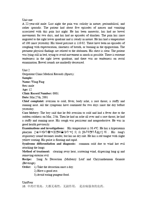
Unit oneA 22-year-old male: Last night the pain was colicky in nature, periumbilical, and rather sporadic. The patient had about five episodes of nausea and vomiting associated with this pain last night. He has been anorectic, has had no bowel movements for two days, and has had no episodes of diarrhea. The pain has since migrated to the right lower quadrant and is steady in nature. He has had a temperature of 100 since yesterday. His blood pressure is 110/82. There have been no episodes of coughing with expectoration, shortness of breath, or burning in the epigastrium. The pertinent physical findings are related to the abdomen. His chest is clear. The patient was lying still in bed, trying to avoid movement as much as possible. There is extreme tenderness in the right lower quadrant, and there was no tenderness on rectal examination. Bowel sounds are markedly decreased.Unit 2Outpatient Clinic Medical Records (9parts):Sample:Name: Wang PingSex: maleAge: 12Clinic Record Number: 0001Date: Mar.27th, 2001Chief complaint:aversion to cold, fever, body ache, a sore throat, a stuffy and running nose. All the symptoms have continued for two days since the day before yesterday.Case history: The boy said that he felt aversion to cold and had a fever due to the sudden coldness on Mar, 25th. Then he had an ache all over and a sore throat, he had a stuffy and running nose. His cough was persistent and nonproductive. He was in good health previously.Examinations and investigations:His temperature is 38.4o C. He has a hyperaemia pharynx [ ♒♋✋☐☜♊❒♓❍✋☜]充血[ ♐✌❒✋☠♦]咽. His lung's respiratory sound becomes louder, but has no dry rale. He has a red tongue with slight yellow coating. His pulse is floating and rapid.Syndrome differentiation and diagnosis:common cold due to wind hot evil attacking the lungsMethod of treatment:clearing away heat, scattering wind, dispersing lung qi and removing exterior evilRecipe:Sang Ju Decoction (Mulberry Leaf and Chrysanthemum Granule (Beverage)Order: 1) Take the decoction once a day.2) Have a good rest.3) Avoid eating pungent food.UnitFour10. 中药疗效高,大都无毒性,无副作用,是治病强身的良药。
N―乙酰基高丝氨酸内酯调节细菌与植物间互作的研究进展[权威资料]
![N―乙酰基高丝氨酸内酯调节细菌与植物间互作的研究进展[权威资料]](https://img.taocdn.com/s3/m/f90c8692ed3a87c24028915f804d2b160b4e8648.png)
N―乙酰基高丝氨酸内酯调节细菌与植物间互作的研究进展摘要:植物促生菌具有抗病和促生的潜能,在农业生产及环境保护方面都具有重要的作用。
在许多革兰氏阴性细菌中,N-乙酰基高丝氨酸内酯(AHLs)介导的群体感应(QS)系统不仅参与对细菌多种生理行为和生物学功能的调控,而且影响植物基因的表达及细菌与宿主植物间的互作。
为了更好地开发利用生防菌,综述了多年来对AHLs 跨界信号转导参与调节植物生长发育和抗逆性的研究,并对该领域今后的研究方向进行了展望,以期为改善植物抗逆性和促进生长提供一些新思路。
关键词:N-乙酰基高丝氨酸内酯(AHLs);群体感应;信号转导;生长发育;抗逆Q935 A 0439-8114(2016)14-3537-06DOI:10.14088/ki.issn0439-8114.2016.14.001Abstract: With the development of biological control, people pay more and more attention to plant promoting bacteria, which can be used as biological agent against most of fungi pathogens play an important role in agricultural production and environmental protection. In most of gram negative(G-) bacteria, N-acylhomoserine lactones (AHLs)-mediated quorum-sensing (QS) systems is not only involved in the regulation of various physiological behavior and biological function, but also affect the interaction between bacterial and host plants and the expression of plant genes. In order to better exploit and utilization of biocontrol bacteria, this paper summarized the studies of AHLs regulating plantgrowth and resistance by crossover signal transduction,while the prospects are forecasted in order to provide some new ideas for the improvementof plant resistance and growth, and hoping to improve the plant resistance and promote growth to provide some new ideas.Key words: N-acyl-homoserine lactone; quorum sensing; signal transduction; plant development;resistance随着生物防治的发展,植物促生菌日益引起人们的关注。
- 1、下载文档前请自行甄别文档内容的完整性,平台不提供额外的编辑、内容补充、找答案等附加服务。
- 2、"仅部分预览"的文档,不可在线预览部分如存在完整性等问题,可反馈申请退款(可完整预览的文档不适用该条件!)。
- 3、如文档侵犯您的权益,请联系客服反馈,我们会尽快为您处理(人工客服工作时间:9:00-18:30)。
For example, some trophically transmitted animal parasites elicit or alter the production of cues that make intermediate hosts more conspicuous to predators that are primary hosts [3]. Similarly, cues from infected hosts can also make them more conspicuous to vectors [5,6,7], including cues from plants infected with pathogens that are transmitted by insect herbivores [6,8,9]. Consequently, selection may be expected to favor plant pathogens that influence plant cues and vector behavior in ways that are conducive to pathogen acquisition and inoculation by vectors. Here we discuss recent findings suggesting that vector behavior is influenced both by pathogen effects on plant cues that facilitate discrimination between infected and healthy hosts and by direct pathogen effects on vector physiology and cue perception.
Pathogen effects on information-mediated interactions among hosts and vectors
The transmission of vector-borne pathogens requires that vectors interact with infected hosts in a manner conducive to pathogen acquisition and then subsequently interact with other, uninfected hosts in ways that lead to inoculation. Pathogens can potentially influence this process via effects on the plant or on the vector that modify the frequency and nature of interactions between them. Furthermore, because such interactions are mediated by sensory cues (Figure 1), pathogen effects on the transfer of information between hosts and vectors are likely to have important implications for transmission. Vectors of plant pathogens include pollinators [10] and herbivores (the focus of the current discussion), which rely on cues such as leaf odors to locate plants and to assess their resource value [11,12]. Pathogen infection can alter host-derived sensory cues, as well as the resource value of the host for vectors, either as part of an adaptive strategy of indirect (host-mediated) manipulation of vector behavior or as a by-product of pathology [6,8,13] (Figure 1). Some pathogens that colonize and persist in vector tissues following acquisition may also exert direct effects on vector responses to plant cues that influence the efficiency of transmission (e.g., [14,15]) (Figure 1). Given that effective transmission is critical to the fitness of vector-borne pathogens, we may assume that pathogens are frequently under selection to produce (or maintain) host phenotypes and effects on vectors that are conducive to transmission. Consequently, we might also predict some degree of convergence in the effects of vector-borne parasites that share similar modes of transmission, and hence are expected to benefit from
Available onlineects of pathogens on sensory-mediated interactions between plants and insect vectors
Kerry E Mauck1, Consuelo M De Moraes and Mark C Mescher
Current Opinion in Plant Biology 2016, 32:53–61
1
Introduction
Parasites frequently alter the phenotypes of their hosts in ways that enhance their own transmission and fitness, and such effects can have profound implications not only for parasite transmission but also for the structure and dynamics of ecological communities [1]. Work exploring the ecological implications of host manipulation has focused primarily on animal parasites [2–4], but, given the importance of plants in terrestrial ecosystems, the manipulative effects of plant parasites on host phenotypes might have equal or greater significance for ecology [1]. A key way in which parasites influence the interactions of their hosts with other organisms, and thus transmission, is by influencing potential sensory cues produced by infected hosts.