Long Noncoding RNA as Modular Scaffold of Histone Modification
Long Non-Coding RNA SNHG6 as a Potential Biomarker for Hepatocellular Carcinoma
Long Non-Coding RNA SNHG6as a Potential Biomarker for Hepatocellular CarcinomaMaryam Tahmasebi Birgani1&Mohammadreza Hajjari2&Arman Shahrisa3&Atefeh Khoshnevisan2&Zahra Shoja1&Paria Motahari4&Baharak Farhangi5Received:27August2016/Accepted:26April2017#Arányi Lajos Foundation2017Abstract Long Non-coding RNAs(lncRNAs)refer to all non-protein coding transcripts longer than200nucle-otides.Their critical roles in different biological path-ways have been already well established.Altered ex-pression of lncRNAs can be involved in the cancer ini-tiation and/or progression.Since patients with hepatocel-lular carcinoma(HCC)are usually diagnosed in late stages,developing diagnostic methods seems to be es-sential.In this study,the expression levels of different lncRNAs were systematically analysed in different ge-nomic and transcriptome datasets.The analyses showed that SNHG6is among the lncRNAs with distinctive dys-regulation of expression and copy number variation in HCC tumors compared with normal tissues.The results also suggest that the dysregulation of SNHG6is highly cancer type specific.Through co-occurrence analyses,we found that SNHG6and its related co-expressed genes on8q are involved in the structural integrity of ribosome and translation.This comprehensive in silico analysis,provides a resource for investigating SNHG6in hepatocellular carcinoma and lays the groundwork for design of next researches.Keywords Hepatocellular carcinoma.Biomarker.Long noncoding RNA.SNHG6.Systematic analysis IntroductionHepatocellular carcinoma(HCC)is one of the most common cancers worldwide[1].The viral infection(HBV and HCV), alcoholism,non-alcoholic fatty liver,and some hereditary metabolic diseases are the main recognized risk factors for HCC[2–5].Since most of the HCC patients are diagnosed at late stages,when medication is no longer effective,the discovery of sensitive and specific biomarkers for early diag-nosis and treatment is of great attention[6].Long non-coding RNAs(lncRNAs)refer to all lengthy functional transcripts which are actively in-volved in numerous biological processes such as regu-lation of transcription,translation,protein localization, and function,as well as orchestration of cellular scaf-fold.Furthermore,lncRNAs can control the cell cycle, differentiation,apoptosis,and DNA repair through the modulation of epigenome[7,8].Owing to these func-tions,it is not surprising if the aberrant expressions of lncRNAs contribute to disease pathogenesis.The altered expression of lncRNAs in different malignancies along with their tissue-specific expression suggests that these long transcripts may be considered as great biomarkers in cancer diagnosis[9–12].The potential role ofMaryam Tahmasebi Birgani,Mohammadreza Hajjari and Arman Shahrisa contributed equally to this work.*Maryam Tahmasebi Birganitahmasebi-ma@ajums.ac.ir*Mohammadreza Hajjarim-hajari@scu.ac.ir1Department of Medical Genetics,School of Medicine,Ahvaz Jundishapur University of Medical Sciences,Ahvaz,Iran2Department of Genetics,Faculty of Sciences,Shahid Chamran University of Ahvaz,Ahvaz,Iran3Department of Molecular Genetics,Faculty of Biosciences,Tarbiat Modares University,Tehran,Iran4Department of Biotechnology,Iranian Research Organization Science&Technology,Tehran,Iran5Cancer Research Center,Tehran University of Medical Sciences, Tehran,IranlncRNAs in liver malignancies has been recently led into promising novel insights in HCC therapeutic strat-egies [13,14].Therefore,more studies are needed to elucidate the role of lncRNAs in HCC.In recent decades,systematic analyses of the genomic,transcriptomic,and proteomic datasets have become powerful tools in the discovery and the validation of tumor markers [15].Due to the lack of enough supporting evidence to asso-ciate the lncRNAs with HCC,the present study was aimed to perform a systematic genotranscriptomic meta-analysis of the issue.We tried to screen different genomic and transcriptomic datasets in order to find the potential of lncRNAs as prognos-tic and diagnostic tools for HCC.The systematic results can help the researchers with further studies on specific lncRNAs in order to develop predictive biomarkers or therapeutic tar-gets.In the current study,we found that SNHG6,PVT1,and GAS5are potential lncRNAs with a significant role in HCC initiation and progression.Materials and MethodsThe Selection of lncRNAs with High Alteration Frequency in HCC SamplesTo investigate the significance of lncRNAs in liver ma-lignancy,we retrieved 189approved lncRNAs from HGNC ( ).All of the genes wereinterrogated into the cBioPortal database ( )[16,17]for geno/transcriptomic analyses.We queried all the samples from TCGA liver hepatocel-lular carcinoma (TCGA,provisional)with RNA-seq v2data (n =373)in our study and considered RNA dys-regulation with Z-score threshold:±2.TCGA data,as one of the major national and international efforts,in-clude the valid comprehensive data derived from large cohorts.Among different HCC data indexed in TCGA,TCGA liver hepatocellular carcinoma constitutes more number of tissues.The lncRNAs which were altered in more than 10%of the patients were considered as B significant lncRNAs ^for further analyses.It should not be forgot-ten that it was important for us to consider the lncRNAs which were altered at both genomic and transcriptomic levels even if the percent of alteration in patients was near to the threshold of 10%.Regarding to these criteria,these lncRNAs included SNHG6,PVT1,and GAS5due to high levels of alter-ation in both genomic and transcriptomic levels.By means of the R package,the frequency of each genetic alteration was calculated among the cases carrying at least one alteration for the desired genes.Additionally,the co-occurrence between genetic alterations was con-sidered for all of the three genes.The Differential Expression of Significant lncRNAs between Tumor and Normal TissuesTCGA RNA-Seq raw data was extracted in R using the cgdsr extension package (/web/packages/cgdsr /)with a threshold of ±2.The data was then presented as Heatmap plot.The selected lncRNAs were examined in several transcriptomic datasets to explore if any significant difference of expression may exist between normal and tumor tissues.The included datasets were Oncomine ( ),Gene Expression Atlas [18–20],Gene Expression Omnibus:GEO (/geo ),Array express (https:///arrayexpress ),and UCSC cancer genome browser (https:// )[21–26]databases.The results with Fold change > 1.5and P -value <0.01between tumor and normal tissues were considered as significant.In the next step,we also considered any correlation for any pair of genes of interest using Pearson ’s method.In strong positive correlation,the linear correlation coefficient (r)is close to +1;while in strong negative,the correlation is close to −1.Fig.1Genomic alterations (Copy number Variations)of lncRNAs among 373patients with hepatocellular carcinoma.The chart was drawn for the genes which were altered in more than 10%of the patients with hepatocellular carcinoma.The data obtained from cBioPortal ( )Birgani M.T.et al.The Association between lncRNAsand the Clinicopathologic Parameters of Hepatocellular CarcinomaUsing the UCSC Cancer Genome Browser,the asso-ciation of different lncRNAs with clinicopathologic parameters (the histological type,pathologically TNM staging,and grade)was evaluated using Student ’s t -test.Besides,the effect of gene expression dysregula-tion on the patient ’s survival was evaluated using the Kaplan-Meier analysis in cBioPortal.The Log-Rank Test P -Value <0.05was considered as statistically significant.The Comparison of the HCC Profile of lncRNAs with the Profiles of Other CancersIn order to evaluate whether significant lncRNAs fol-low an HCC-specific manner,we examined the geno/transcriptomic alteration of these genes in all of the tumor collections of cBioPortal.Thirty cancers with available RNA-seq data were included at this stage,and the expression data corresponding to genes were extracted.The raw data was filtered based on the z-score >+2and <−2.The mean of the expression levels was calculated using R and the data was presented asheatmaps.Fig.2Altered expression of different lncRNAs in hepatocellular carcinoma with Z score >±2.Sixty out of 189genes were altered among 373patients with HCC.The analysis is done by the R statistical software.The raw data was extracted from cBioPortal ( )Long Non-Coding RNA SNHG6as a potential BiomarkerThe Functional Analysis of Selected lncRNAsAmong 373patients with hepatocellular carcinoma,130cases showed alterations for SNHG6,PVT1,and GAS5.Among these 130cases,we evaluated the genes which were co-expressed with significant lncRNAs using Pearson ’s correla-tion analysis.Then,the genes with correlation value >0.70were uploaded into the GSEA dataset ( )to compute the gene set overlaps matrix based on the GO molecular ing the cBioPortal database,we also drew a network to find any potential contribution of the genes which were co-expressed with sig-nificant lncRNAs.Statistical AnalysisAll of the analyses including the t-test,heatmap,and correla-tion analysis were done by the R statistical software and SPSS.In our analysis,the P -values less than 0.05were considered significant.ResultsSignificant lncRNAs with a Potential Role in Liver MalignanciesAlthough we could not find any mutations in lncRNAs of interest,67out of the 189lncRNAs were found to be altered in their copy numbers at least in one patient.LncRNAs,in-cluding CASC8,PCAT1,PVT1,CCAT1,F ALEC,and GAS5,were the genes whose copy numbers were altered in more than 10%of the patients (Fig.1).We also found 60lncRNAs with altered expression patterns at least in 1%of patients (Fig.2).However,SNHG6,PVT1,and GAS5were the lncRNAs with the highest RNA dysregulation among the 373samples of HCC (Fig.3).For further analysis,we focused on SNHG6,PVT1,and GAS5in which both CNVs (Copy number varia-tions)and RNA dysregulation were higher than other lncRNAs.It is necessary to mentioned that we granted an exception for selection of SNHG6due to some reasons;1)among 189lncRNAs,SNHG6allocated the highest score in total alteration regardless of whether alterations occur atge-Fig.3The expression levels of SNHG6,PVT1,and GAS5among 373patients the patients with hepatocellular carcinoma. a.The chart shows the genes which were altered in more than 10%of the patients with hepatocellular carcinoma. b.The expression levels of SNHG6,PVT1,and GAS5among 130patients with Z score >±2was presented as heatmap.These patients have at least on alteration in SNHG6,PVT1,and GAS5lncRNAs.The analysis is done by the R statistical software.The raw data was extracted from cBioPortal ( )Table 1The frequency of genetic alterations (CNV)of SNHG6,PVT1and GAS5among 373cases of hepatocellular carcinoma using R analysisGAS5SNHG6PVT1Not altered Duplication Not altered Duplication Not altered Duplication Homodeletion 3373634033307651Birgani M.T.et al.nomic or transcriptomic level.2)among 189lncRNAs,SNHG6allocated the highest score at transcriptomic level so although SNHG6apportioned the 9%alteration among the patients at genomic level but due to the two previous reasons,we decided to continue our analysis on it as well as PVT1and GAS5.On the other hand,when we compared the data at both level of genomic and transcriptomic,SNHG6,PVT1and GAS5were common in data of two groups.Regarding to your true comments,we explain clearly the reason of SNHG6selection (in spite of 9%alteration)in the manuscript.In our results,around 35%of the cases (n =130)had an RNA dysregulation in at least one gene,and SNHG6was altered in more cases than PVT1and GAS5.We called these patients B target samples ^in the next steps.The co-occurrence of different genetic alterations for each paired loci was calcu-lated among this group (Table 1).SNHG6,PVT1,and GAS5are Differentially Expressed between Tumor and Normal TissuesAccording to the Cancer Genome Browser,SNHG6,PVT1,and GAS5are significantly upregulated in hepatocellular car-cinoma in comparison with the normal counterparts (P -val-ue =0.001)(Fig.4).Furthermore,the oncomine datasets con-firmed the differential expression of these lncRNAs between cancerous and normal tissues (Table 2).The Overexpression of SNHG6is a Novel Indicator of Reduced Survival in Patients with Hepatocellular CarcinomaWe found that the over-expression of SNHG6in hepato-cellular carcinoma was nearly associated with reduced survival to a median of 19.74months in SNHG6over-Fig.4TCGA hepatocellular carcinoma (LIHC)gene expression by RNA seq (IlluminaHiseq N =423)and the differential expression of SNHG6,PVT1,and GAS5betweenthe primary tumor and the solid normal tissue.The statistical track displayed under the genomic heatmap shows the logarithmic plot of p -values for each genomic position,where the center line indicates a p -value of 1.The primary tumor tissue and the solid normal tissue subgroups were illustrated in red and greenrespectively.The student t -Test was performed to analyze the differential-ly expressed genes between the tumor cells and the normal ones,and P <0.05was considered statistically significant.The red bar indicates that the expression levels of genes are significantly higher in cancerous cells than normal ones.The data was extracted from Cancer Genome BrowserTable 2The differential expression of lncRNAs SNHG6,PVT1and GAS5between normal and tumour samples (Oncomine,the gene expression atlas,and GEO)lncRNA Fold change P value Down-regulatedUp-regulated Experiment typeRef SNHG6 1.983 3.43E-5Non-Tumour Tissue (n =10)HCC (n =35)Human Genome U133Plus 2.0Array [27]PVT1 4.258 1.88E-11Non-Tumour Tissue (n =10)HCC (n =35)Human Genome U133Plus 2.0Array [27]GAS52.692.3E-8Non-Tumour Tissue (n =10)HCC (n =35)Human Genome U133Plus 2.0Array[27]Long Non-Coding RNA SNHG6as a potential Biomarkerexpressing cases,compared with the period of over 48.95months for the remaining cases (Logrank Test P -Value:0.0558)(Fig.5a ).Although the value was not sig-nificant for PVT1and GAS5,studying the clinical associ-ations of these lncRNAs with clinicopathologic parame-ters of HCC represented that all the three genes (SNHG6,PVT1and GAS5)were significantly expressed in high-grade HCC samples compared with low-grade tissues (P -value =0.001)(Fig.5b ).The Association of SNHG6,PVT1,and GAS5with the Progression of Other Human CancersWe evaluated the transcriptomic alterations of these genes among different human tumors.We considered the genes at two levels;first,their frequency among the patients and sec-ond,their related expression among them.In comparison with PVT1and GAS5,SNHG6allocated a good value to itself at both levels.Therefore,SNHG6seems to be astrongerFig.5Study of the clinicalassociation of SNHG6,PVT1,and GAS5with the clinicopathologic parameters of hepatocellular carcinoma.a Kaplan-Meier plots comparing the overall survival in cases with or without SNHG6over-expression.The data was recruited from cBioPortal.b The association of expression of lncRNAs with the histological grade of HCC;we divided tissues based on their grade (G1–2and G3–4as green and red bars re-spectively),which shown in right column.The statistical track which is displayed under the ge-nomic heatmap shows the loga-rithmic plot of p -values for each lncRNA,where the centreline in-dicates a p -value of 1.The bar above the line indicates that the red subgroup (G3–4)is greater than the green subgroup (P -val-ue <0.05).The patients with no pathological data (NA)were omitted from the analysis.The data was extracted from Cancer Genome Browser,TCGA LIHC gene expression by RNA seq (IlluminaHiseq n =423)Birgani M.T.et al.potential biomarker for liver hepatocellular carcinoma in com-parison with PVT1and GAS5.However,PVT1and GAS5can be useful for other cancers such as Uterine Carcinosarcoma (Fig.6).SNHG6is Possibly Involved in the Structural Integrity of Ribosome and TranslationThrough co-occurrence analyses,we found that SNHG6and GAS5have a tendency toward co-occurrence among target samples (n =130),although it was not significant (Table 3).We considered all the genes which had been co-expressed with SNHG6and GAS5.Pearson value >0.7was taken as the puting the gene set overlaps matrix based on the GO molecular function showed that 13of these genes act as molecules contrib-uting to the structural integrity of the ribosome (Table 4).Interestingly,drawn from cBioPortal data,we found that 35%(n =130)of the patients among the target samples had the alteration at least for one of these genes.Among the genes,RPL8,TOP1MT,and RPL30were the top ones which were altered among the patients.We then visualized SNHG6,its co-expressed genes,and the most frequently altered neigh-bour genes network to investigate any probable mode of interaction.Although we did not observe any interaction between SNHG6and these genes,most of the high fre-quently altered genes were located on 8q,near the SNHG6locus (data not shown).DiscussionAlthough the dysregulation of lncRNAs has been report-ed in some studies,their functional mechanism remains to be challenging.Here is a report consideringtheFig.6The genetic alterations of SNHG6,PVT1,and GAS5amongdifferent human cancers.a The frequency of SNHG6,PVT1and GAS5genetic alterations among the patients of 30cancers with available RNA-seq on the portal.b The heatmap of the mean expression levels of SNHG6,PVT1and GAS5among 30cancers with RNA-seq.The heatmaps were drawn using the R software and the raw data of cBioPortal.The grey column represents cancer with no data in case of the gene of interest in the portalTable 3The mutual exclusivity analysis of SNHG6,PVT1and GAS5among 130tumor samples with alterations at least in one gene.The data was recruited from cBioPortal Gene Pairp -V alue Log Odds Ratio AssociationSNHG6PVT1<0.001−1.821Tendency towards mutual exclusivity (significant)SNHG6GAS5<0.0010.344Tendency towards co-occurrencePVT1GAS5<0.001−0.189Tendency towards mutual exclusivityLong Non-Coding RNA SNHG6as a potential Biomarkercontribution of lncRNAs to hepatocellular carcinoma using available bioportals.We queried189approved lncRNAs in cancer gene expression datasets.It was ob-served that the expression levels of68lncRNAs were altered at least in one case.We chose SNHG6,PVT1, and GAS5as lncRNAs with high levels of alteration. We also observed that SNHG6,PVT1,and GAS5were differentially expressed between the HCC tumors tissues and normal tissues.It was also found that SNHG6allo-cated the most RNA dysregulation in cancerous tissues. Although all the three genes were associated with high grades of HCC,the SNHG6up-regulation was more correlated with the shorter survival of patients. However,this value was on the borderline of the statis-tical significance.In a report by Liu et al.,the potential involvement of of SNHG6in portal vein tumor throm-bus,tumor stage,metastasis,and the shorter overall sur-vival of HCC patients was experimentally confirmed [28].The expression and mutation analyses of desired lncRNAs in other human tumours showed the SNHG6 as a potential biomarker.To study the possible function of SNHG6,we recruited all the genes that were co-expressed with it.We classified the co-expressed genes based on the GO molecular function.Interestingly,we found that some of these genes were involved in ribo-some structure/translation and altered in around35%of our analysed HCC samples.Additionally,we found that most of the high frequently altered genes were located on8q21–24,near the SNHG6locus.This data showed that8q may be associated with HCC.There are merely a few studies showing the role of some ribosomal genes including RP36A,RP44in HCC progression [29].It seems that ribosomal proteins are capable to control the gene expression by preparing a selectivity for translating ribosomes[30].The role of the chromosomal alteration,espe-cially8q24in HCC samples,has been the subject of several studies[31].It has been confirmed that this region encodes several lncRNAs involved in tumorogenesis[32].Altogether, this data introduced SNHG6as a good candidate for experi-mental works in HCC researches.However,clinical experi-ments are urgent to evaluate the molecular role of SNHG6in HCC progression as well as its specificity and sensitivity as a biomarker of HCC.Acknowledgments This study was affiliated to Ahvaz Jundishapur University of Medical Sciences and Shahid Chamran University of Ahvaz,Iran.Compliance with Ethical StandardsConflict of Interest The authors declare no conflict of interest. References1.Wang C-H,Wey K-C,Mo L-R,Chang K-K,Lin R-C,Kuo J-J(2014)Current trends and recent advances in diagnosis,therapy, and prevention of hepatocellular n Pac J Cancer Prev16:3595–36042.Mazzocca A,Tahmasebi Birgani M,SabbàC,Carloni V(2014)Tetraspanin-enriched Microdomains and hepatocellular carcinoma progression.Cancer Lett351(1):23–293.Su C-H,Lin Y,Cai L(2013)Genetic factors,viral infection,otherfactors and liver cancer:an update on current n Pac J Cancer Prev14:4953–49604.de Oliveria Andrade LJ,D'Oliveira A,Melo RC,De Souza EC,Costa Silva CA,Parana R(2009)Association between hepatitis C and hepatocellular carcinoma.J Glob Infect Dis1:33–375.El-Serag HB(2012)Epidemiology of viral hepatitis and hepatocel-lular carcinoma.Gastroenterology142(1264–1273):e12616.Zhu K,Dai Z,Zhou J(2013)Biomarkers for hepatocellular carci-noma:progression in early diagnosis,prognosis,and personalized therapy.Biomarker research1:107.Hajjari M,Khoshnevisan A,Shin YK(2014)Molecular functionand regulation of long non-coding RNAs:paradigms with potential roles in cancer.Tumor Biol35:10645–10663Table4TheGeneSet analysis of the genes that are commonly co-expressed with SNHG6and GAS5,based on the GO molecular function.The data was extracted from GSEAGene Set Name Genes in GeneSet(K)Description Genes inOverlap(k)k/K p-value FDR q-valueStructural constituent of ribosome 80Genes annotated by the GO term GO:0003735The action of a molecule thatcontributes to the structural integrityof the ribosome130.1625 3.37E-26 1.33E-2Structural molecule activity244Genes annotated by the GO term GO:0005198The action of a molecule thatcontributes to the structural integrityof a complex or assembly within oroutside a cell130.0533 1.21E-19 2.40E-17RNA Binding259Genes annotated by the GO term GO:0003723Interacting selectively with an RNAmolecule or a portion thereof 60.0309 1.64E-10 2.17E-08Birgani M.T.et al.8.Yarmishyn AA,Kurochkin IV(2015)Long noncoding RNAs:apotential novel class of cancer biomarkers.Front Genet6:1–10 9.Ayers D(2013)Long non-coding RNAs:novel emergent bio-markers for cancer diagnostics.Nature1:31–3510.Hajjari M,Behmanesh M,Sadeghizadeh M,Zeinoddini M(2013)Up-regulation of HOTAIR long non-coding RNA in human gastric adenocarcinoma tissues.Med Oncol30:1–411.Hajjari M,Khoshnevisan A(2013)Potential long non-codingRNAs to be considered as biomarkers or therapeutic targets in gas-tric cancer.Front Genet4:1–312.Hajjari M,Khoshnevisan A,Shin YK(2013)Long non-codingRNAs in hematologic malignancies:road to translational research.Front Genet4:1–213.Shibata C,Otsuka M,Kishikawa T,Ohno M,Yoshikawa T,TakataA,Koike K(2015)Diagnostic and therapeutic application of non-coding RNAs for hepatocellular carcinoma.World J Hepatol7:1–6 14.Fang T-T,Sun X-J,Chen J,Zhao Y,Sun R-X,Ren N,Liu B-B(2014)Long non-coding RNAs are differentially expressed in he-patocellular carcinoma cell lines with differing metastatic potential.Asian Pac J Cancer Prev15:10513–1052415.Goossens N,Nakagawa S,Sun X and Hoshida Y(2015)Cancerbiomarker discovery and validation.Transl Cancer Res4(3):256–26916.Cerami E,Gao J,Dogrusoz U,Gross BE,Sumer SO,Aksoy BA,Jacobsen A,Byrne CJ,Heuer ML,Larsson E,Antipin Y,Reva B, Goldberg AP,Sander C,Schultz N(2012)The cBio cancer geno-mics portal:an open platform for exploring multidimensional can-cer genomics data.Cancer Discov2:401–40417.Gao J,Aksoy BA,Dogrusoz U,Dresdner G,Gross B,Sumer SO,Sun Y,Jacobsen A,Sinha R,Larsson E(2013)Integrative analysis of complex cancer genomics and clinical profiles using the cBioPortal.Sci Signal6:l118.Petryszak R,Keays M,Tang YA,Fonseca NA,Barrera E,BurdettT,Füllgrabe A,Fuentes AM-P,Jupp S,Koskinen S(2015) Expression Atlas update—an integrated database of gene and pro-tein expression in humans,animals and plants.Nucleic Acids Res 44(D1):D746–D75219.Petryszak R,Burdett T,Fiorelli B,Fonseca NA,Gonzalez-Porta M,Hastings E,Huber W,Jupp S,Keays M,Kryvych N,McMurry J, Marioni JC,Malone J,Megy K,Rustici G,Tang AY,Taubert J, Williams E,Mannion O,Parkinson HE,Brazma A(2014) Expression atlas update–a database of gene and transcript expres-sion from microarray-and sequencing-based functional genomics experiments.Nucleic Acids Res42:D926–D93220.Kapushesky M,Adamusiak T,Burdett T,Culhane A,Farne A,Filippov A,Holloway E,Klebanov A,Kryvych N,Kurbatova N, Kurnosov P,Malone J,Melnichuk O,Petryszak R,Pultsin N, Rustici G,Tikhonov A,Travillian RS,Williams E,Zorin A, Parkinson H,Brazma A(2012)Gene expression atlas update–avalue-added database of microarray and sequencing-based func-tional genomics experiments.Nucleic Acids Res40:D1077–D1081 21.Cline MS,Craft B,Swatloski T,Goldman M,Ma S,Haussler D,Zhu J(2013)Exploring TCGA pan-cancer data at the UCSC cancer genomics browser.Sci Rep3:265222.Lopez B,Cline M,Broom B,Margolin A,Omberg L,Weinstein J,Axton M(2013)Thread4:data discovery,transparency and visu-alization.Nat Genet45.doi:10.1038/ng.278923.Goldman M,Craft B,Swatloski T,Ellrott K,Cline M,Diekhans M,Ma S,Wilks C,Stuart J,Haussler D,Zhu J(2013)The UCSC cancer genomics browser:update2013.Nucleic Acids Res41: D949–D95424.Sanborn JZ,Benz SC,Craft B,Szeto C,Kober KM,Meyer L,Vaske CJ,Goldman M,Smith KE,Kuhn RM,Karolchik D,Kent WJ,Stuart JM,Haussler D,Zhu J(2011)The UCSC cancer geno-mics browser:update2011.Nucleic Acids Res39:D951–D959 25.Vaske CJ,Benz SC,Sanborn JZ,Earl D,Szeto C,Zhu J,HausslerD,Stuart JM(2010)Inference of patient-specific pathway activities from multi-dimensional cancer genomics data using PARADIGM.Bioinformatics26:i237–i24526.Zhu J,Sanborn JZ,Benz S,Szeto C,Hsu F,Kuhn RM,KarolchikD,Archie J,Lenburg ME,Esserman LJ,Kent WJ,Haussler D, Wang T(2009)The UCSC cancer genomics browser.Nat Methods6:239–24027.Wurmbach E,Yb C,Khitrov G,Zhang W,Roayaie S,Schwartz M,Fiel I,Thung S,Mazzaferro V,Bruix J(2007)Genome-wide mo-lecular profiles of HCV-induced dysplasia and hepatocellular carci-noma.Hepatology45:938–94728.Cao C,Zhang T,Zhang D,Xie L,Zou X,Lei L,Wu D,Liu L(2016)The long non-coding RNA,SNHG6–003,functions as a competing endogenous RNA to promote the progression of hepatocellular car-cinoma.Oncogene36:1112–112229.Kim JH,You KR,Kim IH,Cho BH,Kim CY,Kim DG(2004)Over-expression of the ribosomal protein L36a gene is associated with cellular proliferation in hepatocellular carcinoma.Hepatology 39:129–13830.Wong QW-L,Li J,Ng SR,Lim SG,Yang H,Vardy LA(2014)RPL39L is an example of a recently evolved ribosomal protein paralog that shows highly specific tissue expression patterns and is upregulated in ESCs and HCC tumors.RNA Biol11:33–41 31.Weber RG,Pietsch T,von Schweinitz D,Lichter P(2000)Characterization of genomic alterations in hepatoblastomas:a role for gains on chromosomes8q and20as predictors of poor outcome.Am J Pathol157:571–57832.Xiang J-F,Yin Q-F,Chen T,Zhang Y,Zhang X-O,Wu Z,Zhang S,Wang H-B,Ge J,Lu X(2014)Human colorectal cancer-specific CCAT1-L lncRNA regulates long-range chromatin interactions at the MYC locus.Cell Res24:513–531Long Non-Coding RNA SNHG6as a potential Biomarker。
lncRNA作用机制
lncRNA 在肿瘤中的研究现状长链非编码RNA(long non-coding RNA, lncRNA)是一类转录本长度超过200nt、不编码蛋白的RNA,这类RNA起初被认为是基因组转录的“噪音”,随着2007年Hotair功能的被发掘,lncRNA的功能渐渐明晰。
据计算,约有93%的转录本为lncRNA1,lncRNA通常位于细胞核和细胞质。
但是lncRNA的基因转录水平一般低于蛋白质编码基因,序列保守性差,承受的进化压力小,但promoter序列通常比较保守。
lncRNA与小分子RNA相比,序列更长、空间结构也较为复杂,参与表达调控的机制也更具有多样性和复杂性。
尽管目前只有一小部分lncRNA的功能有相关报道,但可以明确的是lncRNA参与发育、分化、代谢等多方面的调控。
lncRNA能在表观遗传2、转录3及转录后4水平调控基因表达,参与X染色体沉默、基因组印记以及染色质修饰、转录激活与抑制、核内运输等多种重要的调控过程,与人类疾病的发生、发展和防治都有着密切联系。
研究表明,lncRNA的异常表达与肿瘤的诊断、复发及转移相关5。
lncRNA 以外泌体的形式分泌到细胞外,体液中的lncRNA具有作为生物标志物的潜能,指示肿瘤的进展与恶性程度,指导个性化治疗。
PCA3是一个前列腺癌特异表达lncRNA,在前列腺癌患者尿液中异常升高,已经用于临床前列腺癌诊断6。
血浆中稳定存在的lncRNA也有作为生物标志物的潜能,比如胃癌患者血浆中lncRNA H19显著升高7。
在临床,相同癌症患者接受相同的治疗方式,但往往会表现出不同的临床后果,lncRNA的差异表达是造成这一现象的原因之一8。
癌组织中lncRNA的异常表达通常与转移及预后较差相关。
在胰腺癌中lncRNA HULC表达异常升高,其异常高表达与肿瘤体积、高级别的淋巴结转移与血管浸润显著相关,HULC水平与患者的总体生存率相关9。
Hotair在乳腺、结直肠癌、宫颈癌等多种癌症中表达升高;在宫颈癌中,Hotair的高表达与淋巴结转移相关,且总体生存率较低;通过相应的细胞生物学实验表明,敲减Hotair能够显著抑制宫颈癌细胞的增殖、迁移与侵袭,过表达能引起EMT相关表型10。
长链非编码RNA在多发性硬化发病机制中的研究进展
长链非编码RNA在多发性硬化发病机制中的研究进展①巩盼盼柳宁武世萍俞坤王满侠(兰州大学第二医院神经内科,兰州730000)中图分类号R741 文献标志码 A 文章编号1000-484X(2023)09-1993-05[摘要]多发性硬化(MS)作为一种常见的中枢神经系统炎症脱髓鞘疾病,其发病机制研究尚不清楚。
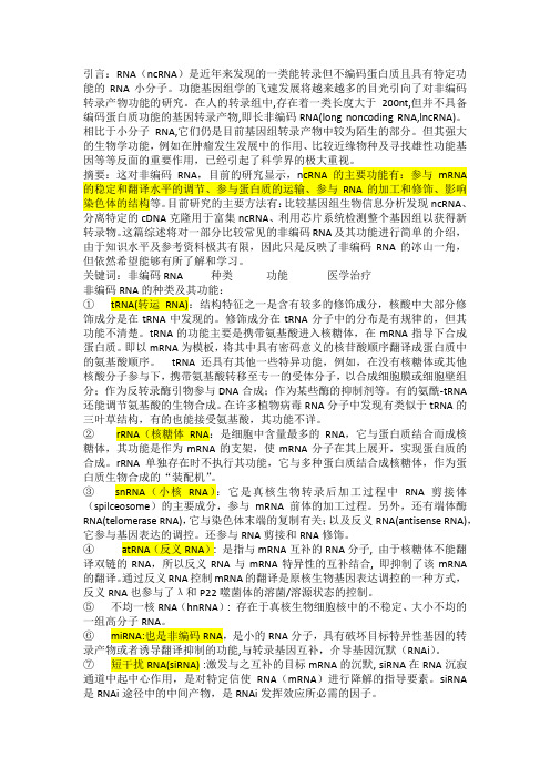
目前长链非编码RNA(lncRNA)虽无蛋白质编码功能,但它在疾病的免疫调节中发挥重要作用。
近年来,在MS患者及实验性自身免疫性脑脊髓炎(EAE)动物模型中发现了大量异常表达的lncRNA。
在本文中,将MS及EAE模型中涉及的lncRNA作一简单小结,着重讨论lncRNA在MS发病机制中的研究现状。
[关键词]长链非编码RNA;多发性硬化;实验性自身免疫性脑脊髓炎;免疫细胞Research progress of long non-coding RNA in pathogenesis of multiple sclerosis GONG Panpan, LIU Ning, WU Shiping, YU Kun, WANG Manxia. Department of Neurology, Lanzhou University Second Hospital, Lanzhou 730000, China[Abstract]Multiple sclerosis (MS) is an inflammatory demyelinating disease of the central nervous system, specific pathogen‐esis of which is still unclear. Long chain non-coding RNA(lncRNA) without protein coding function, which were found in playing im‐portant functions in many diseases. In recent years, some patients with MS and animal model of experimental autoimmune encephalo‐myelitis (EAE) were discovered that expressed a large number of abnormal lncRNA. In this paper, lncRNA involved in MS and EAE models are summarized briefly, focusing on the current research status of lncRNA in the pathogenesis of MS.[Key words]Long non-coding RNA;Multiple sclerosis;Experimental autoimmune encephalomyelitis;Immune cell多发性硬化(multiple sclerosis,MS)是一种慢性中枢神经系统的自身免疫性疾病。
RNA pull-down assay

RNA pull-down assayLong Noncoding RNA as Modular Scaffold of Histone Modification ComplexesMiao-Chih Tsai, et al.RNA pull-down assay using nuclear extract1. Biotin-labeled RNAs were in vitro transcribed with the Biotin RNA Labeling Mix (Roche) and T7 RNA polymerase (Promega), treated with RNase-free DNase I (Promega) and purified with RNeasy Mini Kit (QIAGEN).2. Biotin-HRP Northern blot was followed by manufacture’s manual (NorthernMax kit, Ambion) to demonstrate that all the RNAs are biotinylated and transcribed at the right size.3. Three micrograms of biotinylated RNA was heated to 90°C for 2 minutes, put on ice for 2 minutes, supplied with RNA structure buffer (10 mM Tris pH 7, 0.1 M KCl, 10 mM MgCl2), and then shifted to room temperature (RT) for 20 minutes to allow proper secondary structure formation.4. 107HeLa cell pallets were resuspended in 2 ml PBS, 2 ml nuclear isolation buffer (1.28 M sucrose; 40 mM Tris-HCl pH 7.5; 20 mM MgCl2; 4% Triton X-100), and 6 ml water on ice for 20 min (with frequent mixing).5. Nuclei were pelleted by centrifugation at 2,500 G for 15 min.6. Nuclear pellet was resuspended in 1 ml RIP buffer (150 mM KCl, 25 mM Tris pH7.4, 0.5 mM DTT, 0.5% NP40, 1 mM PMSF and protease Inhibitor (Roche Complete Protease Inhibitor Cocktail Tablets)).7. Resuspended nuclei were mechanically sheared using a dounce homogenizer with 15–20 strokes.8. Nuclear membrane and debris were pelleted by centrifugation at 13,000 RPM for 10 min.9. Folded RNA was then mixed with 1mg of HeLa nuclear extract in RIP buffer and incubated at RT for one hour.10. Sixty microliters washed Streptavidin agarose beads (Invitrogen) were added to each binding reaction and further incubated at RT for one hour.11. Beads were washed briefly five times in Handee spin columns (Pierce) and boiled in SDS buffer, and the retrieved protein was detected by standard western blot technique.RNA pull-down assay using recombinant proteinsPRC2 complex were obtained from BPS Bioscience (5m: 51004; 3m: 51003). LSD1 complex () was purified by tandem affinity purification from HeLa S3 cells as previously described (2). The complex contained known components (LSD1, BHC80, CoREST, HDACs, and BRAF35) in nearly stoichiometric amounts. Recombinant GST-LSD1 and 6xHis- CoREST were purified as described (2). Recombinant Flag-LSD1 was purified from Sf9 insect cells using M2-agarose affinity chromatography as previously described (5).1. Biotin-labeled RNAs were in vitro transcribed with the Biotin RNA Labeling Mix (Roche) and T7 RNA polymerase (Promega), treated with RNase-free DNase I (Promega) and purified with RNeasy Mini Kit (QIAGEN).2. Biotin-HRP Northern blot was followed by manufacture’s manual (NorthernMax kit, Ambion) to demonstrate that all the RNAs are biotinylated and transcribed at the right size.3. Three micrograms of biotinylated RNA was heated to 90°C for 2 minutes, put on ice for 2 minutes, supplied with RNA structure buffer (10 mM Tris pH 7, 0.1 M KCl, 10 mM MgCl2), and then shifted to room temperature (RT) for 20 minutes to allow proper secondary structure formation.4. 107HeLa cell pallets were resuspended in 2 ml PBS, 2 ml nuclear isolation buffer (1.28 M sucrose; 40 mM Tris-HCl pH 7.5; 20 mM MgCl2; 4% Triton X-100), and 6 ml water on ice for 20 min (with frequent mixing).5. Nuclei were pelleted by centrifugation at 2,500 G for 15 min.6. Nuclear pellet was resuspended in 1 ml RIP buffer (150 mM KCl, 25 mM Tris pH7.4, 0.5 mM DTT, 0.5% NP40, 1 mM PMSF and protease Inhibitor (Roche Complete Protease Inhibitor Cocktail Tablets)).7. Resuspended nuclei were mechanically sheared using a dounce homogenizer with 15–20 strokes.8. Nuclear membrane and debris were pelleted by centrifugation at 13,000 RPM for 10 min.9. 0.1 microgram of biotinylated RNA was incubated with different amounts of indicated proteins (2.5 and 5 µg of PRC2-5m and PRC2-3m; 3 and 5 µl of ; 0.4 and 4 µg of GST-LSD1; 1 and 5 µg of Flag-LSD1; 0.4 and 4 µg of His-CoREST).13. RNA and proteins were added in 200 µl binding buffer (50 mM TrisCl 7.9, 10% Glycerol, 100 mM KCl, 5 mM MgCl2, 10 mM β-ME 0.1% NP- 40) and incubated at RT for one hour.10. For Fig S3B experiment, 3 µl DNase I (Invitrogen, 18047- 019) was added incubated at 37 °C for 30 minutes, and then follow the IP process.11. Twenty microliters washed Streptavidin agarose beads (Invitrogen) were added to each binding reaction and further incubated at RT for 30 minutes.12. Beads were washed briefly five times in Handee spin columns (Pierce) and boiled in SDS buffer, and the retrieved protein was detected by standard Western blot technique.。
蔬菜作物中长链非编码RNA的研究进展

※农业科学农业与技术2021, Vol. 41, No. 101蔬菜作物中长链非编码RNA 的研究进展沈锦纯张琳淳李越赵竑博(华南农业大学园艺学院,广东广州510642)摘 要:长链非编码RNA (long non-coding RNA , IncRNA )是指长度超过200个核苷酸,不具备或具备极低编 码蛋白质能力的RNA 分子。
近年研究发现,植物中的lncRNA 数量种类繁多,作用机制复杂,已成为分子生物学 和遗传学研究的热点。
本文介绍了 lncRNA 的类型,概述了 lncRNA 在调控蔬菜作物的生殖发育、果实成熟以及应对生物和非生物胁迫响应等方面的作用,并总结了相关的分子机理。
对未来蔬菜作物lncRNA 研究的发展方向提出了展望。
关键词:长链非编码RNA ;蔬菜;作用机制;生物学功能中图分类号:S-3 文献标识码:A 高通量基因测序和转录组研究表明,真核生物基 因组中只有极少数的转录本可以用于蛋白质的编码,仅占全基因组的1%〜2%,而剩下的大部分转录本是 非编码 RNA (non-codingRNA, ncRNA)⑴。
ncRNA曾被认为是不具有特定生物学功能的“转录噪声”⑵,但随着转越来越多的ncRNA 被发现和鉴定,许多ncRNA 已被证明在生物体的生长发育中具有关键功能[3-6]。
根据转录本的长度,ncRNA 分为小于200 bp 的短链非编码RNA ( small non-coding RNA )和大于200 bp 的长链非编码 RNA (long non-coding RNA , ln- cRNA):7,8:o 与得到广泛研究的短链非编码RNA 相比,长非编码RNA (lncRNA)的研究才刚刚处于起步阶段,属于研究尚不足够的一类非编码转录本⑼。
本文主要介绍了 lncRNA 的类型和生物学特性, 总结了 lncRNA 在蔬菜作物生命历程中发挥的各种生物学功能以及其功能发挥所涉及的调控基因表达的分 子机制,以期为在蔬菜作物上进一步研究lncRNA 提供重要参考依据。
non-coding RNA
引言:RNA(ncRNA)是近年来发现的一类能转录但不编码蛋白质且具有特定功能的RNA小分子。
功能基因组学的飞速发展将越来越多的目光引向了对非编码转录产物功能的研究。
在人的转录组中,存在着一类长度大于200nt,但并不具备编码蛋白质功能的基因转录产物,即长非编码RNA(long noncoding RNA,lncRNA)。
相比于小分子RNA,它们仍是目前基因组转录产物中较为陌生的部分。
但其强大的生物学功能,例如在肿瘤发生发展中的作用、比较近缘物种及寻找雄性功能基因等等反面的重要作用,已经引起了科学界的极大重视。
摘要:这对非编码RNA,目前的研究显示,ncRNA的主要功能有:参与mRNA 的稳定和翻译水平的调节、参与蛋白质的运输、参与RNA的加工和修饰、影响染色体的结构等。
目前研究的主要方法有:比较基因组生物信息分析发现ncRNA、分离特定的cDNA克隆用于富集ncRNA、利用芯片系统检测整个基因组以获得新转录物。
这篇综述将对一部分比较常见的非编码RNA及其功能进行简单的介绍,由于知识水平及参考资料极其有限,因此只是反映了非编码RNA的冰山一角,但依然希望能够有所了解和学习。
关键词:非编码RNA 种类功能医学治疗非编码RNA的种类及其功能:①tRNA(转运RNA):结构特征之一是含有较多的修饰成分,核酸中大部分修饰成分是在tRNA中发现的。
修饰成分在tRNA分子中的分布是有规律的,但其功能不清楚。
tRNA的功能主要是携带氨基酸进入核糖体,在mRNA指导下合成蛋白质。
即以mRNA为模板,将其中具有密码意义的核苷酸顺序翻译成蛋白质中的氨基酸顺序。
tRNA还具有其他一些特异功能,例如,在没有核糖体或其他核酸分子参与下,携带氨基酸转移至专一的受体分子,以合成细胞膜或细胞壁组分;作为反转录酶引物参与DNA合成;作为某些酶的抑制剂等。
有的氨酰-tRNA还能调节氨基酸的生物合成。
在许多植物病毒RNA分子中发现有类似于tRNA的三叶草结构,有的也能接受氨基酸,其功能不详。
长链非编码RNA在免疫系统调节中的作用
长链非编码RNA在免疫系统调节中的作用AbstractLong non-coding RNAs (lncRNAs) have recently emerged as key players in the regulation of immune response and are involved in diverse cellular processes. This review aims to provide an overview of the functions of lncRNAs in immunoregulation, including their roles in inflammatory response, immune cell differentiation and proliferation, and immune checkpoint regulation. Additionally, we discuss how lncRNAs may be involved in the development of immune-related diseases, such as autoimmune disorders and cancer. Improved understanding of the roles of lncRNAs in immune regulation will provide new opportunities to develop targeted therapies for a range of immune-related diseases.Keywords: Long non-coding RNA, Immunoregulation, Inflammatory response, Immune cell differentiation and proliferation, Immune checkpoint regulation, Autoimmune disorders, Cancer.摘要近年来,长链非编码RNA(lncRNA) 在免疫反应调节中已被证明是重要的调节因子,涉及多种细胞过程。
长链非编码RNA在肝胆恶性肿瘤中的研究进展

长链非编码RNA在肝胆恶性肿瘤中的研究进展长链非编码RNA(long noncoding RNA,LncRNA)是指长度超过200个核苷酸、具有调控基因表达作用的非编码RNA。
在各种组织器官肿瘤的发生发展中被认为是重要的启动因素[1]。
LncRNA通过多种调控方式影响肿瘤的生长,参与细胞凋亡调控、肿瘤侵润、肿瘤转移等过程,有希望成为新型肿瘤标志物和肿瘤治疗的靶点。
本文就LncRNA在肝癌及胰腺癌中的研究进展做一综述。
标签:LncRNA;肝胆恶性肿瘤;研究一、LncRNA概述RNA是介于DNA和蛋白质之间的信息物质,人类基因组计划的完成和哺乳类转录组数据的不断积累,说明人类和其他高级真核生物的遗传物质只有极少一部分编码蛋白质,超过97%的转录产物是功能多样的RNA分子,及非编码RNA (noncoding RNA,ncRNA)[2]。
随着高通量测序技术的发展,发现了大量序列比較长的ncRNA,即LncRNA。
LncRNA是一类转录长度超过200个核苷酸的RNA,其本身并不编码蛋白,而是以RNA的形式在表观遗传学调控、转录调控、转录后调控等多种层面上调控基因的表达水平[3]。
某些LncRNA的表达具有组织特异性和时空特异性。
并且许多LncRNA都具有保守的二级机构,提示LncRNA具有重要的生物学功能[4]。
根据LncRNA在基因组上相对于蛋白编码基因的位置,LncRNA分为5种类型:反义长链非编码RNA(antisense lncRNA);内含子非编码RNA(intronic transcript lncRNA);基因间的长链非编码RNA(large intergenic noncoding RNA);正义型LncRNA(sense LncRNA);双向型LncRNA(bidirectional LncRNA)[5]。
LncRNA参与了X染色体沉默、基因组印记以及染色质修饰、转录激活、转录干扰、核内运输等多种重要的调控过程[6]。
- 1、下载文档前请自行甄别文档内容的完整性,平台不提供额外的编辑、内容补充、找答案等附加服务。
- 2、"仅部分预览"的文档,不可在线预览部分如存在完整性等问题,可反馈申请退款(可完整预览的文档不适用该条件!)。
- 3、如文档侵犯您的权益,请联系客服反馈,我们会尽快为您处理(人工客服工作时间:9:00-18:30)。
The lincRNA HOTAIR is transcribed from the HOXC locus and targets Polycomb Repressive Complex 2 (PRC2, comprised of H3K27 methylase EZH2, SUZ12, and EED) to silence HOXD and select genes on other chromosomes (7,11). The genomic regions flanking HOXD are also bound by CoREST/REST repressor complexes (12), which contain LSD1 (KDM1/BHC110), a demethylase that mediates enzymatic demethylation of H3K4me2 (13) and that is required for proper repression of Hox genes in Drosophila (14). We therefore hypothesized that HOTAIR may coordinately interact with both PRC2 and LSD1. Immunoprecipitation (IP)of either endogenous LSD1 or FLAG-tagged LSD1 from primary foreskin fibroblasts or HeLa cells specifically retrieved endogenous HOTAIR RNA with comparable enrichment to EZH2IP, the positive control (Fig. 1A and fig. S1A). IP of three other chromatin proteins did not retrieve HOTAIR (fig. S1A), and LSD1, EZH2, or FLAG-LSD1 IP did not retrieve U1 RNA,a nuclear ncRNA that served as a negative control. Purified biotinylated HOTAIR RNA, but not GFP RNA or an antisense HOTAIR fragment, specifically retrieved EZH2, SUZ12, and LSD1 from HeLa cell nuclear extract (Fig. 1B and fig. S1B). LSD1 forms a complex with CoREST (15), which can bridge LSD1 to the neuronal gene silencer REST (16). REST is believed to mediate silencing through two distinct effector arms: one via LSD1-CoREST, and separately via the adaptor protein CDYL and the H3K9 KMT G9a (17). HOTAIR specifically bound to CoREST and REST, but not CDYL or G9a, nor to the putative PRC1 subunit YY1(Fig. 1B). Further, biotinylated HOTAIR bound to purified PRC2 and LSD1 complexes in vitro (Fig. 1C and fig. S1C). These results suggest that HOTAIR directly interacts with PRC2and LSD1 ing a series of HOTAIR deletion mutants, the PRC2 binding activity mapped to nucleotides 1 to 300 of HOTAIR, while the LSD1 complex binding activity mapped to nucleotides 1500to 2146 (Fig. 1D). Deletion mutants that retained nucleotides 1 to 300 bound EZH2 or SUZ12with equal efficiency as full length HOTAIR, and deletion mutants that retained nucleotides 1500 to 2146 retained LSD1 binding activity. Thus, HOTAIR is a modular bifunctional RNA that has distinct binding domains for PRC2 and LSD1 complexes. Computational analysis andRNA footprinting showed that the PRC2 and LSD1 binding domains of HOTAIR are likelyto possess extensive but distinct secondary structures (fig. S2).The presence of independent binding sites for PCR2 and LSD1 on HOTAIR suggests thatHOTAIR may bridge PRC2 and LSD1 complexes. EZH2 IP retrieved LSD1, and converselyLSD1 IP retrieved EZH2 from foreskin fibroblasts (Fig. 2A). We estimate that less than 5%of the two complexes physically interact with each other, consistent with prior purificationresults that isolated PRC2 and CoREST-LSD1 as separate complexes (18,19). RNAi ofHOTAIR or RNase treatment of the IP abrogated the interaction between EZH2 and LSD1,suggesting that HOTAIR is required to bridge this interaction (Fig. 2A and fig. S3). Wild-typeHeLa cells or HeLa cells stably expressing FLAG-LSD1 (FL-HeLa) expressed ~ten-fold lessHOTAIR than foreskin fibroblasts and showed undetectable endogenous interaction betweenPRC2 and LSD1. Enforced expression of HOTAIR in FL-HeLa cells to a level comparable toforeskin fibroblasts allowed robust interaction between PRC2 and LSD1 (Fig. 2B). Gelfiltration chromatography confirmed that HOTAIR expression shifts PRC2 subunits into ahigher molecular weight complex coincident with the LSD1 complex, suggesting the formationof a higher ordered complex comprised of HOTAIR, PRC2, and LSD1 complexes in HOTAIRoverexpressing cells (fig. S4). Moreover, expression of each HOTAIR mutant that lacked theability to bind either PRC2 or LSD1 in vitro failed to induce PRC2-LSD1 interaction in cells(Fig. 2C and fig. S3C).HOTAIR-mediated bridging of PRC2 and LSD1 complexes also enables their coordinatebinding to target genes on chromatin. HOTAIR is required for H3K27 methylation andtranscriptional silencing across the HOXD locus (7). Therefore, we mapped PRC2 (as indicatedby SUZ12) and LSD1 occupancy across the HOX loci and on promoters genome-wide byNIH-PA Author ManuscriptNIH-PA Author ManuscriptNIH-PA Author Manuscriptchromatin IP followed by microarray analysis (ChIP-chip), in primary foreskin fibroblasts after control RNAi or HOTAIR knock down (Fig. 3 and figs. S5 to S7). HOTAIR knockdown decreased SUZ12 and LSD1 occupancy in a similar pattern across HOXD (Fig. 3, A and B,and fig. S6; R = 0.59, p < 10−9, t -test). Coordinate loss of SUZ12 and LSD1 occupancy caused by HOTAIR knockdown were concentrated in proximal promoters of HOXD genes (Fig. 3B).These regions correspondingly lost H3K27me3 and gained H3K4me2, the respective histone methylation products of PRC2 and LSD1 complexes (Fig. 3, A and C, and figs. S6 and S7; R = 0.40, p < 10−9, t -test). The loss of H3K27me3 occurred across broad domains encompassing multiple HOXD genes and intergenic regions, while the gain of H3K4me2 was concentrated near the transcriptional start sites of HOXD genes (8). Multiple independent siRNAs targeting HOTAIR gave the same results.Examining human promoters genome-wide, ChIP-chip analysis showed that PRC2 and LSD1occupied 4740 and 2116 gene promoters, respectively (Fig. 3D). Nearly one third of LSD1occupied promoters, comprised of 721 genes, were also occupied by SUZ12, revealing a significant overlap (257 overlap expected by chance alone. p = 3.4 × 10−164, hypergeometric distribution). Among these 721 genes co-occupied by SUZ12 and LSD1, the distances between the binding sites of SUZ12 and LSD1 were predominantly less than 500 base pairs, which is the fragmentation size of chromatin in our ChIP assay and the limit of resolution (fig. S8A).HOTAIR knockdown led to concordant loss of SUZ12 and LSD1 occupancy in 289 of the 721genes normally co-occupied by SUZ12 and LSD1 (almost 40%) (Fig. 3E and table S1).Additional genes showed more exclusive loss of LSD1 occupancy (33%) or SUZ12 occupancy (16%), suggesting that HOTAIR may be involved in other LSD1- or SUZ12-dependent pathways. ChIP followed by qPCR confirmed the requirement of HOTAIR for PRC2 and LSD1localization for all six genes tested (fig. S8C). HOTAIR knockdown did not change the chromatin occupancy by PRC2 and LSD1 at hundreds of other genes, nor did it affect the protein or mRNA level of the subunits of PRC2 or LSD1 complexes (Fig. 2A and fig. S9, Ato C). The functional consequence of coordinate targeting of PRC2 and LSD1 by HOTAIR isgene repression: genes co-occupied by SUZ12-LSD1 in a HOTAIR-dependent manner are alsosignificantly induced upon HOTAIR knockdown as measured by microarray or qRT-PCR [p< 0.05, Gene Set Enrichment Analysis (20); Fig. 3F and fig. S8D]. These results suggest thata single lincRNA—HOTAIR—may be required to target both PRC2 and LSD1 to hundredsof genes across the genome in order to coordinate histone modifications for gene silencing.Both PRC2 and LSD1 can bind multiple proteins that are thought to provide DNA targetspecificity (16,21). A possible consequence of the HOTAIR-mediated bridging is that PRC2may be recruited to LSD1-CoREST-REST binding sites, and conversely, LSD1 may berecruited to PRC2 binding sites. Prior genome-scale mapping studies of PRC2 alreadyidentified the REST motif as one of the most enriched DNA sequence motifs within PRC2binding sites but with no mechanistic explanation (22). We searched for enriched sequencemotifs in SUZ12 binding sites lost upon HOTAIR knock down (“HOT-S sites” for short) andidentified several enriched motifs (23), including a motif that corresponds to the right half ofthe canonical REST motif (p = 1.05 × 10⁻¹²;Fig. 4A and fig. S10). REST is able to bind onlyone half site of the canonical REST motif (24), and genes containing HOT-S sites are enrichedfor experimentally measured REST occupancy (p < 1.27 × 10⁻¹⁶, hypergeometric distribution;fig. S9D and table S2) (24). The most significantly enriched motif in LSD1 binding sites thatare lost upon HOTAIR knockdown (termed “HOT-L sites”) is a CG-rich motif (p = 3.66 ×10⁻¹⁰;Fig. 4B and fig. S10), which is important for PRC2 binding (22,25,26). Thus, theenrichment of the CG-rich motif may reflect the HOTAIR-dependent recruitment of LSD1complexes to PRC2 bound sites, which are often in CpG islands. We examined the gain ofSUZ12 and LSD1 occupancy on chromatin when HOTAIR is overexpressed in primary lungfibroblasts, which do not express endogenous HOTAIR. HOTAIR overexpression causedNIH-PA Author ManuscriptNIH-PA Author ManuscriptNIH-PA Author Manuscriptectopic occupancy of LSD1 and SUZ12 that significantly overlapped (p < 7.31 × 10−95).Further, motif analysis of the ectopically gained binding sites recovered an almost identicalCG-rich motif (p = 7.9 × 10−37; Fig. 4C), suggesting that this motif is involved in HOTAIRtarget selection. Nonetheless, the REST half site and the CG-rich motif are currently notsufficient for de novo prediction of all HOTAIR-dependent genes, suggesting that additionalmotifs, binding partners, and/or motif arrangements may be important.In this report, we demonstrate that the lincRNA HOTAIR can link a histone methylase and ademethylase by acting as a modular scaffold (fig. S11). Other lincRNAs may also containmultiple binding sites for distinct protein complexes that direct specific combinations of histonemodifications on target gene chromatin. Some lincRNAs may be “tethers” that recruit severalchromatin modifications to their sites of synthesis (2), while other lincRNAs can act ondistantly located genes as “guides” to affect their chromatin states (2). Based on their dynamicpatterns of expression (27), specific lincRNAs can potentially direct complex patterns ofchromatin states at specific genes in a spatially and temporally organized manner duringdevelopment and disease states.Acknowledgments Microarray data are deposited in Gene Expression Omnibus (/geo/) under accession number GSE22345. We thank members of the D. Herschlag lab for assistance with RNA footprinting; X. Tan, P. Khavari, and J. Wysocka for critical reading of the manuscript. Supported by California Institute for Regenerative Medicine (RN1-00529-1 to H.Y.C.), NIH (R01-HG004361 to H.Y.C. and E.S, R01-CA118487 to Y.S), Susan G. Komen Foundation (M.-C.T.), Azrieli Foundation (O.M.), National Science Foundation (J.K.W.), and A-STAR (Y.W.). E.S.is the incumbent of the Soretta and Henry Shapiro career development chair. Y.S. is co-founder and on the scientific advisory board of Constellation Pharmaceuticals. H.Y.C. is an Early Career Scientist of the Howard Hughes Medical Institute.References and Notes 1. Ponting CP, Oliver PL, Reik W. Cell 2009;136:629. [PubMed: 19239885]2. Lee JT. Genes Dev 2009;23:1831. [PubMed: 19684108]3. Khalil AM, et al. Proc Natl Acad Sci USA 2009;106:11667. [PubMed: 19571010]4. Pandey RR, et al. Mol Cell 2008;32:232. [PubMed: 18951091]5. Nagano T, et al. Science 2008;322:1717. [PubMed: 18988810]6. Zhao J, Sun BK, Erwin JA, Song JJ, Lee JT. Science 2008;322:750. [PubMed: 18974356]7. Rinn JL, et al. Cell 2007;129:1311. [PubMed: 17604720]8. Rando OJ, Chang HY. Annu Rev Biochem 2009;78:245. [PubMed: 19317649]9. Bernstein BE, et al. Cell 2006;125:315. [PubMed: 16630819]10. Mikkelsen TS, et al. Nature 2007;448:553. [PubMed: 17603471]11. Gupta RA, et al. Nature 2010;464:1071. [PubMed: 20393566]12. Lunyak VV, et al. Science 2002;298:1747. [PubMed: 12399542]13. Shi Y, et al. Cell 2004;119:941. [PubMed: 15620353]14. Di Stefano L, Ji JY, Moon NS, Herr A, Dyson N. Curr Biol 2007;17:808. [PubMed: 17462898]15. Lee MG, Wynder C, Cooch N, Shiekhattar R. Nature 2005;437:432. [PubMed: 16079794]16. Ooi L, Wood IC. Nat Rev Genet 2007;8:544. [PubMed: 17572692]17. Mulligan P, et al. Mol Cell 2008;32:718. [PubMed: 19061646]18. Shi YJ, et al. Mol Cell 2005;19:857. [PubMed: 16140033]19. Kuzmichev A, Nishioka K, Erdjument-Bromage H, Tempst P, Reinberg D. Genes Dev 2002;16:2893.[PubMed: 12435631]20. Subramanian A, et al. Proc Natl Acad Sci USA 2005;102:15545. [PubMed: 16199517]21. Kerppola TK. Trends Cell Biol 2009;19:692. [PubMed: 19889541]22. Ku M, et al. PLoS Genet 2008;4:e1000242. [PubMed: 18974828]NIH-PA Author ManuscriptNIH-PA Author ManuscriptNIH-PA Author Manuscript23. Sharon E, Lubliner S, Segal E. PLoS Comput Biol 2008;4:e1000154. [PubMed: 18725950]24. Johnson DS, Mortazavi A, Myers RM, Wold B. Science 2007;316:1497. [PubMed: 17540862]25. Peng JC, et al. Cell 2009;139:1290. [PubMed: 20064375] NIH-PA Author Manuscript26. Li G, et al. Genes Dev 2010;24:368. [PubMed: 20123894]27. Guttman M, et al. Nature 2009;458:223. [PubMed: 19182780] NIH-PA Author ManuscriptNIH-PA Author ManuscriptFig. 1.5′ domain of HOTAIR binds PRC2 and 3′ domain of HOTAIR binds LSD1. (A ) LSD1 IP specifically retrieves HOTAIR RNA. Data (mean ± SD, n = 3) is relative to mock-IP (IgG or FLAG). ND, not-detectable. (B ) In vitro transcribed (IVT), biotinylated HOTAIR retrieves EZH2, LSD1, CoREST, and REST, but not G9a, CDYL, or YY1. (C ) IVT biotinylated HOTAIR binds to purified PRC2 and LSD1 complexes. PRC2_3m: recombinant purified core PRC2 complex with 3 members (EZH2, SUZ12, EED). PRC2_5m: recombinant purified PRC2complex with 5 members (+RbAP48, AEBP2). : Tandem affinity purified protein complex associated with FLAG-HA-LSD1 from HeLa cells. Composition of protein complexes are shown in fig. S1C. (D )The first 300 bp (lined boxes) of HOTAIR is necessary and sufficient to bind PRC2; the last 646 bp (meshed boxes) is necessary and sufficient to bind LSD1 complex. The profiles are established by RNA pull-down of HeLa extract; retrieved proteins are detected by immunoblotting.NIH-PA Author ManuscriptNIH-PA Author ManuscriptNIH-PA Author ManuscriptFig. 2.HOTAIR is necessary and sufficient for interaction between EZH2 and LSD1. (A ) In foreskin fibroblasts, EZH2 interacts with LSD1 (lanes 1, 4). Knockdown of HOTAIR (lanes 3, 6), but not GFP (lanes 2, 5), abolishes this interaction. HOTAIR levels (mean ± SD) are shown on the right. (B ) HOTAIR expression in FLAG-LSD1 HeLa cells induces EZH2 and LSD1 interaction (lanes 3 and 6). (C ) Full length HOTAIR induces EZH2 and LSD1 interaction (lanes 3, 10)but not HOTAIR mutants lacking either 5′ or 3′ domain (lanes 4 to 7 and 11 to 14). Presence of indicated RNA domains is confirmed by RT-PCR (bottom panel).NIH-PA Author ManuscriptNIH-PA Author ManuscriptNIH-PA Author ManuscriptFig. 3.HOTAIR coordinates localization of PRC2 and LSD1 genome-wide. (A ) Changes in mRNA and occupancy of H3K4me2, H3K27me3, LSD1, and SUZ12 across HOXD locus after RNAi of HOTAIR in foreskin fibroblasts. Yellow boxes indicate regions of notable correlation between gain of H3K4me2 and concordant loss of LSD1, H3K27me3, and SUZ12. (B ) The patterns of change in LSD1 (x axis) and SUZ12 occupancy (y axis) upon HOTAIR knockdown across the HOXD locus are significantly correlated (Pearson correlation, R = 0.59, p < 10−9,t -test). This correlation is concentrated in proximal promoters of HOXD genes (R = 0.86).(C ) Positive correlation of changes in SUZ12 (x axis) and H3K27me3 occupancy (y axis) and negative correlation of LSD1 (x axis) and H3K4me2 occupancy (y axis). (D ) Venn diagram shows the genes occupied by SUZ12 (4740 genes), LSD1 (2116 genes), or both (721 genes).(E ) Heatmap of SUZ12 and LSD1 co-occupied genes (721 genes). Each column is anexperiment; each row is a gene. HOTAIR knockdown led to concordant loss of SUZ12 and LSD1 occupancy. Chromatin occupancy is indicated in blue per the scale bar. (F ) HOTAIR knockdown leads to transcription de-repression of target genes. Mean ± SD of qRT-PCR data are shown.NIH-PA Author ManuscriptNIH-PA Author ManuscriptNIH-PA Author ManuscriptFig. 4.HOTAIR-dependent SUZ12 and LSD1 binding motifs. (A) SUZ12 occupancy sites lost uponHOTAIR knockdown (HOT-S sites) are enriched for a DNA motif very similar to the righthalf of canonical REST motif. (B) LSD1 occupancy sites lost upon HOTAIR knockdown(HOT-L sites) are enriched for a CG-rich motif. (C) A nearly identical CG-rich motif isenriched in LSD1/SUZ12 binding sites gained upon HOTAIR overexpression, suggesting thatthis motif is involved in HOTAIR target selection. NIH-PA Author ManuscriptNIH-PA Author Manuscript。