帕金森和多发性硬化
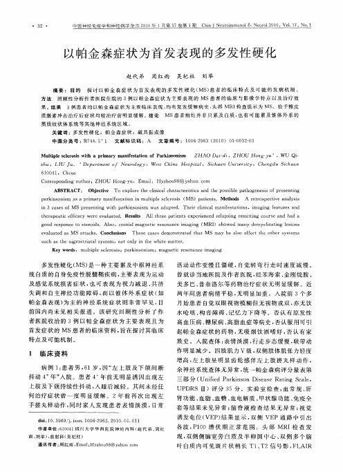
以帕金森症状为首发表现的多发性硬化

质 激 素 冲 击 治疗 后 症 状 均 较 治疗 前 明显 缓 解 。结 论 黑质纹状体系统等其他神经系统区域 。
MS患 者 病 灶 并 非 只 累及 白质 , 有 可 能 累 及 锥 体 外 系 的 也
关 键 词 : 发 性 硬 化 ;帕 金 森 症 状 ;磁 共 振 成 像 多
中 图分 类 号 : 7 4 5 文献 标 识 码 :A 文 章 编 号 :1 0 9 3( 0 0 10 3 —3 R 4 . 1 0 6 2 6 2 1 )0 — 0 20
Co rs o dn uh r re p n ig a t o :ZHOU n — u, E i: H y h u 8 y h o c m Ho g y mal zo 9 @ a o .o
AB T S RAC Ob et e To e po et eciia c aa trsisa dt ep s il p to e e i o rs nig T: jci v x lr h l c l h rceit n h o sbe ab g n ss fp e e t n c n
中国神经免疫学和神经病学杂志 21 年 1 00 月第 1 卷第 塑 c i 7 h
!
望
坠 !!!Байду номын сангаас
! ! ! : :!
以帕金 森 症 状 为 首发 表 现 的多 发性 硬 化
神经系统疾病的病理生理学机制

神经系统疾病的病理生理学机制神经系统疾病涉及到许多不同类型的疾病,其中包括多发性硬化症、帕金森病、阿尔茨海默病等。
这些疾病会影响到神经元、突触和神经传导通路,导致一系列复杂的病理生理学变化。
一、多发性硬化症多发性硬化症是一种自身免疫性疾病,会影响到中枢神经系统。
其病理生理学变化包括神经髓鞘损伤和神经元细胞死亡。
在病变区域,髓鞘的破坏会导致电信号传递的阻碍和神经元变形。
神经元的死亡则会导致神经功能损害,例如思维障碍、肢体运动障碍等。
此外,多发性硬化症还会导致脑组织炎症反应,增加细胞因子和炎症介质水平,进一步破坏神经细胞和突触连接。
二、帕金森病帕金森病是一种退行性神经疾病,主要影响运动系统。
其病理生理学变化包括多巴胺能神经元的死亡和运动控制通路的损伤。
多巴胺能神经元的死亡会导致多巴胺水平的下降,进而导致肌肉僵硬、震颤和身体不协调等症状。
此外,还会导致神经系统的发炎和氧化应激,进一步破坏神经细胞功能。
三、阿尔茨海默病阿尔茨海默病是一种逐渐发展的退行性神经疾病,会影响到记忆和认知。
其病理生理学变化包括神经元死亡、神经元连接断裂与神经系统内过度的神经元化学物质。
神经元死亡会导致认知和记忆功能损害,如易忘、失误等。
神经元连接断裂会影响到神经网络通信,导致不同脑区之间相互影响的能力降低。
过度的神经元化学物质会导致神经元过度激活,增加神经元死亡的风险。
四、病毒感染引起的神经系统疾病病毒感染也是一个常见的神经系统疾病的诱因。
例如,带状疱疹病毒可以引发带状疱疹和带状疱疹后神经痛等神经系统疾病。
病毒和细菌感染会导致炎症和免疫反应,破坏神经元和突触连接。
此外,病毒感染还可以直接侵入神经系统组织,影响神经元编码和传递信息的能力。
总之,神经系统疾病的病理生理学机制是复杂的,涉及到多个细胞类型和分布广泛的神经系统结构。
这些疾病的治疗需要综合考虑病理生理学的变化,采用相应的治疗手段来保护和恢复神经系统的功能。
神经内科病人的饮食指导与营养支持

神经内科病人的饮食指导与营养支持神经内科疾病是指影响神经系统的一类疾病,如脑卒中、帕金森病、多发性硬化症等。
这些疾病对患者的身体和生活带来了很大的影响,因此,对于神经内科病人的饮食指导与营养支持显得尤为重要。
本文将从不同疾病的特点出发,介绍神经内科病人在饮食方面的相关指导和营养支持。
一、脑卒中病人的饮食指导与营养支持脑卒中是一种常见的神经内科疾病,患者在恢复期间需要特殊的饮食指导和营养支持。
首先,对于脑卒中患者来说,保持适宜的体重是至关重要的。
因此,医生通常会根据患者的身体状况和病情,制定合理的饮食计划。
在饮食中,应注意提供足够的蛋白质,以促进肌肉的恢复和维持。
此外,适量的碳水化合物和脂肪也是必须的,以提供能量。
同时,脑卒中患者还需要关注摄入钠的量。
高钠饮食会增加血压,进而增加脑卒中再发的风险。
因此,应避免食用高盐或高钠食物,如咸菜、腌制食品等。
此外,还应限制饮食中的胆固醇和饱和脂肪酸的摄入,以预防动脉堵塞。
二、帕金森病患者的饮食指导与营养支持帕金森病是一种慢性进行性神经系统疾病,主要表现为肌肉僵硬、震颤和运动障碍等。
对于帕金森病患者来说,饮食的选择和营养的摄入可以帮助缓解症状,提高生活质量。
第一,帕金森病患者应保持充足的蛋白质摄入。
蛋白质是维持肌肉功能和修复组织的重要营养素,对于帕金森病患者来说尤为重要。
但是,过多的蛋白质摄入也可能干扰药物对身体的吸收和利用,因此,应根据医生的建议合理安排饮食。
第二,帕金森病患者还应摄入足够的纤维。
纤维有助于保持肠道的正常运转,防止便秘和消化不良。
常见的富含纤维的食物有水果、蔬菜、全谷类等。
此外,帕金森病患者还应多摄入富含维生素B6和镁的食物,以缓解症状。
三、多发性硬化症患者的饮食指导与营养支持多发性硬化症是一种免疫介导的神经系统疾病,主要影响中枢神经系统,导致肌力减退、感觉障碍等症状。
对于多发性硬化症患者来说,合理的饮食指导和营养支持可以帮助调节免疫系统、维持健康状况。
nd疾病名词解释(一)

nd疾病名词解释(一)1. 什么是ND疾病?•ND疾病是一类与神经系统相关的疾病,包括多种病理和功能异常。
2. 常见的ND疾病名词及解释:•阿尔茨海默病(Alzheimer’s Disease):一种进行性神经系统退化性疾病,导致记忆力和认知能力的丧失。
例如,患者可能逐渐忘记亲人的名字和日常生活活动。
•帕金森病(Parkinson’s Disease):中枢神经系统退行性疾病,导致患者肌肉僵硬、震颤以及运动障碍。
例如,患者可能出现手部颤抖和行走困难的症状。
•脑卒中(Stroke):由于脑血管破裂或阻塞导致的大脑区域血液供应不足而引起的疾病。
例如,患者可能出现面部麻木、言语障碍和肢体瘫痪的症状。
•帕金森症候群(Parkinsonism Syndrome):一组由各种原因引起的症状类似于帕金森病的疾病。
例如,某些药物副作用或其他疾病(如帕金森病的变异形式)可能导致帕金森症候群。
•亨廷顿舞蹈病(Huntington’s Disease):遗传性神经系统疾病,导致运动、认知和行为的障碍。
例如,患者可能出现不自主的舞蹈样动作、认知功能下降和情绪不稳定的症状。
•多发性硬化症(Multiple Sclerosis,简称MS):一种自身免疫性疾病,影响中枢神经系统。
例如,患者可能经历感觉和运动障碍、视力受损以及疲劳等症状。
•渐冻人综合症(ALS,即肌萎缩侧索硬化症):一种进展性神经肌肉疾病,导致肌肉无力和萎缩。
例如,患者可能逐渐丧失肌肉控制能力,导致肢体无法运动和言语困难。
•格林-巴利综合症(Guillain-Barre Syndrome):一种罕见的自身免疫疾病,主要影响神经系统。
例如,患者可能出现感觉异常、肌肉无力和脚趾和手指麻木的症状。
•帕金森变异性(Parkinsonism Variants):一类疾病,其症状类似于帕金森病,但与其不同的病因和病理机制相关。
例如,多系统萎缩(MSA)和普罗卡齐恩体病(PSP)等都是帕金森变异性。
神经内科学知识点

神经内科学知识点神经内科学是医学的一个分支,主要研究神经系统的疾病,包括脑、脊髓、周围神经和神经肌肉接头等方面的疾病。
下面将介绍一些神经内科学的知识点。
一、脑卒中脑卒中是指突发性脑血管意外,造成脑血管的急性缺血或出血性病变。
常见的症状包括突发性头痛、嘴歪眼斜、肢体无力等。
脑卒中应该尽早就诊,并接受头部CT或MRI检查,确定病变类型,以便制定合理的治疗方案。
二、帕金森病帕金森病是一种慢性进行性疾病,以静止性震颤、肌肉强直、运动迟缓等为主要症状。
该病的病因尚不明确,目前主要通过药物治疗和物理治疗来缓解症状,有时候还需要外科手术治疗。
三、脑膜炎脑膜炎是由细菌、病毒、真菌等感染脑膜组织引起的疾病。
患者常表现为高热、头痛、呕吐、意识障碍等症状。
治疗上应该及时使用抗生素、抗病毒药物等进行治疗,预防并发症的发生。
四、癫痫癫痫是一种常见的神经系统疾病,患者会出现反复发作的癫痫发作。
治疗上主要通过抗癫痫药物来控制症状,部分患者还需要进行手术治疗,切除病变部位。
五、多发性硬化多发性硬化是一种自身免疫性疾病,主要累及中枢神经系统。
患者表现为多发性的中枢神经系统损害症状,包括感觉障碍、运动障碍等。
治疗上主要通过激素治疗和免疫调节剂来控制病情。
六、脑肿瘤脑肿瘤是指生长在脑组织中的一类瘤,可以分为原发性和继发性两种。
临床上常表现为头痛、呕吐、意识障碍等症状。
治疗上主要通过手术切除、放疗和化疗等综合治疗手段来控制病情。
以上就是关于神经内科学的一些知识点,希望对大家有所帮助。
神经内科学是一个广阔而又复杂的领域,需要医务人员不断学习和提升专业技能,为神经系统疾病的治疗提供更好的服务。
祝愿所有患者早日康复!。
神经疾病导致的神经元死亡的机理研究

神经疾病导致的神经元死亡的机理研究神经疾病是指被破坏或改变了神经组织结构和功能的一类疾病,如阿尔茨海默病、帕金森病、多发性硬化症等。
这些疾病的共同点是导致神经元死亡,从而影响神经系统的功能。
因此,研究神经疾病导致的神经元死亡的机理对于疾病的治疗和预防具有重要意义。
神经元死亡是由多种因素引起的,包括兴奋性毒性、细胞凋亡、氧化应激等。
其中,兴奋性毒性是最为常见的一种。
神经元在工作时需要释放神经递质,而过量的神经递质会对神经元产生毒性作用,造成神经元死亡。
例如,帕金森病的发病机理与多巴胺神经元死亡有关。
多巴胺神经元是运动控制和情绪调节的关键神经元,而帕金森病会导致多巴胺神经元数量减少,从而引起肢体僵硬、震颤等症状。
细胞凋亡也是导致神经元死亡的重要机制。
凋亡是一种正常的细胞死亡方式,它与细胞的生长、发育和代谢密切相关。
在神经系统中,细胞凋亡是一种保护性机制,可用来清除有问题的神经元,并维持神经系统的稳态。
然而,在某些疾病中,细胞凋亡会变得过度或异常,导致神经元数量的大量减少。
例如,阿尔茨海默病的发病机制涉及神经元凋亡,它会引起神经元内的tau蛋白异常聚集和β淀粉样蛋白的沉积,从而导致神经元凋亡和认知功能的损失。
氧化应激是另一种导致神经元死亡的机制。
氧化应激是指细胞内自由基(ROS)或反应性氮物质(RNS)超过细胞清除机制的能力所导致的一种累积现象。
在神经系统中,大量的氧化应激会导致神经元膜的氧化损伤和线粒体功能不良,从而引起神经元死亡。
多发性硬化症是一种典型的氧化应激性神经疾病,它的发病机制涉及脊髓和脑神经纤维的炎症和氧化应激反应,导致神经元死亡和脱髓鞘。
总体而言,神经疾病导致的神经元死亡是复杂的、多因素的过程。
它不仅涉及兴奋性毒性、细胞凋亡和氧化应激等机制,还和许多因素如遗传、环境和压力等有关。
因此,探究神经元死亡的机理必须从多个维度入手,建立全面的研究体系,以期为神经疾病的治疗和预防提供更为有效的策略。
神经退行性疾病的诊断与治疗研究

神经退行性疾病的诊断与治疗研究神经退行性疾病是指发生于中枢神经系统和周围神经系统的一类疾病,这些疾病包括帕金森病、阿尔茨海默症、亨廷顿舞蹈症、多发性硬化症等。
这些疾病都有一个共同点,即神经细胞逐渐死亡,从而导致神经元的功能损失,严重干扰了机体正常的生理功能和行为表现。
因此,对于神经退行性疾病及其治疗研究的深入,对于促进人类健康都是非常重要的。
一、神经退行性疾病的分类1.阿尔茨海默病阿尔茨海默病(AD)是一种进行性神经退行性疾病,其典型症状为认知障碍、记忆力下降、行为异常等。
严重的AD患者可能会失去自理能力,成为社会负担。
2.亨廷顿舞蹈症亨廷顿舞蹈症(HD)是德国医生乔治-亨廷顿在19世纪末首次描述的一种遗传性神经退行性疾病。
它主要影响到个体的智力、感情和运动控制能力。
此病是由外部射突氨基酸组成的蛋白质的不正常聚集引起的。
3. 帕金森病帕金森病(PD)是一种运动系统退行性疾病,表现为运动控制失调,剧烈的震颤、僵硬以及运动迟缓等症状。
此病是由于丧失脑内多巴胺(A dopaminergic)神经元的数目造成的。
4.多发性硬化症多发性硬化症(MS)是一种中枢神经系统疾病,表现为全身和运动障碍、感觉障碍等症状,同时还有运动神经元病变和灰质损失。
二、神经退行性疾病的诊断1.家族史询问对于某些神经退行性疾病,例如亨廷顿舞蹈症,家族史是确定诊断的重要依据。
在许多神经退行性疾病患者中,超过10%的受到疾病影响患者的亲属也出现了类似的症状。
2.临床表现临床表现是诊断神经退行性疾病的重要标记,具体表现包括但不限于视力丧失、震颤、行为异常、认知障碍等。
3.分子生物学分析在一些特定的神经退行性疾病中,如亨廷顿舞蹈症、肌萎缩性侧索硬化症等,可能存在某些特定的基因突变引起的,因此在这些病例中应进行基因分析。
4.影像学检查神经影像学检查,如核磁共振成像(MRI)等,对于神经退行性疾病的诊断也是非常有帮助的。
MRI能够检测出透明外质、重复运动性损伤等,还能观察到深内侧核等神经核的异常。
神经系统疾病

神经系统疾病神经系统是人体重要的调节和控制系统,但它也容易受到各种疾病的影响。
神经系统疾病是指那些影响神经组织和神经功能的疾病,包括神经退行性疾病、神经炎症性疾病和神经肿瘤等。
一、神经退行性疾病1. 阿尔茨海默病阿尔茨海默病是老年人中最常见的神经退行性疾病。
其特征是大脑神经细胞的逐渐退化和死亡,导致记忆力、思维能力和行动能力的丧失。
阿尔茨海默病的早期症状包括记忆力下降、注意力不集中、迷失方向等,随着病情的进展,患者会出现言语障碍、认知障碍和行为变化等。
2. 帕金森病帕金森病是一种常见的神经运动障碍性疾病,主要由脑内多巴胺神经元的损失引起。
其症状包括肌肉僵硬、震颤、动作迟缓等。
帕金森病的严重性逐渐加重,严重影响患者的日常生活和生活质量。
二、神经炎症性疾病1. 脊髓灰质炎脊髓灰质炎是由脊髓灰质炎病毒引起的急性传染病,主要通过肠道传播。
其症状包括发热、头痛、呕吐、肌肉无力等,严重时可导致四肢瘫痪和呼吸肌麻痹。
2. 多发性硬化症多发性硬化症是一种自身免疫性疾病,主要影响中枢神经系统的脑和脊髓。
多发性硬化症的症状多种多样,包括视力模糊、肢体无力、平衡障碍等。
病情逐渐加重,可导致行动困难和身体功能障碍。
三、神经肿瘤1. 脑肿瘤脑肿瘤是指在脑组织中发生的肿瘤,其种类繁多。
脑肿瘤的症状取决于肿瘤的位置和大小,常见症状包括头痛、恶心、呕吐、意识改变等。
脑肿瘤的治疗方法包括手术切除、放疗和化疗等。
2. 神经纤维瘤神经纤维瘤是一种起源于神经鞘细胞的良性肿瘤,常见于神经系统的周边神经。
神经纤维瘤的症状包括局部皮肤颜色改变、肿块感、疼痛、神经功能障碍等。
治疗方法包括手术切除和放疗等。
综上所述,神经系统疾病是一类严重影响人们生活质量和健康的疾病。
了解其症状和治疗方法对于预防和治疗神经系统疾病具有重要意义。
鉴于每种疾病的特点不同,患者在面临疑似神经系统疾病时,应及时就医并接受专业医生的诊断和治疗。
- 1、下载文档前请自行甄别文档内容的完整性,平台不提供额外的编辑、内容补充、找答案等附加服务。
- 2、"仅部分预览"的文档,不可在线预览部分如存在完整性等问题,可反馈申请退款(可完整预览的文档不适用该条件!)。
- 3、如文档侵犯您的权益,请联系客服反馈,我们会尽快为您处理(人工客服工作时间:9:00-18:30)。
Research letterParkinson disease and multiple sclerosis are not associated with autoantibodies against structural proteins of the dermal –epidermal junctionDOI:10.1111/bjd.14538D EARE DITOR ,Bullous pemphigoid (BP),the most frequent autoimmune blistering disease,is associated with autoantibod-ies against two proteins of the dermal –epidermal junction (DEJ),BP180(type XVII collagen)and BP230[BP antigen 1(BPAG1)].1Two peculiar clinical features of BP are the advanced age of patients,with a mean age of between 75and 80years at disease onset,and its association with neurological disease.1Neurological diseases can be diagnosed in 30–50%of patients with BP,including cognitive impairment,stroke,epilepsy,Parkinson disease (PD)and multiple sclerosis (MS),with odds ratios (ORs)of 2Á2,1Á8–3Á3,1Á7–4Á0,2Á16–3Á50and 10Á7,respectively.2–7In addition,patients with MS are more likely to develop BP (OR 6Á7).8These findings are par-ticularly intriguing as the cutaneous target antigens of BP –BP180and BP230–are also expressed in the central nervous system (CNS).BP180expression was found in the cerebellum of rats and in autopsy samples of various neuroanatomical regions of human brain.9,10Mice with mutations in the dys-tonin gene encoding for various isoforms of BPAG1,includingthe epithelial isoform BP230,develop severe dystonia and sensory nerve degeneration.11While PD is thought to be a primary neurodegenerative disorder,inflammatory responses seem to be secondary.12,13MS is believed to be initially induced via a peripheral immune response,and further driven by immune reactions within the CNS with secondary neurode-generation.14During the process of neurodegeneration the two BP autoantigens in the CNS may have been exposed to the immune system,leading to the break of tolerance and,subsequently,to the generation of anti-BP180and anti-BP230antibodies,and,finally,to BP.9,15In the present study,according to this hypothesis,we expected to detect serum autoantibodies against BP180and BP230more frequently in patients with PD and MS compared with age-and sex-matched controls.Alternatively,or addition-ally,environmental factors that increase susceptibility to the development of neurological disorders might similarly change the risk of autoimmune blistering dermatoses.8We compared three age-and sex-matched groups of patients with PD (n =75,cohort A1),other neurological diseases [n =75,cohort A2;detailed information is given in Table S1(see Supporting Information)]and healthy controls (n =75,cohort A3)(Table 1).All sera from cohort A were prospec-tively collected and matched for age and sex.Furthermore,prospectively collected sera from consecutive patients with PD at another academic site (L €u beck;n =50,cohort C),a cohortTable 1Overview of serological results©2016British Association of Dermatologists British Journal of Dermatology (2016)1of patients with MS(n=50,cohort D)and patients with non-inflammatory skin diseases and older than75years(n=65, cohort B;Table1)were analysed.Cohort B was included to mirror the age group of patients with BP.To determine the sample sizes necessary to compare the frequency of reactive sera in patients and controls,a power calculation based on Fisher’s exact test was performed.To be clinically relevant,weassumed that the odds of detecting serumautoantibodies inpatients with PD and MS would be increased at leastfive times compared with controls,and that anti-BP180/BP230antibod-ies would be detected in1–2%of controls.16–18Based on these assumptions,the power(p)for detecting significantly different amounts of BP180/BP230-reactive sera between patients with PD/MS(n=175)and controls(n=215)was0Á51–0Á86.To detect reactivity against BP180and BP230,all sera were subjected to a panel of diagnostic assays,including(i)indirect immunofluorescence(IIF)microscopy on a BIOCHIPâmosaic (monkey oesophagus,split human skin,recombinant BP180 NC16A,HEK293expressing the BP180ectodomain,the BP230globular domain and full-length BP230;Euroimmun, L€u beck,Germany);(ii)BP180NC16A-based enzyme-linked immunosorbent assay(ELISA);(iii)BP230-based ELISA(both from Euroimmun);(iv)Western blotting with extracellular matrix of cultured human keratinocytes(for the detection of laminin332and full-length cell-derived BP180);(v)IIF microscopy on monkey oesophagus;and(vi)1mol LÀ1 NaCl-split human skin(both in-house tests).16,18–20No signif-icant differences in the frequency of detecting serum autoanti-bodies against proteins of the DEJ were found in patients with PD and MS compared with controls(Tables1and2).Reactiv-ities in all cohorts are detailed in Table1.In none of the sam-ples could reactivity against BP180or BP230be demonstrated by all test methods;however,the BP180NC16A ELISA was more often positive(seven of the total390samples)than the corresponding BIOCHIP mosaic substrate(one of the total390samples).Altogether,antibodies against the DEJ were observed in four of175PD/MS sera[2Á3%;95%confidence interval (CI)0Á9–5Á7]and16of215control sera(7Á4%;95%CI 4Á6–11Á7),which is in line with the known specificities of 98–99%of the employed test systems.16,18–20Our results indicate that patients with PD and MS do not show a clinically relevant increased incidence of autoreactivity against BP180,BP230and laminin332.If there is a higher risk of patients with these neurological disorders developing BP,this is not reflected by our serologicalfindings.This notion indicates that serum autoreactivity against proteins of the DEJ does not precede the clinical manifestation of BP in patients with PD/MS.Two other risk factors for developing antibodies to BP180and BP230–old age and chronic pruritus –have been described;however,these additional risk factors remain disputed.19,21,22It is feasible that as-yet-uncovered environmental factors orchestrate the initiation of the neuro-logical disorders,the onset of pruritus,and the loss of toler-ance against BP180and BP230in the elderly.One might speculate that such a factor could be a viral infection that affects both skin and CNS,such as varicella zoster virus.23It has been hypothesized that the development of autoimmunity against CNS antigens may be associated with repairing mecha-nisms of the CNS.24In this context,the autoimmune skin dis-ease may be regarded as a casualty of the organism in its efforts to preserve the brain.AcknowledgmentsWe thank Vanessa Krull for her assistance with the autoim-mune diagnostic procedures.This study was approved by the ethics committee of the University of L€u beck(10-229).A.R E C K E1,2A.O E I1F.H€U B N E R2K.F E C H N E R3J.G R A F4J.H A G E N A H4C.M A Y5D.W O I T A L L A6A.S A L M E N7D.Z I L L I K E N S2R.G O L D8W.S C H L U M B E R G E R3E.S C H M I D T1 1L€u beck Institute of ExperimentalDermatology(LIED),and Departments of2Dermatology and4Neurology,University ofL€u beck,L€u beck,Germany3Institute of Experimental Immunology,Euroimmun Inc.,L€u beck,Germany5Medizinisches Proteom-Center,Ruhr-Universit€a t Bochum,Bochum,Germany6Department of Neurology,KatholischeKliniken Ruhrhalbinsel GmbH,Essen,Germany7Department of Neurology,Inselspital,BernUniversity Hospital,Bern,Switzerland8Department of Neurology,St.JosefHospital,Ruhr-University Bochum,GermanyCorrespondence:Enno Schmidt.E-mail:enno.schmidt@uksh.deReferences1Schmidt E,Zillikens D.Pemphigoid ncet2013;381:320–32.Table2Association of positive serologicalfindings with neurologicaldiseases©2016British Association of Dermatologists British Journal of Dermatology(2016)2Research letter2Taghipour K,Chi C-C,Vincent A et al.The association of bullous pemphigoid with cerebrovascular disease and dementia:a case-control study.Arch Dermatol2010;146:1251–4.3Chen YJ,Wu CY,Lin MW et orbidity profiles among patients with bullous pemphigoid:a nationwide population-based study.Br J Dermatol2011;165:593–9.4Langan SM,Groves RW,West J.The relationship between neuro-logical disease and bullous pemphigoid:a population-based case–control study.J Invest Dermatol2011;131:631–6.5Bastuji-Garin S,Joly P,Lemordant P et al.Risk factors for bullous pemphigoid in the elderly:a prospective case–control study.J Invest Dermatol2011;131:637–43.6Brick KE,Weaver CH,Savica R et al.A population-based study of the association between bullous pemphigoid and neurologic disor-ders.J Am Acad Dermatol2014;71:1191–7.7Cordel N,Chosidow O,Hellot M-F et al.Neurological disorders in patients with bullous pemphigoid.Dermatology2007;215:187–91. 8Langer-Gould A,Albers KB,Van Den Eeden SK,Nelson LM.Autoimmune diseases prior to the diagnosis of multiple sclerosis:a population-based case–control study.Mult Scler2010;16:855–61. 9Sepp€a nen A,Miettinen R,Alafuzoff I.Neuronal collagen XVII is localized to lipofuscin granules.NeuroReport2010;21:1090–4.10Claudepierre T,Manglapus MK,Marengi N et al.Collagen XVII and BPAG1expression in the retina:evidence for an anchoring complex in the central nervous system.J Comp Neurol2005;487:190–203.11Guo L,Degenstein L,Dowling J et al.Gene targeting of BPAG1: abnormalities in mechanical strength and cell migration in strati-fied epithelia and neurologic degeneration.Cell1995;81:233–43. 12Sanchez-Guajardo V,Barnum CJ,Tansey MG,Romero-Ramos M.Neuroimmunological processes in Parkinson’s disease and their relation to a-synuclein:microglia as the referee between neuronal processes and peripheral immunity.ASN Neuro2013;5:113–39.13Pradhan S,Andreasson mentary:progressive inflammation as a contributing factor to early development of Parkinson’s dis-ease.Exp Neurol2013;241:148–55.14Hemmer B,Kerschensteiner M,Korn T.Role of the innate and adaptive immune responses in the course of multiple n-cet Neurol2015;14:406–19.15Taghipour K,Chi C-C,Bhogal B et al.Immunopathological charac-teristics of patients with bullous pemphigoid and neurological dis-ease.J Eur Acad Dermatol Venereol2014;28:569–73.16Bl€o cker IM,D€a hnrich C,Probst C et al.Epitope mapping of BP230 leading to a novel enzyme-linked immunosorbent assay for autoantibodies in bullous pemphigoid.Br J Dermatol2012;166:964–70.17Tampoia M,Giavarina D,Di Giorgio C,Bizzaro N.Diagnostic accuracy of enzyme-linked immunosorbent assays(ELISA)to detect anti-skin autoantibodies in autoimmune blistering skindiseases:a systematic review and meta-analysis.Autoimmun Rev 2012;12:121–6.18Pr€ußmann J,Pr€ußmann W,Recke A et al.Co-occurrence of autoan-tibodies in healthy blood donors.Exp Dermatol2014;23:519–21. 19van Beek N,Dohse A,Riechert F et al.Serum autoantibodies against the dermal-epidermal junction in patients with chronic pruritic disorders,elderly individuals and blood donors prospec-tively recruited.Br J Dermatol2014;170:943–7.20van Beek N,Rentzsch K,Probst C et al.Serological diagnosis of autoimmune bullous skin diseases:prospective comparison of the BIOCHIP mosaic-based indirect immunofluorescence technique with the conventional multi-step single test strategy.Orphanet J Rare Dis2012;7:49.21Schmidt T,Sitaru C,Amber K,Hertl M.BP180-and BP230-specific IgG autoantibodies in pruritic disorders of the elderly:a preclinical stage of bullous pemphigoid?Br J Dermatol2014;171:212–19.22Meijer JM,Lamberts A,Pas HH,Jonkman MF.Significantly higher prevalence of circulating bullous pemphigoid(BP)-specific IgG autoantibodies in elderly patients with a nonbullous skin disorder.Br J Dermatol2015;173:1274–6.23Kamiya K,Aoyama Y,Suzuki T et al.Possible enhancement of BP180autoantibody production by herpes zoster.J Dermatol2016;43:197–9.24Schwartz M,Baruch K.Breaking peripheral immune tolerance to CNS antigens in neurodegenerative diseases:boosting autoimmu-nity tofight-off chronic neuroinflammation.J Autoimmun2014;54:8–14.Supporting InformationAdditional Supporting Information may be found in the online version of this article at the publisher’s website:Appendix S1.Supplemental methods.Table S1.Group of patients with‘other neurologic diseases’(subcohort A2).Funding sources:This work was supported by Deutsche Forschungsge-meinschaft KFO303/1,and received infrastructural support from Excellence Cluster Inflammation at Interfaces(EXC306/2)andP.U.R.E.(Protein Unit for Research in Europe),a project of the federal German state Nordrhein-Westfalen.Conflicts of interest:A.R.,D.Z.and E.S.have received a scientific award from Euroimmun.D.Z.and E.S.have a scientific cooperation withEuroimmun.©2016British Association of Dermatologists British Journal of Dermatology(2016)Research letter3。
