20140603-Role of Leukocyte Cell-Derived Chemotaxin 2 as a Biomarker in Hepatocellular Carcinoma
CD and CAM talk - english

Cell Adhesion Molecules
Lie Wang (汪洌)
Institute of Immunology Zhejiang University School of Medicine Medical Research Building A810
CD8 T cells Peptide+MHC I
CD8+
Perforin Granzyme
CD4 T cells Peptide+MHC II
CD4+ CD40L
Major histocompatibility complex,
CD40
B cell
Virally infected cell
CD8 Cytokines IFN
Tel: 88981392 E-mail:wanglie@
Contents
• Concept of leukocyte differentiation antigens
• Concept of cluster of differentiation • Cell adhesion molecules
Rules for assigning a CD number
• at least one Workshop—characterized antibody and good molecular data.
• the antigen is expressed on the surface of cells involved in immune reactions.
Leukocyte differentiation antigens
肠神经胶质细胞在炎症性肠病中的作用研究进展
综述China &Foreign Medical Treatment 中外医疗肠神经胶质细胞在炎症性肠病中的作用研究进展杜俊艳1,骆淑娥2,温萌11.甘肃卫生职业学院附属医院门诊部,甘肃兰州 730207;2.解放军96640部队医院外一科,甘肃兰州730030[摘要] 近年来,肠神经胶质细胞(enteric glial cell, EGC )在炎症性肠病(inflammatory bowel disease, IBD )发病机制中引起广泛关注。
在IBD 患者中,肠道EGC 通过释放细胞因子和介质参与调节肠道炎症反应,EGC 与肠道上皮细胞、免疫细胞和神经元等相互作用,参与调节肠道的免疫平衡和功能,EGC 在炎症性肠病治疗中的潜在应用,调控和干预EGC 有望为治疗提供新思路和策略。
[关键词] 肠神经系统;肠神经胶质细胞;胶质细胞源性神经营养因子-胶质细胞系源性神经营养因子受体α1/原癌基因自分泌环路[中图分类号] R4 [文献标识码] A [文章编号] 1674-0742(2023)09(a)-0195-04Advances in the Study of the Role of Enteric Glial Cells in Inflammatory Bowel DiseaseDU Junyan 1, LUO Shu'e 2, WEN Meng 11.Outpatient Department, Affiliated Hospital of Gansu Health Vocational College, Lanzhou, Gansu Province, 730207 China;2.Department of Surgery, PLA 96640 Army Hospital, Lanzhou, Gansu Province, 730030 China[Abstract] In recent years, enteric glial cells (EGC) have attracted widespread attention in the pathogenesis of inflam⁃matory bowel disease (IBD). In IBD patients, intestinal EGCs are involved in the regulation of intestinal inflammatory responses through the release of cytokines and mediators, and EGCs interact with intestinal epithelial cells, immune cells, and neurons to participate in the regulation of intestinal immune homeostasis and function. The potential appli⁃cation of EGC in the treatment of inflammatory bowel disease, modulation and intervention of EGC is expected to pro⁃vide new ideas and strategies for treatment.[Key words] Enteric nervous system; Enteric glial cells; GDNF-GFRa1/RET autocrine loop溃疡性结肠炎(ulcerativecolitis, UC )是一种与克罗恩病(crohn's disease, CD )同属炎性肠病(in⁃flammatory bowel disease, IBD )的慢性反复发作的肠炎[1]。
内皮祖细胞膜微粒与脑卒中治疗相关性研究进展

内皮祖细胞膜微粒与脑卒中治疗相关性研究进展邱文姬;陈煜森【摘要】Vascular endothelial dysfunction is related to the occurrence and development of stroke.Endothelial progenitor cells-derived microvesicles (EPC-MVs) can slow the progression of stroke and improve the prognosis of stroke by repairing the damaged vascular endothelium,providing a new method for the treatment of stroke.This article reviews the correlation between EPC-MVs and stroke treatment.%血管内皮功能障碍与脑卒中的发生发展互为因果.内皮祖细胞膜微粒可以通过修复损伤的血管内皮来延缓脑卒中的进展和改善脑卒中患者的预后,为脑卒中的治疗提供了新的方法.本文就内皮祖细胞膜微粒与脑卒中治疗相关性进行综述.【期刊名称】《海南医学》【年(卷),期】2018(029)005【总页数】4页(P674-677)【关键词】内皮祖细胞;内皮祖细胞膜微粒;脑卒中;修复;治疗【作者】邱文姬;陈煜森【作者单位】广东医科大学,广东湛江524000;广东医科大学附属医院,广东湛江524000【正文语种】中文【中图分类】R743.3脑卒中俗称“中风”,是以高发生率、高致残率、高致死率和高复发率为特点的急性脑血管循环障碍性疾病。
一般分为缺血性卒中(如脑梗死)和出血性卒中(如脑出血)两大类。
由于脑供血动脉闭塞,使该动脉的供血区得不到血液中的氧气和营养而发生坏死,被称为缺血性脑卒中,约占80%~85%;而由于脑动脉硬化而血管破裂,血液进入脑内和脑周围间隙,使脑细胞得不到正常血管内运输的氧气和营养供应而发生坏死,另被称为出血性脑卒中,占15%~20%。
中山大学博士学位论文金属卟啉模拟...

生催化活性的增强机制,即:通过 4A 分子筛的 Al3+路易斯酸活性位与环己酮、苯甲醛 中羰基的氧配位,同时活化了底物环己酮和共还原剂苯甲醛,并同时促进了金属卟啉高 价活性物种和 Criegee 加合物的形成(环己酮 B–V 氧化反应的关键步骤),实现了仿生 催化活性的增强。
4. 在金属卟啉仿生催化体系中加入羧基化多壁碳纳米管作为助催化剂,通过对催化 过程中自由基稳定性的提高增强了金属卟啉仿生催化的活性。在反应温度为 50 oC,1,2二氯乙烷为溶剂,FeTPPCl 为催化剂,羧基化多壁碳纳米管为助催化剂的优化条件下, 环己酮的转化率达到 95%,ε-己内酯的收率为 95%。该反应体系对于其它酮类化合物的 B–V 氧化同样也表现出了良好的催化活性。通过原位电子顺磁共振波谱方法、原位拉曼 光谱技术、原位紫外光谱方法等系统研究了反应历程,阐明了羧基化多壁碳纳米管有效 地提高了自由基的稳定性,加快了过氧酸的生成,从而促进了金属卟啉高价活性物种的 形成(B–V 氧化反应的关键步骤),显著增强了仿生催化的活性。
中山大学博士学位论文
金属卟啉模拟酶的特异性及其催化 Baeyer–Villiger 氧化的 活性增强机制研究
Investigation into the specificity of metalloporphyrin as mimic enzyme and mechanisms of enhancing catalytic
1. The nature of efficiency substrate specificity in biomimetic catalytic oxidation was clarified from the viewpoints of kinetic and mechanism by using styrene and cyclohexanone as model substrates. Kinetic results showed that the metalloporphyrins-catalyzed styrene oxidation exhibited excellent efficiency as enzyme-like catalysis and followed Michaelis-Menten equation, while cyclohexanone oxidation was in accordance with the general rules of chemical catalysis and belonged to first-order rate kinetic equation. Different catalytic mechanisms of two substrates were discussed by means of in situ EPR spectroscopy and in situ UV-vis spectroscopy. The remarkable efficiency difference for the two kinds of substrates was attributed to the stability of free radical and high-valence species.
生物质谱技术在腺相关病毒(AAV)载体制剂质量控制中的应用

学 报Journal of China Pharmaceutical University 2023,54(6):682 - 694682生物质谱技术在腺相关病毒(AAV)载体制剂质量控制中的应用李孟效,李惠琳*(中山大学药学院,广州 510006)摘 要 腺相关病毒(adeno-associated virus,AAV)是一种基因治疗中常用的病毒载体。
由于其安全性较高且能够靶向多种细胞,在临床前和临床研究中得到了较多的应用。
不过在设计和生产的过程中,AAV载体有着诸多会影响其安全性和疗效的关键质量属性。
生物质谱技术的发展和应用为生物大分子的研究提供了一个便捷的平台,尤其是在蛋白质序列、结构和相互作用方面。
对于AAV载体而言,质谱技术可以实现衣壳蛋白比率、翻译后修饰、血清型、空衣壳比率的测定或表征,从而协助对AAV载体的质量控制。
与现有方法相比,质谱技术具有样品需求量少、分析快速灵敏、适用于完整AAV载体的分析和质量分辨率高的优点,并且可以区分空衣壳、满衣壳和部分包封的衣壳。
未来,通过将更加高效的蛋白质分离技术与质谱技术联用、开发新的信息处理软件平台和新的质谱检测方法,质谱技术有望在AAV载体的设计和生产中发挥更加重要的作用。
关键词腺相关病毒;质谱;质量控制;非变性质谱;电荷检测质谱中图分类号R392 文献标志码 A 文章编号1000 -5048(2023)06 -0682 -13doi:10.11665/j.issn.1000 -5048.2023062901引用本文李孟效,李惠琳.生物质谱技术在腺相关病毒(AAV)载体制剂质量控制中的应用[J].中国药科大学学报,2023,54(6):682–694.Cite this article as:LI Mengxiao,LI Huilin. Application of biological mass spectrometry in quality control of adeno-associated virus carrier preparations[J].J China Pharm Univ,2023,54(6):682–694.Application of biological mass spectrometry in quality control of adeno-asso⁃ciated virus carrier preparationsLI Mengxiao, LI Huilin*School of Pharmaceutical Science, Sun Yat-Sen University, Guangzhou 510006, ChinaAbstract Adeno-associated virus (AAV) is a common viral vector used in gene therapy.Because of its high safe⁃ty and its ability to target a variety of cells, it has been widely used in preclinical and clinical studies.However, during the design and production, AAV vectors have many key quality attributes that affect their safety and efficacy. The development and application of biological mass spectrometry technology provides a convenient platform for the research on biological macromolecules, especially in the aspects of protein sequence, structure and interac⁃tion.For AAV vectors, mass spectrometry can facilitate the determination or characterization of capsid protein ratio, post-translational modification, serotype, and empty capsid ratio, thus assisting in the quality control of AAV pared with the existing methods, mass spectrometry has the advantages of smaller amount of sample size, faster and more sensitive analysis, being more suitable for the analysis of complete AAV vectors with higher mass resolution, and can distinguish empty capsids, full capsids and partial capsids.In the future, mass spectrometry technology is expected to play a more important role in the design and production of AAV vectors through the coupling of more efficient protein separation technology with mass spectrometry, the development of new information processing software platforms and new mass spectrometry detection techniques.Key words adeno-associated virus (AAV); mass spectrometry (MS); quality control; native MS; charge detec⁃tion MS收稿日期2023-06-29 *通信作者Tel:138****4968E-mail:lihlin6@基金项目国家自然科学基金资助项目(No.81872836,No.91953102);广东省自然科学基金资助项目(No.2019A1515011265)第 54 卷第 6 期李孟效,等:生物质谱技术在腺相关病毒(AAV )载体制剂质量控制中的应用This study was supported by the National Natural Science Foundation of China (No.81872836, No.91953102) and the National Nat⁃ural Science Foundation of Guangdong Province (No.2019A1515011265)1 腺相关病毒(AAV)概述基因治疗是指为了治疗目的而修改操纵基因表达或改变活细胞的基因,基因治疗的方式可以划分基因补充和基因编辑,基因补充是指将遗传物质导入需要治疗的靶细胞,基因编辑是指对细胞已有的缺陷基因进行修改和调控,但无论哪种方式都需要依赖特定的递送载体才能完成[1]。
白血病干细胞表面分子标志物及分化抗原高表达与难治性急性白血病
白血病干细胞表面分子标志物及分化抗原高表达与难治性急性白血病王荣华;陈信义;褚雨霆;侯丽;王婧【摘要】Leukemia is one of the malignant cloning diseases derived from hematopoietic stem cell. There are more and more researches showing that leukaemia stem cell( LSC )is the origin of the recurrence of leukemia, which has the similarity with the normal haemopoietic stem cell in infinite proliferation and self-renewing. And LSC can get away with the effect of drugs in its resting stage and lead to the recurrent and refractory leukemia. The relapse and refractory of leukemia are closely related to the high expression of differentiation antigens and the surface molecular markers of LSC. Consequently,the biological target drug,which targets specific surface antigens or molecular markers of LSC ,has become the new emphasis in the research of leukemia.%白血病是一类起源于造血干细胞的恶性克隆性疾病,越来越多的研究证明,白血病干细胞(LSC)是白血病复发的根源,其具有与正常造血干细胞类似的无限增殖和自我更新能力,且LSC处于静止期,能逃逸化学药物的治疗作用,从而导致白血病复发和难治.其中,白血病复发及其难治与LSC表面分子标志物及分化抗原表达密切相关.因此,以LSC特异性表面抗原或分子标志物为靶点,寻找和发现治疗白血病的生物靶向药物已成为可能治愈白血病研究的重点.【期刊名称】《医学综述》【年(卷),期】2012(018)013【总页数】4页(P1973-1976)【关键词】白血病干细胞;分子标志物;分化抗原;难治性急性白血病【作者】王荣华;陈信义;褚雨霆;侯丽;王婧【作者单位】北京中医药大学东直门医院血液肿瘤科,北京100700;北京中医药大学东直门医院血液肿瘤科,北京100700;北京中医药大学东直门医院血液肿瘤科,北京100700;北京中医药大学东直门医院血液肿瘤科,北京100700;北京中医药大学东直门医院血液肿瘤科,北京100700【正文语种】中文【中图分类】R733.711.1 LSC表面分化抗原高表达目前的研究发现,M0、M1、M2、M4、M5 的LSC 细胞表面抗原表型是从HSC恶性转化而来,因而,具有许多HSC的表型标记,如表达 C、CD133、HLA-DR-等。
卵泡抑素样蛋白1在自身免疫性疾病中的研究进展
卵泡抑素样蛋白1在自身免疫性疾病中的研究进展张艳;彭肖;徐凤金【摘要】卵泡抑素样蛋白1(FSTL1)是一种新近发现的细胞外基质糖蛋白,其可能通过转化生长因子β信号转导通路及雌激素受体信号转导通路刺激产生.FSTL1有其特殊的结构,在胚胎发育的调控炎症反应、细胞增殖、迁移、组织重塑、肿瘤免疫等一系列病理生理过程中发挥重要作用.其参与自身免疫疾病的发生与发展,如类风湿关节炎(RA)、系统性红斑狼疮等.血清FSTL1水平与RA疾病活动的生化及临床指标有关,其可作为RA活动的一项血清学指标.FSTL在狼疮肾炎的发生、发展中扮演重要角色,可能成为自身免疫性疾病治疗的新靶点.%Follistatin-like protein1(FSTL1) is a newly discovered extracellular matrixglycoprotein.Transforming growth factor β signal transduction pathways and estrogen receptor signal transduction pathways can promote FSTL1's production.It has the special structure and it plays an important role in regulation of embryonic development,inflammatory reaction,cell proliferation,tissue remodeling,tumor immunity and so on.It involves in the occurrence and development of autoimmune diseases,such as rheumatoid arthritis(RA) and systemic lupus erythematosus.Serum FSTL1 level is associated with the disease activity of RA and it can be used as a serological indicator of RA activity.FSTL1 plays an important role in the occurrence and development in lupus nephritis,which may be a new target for the autoimmune disease treatment.【期刊名称】《医学综述》【年(卷),期】2017(023)008【总页数】5页(P1496-1499,1504)【关键词】卵泡抑素样蛋白1;信号通路;自身免疫【作者】张艳;彭肖;徐凤金【作者单位】哈励逊国际和平医院风湿免疫科,河北衡水 053000;哈励逊国际和平医院风湿免疫科,河北衡水 053000;哈励逊国际和平医院风湿免疫科,河北衡水053000【正文语种】中文【中图分类】R593.2卵泡抑素样蛋白1(follistatin-like protein 1,FSTL1)是一种新近发现的细胞外基质糖蛋白,因其与卵泡抑素共同拥有富含半胱氨酸的卵泡抑素位区而归属于卵泡抑素家族。
聚乙二醇化重组人粒细胞集落刺激因子用于多发性骨髓瘤自体造血干细胞动员的研究

聚乙二醇化重组人粒细胞集落刺激因子用于多发性骨髓瘤自体造血干细胞动员的研究丁筱1黄文阳2刘雪莲'杨艳萍1樊红琼1岳婷婷1邹德慧2邱录贵2靳凤艳1 '吉林大学第一医院肿瘤中心血液科,长春130021;2中国医学科学院血液病医院(中国 医学科学院血液学研究所)实验血液学国家重点实验室国家血液系统疾病临床研究 中心,天津 300020通信作者:斩凤艳,Email:fengyanjin@【摘要】目的探讨聚乙二醇化重组人粒细胞集落刺激因子(PEG-rhG-CSF)用于多发性骨髓瘤(MM)患者外周血造血干细胞动员(PBSCM)的效果及药物经济学价值。
方法回顾性分析2015年1月至2017年10月在吉林大学第一医院和中国医学科学院血液病医院住院治疗的9丨例初治MM患者资料。
根据患者意愿,采用大剂量化疗结合皮下注射PEG-rhG-CSF或重组人粒细胞集落刺激因子UhG-CSF)进 行干细胞动员,分别为42、49例。
分析两组动员后采集单个核细胞(MNC)数、采集物CD34+细胞数、动员 中最高中性粒细胞(mANC)数、动员的费用以及移植后白细胞和血小板植人时间,,结果PEG-rhG-CSF 组和rhG-CSF组的中位采集MNC 数分别为 5.86x10s / kg[ ( 1.08 ~ 24.54)x l〇8 / kg]和 6.61x l〇« / kg [(0.83 ~ 33.80)x l〇V kg],差异无统计学意义(t/= 883.00, P= 0.245); PEG-rhG-CSF组的中位采集物CD34+细胞数高于rhG-CSF组,分别为5.56 x l〇6/kg[(0.94~ 19.90) x l〇V kg]和4.82x l〇6/kg[(丨.12~ 14.61) x l〇V kg],差异有统计学意义((7= 732.00, P= 0.038)。
PEG-rhG-CSF组动员期间中位mANC数 较 rhG-CSF组低,分别为 20.50x l09/L[(7.26~61.30)x l0V L;^32.08x l0V L[(6.92~69.99)x l0V L],差异有统计学意义(i/= 490.00, P= 0.001)。
- 1、下载文档前请自行甄别文档内容的完整性,平台不提供额外的编辑、内容补充、找答案等附加服务。
- 2、"仅部分预览"的文档,不可在线预览部分如存在完整性等问题,可反馈申请退款(可完整预览的文档不适用该条件!)。
- 3、如文档侵犯您的权益,请联系客服反馈,我们会尽快为您处理(人工客服工作时间:9:00-18:30)。
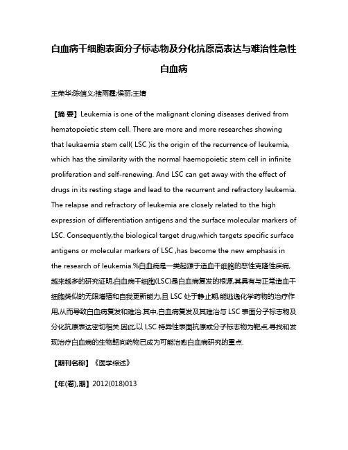
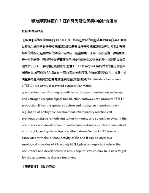
Role of Leukocyte Cell-Derived Chemotaxin2as a Biomarker in Hepatocellular CarcinomaHirohisa Okabe1,2,Evan Delgado1,Jung Min Lee1,Jing Yang1,Hiroki Kinoshita2,Hiromitsu Hayashi2, Allan Tsung3,Jaideep Behari4,Toru Beppu2,5,Hideo Baba2,Satdarshan P.Monga1,4*1Department of Pathology,University of Pittsburgh,Pittsburgh,Pennsylvania,United States of America,2Department of Gastroenterological Surgery,Graduate School of Life Sciences,Kumamoto University,Kumamoto,Japan,3Department of Surgery,University of Pittsburgh Medical Center,Pittsburgh,Pennsylvania,United States of America,4Department of Medicine,University of Pittsburgh,Pittsburgh,Pennsylvania,United States of America,5Department of Multidisciplinary Treatment for Gastroenterological Cancer,Kumamoto University Hospital,Kumamoto,JapanAbstractWe sought to identify a secreted biomarker for b-catenin activation commonly seen in hepatocellular carcinoma(HCC).By examination of our previously published genearray of hepatocyte-specific b-catenin knockout(KO)livers,we identified secreted factors whose expression may be b-catenin-dependent.We verified expression and secretion of the leading factor in HCC cells transfected with mutated(Hep3B S33Y)-b-catenin.Serum levels of biomarker were next investigated in a mouse model of HCC with b-catenin gene(Ctnnb1)mutations and eventually in HCC patients.Leukocyte cell-derived chemotaxin-2 (LECT2)expression was decreased in KO livers.Hep3B S33Y expressed and secreted more LECT2in media as compared to Hep3B WT.Mice developing HCC with Ctnnb1mutations showed significantly higher serum LECT2levels.However patients with CTNNB1mutations showed LECT2levels of54.28622.32ng/mL(Mean6SD;n=8)that were insignificantly different from patients with non-neoplastic chronic liver disease(32.8621.1ng/mL;n=15)or healthy volunteers(33.267.2ng/mL;n=11).Intriguingly,patients without b-catenin mutations showed significantly higher serum LECT2levels(54.26622.25ng/mL;n=46).While b-catenin activation was evident in a subset of non-mutant b-catenin HCC group with highLECT2expression,serum LECT2was unequivocally similar between b-catenin-active and-normal group.Further analysis showed that LECT2levels greater than50ng/ml diagnosed HCC in patients irrespective of b-catenin mutations with specificity of96.1%and positive predictive value of97.0%.Thus,LECT2is regulated by b-catenin in HCC in both mice and men,but serum LECT2reflects b-catenin activity only in mice.Serum LECT2could be a potential biomarker of HCC in patients.Citation:Okabe H,Delgado E,Lee JM,Yang J,Kinoshita H,et al.(2014)Role of Leukocyte Cell-Derived Chemotaxin2as a Biomarker in Hepatocellular Carcinoma.PLoS ONE9(6):e98817.doi:10.1371/journal.pone.0098817Editor:Diego Calvisi,Institut fu¨r Pathologie,Greifswald,Germany,GermanyReceived February19,2014;Accepted May6,2014;Published June3,2014Copyright:ß2014Okabe et al.This is an open-access article distributed under the terms of the Creative Commons Attribution License,which permits unrestricted use,distribution,and reproduction in any medium,provided the original author and source are credited.Funding:This study was funded by NIH grants1R01DK62277,R01DK100287and Endowed Chair for Experimental Pathology to SPM.The funders had no role in study design,data collection and analysis,decision to publish,or preparation of the manuscript.Competing Interests:SPM is a consultant for Abbvie and PhaseRx.None of the content in the current manuscript was affected by the consultation relationship.In addition,this does not alter the authors’adherence to PLOS ONE policies on sharing data and materials.*E-mail:smonga@IntroductionPrimary liver cancer,which is predominantly hepatocellular carcinoma(HCC),is the sixth most common cancer worldwide and the third most frequent cause of cancer mortality[1].b-Catenin gene(CTNNB1)mutations are one of the major oncogenic gene alterations in HCC seen in10–40%,while mutations affecting Axin1are seen in around10%of all HCCs[2].CTNNB1 mutations are observed in exon-3that contain phosphorylation sites essential for b-catenin degradation leading to its stabilization and enhanced expression of target genes such as glutamine synthetase (GS),axin2and regucalcin[3,4,5,6].This mutation is mutually exclusive to p53mutation,which is the most common mutation in HCC[7,8].No routine test is currently available that can yield any genetic information relevant to HCC.Since biopsies for HCC carry high risk because HCCs are usually associated with an underlying liver disease,serum biomarkers specific for a molecular aberration may be highly relevant in personalized medicine in Oncology.LECT2is a16kDa chemotactic protein purified from human T-cell line[9].Since hepatocytes are the chief source,expression of LECT2is specific for liver[10].LECT2is a direct target of b-Catenin and has been shown to have a role in the pathogenesis of HCC[11].In a mouse liver tumor model,LECT2prohibits tumor progression by regulating Th2-based inflammation[12].Although its role is well known as chemokine-like secreted protein involved in inflammation,there is no investigation about its serum levels especially in the setting of liver tumor development.Here,screening a previously published Affymetrix gene array, we identify Lect2expression to be decreased in hepatocyte-specific b-catenin knockout livers[13].Next,using an in vitro analysis in human HCC cells,we demonstrate that indeed LECT2expression and its protein levels reflect b-catenin activity and hence hypothesize that it may be a good biomarker for HCC with b-catenin activation.The utility of LECT2as a biomarker was validated first in a mouse liver tumor model where exon-3 mutation in b-catenin gene and ensuing b-catenin activation is implicated in HCC pathogenesis[14,15].However,in HCC patients,serum LECT2levels were not significantly different intumor with CTNNB1or without CTNNB1mutations when compared to patients with chronic liver disease or healthy volunteers.Furthermore,despite b-catenin activation observed in an additional subset of non-CTNNB1mutated HCC,which showed high LECT2expression,serum LECT2levels were not predictive for active b-catenin signaling in the tumor.Interestingly though,irrespective of molecular aberrations,LECT2levels were significantly higher in all HCC patients versus patients with cirrhosis or healthy controls.In fact,serum LECT2$50ng/ml indicated HCC with high specificity and positive predictive value. Materials and MethodsCell lines and treatmentHuman HCC cell lines,Hep3B,HepG2,SNU449,SNU398, and HuH7,were obtained from the American Type Culture Collection(Manassas,VA).Cells were cultured in Eagle’s minimal essential medium(EMEM)or RPMI supplemented with10%vol/ vol FBS at37u C in a humidified5%carbon dioxide atmosphere. For siRNA knockdown experiment,the cells were transfected using Lipofectamine2000(Life Technologies,Grand Island,NY) with b-catenin(CTNNB1)or scrambled Negative Control siRNAs (Ambion,Grand Island,NY)as previously described[16].Wild type b-Catenin gene(WT)or b-Catenin gene mutated at serine33 to tyrosine(S33Y),which is constitutively active,were stably transfected into Hep3B cells to generate Hep3B WT and Hep3B S33Y,respectively,as described previously[17].Animal studiesAll animal experiments were performed under the guidelines of the National Institutes of Health and the Institutional Animal Use and Care Committee at the University of Pittsburgh and approved by the Institutional Animal Use and Care Committee at the University of Pittsburgh.C3H/He mice were injected intraperi-toneally with DEN(Sigma-Aldrich,Inc.)at a dose of90m g/gram body weight at6weeks of age and3weeks later putting them on a diet containing0.05%phenobarbital(PB)until sacrifice at6–8 months.Serum was collected at time of euthanasia and simultaneously,liver tissues were collected for histology and protein analysis.Mutation analysisGenomic DNA was extracted using DNA Micro Kit(Qiagen, Valencia,CA)from both the frozen liver sections after hematoxilin and eosin(HE)staining(Sigma-Aldrich,Inc.)to identify the tumor and from patient’s frozen liver tumor tissues.Amplification of exon 3of b-catenin gene(CTNNB1)using polymerase chain reaction (PCR),gel extraction,purification of PCR products,and direct sequencing were performed as described previously[18]. Immunohistochemistry and Western blot Immunohistochemical staining and Western blot was performed as described previously[19].Rabbit polyclonal anti-GS(Santa Cruz,1:100dilution)was used in immunohistochemistry.Anti-bodies used in Western blot were mouse monoclonal anti-b-Catenin antibody(Santa Cruz,1:1000dilution),goat polyclonal anti-LECT2antibody(Santa Cruz,1:100dilution),and rabit polyclonal anti-GAPDH(Santa Cruz,1:1000dilution).Real-time polymerase chain reactionIn mice analyses,total RNA was extracted from frozen sections after H&E staining using a mirVana microRNA isolation kit (Ambion)in accordance with the manufacturer’s instructions. After reverse transcription and DNase treatment were performed,qRT-PCR was performed on a StepOne Plus using26SYBR Green PCR Master Mix(Applied Biosystems,Foster,CA).The primers’sequences are as follows:Glutamine Synthetase(encoded by Glul)forward,59-CTCGCTCTCCTGACCTGTTC-39and reverse,59-TTCAAGTGGGAACTTGCTGA-39;LECT2for-ward,59-CCCACAACAATCCTCATTTCA-39and reverse,59-GTTAGCCCATGGTCCTGCTA-39;GAPDH was used as an internal control.In human analyses,total RNA was extracted from frozen tissues and qRT-PCR analysis performed as described previously[20]. Enzyme-linked immunosorbent assay(ELISA)Serum LECT2levels were measured by either human or mouse LECT2ELISA kit(Medical&Biological Laboratories(MBL)Co, Ltd,Niigata,Japan)according to the manufacture’s protocol. Clinical tissue and serum samplesAll tissues and materials used in this study were obtained under an approved Institutional Review Board protocol at the University of Pittsburgh and Kumamoto University.Specifically,frozen tissues and serum samples were obtained from HCC patients in the Department of Surgery,University of Pittsburgh(Pittsburgh, PA;n=20)with a written informed consent approved by the University of Pittsburgh Institutional Review Board.Frozen tissues and serum samples from HCC patients were also collected by the Department of Gastroenterological Surgery,Kumamoto Univer-sity(Kumamoto,Japan;n=45),with a written informed consent approved by the Institutional Review Board at the Kumamoto University.Additional serum samples were obtained from patients with chronic liver disease(n=15)in the Department of Medicine that did not have any evidence of HCC as determined by normal serum a-fetoprotein and negative abdominal ultrasound or CT scan within6months of serum collection.These patients also signed informed consent prior to providing serum samples and the University of Pittsburgh Institutional review board approved the study.Chromatin immunoprecipitation(ChIP)ChIP Assay was performed using Hep3B WT and Hep3B S33Y cells(Cambridge,MA).36106cells were fixed with1% formaldehyde for10min,incubated with glycine(0.125M)for 5min,and then washed twice with PBS.After a short spin,the pellets were resuspended in cell lysis buffer(5mM PIPES,pH8.0, 85mM KCl,0.5%NP-40)by pipetting.After centrifugation,the pellets were resuspended in nuclear lysis buffer(50mM Tris, pH8.1,10mM EDTA,1%SDS)containing protease inhibitors by pipetting,and sonicated to break chromatin into fragments of around0.3–1.0kb length.Subsequent IP experiment was performed with ChIP-IT High sensitivity kit(Active motif, Carlsbad,CA).The diluted DNA-protein complex(25m g of protein)was incubated overnight at4u C with different antibodies (rabbit anti-TCF4,Cell Signaling Technology;rabbit IgG and goat anti-HNF1a,Santa Cruz)in the presence of herring sperm DNA and protein A/G agarose beads.PCR was performed using primers for LECT2promoter region:59-CAGCCCAGAA-GACTGTCGAT-39(forward)and59-GATTAGAGTTGC-CCCCACAC-39(reverse);albumin promoter region,59-TGGAGAAAACAGTTCCAGATGGT-39(forward)and59-CGTGTGGGGTTGACAGAAGA-39(reverse).Cell culture and luciferase AssaySNU449cells were plated in24well plate and treated with either50nM control si-RNA or50nM b-Catenin si-RNA withlipofectamine iMAX (Invitrogen).TOPFLASH reporter plasmid was transfected using the lipofectamine 2000(Invitrogen)a day after the siRNA treatment.Subsequently,cells were treated with TGF-b 1(5ng/mL)(R&D system)for 24hours.Cells were lysed and prepared using the luciferase reporter assay system (Promega,Madison,WI).Reporter activity was read on a luminometer (Lumat;EG &G Berthold).Statistical analysisStatistical analysis was performed using the JMP 8.0software (SAS Institute,Cary,NC,USA).Values were presented as the mean 6standard deviation (SD).Differences between two variables were calculated using the Wilcoxon test.Multiple samples were compared by ANOVA followed by Tukey-Kramer post hoc test.P ,0.05was considered to indicate a statistically significant difference.ResultsIdentification of LECT2regulation by b -CateninTo identify specific biomarkers for b -catenin upregulation,we utilized our previous dataset of microarray analysis comparing gene expression of liver tissue of hepatocyte-specific b -catenin knockout (KO)with that of wild type (WT)[13].From a list of 2963upregulated genes in WT as compared to KO (withfoldFigure 1.Regulation of LECT2expression by b -catenin.A.Strategy to identify biomarker for b -catenin activation.Microarray analysis was performed using liver tissue from hepatocyte-specific b -catenin knockout (KO)and wild-type (WT)mice,which identified 14secreted targets.Lect 2expression was 117-fold lower in KO livers.B.b -Catenin expression in Hep3B cells and stable cell lines established with wild-type b -catenin (Hep3B WT)-or mutated b -catenin (Hep3B S33Y)-transfected cells.C.Representative Western blot shows increased LECT2protein levels in Hep3B S33Y cells as compared to Hep3B WT.D.Hep3B S33Y cells transfected with either b -catenin or control siRNA showed decreased b -catenin and LECT2protein levels in a representative Western blot.E.Increased LECT2protein levels were observed in culture media collected from Hep3B S33Y cells as compared to Hep3B WT as analyzed by ELISA.Basal media was used as a negative control.F.Occupation of Lect2promoter by TCF4especially in Hep3B S33Y cells was as assessed by ChIP.Albumin promoter is not regulated by b -catenin but by HNF1a ,which is used as quality control for chromatin.doi:10.1371/journal.pone.0098817.g001Table 1.Candidate genes encoding secreted protein.Gene NameLect2leukocyte cell-derived chemotaxin 2AFP alpha fetoproteinSlpi secretory leukocyte protease inhibitor Del1del1minor splice variantClecsf8C-type lectin,superfamily member 8Sh2d1a SH2domain protein 1AWnt10a wingless related MMTV integration site 10a IL22b interleukin-22bEgf epidermal growth factorFgf13fibroblast growth factor-related protein FGF-13Wnt4wingless-related MMTV integration site 4Igf2insulin-like growth factor 2Fdp fibrocyte-derived derived protein Scya24eotaxin-2doi:10.1371/journal.pone.0098817.t001change .5),we identified 14genes that encode for secreted proteins (Table 1).The leading candidate lect2showed a 117-fold lower expression in KO as compared to WT (Figure 1A).To validate if LECT2expression could be induced by b -catenin activation,we used two previously generated stable human cell lines,Hep3B cells expressing wild type b -catenin (Hep3B WT )and Hep3B cells expressing S33Y-mutated b -catenin (Hep3B S33Y ),the latter showing highest b -catenin levels (Figure 1B)[17].We compared LECT2expression between these cell lines using qRT-PCR analysis and detected a notable increase in its expression in Hep3B S33Y cells (data not shown).Whole cell lysates also showed increased LECT2protein levels in Hep3B S33Y as compared toHep3B WT cells (Figure 1C).To see if LECT2upregulation is caused by active b -catenin,Hep3B S33Y were transiently transfect-ed with b -catenin or control siRNA.b -Catenin knockdown led to a notable decrease in LECT2protein levels (Figure 1D).Next,to assess if LECT2is being secreted,conditioned media was collected from Hep3B WT and Hep3B S33Y cells and subjected to ELISA assay.Significantly higher LECT2protein was detected in conditioned media from Hep3B S33Y (0.2960.13ng/mL)as compared to Hep3B WT cells (0.02360.046ng/mL)(Figure 1E).Basal media,used a negative control,also showed lack of any LECT2protein (0.01760.057ng/mL).Figure 2.Serum LECT2levels in mice with b -catenin gene mutated HCC.A.Representative picture of b -catenin (left panel)and GS (right panel)immunohistochemistry of liver of a tumor bearing mouse at 8months after DEN/PB treatment.Magnification,ing frozen tissue from a representative tumor,b -catenin gene exon-3mutation affecting codon 33(red box)was confirmed by direct sequencing.C.Serum LECT2levels were significantly (*)increased in tumor bearing versus non-tumor bearing DEN/PB treated mice as analyzed by ELISA.(*p ,0.01).D.Representative pictures of frozen sections from which tumors (T1-T3)were scraped for direct sequencing.E.Sequence analysis from three tumor lesions (T1-T3)show S33Y-b -catenin gene mutations in codon 33(red boxes)by direct sequencing.F.Glutamine Synthetase (Glul)and Lect2expression in three tumor lesions (T1-T3)were assessed by qRT-PCR.Gene expression of background liver tissues surrounding tumor are shown as N.doi:10.1371/journal.pone.0098817.g002Figure3.No correlation of b-catenin mutations or b-catenin activation to serum LECT2levels in patients.A.Serum LECT2levels in patients with HCC with CTNNB1mutations,absent CTNNB1mutations,patients with chronic liver fibrosis(CH/LC),and healthy volunteer(HV)as assessed by ELISA.(*p,0.05).B.No correlation observed between LECT2expression in tumor and serum levels of LECT2in HCC patients(n=28).C.Nocorrelation observed between LECT2expression in b-catenin mutated tumors and serum levels of LECT2in these HCC patients(n=4).D.NoLastly,to specifically determine if b-catenin-T cell factor(TCF) signaling was indeed regulating LECT2expression,chromatin immunoprecipitation(ChIP)analysis was performed on extracts from Hep3B S33Y and Hep3B WT cells.As shown in figure1F, LECT2was pulled down with TCF4and not control IgG in Hep3B S33Y cells.This indicates promoter activity driven by TCF4-b-catenin in Hep3B S33Y cells.Albumin,whose expression is b-catenin-TCF independent,was pulled down by HNF1a in both Hep3B S33Y and Hep3B WT cells.These results show occupancy of Lect2promoter by TCF4,thus demonstrating its regulation by the b-catenin-TCF signaling.HCC with b-Catenin mutation increases serum LECT2 level in miceTo investigate if Lect2could be a serum biomarker in mice,we used a murine HCC model,which utilizes b-catenin signaling as a major mechanism of carcinogenesis.Tumor induction by a single injection of diethylnitrosamine followed by exposure to PB as described in methods and elsewhere has been shown to select for exon-3mutations in b-catenin gene to give rise to HCC at6-8 months[14,15].Indeed,HCC observed in mice with this protocol were strongly GS-positive and had nuclear b-catenin accumula-tion as observed by immunohistochemical staining(Figure2A). Genetic alteration in b-catenin gene contributing to b-catenin activation was confirmed by direct sequencing(Figure2B).We could recognize liver tumor formation in9mice out of13that were subjected to DEN/PB protocol.Strikingly,the9mice with evidence of histological tumor burden showed significantly(p, 0.01)higher serum Lect2levels(55.9619.9ng/mL)as compared to the4non-tumor bearing mice(24.965.5ng/mL)(Figure2C). To confirm mutations in b-catenin gene due to existing tumor heterogeneity and also verify corresponding Lect2expression,we extracted both genomic DNA and total RNA from same nodules (Figure2D).Direct sequencing showed a common mutation (S33Y)in T1,T2and T3nodules(Figure2E).We next examined Lect2mRNA expression as well as Glul mRNA(encoding GS protein)expression in these tumor nodules.All3-tumor nodules had high Lect2and Glul expressions as compared to the background liver(Figure2F).Thus,in mice Lect2expression and eventually its secretion is upregulated by b-catenin mutations in HCC,which can be detected in serum and hence may be a useful biomarker in this mouse model.Serum LECT2level is inconsistent with b-Catenin mutations in human HCCSera were available from54HCC patients through appropriate IRB approvals.Eight of the54patients showed b-catenin gene (CTNNB1)alterations in the form of missense mutations in exon-3 (Table2).Remaining46patients lacked any genetic alterations in exon-3of CTNNB1.Additionally,we enrolled healthy volunteers and also patients with cirrhosis due to chronic liver disease (Table3)to determine serum LECT2levels and address its efficacy as a tumor marker.Based on Tukey-Kramer post hoc test, serum LECT2levels in patients with mutated b-Catenin (54.28622.32ng/mL;n=8)were not statistically different from either patients with cirrhosis(32.8621.1ng/mL,p=0.091; n=15)or healthy volunteers(33.267.2ng/mL,p=0.137; n=11).On the other hand,patients who did not harbor CTNNB1 mutations showed significantly higher LECT2level (54.26622.25ng/mL;n=46)than those with cirrhosis and from healthy volunteers(p=0.0044and0.0176,respectively) (Figure3A).To address if serum LECT2levels showed any correlation with LECT2expression in the tumors,we selected a group of28 patients for which had serum and corresponding tumor tissue (Table2).Intriguingly,no correlation of serum LECT2to its mRNA expression was detectable(Figure3B).Even upon stratification of tumors for presence(n=4)or absence(n=24)of b-catenin gene mutations,no correlation was evident between serum LECT2and its gene expression(Figure3C–D).This led us to further investigate if a subset of these tumors with WT b-catenin gene may still have b-catenin activation.We examined expression of several surrogate Wnt target genes such as AXIN2,REGUCALCIN,LECT2and GLUL in all28tumors using qRT-PCR.Based on the known heterogeneity in Wnt signaling in HCC,we labeled a tumor to be b-catenin-active if at least2of4 target genes were simultaneously upregulated.In addition to the4 tumors with CTNNB1mutations,12more samples showed increased expression of target genes indicative of b-catenin activation as shown in a heat map(Figure3E).Based on this classification,we next compared serum LECT2levels between patients with WT b-catenin gene with active Wnt signaling(+)and with WT b-catenin gene with absent Wnt signaling(2).While some HCC patients in the former group showed high serum LECT2levels,there was no statistical difference between the two groups(Figure3F).Lastly,we compared b-catenin target gene expression among the three HCC groups;1)patients with CTNNB1mutations(MT), 2)patients with WT b-catenin gene but active Wnt signaling(+), and3)those with WT b-catenin gene but lacking Wnt signaling (2).Patients in MT and(+)groups were insignificantly different from each other in expression values of AXIN2,REGUCALCIN, LECT2and GLUL,although the expression was always highest in the MT group(Figure3G).However,both these groups showed significantly higher expression of these four target genes when compared to the(2)group clearly demonstrating lack of active Wnt signaling in this subgroup of HCCs(Figure3D).Intriguingly, (2)group also showed significantly lower LECT2expression than MT and(+)(Figure3G).Taken together,these data indicate that despite some HCC patients showing low LECT2expression in tumors,they unex-pectedly still showed high serum LECT2level with the values ranging from50–80ng/mL.Increased serum LECT2is a biomarker of HCC-independent of b-catenin mutation/activation in tumors Next,due to heterogeneity in HCC,we investigated if serum LECT2levels may be a biomarker of HCC irrespective of acorrelation observed between LECT2expression in non-b-catenin mutated tumors and serum levels of LECT2in these HCC patients(n=24).E.Heat map shows expression of b-catenin target genes in b-catenin-mutated(MT)and non-mutated wild-type(WT)HCC patients(n=28).Genes assessed included AXIN2,REGUCALCIN,LECT2,and GLUL.(+)indicates b-catenin activity as seen by increased expression of at least2target genes whereas(2) indicates absent b-catenin activation reflected by lack of target gene expression.F.Serum LECT2levels showed insignificant difference in HCC patients that lacked b-catenin gene mutations but showed high expression of b-catenin target genes versus patients who have neither b-catenin gene mutations nor any increase in b-catenin target gene expression.G.b-Catenin target gene expression shown by qRT-PCR.Steel-Dwass test was performed to compare the values among three groups.*,p,0.05.(+)indicates b-catenin activity as seen by increased expression of at least2target genes whereas(2)indicates lack of b-catenin activity due lack of target gene expression.doi:10.1371/journal.pone.0098817.g003T a b l e 2.C l i n i c a l c h a r a c t e r i s t i c s o f p a t i e n t s w i t h H C C .P a t i e n tA g e G e n d e r T u m o r s i z e (c m )D i f f e r e n t i a t i o n s t a t u s S e r u m L E C T 2l e v e l (n g /m L )M u t a t i o n R N A a v a i l a b l eK 28481M4.1W e l l 93.2W T #K 28564M1.7W e l l 87.9W T 6K 28669F4.9M o d e r a t e 48.7W T #K 28762M1.6W e l l 62.6W T #K 28863M1.7W e l l 52.6W T #K 28958M3.8M o d e r a t e 83.3W T 6K 29077M1.5M o d e r a t e 64.6W T #K 29152M7.8M o d e r a t e 63.4W T #K 29339M2.0M o d e r a t e 69.3W T #K 29436M2.5M o d e r a t e 32.8W T6K 29574M4.0M o d e r a t e 78.2W T#K 29676M2.4M o d e r a t e 46.9W T#K 29770M4.4N e c r o s i s (T A C E )95.1W T6K 29866M4.3M o d e r a t e 54.4W T6K 29967M2.5M o d e r a t e 61.3W T#K 30074F5.5M o d e r a t e 32.8W T6K 30164M3.5M o d e r a t e 57.2M T6K 30286M5.5P o o r 51.8M T#K 30365M2.3M o d e r a t e 41.8W T#K 30476F1.0W e l l 56.1W T#K 30577F2.5M o d 34.9M T#K 30673M1.0W e l l 56.1W T#K 30772M2.3M o d e r a t e 64.2W T6K 30866M11.0M o d e r a t e 66.1W T#K 30972M6.8M o d e r a t e 58.7W T#K 31076M4.6W e l l 101.3M T#K 31168F9.0M o d e r a t e 45.6W T6K 31260M1.3P o o r49.3W T6K 31382M4.0M o d e r a t e34.7W T6K 31483F4.5M o d e r a t e59.5M T6K 31562M4.5M o d e r a t e57.7W T#K 31675M4.0M o d e r a t e68.4W T#K 31782F7.0P o o r56.0W T#K 31869M3.0M o d e r a t e58.8M T#K 31963M4.0M o d e r a t e55.5W T#T a b l e 2.C o n t .P a t i e n tA g e G e n d e r T u m o r s i z e (c m )D i f f e r e n t i a t i o n s t a t u s S e r u m L E C T 2l e v e l (n g /m L )M u t a t i o n R N A a v a i l a b l eK 32067M2.2W e l l 60.3M T #K 32159M2.2M o d e r a t e 88.0W T #K 32263M6.0M o d e r a t e 13.4W T #K 32369M3.3M o d e r a t e 68.8W T #K 32464M3.9M o d e r a t e 56.7W T #K 32566M3.0M o d e r a t e 61.3W T 6K 32677M2.5P o o r 47.8W T 6K 32770M7.6M o d e r a t e 31.9W T 6K 32856M6.1M o d e r a t e 103.3W T 6K 32976M4.0M o d e r a t e 56.1W T6P 31858M10.6M o d e r a t e 17.2W T6P 33676F2.5M o d e r a t e 10.2W T6P 32857F4.2M o d e r a t e 5.5W T6P 51372M-M o d e r a t e 38.9W T6P 52069M5.0M o d e r a t e 18.5W T6P 78856M14.0M o d e r a t e 42.7W T6P 95871M17.0M o d e r a t e 28.4M T6P 133564M2.5W e l l 49.8W T6P 134268M2.2M o d e r a t e 26.2W T6d o i :10.1371/j o u r n a l.p o ne .0098817.t 002。