Transgenic Expression of the Recombinant Phytase in Rice (Oryza sativa)
EXPRESSION OF RECOMBINANT HEMOGLOBIN IN YEAST

专利名称:EXPRESSION OF RECOMBINANTHEMOGLOBIN IN YEAST发明人:DE ANGELO, Joseph,MOTWANI, Nalini, M.,BAJWA, Wajeeh申请号:EP91908364.2申请日:19910415公开号:EP0537160B1公开日:20020821专利内容由知识产权出版社提供摘要:the invention relates to a substantially pure mammalian globins chain or a fragment of heme in the chain, to recombinant dna vectors capable of expressing at least one channel or a substantially homologous variant globins of the chain in the yeast.as well as the methods of expression of at least one channel or a substantially homologous variant globins of the chain in the yeast. the chains of globins of the alpha and beta globins of expressed or variants of these chains may be combined with a source of heme to produce hemoglobin or a hemoglobin variant in counterpart.chains of globins gamma expressed may be further combined with a source of heme to produce hemoglobin or a hemoglobin variant in counterpart.the invention also relates to methods of expression of hemoglobin or hemoglobin variants in yeast, in which the heme is produced by the yeast and linked to the globins to form of hemoglobin in vivo.the hemoglobin produced by such methods can be used in applications requiring a physiological oxygen carrier, for example, alternatives and substitutes for blood plasma or in applications requiring an oxygen carrier ph ysiologique.申请人:APEX BIOSCIENCE, INC.代理机构:Hirsch, Marc-Roger 更多信息请下载全文后查看。
targetedinsertio...

Targeted Insertion of Cre Recombinase into the TNAP Gene:Excision in Primordial Germ CellsHilda Lomelı´,1Vero´nica Ramos-Mejı´a,1Marina Gertsenstein,2Corrinne G.Lobe,3,4and Andras Nagy2,5*1Departamento de Gene´tica y Fisiologia Molecular,Instituto de Biotecnologı´a.Universidad Nacional Auto´noma de Mexico,Cuernavaca,Mexico2Samuel Lunenfeld Research Institute,Mount Sinai Hospital,Toronto,Ontario,Canada3Cancer Research Division,Sunnybrook Health Science Centre,Toronto,Ontario,Canada4Department of Biophysics,University of Toronto,Toronto,Ontario,Canada5Department of Molecular and Medical Genetics,University of Toronto,Toronto,Ontario,CanadaReceived3November1999;Accepted8November1999Cell type-specific genomic alterations afforded by theCre/loxP system are a powerful approach to study thefunction of genes in particular cell lineages and forwhich generalized null mutations result in early lethalphenotypes(Lobe and Nagy,1998).Here we report thecreation of a primarily PGC(primordial germ cell)spe-cific Cre recombinase transgenic line,designated TNAP-Cre.In this strain,the Cre recombinase has beenknocked into the locus of the TNAP(Tissue Non-SpecificAlkaline Phosphatase)gene.The structure of the TNAP-Cre targeting construct isdepicted in Figure1a.After electroporation into R1ES(Nagy et al.,1993a)cells,about30%of the clonesscreened by Southern analysis had undergone homolo-gous recombination(Fig.1b).Two clones were used toderive germ-line chimaeras(Nagy et al.,1993b).Bothlines gave germline transmission and the two transgeniclines did not differ from each other in any aspects stud-ied.Our heterozygous TNAP-Cre mice on a hybrid back-ground did not show any abnormality,in agreementwith previous report of the null mutation of the TNAPgene(MacGregor et al.,1995).To test the in vivo specificity of excision achieved bythe TNAP-Cre mouse lines,we crossed them with thedouble-reporter line,Z/AP(Lobe et al.,1999).The Z/APtransgene expresses lacZ before Cre-mediated excisionand the heat-resistant human alkaline phosphatase(hAP)gene after Cre excision.Double transgenic TNAP-Cre/Z/AP embryos showed the initial hAP activity was spe-cific to the PGCs at E9.5–10.5(Fig.2).After midgesta-tion,however,it was also expressed in the labyrinthineregion of the placenta,the intestine and the neural tube.A detailed analysis of E13embryos from crosses betweenTNAP-Cre males and Z/AP females showed8out of16embryos were positive exclusively in PGCs.Three em-bryos showed additional activity in ventral neural tubeand/or placenta although always at a lower level than inthe PGCs.Five presented a mosaic type of ectopic activ-ity all over the embryos with a strong signal in PGCs.In the offspring of reciprocal crosses(Z/AP male,TNAP-Cre*Correspondence to:Andras Nagy,Samuel Lunenfeld Research Institute, Mount Sinai Hospital,600University Ave.,Toronto,Ontario M5G1X5, Canada.E-mail:*************.caFIG.1.Generation of a TNAP Cre allele by homologous recombina-tion.(a)The wild type TNAP locus,the targeting construct and the predicted structure of the TNAP Cre allele are represented.Exons are depicted by numbered boxes,shaded boxes represent the knocked in DNA.I is IRES(Jang et al.,1988),Cr is Cre,N-pA is neomycin and polyA sequence.Sizes of the diagnostic restriction fragment(in kb) of the endogenous and targeted allele obtained with BfrI are indi-cated with arrows.The probe used in the southern analysis is denoted with a shaded box,called pro3.(b)Southern analysis on DNA from129wild type and the two different clones(12and45).©2000Wiley-Liss,Inc.genesis26:116–117(2000)female),the fraction of embryos presenting ectopic ac-tivity was higher,suggesting either the presence of ma-ternally produced Cre in the oocytes or different speci-ficity of the maternal TNAP-Cre allele.To evaluate the actual excision activity of the Crerecombinase in germ cells,the gonads from separateE13.5double transgenic embryos were dissected anddisaggregated.For each gonad,half the cells werestained for endogenous TNAP activity to indicate thetotal number of germ cells.The other half was stainedfor hAP activity to estimate the number of cells with Creexcision.This experiment indicated 59.8%Ϯ16.6,(n ϭ6)of the germ cells had Cre activity.We next examined double transgenic embryos (Cre ϩ/Z/AP ϩ)at E6–E8to determine the earliest stage of primordialgerm cell specific excision.In most of the cases,E6,E7,andE8double transgenic embryos did not show significant hAPactivity in prospective PGCs or elsewhere (not shown).Some embryos in which hAP activity was detected at theseearly stages presented mosaic ectopic patterns.At E9,how-ever,the PGCs were positive for hAP activity in most of theembryos.This observation indicates that Cre excision oc-curs before E9,which already results in detectable hAPactivity at this stage.We believe that the TNAP-Cre mouse will facilitate theproduction of germ cell-specific mutations in mice.Al-though excision of floxed DNA is not always exclusive tothe PGCs,the frequency of specific excision is highenough for feasible use of this reagent in many studiesaddressing functions of genes in PGC development.ACKNOWLEDGMENTS We thank George MacGregor for providing the genomic clone of the TNAP gene,and Gail Martin and Mark Lewandoski for providing plasmids PML78containing the NLS-Cre and plasmid pK11containing the FRT flanked Neo.REFERENCES Jang SK,Krausslich HG,Nicklin MJ,Duke GM,Palmenberg AC,Wim-mer E.1988.A segment of the 5Јnontranslated region of encepha-lomyocarditis virus RNA directs internal entry of ribosomes during in vitro translation.J Virol 62:2636–2643.Lobe CG,Nagy A.1998.Conditional genome alteration in mice.BioEs-says 20:200–208.Lobe CG,Koop KE,Kreppner W,Lomeli H,Gertsenstein M,Nagy A.1999.Z/AP,a double reporter for Cre-Mediated Recombination.Dev Biol 208:281–292.MacGregor GR,Zambrowicz BP,Soriano P.1995.Tissue non-specific alkaline phosphatase is expressed in both embryonic and extraem-bryonic lineages during mouse embryogenesis but is not required for migration of primordial germ cells.Development 121:1487–1496.Nagy A,Rossant J,Nagy R,Abramow-Newerly W,Roder J.1993a.Derivation of completely cell culture-derived mice from early-passage embryonic stem cells.Proc Natl Acad Sci USA 90:8424–8428.Nagy A,Rossant J.1993b.Production of completely ES cell-derived fetuses.In:Joyner A,editor.Practical approach series:Gene tar-geting:A practical approach.Oxford:IRL.p147–179.FIG.2.Alkaline phosphatase gene expression after Cre excision in double transgenic Z/AP-TNAP Cre embryos.(A )A whole-mount (headless)E9.5embryo stained for hAP activity.The staining is indicated with arrowheads and localized within the developing gut region,where germ cells are found at this time,and in the neural tube.(B )A whole-mount E10.5double transgenic embryo showing staining in the dorsal mesentery region,where germ cells are migrating at this time.(C )A dissected lower half of an E13double transgenic embryo stained for hAP.Reporter expression is localized in primordial germ cells now present in the genital ridge (gr).For whole-mount lacZ and alkaline phosphatase staining of embryos and tissues,we followed the procedure described in Lobe et al.,1999.117INSERTION OF CRE RECOMBINASE INTO TNAP GENE。
矮牵牛转基因

Background and Significance
4. Nevertheless, efficiency can be effectively improved by way of the transgenic technique. 5. Cytochrome P450 2E1 is a decomposition enzyme in mammalian liver, with the strong and specific capacity of decomposing organic pollutants in animal bodies.
Transgenic plants of Petunia hybrida harboring the CYP2E1 gene efficiently remove benzene and toluene pollutants and improve resistance to formaldehyde
Thanks!
ห้องสมุดไป่ตู้
Material and Methods
Methods 1.Transformation of pSLD50-6 and pKH200 plasmids into Agrobacterium rhizogenes K599 2. Induction of transgenic hairy roots and regeneration of Petunia plants. 3. Fluorescent quantitative RT-PCR analysis of CYP2E1 genes in transgenic Petunia plants. 4. Gas chromatography analysis of Petunia transgenic-plant benzene and toluene absorption Capacity.
EMT有关基因

MINI REVIEW ARTICLEpublished:17September2013doi:10.3389/fonc.2013.00221 Role of epithelial-mesenchymal transition in pancreatic ductal adenocarcinoma:is tumor budding the missing link? Eva Karamitopoulou1,2*1Clinical Pathology Division,Institute of Pathology,University of Bern,Bern,Switzerland2Translational Research Unit,Institute of Pathology,University of Bern,Bern,SwitzerlandEdited by:Inti Zlobec,University of Bern, SwitzerlandReviewed by:Parham Minoo,University of Calgary, CanadaQianghua Xia,The Children’s Hospital of Philadelphia,USA*Correspondence:Eva Karamitopoulou,Clinical Pathology Division,Institute of Pathology,University of Bern, Murtenstrasse31,CH-3010Bern, Switzerlande-mail:eva.diamantis@pathology.unibe.ch Pancreatic ductal adenocarcinoma(PDAC)ranks as the fourth commonest cause of cancer death while its incidence is increasing worldwide.For all stages,survival at5years is<5%. The lethal nature of pancreatic cancer is attributed to its high metastatic potential to the lymphatic system and distant ck of effective therapeutic options contributes to the high mortality rates of PDAC.Recent evidence suggests that epithelial-mesenchymal transition(EMT)plays an important role to the disease progression and development of drug resistance in PDAC.Tumor budding is thought to reflect the process of EMT which allows neoplastic epithelial cells to acquire a mesenchymal phenotype thus increasing their capacity for migration and invasion and help them become resistant to apoptotic signals. In a recent study by our own group the presence and prognostic significance of tumor budding in PDAC were investigated and an association between high-grade budding and aggressive clinicopathological features of the tumors as well as worse outcome of the patients was found.The identification of EMT phenotypic targets may help identifying new molecules so that future therapeutic strategies directed specifically against them could potentially have an impact on drug resistance and invasiveness and hence improve the prognosis of PDAC patients.The aim of this short review is to present an insight on the morphological and molecular aspects of EMT and on the factors that are involved in the induction of EMT in PDAC.Keywords:pancreatic cancer,epithelial-mesenchymal transition,tumor budding,prognosis,biomarkerPANCREATIC CANCERPancreatic ductal adenocarcinoma(PDAC)is a common can-cer with dismal prognosis(1)that escapes early detection and resists treatment(2).Most patients have advanced stage dis-ease at presentation with a median survival of less than1year (1,3).Surgical resection is the only potentially curative treat-ment of PDAC(3).Classical histomorphological features like tumor size,blood vessel,or lymphatic invasion,and presence of lymph node metastases constitute essential prognostic deter-minants in pancreatic cancer and are invariably included in the pathology reports,with tumor stage being the most important of all(3).The lethal nature of PDAC has been attributed to the propensity of PDAC cells to rapidly disseminate to the lym-phatic system and distant organs(4).However,even patients with completely resected,node-negative PDACs eventually die of their disease.Within this context and considering the fact that the management of PDAC remains suboptimal and that adjuvant therapy has resulted to limited progress,the identification of addi-tional reliable and reproducible prognostic markers that would enable better patient stratification and eventually provide a guide toward a more successful and individualized therapy,is mandatory (1,5).EPITHELIAL-MESENCHYMAL TRANSITIONEpithelial-mesenchymal transition is a biologic process that allows epithelial cells to undergo the biochemical changes that enable them to acquire a mesenchymal phenotype,including enhanced migratory capacity,invasiveness,elevated resistance to apoptosis, and increased production of extracellular matrix(ECM)compo-nents(6,7).EMT is characterized by loss of cell adhesion,down regulation of E-cadherin expression,acquisition of mesenchy-mal markers(including N-cadherin,Vimentin,and Fibronectin), and increased cell motility(6).Both EMT and mesenchymal-epithelial transition(MET),the reversion of EMT,are essential for developmental and repair processes like implantation,embryo for-mation,and organ development as well as wound healing,tissue regeneration,and organfibrosis(8).However,EMT also occurs in neoplastic cells that have undergone genetic and epigenetic changes.These changes affect both oncogenes and tumor sup-pressor genes that enable cancer cells to invade and metastasize. Moreover,some neoplastic cells may go through EMT retaining many of their epithelial properties while other cells are becoming fully mesenchymal(9).Many molecular processes are involved in the initiation of EMT including activation of transcription factors,expression of specific cell-surface proteins,reorganization and expression of cytoskeletal proteins,production of ECM-degrading enzymes,and changes in the expression of specific microRNAs(miRNAS).The above fac-tors can also be used as biomarkers to detect cells in EMT state(10). EMT has been linked to cellular self-renewal programs of cancer stem cells and apoptosis-anoikis resistance,which are features of therapeutic resistance(11).The zincfinger transcription factors Snail,Slug,Zeb1,and Twist repress genes responsible for the epithelial phenotype and represent important regulators of EMT(6,7,12).In PDAC Snail expression has been reported to be seen in nearly80%of the cases and Slug expression in50%(13).Snail expression was inversely correlated with E-cadherin expression and decreased E-cadherin expression was associated with higher tumor grade. Similarly,poorly differentiated pancreatic cancer cell lines showed higher levels of Snail and lower levels of E-cadherin compared with moderately differentiated cell lines(13)while silencing of Zeb1leaded to up-regulation of E-cadherin and restoration of an epithelial phenotype(14).Zeb1expression in PDAC also corre-lated with advanced tumor grade and worse outcomes(14–16) and was shown to be primarily responsible for the acquisition of an EMT phenotype,along with increased migration and inva-sion in response to NF-κB signaling in pancreatic cancer cells (16).EMT AND TUMOR BUDDINGTumor budding reflects a type of diffusely infiltrative growth con-sisting of detached tumor cells or small cell clusters of up tofive cells at the invasive front of gastrointestinal carcinomas(17–22). Tumor buds represent a non-proliferating,non-apoptotic,highly aggressive subpopulation of tumor cells that display migratory and invasive capacities(23).The aim of tumor buds seems to be the invasion of the peritumoral connective tissue,the avoidance of the host’s defense andfinally the infiltration of the lymphatic and blood vessels with the consequence of local and distant metastasis. The EMT process by allowing a polarized cell to assume a more mesenchymal phenotype with increased migratory capacity,inva-siveness,and resistance to apoptosis seems to play a major role in the development of tumor buds.In fact,tumor buds are thought to result from the process of EMT.Thus,although formally tumor budding cannot be equated with EMT,several similarities between the two processes,including activation in WNT signaling,can be shown(24).The detachment of tumor buds from the main tumor body is accomplished by loss of membranous expression of the adhesion molecule E-cadherin.Activation of WNT sig-naling is further suggested by nuclear expression of b-catenin in tumor-budding cells,as well as increase of laminin5gamma2and activation of Slug and Zeb1(24,25).The presence of high-grade tumor budding has been consis-tently associated with negative clinicopathologic parameters in gastrointestinal tumors(26–30).In a previous study from our group we could show that tumor budding occurs frequently in pancreatic cancer and is a strong,independent,and reproducible, highly unfavorable prognostic factor that may be used as a para-meter of tumor aggressiveness and as an indicator of unfavorable outcome,even within this group of patients with generally poor prognosis.Moreover,tumor budding was proven to have a more powerful prognostic ability than other more classic prognostic fac-tors including TNM stage,thus adding relevant and independent prognostic information(31).EMT AND miRNAsMicroRNAS are small non-coding RNAs of18–25nucleotides, excised from60to110nucleotide RNA precursor structures (32).MiRNAs are involved in crucial biological processes, including development,differentiation,apoptosis,and pro-liferation,through imperfect pairing with target messenger RNAs of protein-coding genes and the transcriptional or post-transcriptional regulation of their expression(33,34).Recent studies illustrate the role of miRNAs on the regula-tion of gene expression and proteins in metastasis.For exam-ple,it has been shown that miR-10b,which is up-regulated by EMT transcription factor Twist,is associated with increased invasiveness and metastatic potential(35,36).Furthermore,it was shown that the miR-200family(miR-200a,miR-200b,miR-200c,miR-141,and miR-429)and miR-205play critical roles in regulating EMT by directly targeting the mRNAs encoding E-cadherin repressors Zeb1and Zeb2(37).Moreover,recent studies showed that members of the miR-200family by induc-ing EMT can regulate the sensitivity to epidermal growth fac-tor receptor(EGFR)in bladder cancer cells and to gemcitabine in pancreatic cancer cells(38).Conversely,Zeb1represses the transcription of miR-200genes by directly binding to their promoter region,thereby forming a double-negative feedback loop(39).On the other hand,miR-200family can also pro-mote the conversion of mesenchymal cells to epithelial-like cells (MET)suggesting that these miRNAs may also favor metastatic outgrowth.Recent studies aiming at the evaluation of miRNAs in pan-creatic cancer have shown that specific miRNAs are dysregulated in PDAC while the higher expression of some miRNA species was able to distinguish between benign and malignant pancre-atic tissue(40).For example,miR-21was shown to be over-expressed in79%of pancreatic cancers as opposed to27%of chronic pancreatitis(41).In resected PDAC specimens high lev-els of miR-200c expression strongly correlated with E-cadherin levels and were associated with significantly better survival rates compared with patients whose tumors had low levels of miR-200c expression(42).CHEMORESISTANCE AND EMTCells undergoing EMT become invasive and develop resistance to chemotherapeutic agents.Moreover,EMT can be induced by chemotherapeutic agents,and stress conditions such as exposure to radiation or hypoxia(43,44).Up-regulation of Twist has been shown to be associated with resistance to paclitaxel in nasopharyngeal,bladder,ovarian,and prostate cancers(45).In colorectal cancer cell lines,chronic expo-sure to oxaliplatin leaded to the development of the ability to migrate and invade with phenotypic changes resembling EMT(spindle-cell shape,loss of polarity,intercellular separa-tion,and pseudopodia formation)by the oxaliplatin-resistant cells(46).Pancreatic cancer remains today an extremely lethal disease largely because of its resistance to existing treatments(47).EMT has been shown to contribute significantly to chemoresistance in several cancers,including pancreatic cancer(30,48,49).Induction of gemcitabine resistance in previously sensitive cell lines resulted in development of an EMT phenotype and was associated with an increased migratory and invasive ability compared to gemc-itabine sensitive cells(49).Moreover,gene expression profiling ofchemoresistant cells showed a strong association between expres-sion of the EMT transcription factors Zeb1,Snail,and Twist and decreased expression of E-cadherin(39,50).Silencing of Zeb1 with siRNA resulted to MET(51)and restored chemosensitivity (14).Interestingly,maintenance of chemoresistance in cell lines that have undergone EMT is dependent on Notch and NF-κB signaling(30).Inhibition of Notch-2down regulates Zeb1,Snail, and Slug expression,attenuates NF-κB signaling,and reduces the migratory and invasive capacity of the gemcitabine resistant cells(30).Epithelial-mesenchymal transition can also confer resistance to targeted agents.For example,lung cancer cell lines that have undergone EMT,became resistant to the growth inhibitory effects of EGFR kinase inhibition(erlotinib)in vitro and in xenografts(47)as well as other EGFR inhibitors such as gefitinib and cetuximab(48)Thus,EMT can lead to resis-tance to multiple agents and result to rapid progression of the tumor.Clarifying the correlation between EMT and drug resistance may help clinicians select an optimal treat-ment.CONCLUSIONPancreatic cancer remains an extremely lethal disease partly because of the poor response to existing treatments.Accumulat-ing evidence suggests that EMT plays an important role in PDAC progression,is associated with stem cell features of the PDAC cells and seems to significantly contribute to the chemoresistance of pancreatic cancer.Moreover,is associated with more aggressive tumor characteristics and with poor patient survival.Because of its role in therapy response and tumor progression,targeting EMT could potentially reduce drug resistance and have a great impact in the survival of PDAC patients.Tumor budding thought to be the result of the EMT process is commonly observed in PDAC and high-grade tumor budding has been proven to have an independent adverse prognostic impact in the survival of PDAC patients.Figure1depicts tumor bud-ding as a possible transition between a fully epithelial and a fully mesenchymal phenotype of the tumor cells in PDAC.Moreover, cancer cells in tumor buds have been shown to have EMT and cancer stem cell characteristics.The further characterization of the budding cells at a protein and gene level in order to iden-tify a“molecular budding-promoting profile”will lead to a better understanding of the tumor-stroma interaction at the area of the invasive front and help to further elucidate the similarities between budding cells,EMT process and cancer stem cells in pancreatic cancer.Investigating these issues will allow us to gain further insight into pancreatic carcinogenesis,and provide us with a platform on which to build future studies leading to the identification of new therapeutic interventions.REFERENCES1.Hidalgo M.Pancreatic cancer.NEngl J Med(2010)362:1605–17.doi:10.1056/NEJMra09015572.Tuveson DA,Hingorani SR.Duc-tal pancreatic cancer in humans and mice.Cold Spring Harb Symp Quant Biol(2005)70:65–72.doi:10.1101/ sqb.2005.70.0403.Fernandez-del-Castillo C,JimenezRE,Steer ML.Surgery in the treatment of exocrine pancreas and prognosis.In:Tanabe KK,edi-tor(2013).Available from:www.4.Li Y,Kong D,Ahmad A,Bao B,Sarkar FH.Pancreatic cancer stem cells:emerging target for designing novel therapy.Cancer Lett(2012).Available from:/10.1016/j canlet.2012.03.018,5.Welsch T,Kleeff J,Friess H.Molecu-lar pathogenesis of pancreatic can-cer:advances and challenges.Curr Mol Med(2007)7:504–21.doi:10.2174/1566524077813870826.Kalluri R,Weinberg RA.The basicsof epithelial-mesenchymal transi-tion.J Clin Invest(2009)119:1420–8.doi:10.1172/JCI391047.Thiery JP,Acloque H,Huang RYJ,Nieto MA.Epithelial-mesenchymal transitions in development and dis-ease.Cell(2009)139:871–90.doi:10.1016/j.cell.2009.11.0078.Rasheed ZA,Yang J,Wang Q,Kowal-ski J,Freed I,Murter C,et al.Prog-nostic significance of tumorigenic cells with mesenchymal features in pancreatic adenocarcinoma.J Natl Cancer Inst(2010)102:340–51.doi:10.1093/jnci/djp5359.Iwatsuki M,Mimori K,YokoboriT,Ishi H,Beppu T,Nakamori S.Epithelial–mesenchymal transi-tion in cancer development and its clinical significance.Cancer Sci (2010)101(2):293–9.doi:10.1111/j.1349-7006.2009.01419.x10.Kalluri R.EMT:when epithelial cellsdecide to become mesenchymal-like cells.J Clin Invest(2009)119:1417–9.doi:10.1172/JCI3967511.Krantz SB,Shields MA,Dangi-Garimella S,Munshi HG,Bentrem DJ.Contribution of epithelial-to-mesenchymal transition and cancer stem cells to pancreatic cancer progression.J Surg Res(2012)173: 105–12.doi:10.1016/j.jss.2011.09.02012.Lee JM,Dedhar S,Kalluri R,Thompson EW.The epithelial-mesenchymal transition:new insights in signaling,devel-opment,and disease.J Cell Biol(2006)172:973–81.doi:10.1083/jcb.20060101813.Hotz B,Arndt M,Dullat S,BhargavaS,Buhr HJ,Hotz HG.Epithelial tomesenchymal transition:expressionof the regulators snail,slug,andtwist in pancreatic cancer.ClinCancer Res(2007)13:4769–76.doi:10.1158/R-06-292614.Arumugam T,Ramachandran V,Fournier KF,Wang H,MarquisL,Abbruzzese JL,et al.Epithe-lial to mesenchymal transition con-tributes to drug resistance in pan-creatic cancer.Cancer Res(2009)69:5820–8.doi:10.1158/0008-5472.CAN-08-281915.Buck E,Eyzaguirre A,Barr S,Thompson S,Sennello R,Y oung D,et al.Loss of homotypic cell adhe-sion by epithelial-mesenchymaltransition or mutation limits sen-sitivity to epidermal growth factorreceptor inhibition.Mol CancerTher(2007)6:532–41.doi:10.1158/1535-7163.MCT-06-046216.Maier HJ,Schmidt-Strassburger U,Huber MA,Wiedemann EM,BeugH,Wirth T.NF-kappaB promotesepithelial-mesenchymal transition,migration and invasion of pancre-atic carcinoma cells.Cancer Lett(2010)295:214–28.doi:10.1016/j.canlet.2010.03.00317.Prall F.Tumour budding in col-orectal carcinoma.Histopathology(2007)50:151–62.doi:10.1111/j.1365-2559.2006.02551.x18.Brown M,Sillah K,Griffiths EA,Swindell R,West CM,Page RD,et al.Tumour budding and a lowhost inflammatory response areassociated with a poor progno-sis in oesophageal and gastro-oesophageal junction cancers.Histopathology(2010)56:893–9.doi:10.1111/j.1365-2559.2010.03559.x19.Koike M,Kodera Y,Itoh Y,Nakayama G,Fujiwara M,Hamajima N,et al.Multivariateanalysis of the pathologic featuresof esophageal squamous cell cancer:tumor budding is a significantindependent prognostic factor.AnnSurg Oncol(2008)15:1977–82.doi:10.1245/s10434-008-9901-620.Miyata H,Y oshioka A,YamasakiM,Nushijima Y,Takiguchi S,Fujiwara Y,et al.Tumor bud-ding in tumor invasive front pre-dicts prognosis and survival ofpatients with esophageal squa-mous cell carcinomas receivingneoadjuvant chemotherapy.Cancer(2009)115:3324–34.doi:10.1002/cncr.2439021.Roh MS,Lee JI,Choi PJ.Tumorbudding as a useful prognosticmarker in esophageal squamous cellcarcinoma.Dis Esophagus(2004)17:333–7.doi:10.1111/j.1442-2050.2004.00436.x22.Ohike N,Coban I,Kim GE,Bas-turk O,Tajiri T,Krasinskas A,etal.Tumor budding as a strongprognostic indicator in invasiveampullary adenocarcinomas.Am JSurg Pathol(2010)34:1417–24.doi:10.1097/PAS.0b013e3181f0b05a23.Zlobec I,Lugli A.Epithelial mes-enchymal transition and tumorbudding in aggressive colorectalcancer:tumor budding as oncotar-get.Oncotarget(2010)1:651–61.24.Muto T,Mochizuki H,Masaki T edi-tors.Tumor Budding in ColorectalCancer:Recent Progress in ColorectalCancer Research(Horizons in Can-cer Research).(Vol.8).Tokyo:Nova(2006).25.Schmalhofer O,Brabletz S,Brabletz T.E-cadherin,beta-catenin,and ZEB1in malignantprogression of cancer.CancerMetastasis Rev(2009)28:151–66.doi:10.1007/s10555-008-9179-y26.Karamitopoulou E,Zlobec I,KölzerV,Kondi-Pafiti A,Patsouris ES,Gen-natas K,et al.Proposal for a10-high-power-fields scoring methodfor the assessment of tumor bud-ding in colorectal cancer.ModPathol(2013)26:295–301.doi:10.1038/modpathol.2012.15527.Nakamura T,Mitomi H,KikuchiS,Ohtani Y,Sato K.Evaluationof the usefulness of tumor bud-ding on the prediction of metasta-sis to the lung and liver after cura-tive excision of colorectal cancer.Hepatogastroenterology(2005)52:1432–5.28.Ueno H,Mochizuki H,HashiguchiY,Hatsuse K,Fujimoto H,HaseK.Predictors of extrahepatic recur-rence after resection of colorectalliver metastases.Br J Surg(2004)91:327–33.doi:10.1002/bjs.442929.Ueno H,Murphy J,Jass JR,Mochizuki H,Talbot IC.Tumour‘budding’as an indexto estimate the potential ofaggressiveness in rectal cancer.Histopathology(2002)40:127–32.doi:10.1046/j.1365-2559.2002.01324.x30.Wang LM,Kevans D,Mulcahy H,O’Sullivan J,Fennelly D,Hyland J,et al.Tumor budding is a strong andreproducible prognostic marker inT3N0colorectal cancer.Am J SurgPathol(2009)33:134–41.doi:10.1097/PAS.0b013e318184cd5531.Karamitopoulou E,Zlobec I,BornD,Kondi-Pafiti A,Patsouris E,Gen-natas K,et al.Tumor budding isa strong and independent prognos-tic factor in pancreatic cancer.EurJ Cancer(2013)49:1032–9.doi:10.1016/j.ejca.2012.10.02232.Calin GA,Croce CM.Micro RNAsignatures in human cancers.NatRev Cancer(2006)6:857–66.doi:10.1038/nrc199733.McShane L,Altman DG,SauerbreiW,Taube SE,Gion M,Clark GM,etal.REporting recommendations fortumor MARKer prognostic studies(REMARK).Eur J Cancer(2005)41:1690–6.doi:10.1016/j.ejca.2005.03.03234.Pasquinelli AE,Hunter S,BrachtJ.MicroRNAs:a developing story.Curr Opin Gen Dev(2005)15:200–5.doi:10.1016/j.gde.2005.01.00235.Peter ME.Let-7and miR-200microRNAs:guardians againstpluripotency and cancer progres-sion.Cell Cycle(2009)8:843–52.doi:10.4161/cc.8.6.790736.Preis M,Gardner TB,Gordon SR,Pipas JM,Mackenzie TA,Klein EE,et al.MicroRNA10b expression cor-relates with response to neoadju-vant therapy and survival in pan-creatic ductal adenocarcinoma.ClinCancer Res(2011)17:5812–21.doi:10.1158/R-11-069537.Gregory PA,Bert AG,Paterson EL,Barry SC,Tsykin A,Farshid G,et al.The miR-200family and miR-205regulate epithelial to mesenchymaltransition by targeting ZEB1andSIP1.Nat Cell Biol(2008)10:593–601.doi:10.1038/ncb172238.Burk U,Schubert J,Wellner U,Schmalhofer O,Vincan E,SpadernaS,et al.A reciprocal repressionbetween ZEB1and members of themiR-200family promotes EMT andinvasion in cancer cells.EMBO Rep(2008)9:582–9.doi:10.1038/embor.2008.7439.Wellner U,Schubert J,Burk UC,Schmalhofer O,Zhu F,Sonntag A,et al.The EMT-activator ZEB1pro-motes tumorigenicity by repress-ing stemness-inhibiting microR-NAs.Nat Cell Biol(2009)11:1487–95.doi:10.1038/ncb199840.Panarelli NC,Chen YT,ZhouXK,Kitabayashi N,Yantiss RK.MicroRNA expression aids thepreoperative diagnosis of pancre-atic ductal adenocarcinoma.Pan-creas(2012)41:685–90.doi:10.1097/MPA.0b013e318243a90541.Dillhoff M,Liu J,Frankel W,CroceC,Bloomston M.MicroRNA-21is overexpressed in pan-creatic cancer and a potentialpredictor of survival.J Gas-trointest Surg(2008)12:2171–6.doi:10.1007/s11605-008-0584-x42.Yu J,Ohuchida K,Mizumoto K,Sato N,Kayashima T,Fujita H, et al.MicroRNA,hsa-miR-200c, is an independent prognos-tic factor in pancreatic cancer and its upregulation inhibits pancreatic cancer invasion but increases cell prolifera-tion.Mol Cancer(2010)9:169.doi:10.1186/1476-4598-9-169 43.Li C,Heidt DG,Dalerba P,BurantCF,Zhang L,Adsay V,et al.Identification of pancreatic can-cer stem cells.Cancer Res(2007) 67:1030–7.doi:10.1158/0008-5472.CAN-06-203044.Lee CJ,Dosch J,Simeone DM.Pan-creatic cancer stem cells.J Clin Oncol(2008)26:2806–12.doi:10.1200/JCO45.Hong SP,Wen J,Bang S,ParkS,Song SY.CD44-positive cells are responsible for gemc-itabine resistance in pancreatic cancer cells.Int J Cancer(2009)125:2323–31.doi:10.1002/ijc.2457346.Hermann PC,Huber SL,HerrlerT,Aicher A,Ellwart JW,GubaM,et al.Distinct populations ofcancer stem cells determine tumorgrowth and metastatic activity inhuman pancreatic cancer.Cell StemCell(2007)1:313–23.doi:10.1016/j.stem.2007.06.00247.Li Y,VandenBoom TG,Kong D,Wang Z,Ali S,Philip PA,et al.Up-regulation of miR-200and let-7bynatural agents leads to the reversalof epithelial-to-mesenchymal tran-sition in gemcitabine-resistant pan-creatic cancer cells.Cancer Res(2009)69:6704–12.doi:10.1158/0008-5472.CAN-09-129848.Yang AD,Fan F,Camp ER,vanBuren G,Liu W,Somcio R,etal.Chronic oxaliplatin resistanceinduces epithelial-to-mesenchymaltransition in colorectal cancercell lines.Clin Cancer Res(2006)12:4147–53.doi:10.1158/1078-R-06-003849.Shah AN,Summy JM,Zhang J,Park S,Parikh N,Gallick GE.Development and characterizationof gemcitabine-resistant pancre-atic tumor cells.Ann Surg Oncol(2007)14:3629–37.doi:10.1245/s10434-007-9583-550.Shimono Y,Zabala M,ChoRW,Lobo N,Dalerba P,QianD,et al.Downregulation ofmiRNA-200c links breast cancerstem cells with normal stemcells.Cell(2009)138:592–603.doi:10.1016/j.cell.2009.07.01151.Conroy T,Paillot B,Francois E,Bugat R,Jacob JH,Stein U,etal.Irinotecan plus oxaliplatin andleucovorin-modulatedfluorouracilin advanced pancreatic cancer–aGroupe Tumeurs Digestives of theFederation Nationale des Centres deLutte Contre le Cancer study.J ClinOncol(2005)23:1228–36.doi:10.1200/JCO.2005.06.050Conflict of Interest Statement:Theauthor declares that the research wasconducted in the absence of anycommercial orfinancial relationshipsthat could be construed as a potentialconflict of interest.Received:24July2013;accepted:11August2013;published online:17Sep-tember2013.Citation:Karamitopoulou E(2013)Role of epithelial-mesenchymal tran-sition in pancreatic ductal adenocar-cinoma:is tumor budding the miss-ing link?Front.Oncol.3:221.doi:10.3389/fonc.2013.00221This article was submitted to Gastroin-testinal Cancers,a section of the journalFrontiers in Oncology.Copyright©2013Karamitopoulou.Thisis an open-access article distributed underthe terms of the Creative CommonsAttribution License(CC BY).The use,distribution or reproduction in otherforums is permitted,provided the origi-nal author(s)or licensor are credited andthat the original publication in this jour-nal is cited,in accordance with acceptedacademic practice.No use,distribution orreproduction is permitted which does notcomply with these terms.。
分子生物学常见名词解释(中英文对照)
分子生物学重要概念AAbundance (mRNA 丰度):指每个细胞中mRNA 分子的数目。
Abundant mRNA(高丰度mRNA):由少量不同种类mRNA组成,每一种在细胞中出现大量拷贝。
Acceptor splicing site (受体剪切位点):内含子右末端和相邻外显子左末端的边界。
Acentric fragment(无着丝粒片段):(由打断产生的)染色体无着丝粒片段缺少中心粒,从而在细胞分化中被丢失。
Active site(活性位点):蛋白质上一个底物结合的有限区域。
Allele(等位基因):在染色体上占据给定位点基因的不同形式。
Allelic exclusion(等位基因排斥):形容在特殊淋巴细胞中只有一个等位基因来表达编码的免疫球蛋白质。
Allosteric control(别构调控):指蛋白质一个位点上的反应能够影响另一个位点活性的能力。
Alu-equivalent family(Alu 相当序列基因):哺乳动物基因组上一组序列,它们与人类Alu家族相关。
Alu family (Alu家族):人类基因组中一系列分散的相关序列,每个约300bp长。
每个成员其两端有Alu 切割位点(名字的由来)。
α-Amanitin(鹅膏覃碱):是来自毒蘑菇Amanita phalloides 二环八肽,能抑制真核RNA聚合酶,特别是聚合酶II 转录。
Amber codon (琥珀密码子):核苷酸三联体UAG,引起蛋白质合成终止的三个密码子之一。
Amber mutation (琥珀突变):指代表蛋白质中氨基酸密码子占据的位点上突变成琥珀密码子的任何DNA 改变。
Amber suppressors (琥珀抑制子):编码tRNA的基因突变使其反密码子被改变,从而能识别UAG 密码子和之前的密码子。
Aminoacyl-tRNA (氨酰-tRNA):是携带氨基酸的转运RNA,共价连接位在氨基酸的NH2基团和tRNA 终止碱基的3¢或者2¢-OH 基团上。
动物乳腺反应器

blot results of the different samples as they correspond to SDSPAGE
17
6 The stability of the rhLF in artificial gastric solution and intestinal juice
Purification of the rhLF, using cation exchange chromatography: peak 1 and peak 2, protein peak
15
M: marker; 1: skimmed milk; 2:defatted whey without casein; 3: sample of peak 1; 4: sample of peak 2; P: natural hLF (5 μg).
SDS-PAGE results of the hLF (a) and rhLF (b) digested in stimulated gastric solution
18
M:
marker; lanes 1–2: 10 min; lanes 3–4: 30 min; lanes 5–6: 1 h; lanes1, 3, 5: hLF; lanes 2, 4, 6: rhLF
28
bovine lactoferrin
no statistical differences were observed between the groups.
transgenic cows ( ) control cows ( )
29
Serum amyloid A
• In the transgenic group, mean SAA was lower throughout the study period and never exceeded 15 mg/l
大肠杆菌包涵体表达重组蛋白的纯化和复性

Purification and renaturation of recombinant proteins produced in Escherichia coli as inclusion bodies
Application Note
Key words: Purification, renaturation, eukaryotic genes, recombinant proteins, inclusion bodies, E. coli, gel filtration,
Ionexchange
HIC
Dilution or Dialysis
Fig. 1. General scheme for the extraction, solubilization and re-naturation (refolding) of eukaryotic proteins (recombinant proteins) produced as insoluble inclusion bodies in the cytoplasm of Escherichia coli cells. In some instances, the procedure used for refolding can result in a significant purification of the solubilized and denatured recombinant protein. The dotted squares indicate alternative methods for the refolding process.
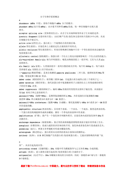
酵母双杂交(自激活)

一、原理
真核生物的转录因子是由两个可以分开的、 功能上相互独立的结构域(domain)组成的。
酵母的转录激活因子GAL4,在N端有一个 由147个氨基酸组成的DNA结合域(DNA binding domain,BD),C端有一个由113个 氨基酸组成的转录激活域(transcription activation domain,AD)。
g e n e
D o th e s e p ro te in s b in d ?
Y
X
X
X Y
o u r t w o h y b r i d p r o t e i n s b i n d !
yes, we have expression of the reporter indicating that the proteins bind!
D N A b i n d i n g d o m a i n
U A S ( u p s t r e a m a c t i v a t i o n s e q u e n c e )
t r a n s c r i p t i o n m a c h i n e r y
g e n e
Yeast two-hybrid system:
0.2g ade定容至 100ml → 0.2%的ade母液
pH 6.5
灭菌 121℃ 15: pBD-GAL4
酵母菌株感受态细胞制备:
1. 将-70℃下冻存的酵母菌在YPAD平板上划线,倒置于30℃培养2-3天。 2. 挑取2-4个大菌落(直径2-3mm)于1.5ml YPAD液体培养基中,剧烈震 荡5min,打散菌落,接种于50ml YPAD液体培养基中(250ml三角 瓶),230-250转/min,30℃培养18-24hr。 3. 取菌液测定其OD600值,需达到1.5。若不到,再培养1~2hr,若还不到, 换单克隆重摇。 4. 取50ml菌液于300mlYPAD培养基中(1-2L大三角瓶),30℃, 230rpm,摇培3hr,至 OD600值为0.4~0.6。 5. 4000rpm 5min 于常温收集菌体,重复一次。 6. 室温下1000g离心5min,去上清,加入50ml 超纯水清洗悬浮3次。 7. 1000g离心5min,弃上清,用新配的1.5ml 1×TE/LiAc重悬细胞,置冰