彩虹130广谱蛋白marker说明书
蛋白Marker说明书
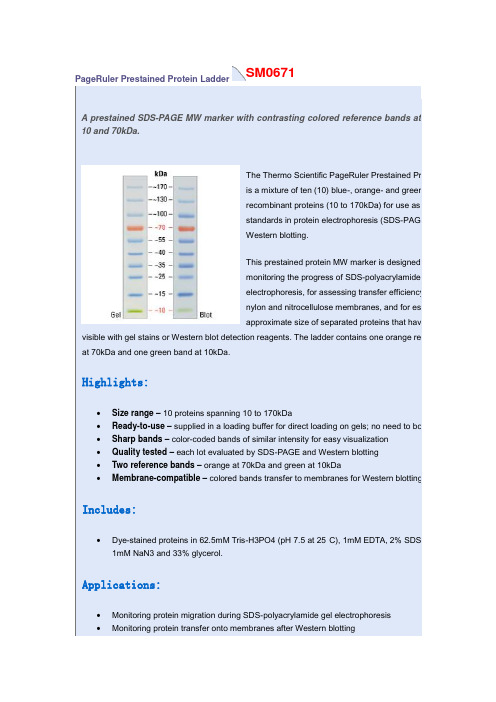
SM0671PageRuler Prestained Protein LadderA prestained SDS-PAGE MW marker with contrasting colored reference bands at10 and 70kDa.The Thermo Scientific PageRuler Prestained Proteis a mixture of ten (10) blue-, orange- and green-srecombinant proteins (10 to 170kDa) for use as sizstandards in protein electrophoresis (SDS-PAGE)Western blotting.This prestained protein MW marker is designed fomonitoring the progress of SDS-polyacrylamide geelectrophoresis, for assessing transfer efficiency onylon and nitrocellulose membranes, and for estimapproximate size of separated proteins that have b visible with gel stains or Western blot detection reagents. The ladder contains one orange refer at 70kDa and one green band at 10kDa.Highlights:•Size range– 10 proteins spanning 10 to 170kDa•Ready-to-use– supplied in a loading buffer for direct loading on gels; no need to boil •Sharp bands– color-coded bands of similar intensity for easy visualization•Quality tested – each lot evaluated by SDS-PAGE and Western blotting•Two reference bands– orange at 70kDa and green at 10kDa•Membrane-compatible– colored bands transfer to membranes for Western blottingIncludes:•Dye-stained proteins in 62.5mM Tris-H3PO4 (pH 7.5 at 25°C), 1mM EDTA, 2% SDS, 1 1mM NaN3 and 33% glycerol.Applications:•Monitoring protein migration during SDS-polyacrylamide gel electrophoresis•Monitoring protein transfer onto membranes after Western blotting•Sizing of proteins on SDS-polyacrylamide gels and Western blotsProduct Details:SDS-PAGE band profile of the Thermo ScientificPageRuler Prestained Protein Ladder. Images arefrom a 4-20% Tris-glycine gel (SDS-PAGE) andsubsequent transfer to membrane.Sm1841Spectra Multicolor Broad Range Protein LadderA prestained, 4-color, SDS-PAGE MW marker especially for proteins between 10 and 260kDa.The Thermo Scientific Spectra Multicolor Broad RangeProtein Ladder is a 4-color protein standard containing 10prestained recombinant prokaryotic proteins (10 to 260kDa)for use as size standards in gel electrophoresis and Westernblotting.This prestained protein MW marker is designed formonitoring the progress of SDS-polyacrylamide gelelectrophoresis, for assessing transfer efficiency onto PVDF,nylon and nitrocellulose membranes, and for estimating theapproximate size of separated proteins that have been made visible with gel stains or Western blot detection reagents. Four different chromophores (blue, orange, green, pink) are bound to the different component proteins, producing a brightly colored ladder with an easy-to-remember pattern.Highlights:•Size range– 10 proteins spanning 10 to 260kDa•Multicolor– four different colors for unambiguous band-size assignment•Ready-to-use– supplied in a loading buffer for direct loading on gels; no need to boil•Sharp bands– color-coded bands of similar intensity for easy visualization•Quality tested – each lot evaluated by SDS-PAGE and Western blotting•Membrane-compatible– colored bands transfer to membranes for Western blotting Includes:•Dye-stained proteins in 62.5mM Tris-H3PO4 (pH 7.5 at 25°C), 1mM EDTA, 2% SDS, 10mM DTT, 1mM NaN3 and 33% glycerol.Applications:•Monitoring protein migration during SDS-polyacrylamide gel electrophoresis•Monitoring protein transfer onto membranes after Western blotting•Sizing of proteins on SDS-polyacrylamide gels and Western blotsProduct Details:SDS-PAGE band profile of the Thermo ScientificSpectra Multicolor Broad Range Protein Ladder.Images are from a 4-20% Tris-glycine gel(SDS-PAGE) and subsequent transfer to membrane.S m1811PageRuler Plus Prestained Protein LadderA prestained SDS-PAGE MW marker with contrasting colored reference bands at 10, 25 and 70kDa.The Thermo Scientific PageRuler Plus Prestained ProteinLadder is a mixture of nine (9) blue-, orange- andgreen-stained recombinant proteins (10 to 250kDa) for useas size standards in protein electrophoresis (SDS-PAGE)and Western blotting.This prestained protein MW marker is designed formonitoring the progress of SDS-polyacrylamide gelelectrophoresis, for assessing transfer efficiency onto PVDF,nylon and nitrocellulose membranes, and for estimating theapproximate size of separated proteins that have been made visible with gel stains or Western blot detection reagents. A blue chromophore is bound to all proteins, except proteins of two reference bands of 70kDa and 25kDa that are colored with an orange dye and one green reference band of 10kDa. PageRuler Plus Prestained Protein Ladder is ready to use: no heating, further dilution or addition of a reducing agent is required before loading onto a gel.Highlights:•Size range– nine proteins spanning 10 to 250kDa•Ready-to-use– supplied in a loading buffer for direct loading on gels; no need to boil•Sharp bands– color-coded bands of similar intesity for easy visualization•Quality tested – each lot evaluated by SDS-PAGE and Western blotting•Bright reference bands– orange at 70 and 25kDa, and green at 10kDa•Membrane-compatible– colored bands transfer to membranes for Western blotting Includes:•Dye-stained proteins in 62.5mM Tris-H3PO4 (pH 7.5 at 25°C), 1mM EDTA, 2% SDS, 10mM DTT, 1mM NaN3 and 33% glycerol.Applications:•Monitoring protein migration during SDS-polyacrylamide gel electrophoresis•Monitoring protein transfer onto membranes after Western blotting•Sizing of proteins on SDS-polyacrylamide gels and Western blotsProduct Details:SDS-PAGE band profile of the Thermo Scientific PageRuler Plus Prestained Protein Ladder. Images are from a 4-20% Tris-glycine gel(SDS-PAGE) and subsequent transfer to membrane.。
WB实验步骤
蛋白电泳(制胶、SDS -PAGE 电泳)实验材料:SDS-PAGE 凝胶制备试剂盒、电泳缓冲液、提取的蛋白或全细胞裂解液、广谱彩虹预染中分子量蛋白Marker 、SDS-PAGE Loading Buffer (还原,5×)、G250蛋白快速染色试剂、若干蒸馏水 实验仪器、耗材:蛋白电泳仪、制胶仪、水平摇床、15ml 离心管、5ml 移液器、微量移液器、1.5ml 离心管、塑料饭盒(可在微波炉里加热使用) 配制溶液:● 配制10%APS 溶液:-20度保存0.5g APS + 5ml 蒸馏水、2.5g APS + 25ml 蒸馏水● 配制200 ml 电泳缓冲液:CW0045 Tris-Glycine SDS (ph8.3,10×)20 ml Tris-Glycine SDS + 180 ml 蒸馏水 ● 配制1 ml loading buffer :200 ul 5×loading buffer +800 ul 蒸馏水 实验步骤: 一.制胶:1. 参照凝胶模具说明书,装配好凝胶模具。
2. 根据分离蛋白的大小配制分离胶和浓缩胶。
3. 配制10%分离胶:将不同体积的30%Acr-Bis(29:1)、分离胶缓冲液和双蒸水在小烧杯或试管中混合。
加入10%APS 和TEMED ,轻轻搅拌使其混匀,避免产生气泡。
4.待胶灌至距离玻璃板顶端1.5cm的时候停止灌胶,加入蒸馏水水封。
静置40分钟至1小时。
5.待分离胶凝固后(水层胶层中间出现折线),倒掉蒸馏水,用滤纸将水溶液吸干。
6.配制5%浓缩胶:7.将浓缩胶溶液加至分离胶的上面,直至凝胶溶液到达前玻璃板的顶端。
注意:灌胶速度要快,防止凝胶。
8.将梳子插入凝胶内,避免产生气泡。
静置20分钟,等待浓缩胶聚合。
9.待凝胶聚合后,小心地拔出梳子,以免破坏加样孔。
赶走气泡。
二.SDS-PAGE电泳:1. 在电泳槽的内外槽灌至电泳缓冲液。
彩虹130广谱蛋白marker说明书
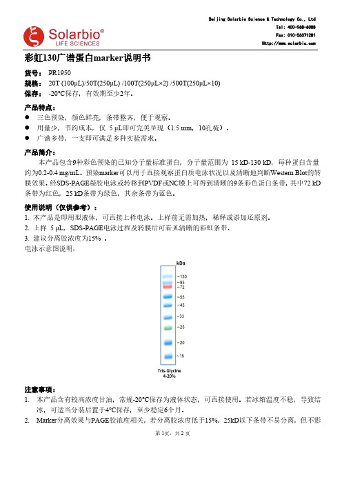
第1页,共2页彩虹130广谱蛋白marker 说明书货号:PR1950规格:20T (100μL)/50T(250μL)/100T(250μL×2)/500T(250μL×10)保存:-20℃保存,有效期至少2年。
产品特点:●三色预染,颜色鲜亮,条带整齐,便于观察。
●用量少,节约成本,仅5μL 即可完美呈现(1.5mm ,10孔梳)。
●广谱多带,一支即可满足多种实验需求。
产品简介:本产品包含9种彩色预染的已知分子量标准蛋白,分子量范围为15kD-130kD ,每种蛋白含量约为0.2-0.4mg/mL 。
预染marker 可以用于直接观察蛋白质电泳状况以及清晰地判断Western Blot 的转膜效果。
经SDS-PAGE 凝胶电泳或转移到PVDF 或NC 膜上可得到清晰的9条彩色蛋白条带,其中72kD 条带为红色,25kD 条带为绿色,其余条带为蓝色。
使用说明(仅供参考):1.本产品是即用型液体,可直接上样电泳。
上样前无需加热,稀释或添加还原剂。
2.上样5μL ,SDS-PAGE 电泳过程及转膜后可看见清晰的彩虹条带。
3.建议分离胶浓度为15%。
电泳示意图说明:注意事项:1.本产品含有较高浓度甘油,常规-20℃保存为液体状态,可直接使用。
若冰箱温度不稳,导致结冰,可适当分装后置于4℃保存,至少稳定6个月。
2.Marker 分离效果与PAGE 胶浓度相关,若分离胶浓度低于15%,25kD 以下条带不易分离,但不影Beijing Solarbio Science &Technology Co.,LtdTel:400-968-6088Fax:响绿色(25KD)及以上条带。
如果目的条带小于25KD,建议分离胶浓度大于或等于15%。
3.转膜效果与转膜时间有关,需根据客户目的条带大小而定,如果转膜后较大分子量条带有部分未转膜成功,属于正常现象。
相关产品:P10154×蛋白上样缓冲液(含DTT)P1200SDS-PAGE凝胶制备试剂盒T10705×Tris-甘氨酸电泳缓冲液D106010×电泳转移缓冲液PR1920彩虹245广谱蛋白markerPR1700预染次高分子量蛋白markerSW3010膜封闭液DA1010DAB显色试剂盒(20×)PE0010ECL Plus荧光检测试剂(ECL超敏发光液)第2页,共2页。
marker说明书
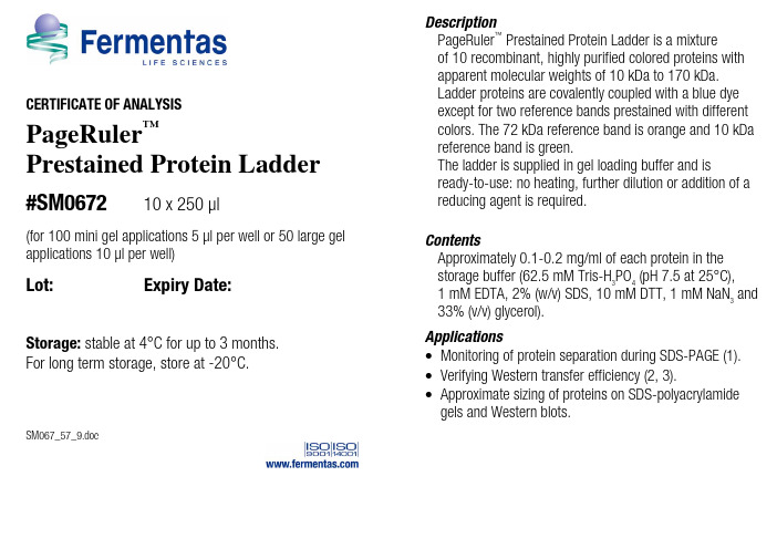
CERTIFICATE OF ANALYSISPageRuler ™Prestained Protein Ladder#SM067210 x 250 µl(for 100 mini gel applications 5 µl per well or 50 large gel applications 10 µl per well)Lot: Expiry Date:Storage: stable at 4°C for up to 3 months. For long term storage, store at -20°C.SM067_57_9.docDescriptionPageRuler ™Prestained Protein Ladder is a mixture of 10 recombinant, highly purified colored proteins with apparent molecular weights of 10 kDa to 170 kDa. Ladder proteins are covalently coupled with a blue dye except for two reference bands prestained with different colors. The 72 kDa reference band is orange and 10 kDa reference band is green.The ladder is supplied in gel loading buffer and isready-to-use: no heating, further dilution or addition of a reducing agent is required.ContentsApproximately 0.1-0.2 mg/ml of each protein in the storage buffer (62.5 mM Tris-H 3PO 4 (pH 7.5 at 25°C), 1 mM EDTA, 2% (w/v) SDS, 10 mM DTT, 1 mM NaN 3 and 33% (v/v) glycerol).Applications∙ Monitoring of protein separation during SDS-PAGE (1). ∙ Verifying Western transfer efficiency (2, 3).∙ Approximate sizing of proteins on SDS-polyacrylamidegels and Western blots.Instruction for Use❶ Thaw the ladder at room temperature for a few minutes to dissolve precipitated solids. DO NOT BOIL!❷ Mix gently, but thoroughly, to ensure the solution is homogeneous.❸ Load the following volumes of the ladder on an SDS-polyacrylamide gel:– 5 µl per well for mini gel,– 10 µl per well for large gel.Use the same volumes for Western blotting.❹ After the run is complete, stain the gel or perform Western transfer procedure as desired.Note•Each lot of the PageRuler™ Prestained Protein Ladder is calibrated against a precisely sized, PageRuler™ Unstained Protein Ladder and calculated apparent molecular weights are reported in the picture.•For precise molecular weight determinations use PageRuler™ Unstained Protein Ladder, #SM0661, see.•In 8 or 10% gels low molecular weight proteins may migrate with the dye front.•Loading volumes are intended for use in gels with a thickness of 0.75 mm. For thicker gels, the recommended loading volume should be increased.•PageRuler™ Prestained Protein Ladder could be used in Western blotting with all common membranes: PVDF, nylon and nitrocellulose.•Longer transfer times or higher transfer voltages may be required for Western blotting of large (>100 kDa) proteins.Lot specific MW, kDa4-20% Tris-glycine SDS-PAGEcontinued on back pageQUALITY CONTROL5 µl of PageRuler™ Prestained Protein Ladder resolves 10 bands of equal intensities in 4-20% SDS-PAGE (Tris-glycine buffer) and after Western blotting onto PVDF membrane.Quality authorized by: Jurgita Zilinskiene References1. Laemmli, U.K., Cleavage of structural proteins during the assembly of the head of bacteriophage T4, Nature, 227, 680-685, 1970.2. Burnette, W.N., "Western blotting": electrophoretic transfer of proteins from sodium dodecyl sulfate – polyacrylamide gels to unmodified nitrocellulose and radiographic detection with antibody and radioiodinated protein A, Anal. Biochem., 112 (2), 195-203, 1981.3. Towbin, H., et al., E lectrophoretic transfer of proteins from polyacrylamide gels to nitrocellulose sheets: procedure and some applications, Proc. Natl. Acad. Sci. USA, 76, 4350-4354, 1979.This product is manufactured under the license forStrep-tag® technology covered by US patents Nos.5,506,121, 6,103,493 and foreign counterparts. Related Products∙DualColor™ Protein Loading Buffer Pack #R1011∙Loading Buffer Pack #R0891∙Spectra™ Multicolor Broad Range Protein Ladder #SM1841∙PageRuler™ Unstained #SM0661∙PageRuler™ Plus Prestained Protein Ladder #SM1811∙PageSilver™ Silver Staining Kit #K0681∙PageBlue™ Protein Staining Solution #R0571∙10X Tris-glycine-SDS Buffer #B46∙10X Tris-tricine-SDS Buffer #B48∙DTT #R0861 ∙ProteoJET™ Mammalian Cell Lysis Reagent #K0301∙ProteoJET™ Cytoplasmic and Nuclear ProteinExtraction Kit #K0311∙Bradford Reagent, ready-to-use #R1271∙Bovine Serum Albumin Standard Set, ready-to-use #R1281∙Bovine Gamma Globulin Standard Set, ready-to-use #R1291PRODUCT USE LIMITATION.This product is developed, designed and sold exclusively for research purposes andin vitro use only. The product was not tested for use in diagnostics or for drugdevelopment, nor is it suitable for administration to humans or animals.Please refer to for Material Safety Data Sheet of the product.。
蛋白marker标记颜色方法

蛋白marker标记颜色方法全文共四篇示例,供读者参考第一篇示例:蛋白marker标记颜色方法在生物科学研究中,标记蛋白是非常常见的实验技术之一。
蛋白marker的标记可以帮助研究人员在实验中追踪和定位蛋白,从而更好地理解蛋白的功能和相互作用。
而标记蛋白的颜色选择也是非常重要的,不同颜色的标记能够帮助实验者在实验过程中更清晰地识别和分析样品。
本文将介绍一些常用的蛋白marker标记颜色方法,希望能够帮助读者更好地进行蛋白标记实验。
一、荧光标记荧光标记是蛋白标记颜色中最常用的方法之一。
通过将蛋白与荧光物质结合,可以在光学显微镜下直接观察到蛋白标记的颜色。
常用的荧光标记包括FITC(荧光异硫氰酸酯),TRITC(罗丹明异硫氰酸酯)和Cy5(Cyanine5)。
这些荧光物质在实验中常用于免疫荧光染色、融合蛋白标记等实验中。
荧光标记的优势是信号强度高、灵敏度高且不受环境影响,适用于单细胞和组织的标记。
在实验中,荧光标记的颜色可以根据荧光物质的不同而有所区分。
比如,FITC标记的蛋白呈现绿色,TRITC标记呈现橙红色,Cy5标记呈现红色。
实验者可以根据实验需要选择不同的荧光标记物质,并通过显微镜观察到标记蛋白的颜色。
二、生物素标记生物素标记是一种基于生物素-亲和素相互作用的蛋白标记方法。
生物素是一种维生素H成分,具有与亲和素结合的特性。
在实验中,生物素可以与生物素素结合并形成稳定的结合物,从而实现蛋白的标记。
生物素标记的颜色在实验中常表现为紫色,实验者可以通过观察颜色变化来判断蛋白是否标记成功。
生物素标记的优势是其结合力强、稳定性高,适用于长时间的实验操作。
另外,生物素标记也可以用于有机溶剂、高温、酸碱环境等多种条件下的蛋白标记。
因此,在一些特殊实验条件下,生物素标记是一种非常理想的标记方法。
三、酶标记酶标记是另一种常用的蛋白标记方法。
通过将蛋白与酶结合,可以在实验中通过酶的催化作用来识别蛋白的存在。
常用的酶标记包括辣根过氧化物酶(HRP)、碱性磷酸酶(AP)等。
中分子量蛋白Marker

中分子量蛋白Marker I使 用 说 明 书包 装 量:浓 度:每种蛋白约0.1-0.2mg/ml 在1×loading Buffer储运温度:收到产品后分装冻存,避免反复冻融降解蛋白,-20℃ (长期保存请置于-70℃ )(用于小胶5µl/次可用20次 用于大胶10µl /次 可用10次)制品说明:本产品是由6种蛋白质分别纯化后混合而成的蛋白质溶液,分子量范围为14KD -94KD ,经SDS -聚丙烯酰胺凝胶电泳后,用考马斯亮蓝R -250(Coomassie Blue R-250)染色后可得清晰的7条蛋白带。
建议使用分离浓度为12%~15%。
适用胶浓度:最优适用于12% (37.5:1 丙烯酰胺:双丙烯酰胺)的聚丙烯酰胺凝胶。
该Marker 也可用于8-15%的胶。
胶浓度为8-10% 时蛋白Marker 中低分子量的蛋白易于同染料前沿跑在一条线上不易区分,在12-15%的胶上及梯度电泳中,所有的条带都能锐利清晰分开。
推荐上样量:上样量胶厚度大小 5µl 0.75mm thick mini 10µl1.5mm thick mini 0.75mm thick large 20µl 1.5mm thick large操作步骤:1.室温融解Marker 或37- 40°C 温浴几分钟使之融解,混匀均一,不要高温加热。
2.为避免污染最好分装保存以备使用,取所需体积的Marker 置于一干净离心管中并封好口。
3. 上样并进行 SDS-PAGE 电泳。
4.该Marker适用于考马斯亮蓝, 银染或其他蛋白染色方法。
注意:银染比考马斯亮蓝染色敏感度高10~100 倍,相应的银染需要减少用量。
5.变性蛋白Marker储存于-20°C。
6.在非变性聚丙烯酰胺凝胶电泳中不要使用该Marker,因为在该Marker的储存缓冲液中存在SDS。
爱必信WB彩色凝胶试剂盒上新啦!让您的WB实验多姿多彩

爱必信WB彩色凝胶试剂盒上新啦!
让您的WB实验多姿多彩
蛋白免疫印迹实验(Western Blot)实验大家肯定都不陌生,要想做出好看的条带,从制胶开始就不容半点马虎。
WB制胶可谓实验室新手的必修课了,想想小编我在当初实验室的时候,就没少帮师兄师姐配置WB凝胶,泛黄的配方表小编还记忆犹新。
WB凝胶配
方表
说起WB凝胶配置,小编可有一肚子苦水要倒,第一个让人头疼的一点就是多种溶液都需要计算量取,每次调移液器就要调半天。
还有各种试剂的保存条件也不一样,配胶之前还需要从冰箱,室温柜子里分别拿出不同的溶液。
这还不算,最容易出问题的促凝剂APS,需要新鲜配置,4度保存不能超过一周,关键是粉末还特容易吸水,打开后需要密封保存。
小编之前就踩过一次坑,APS粉末吸水结块,称量配置后的APS含量不够,凝胶凝结不好,整个WB实验条带跑的七扭八歪。
蛋白质印迹(Western Blot)实验中产品推荐:。
细胞角蛋白(广谱)抗体试剂(免疫组织化学)说明书
12mL
【预期用途】
体外诊断用途。 在常规染色(如:HE染色)基础上进行免疫组织化学染色,为医师提供诊断的辅助信息。 细胞角蛋白(广谱)抗体试剂(免疫组织化学),用于在 Dako Omnis 仪器上进行的免疫组化分析。该抗 体有助于判定肿瘤是否为恶性或者上皮来源 (1-3)。临床判定任何染色的着色或缺失都必须有恰当对照的 形态学结果作为补充,基于患者的临床病史和其它诊断结果,由有资质的病理医生诊断。
3. Moll R, Franke WW, Schiller Dl. The catalog of human cytokeratins: patterns of expression in normal epithelia, tumors and cultured cells. Cell 1982; 31:11
2. Woodcock-Mitchell J, Eichner R, Nelson WG, Sun TT. Immunolocalization of keratin polypeptides in human epidermis using monoclonal antibodies. J Cell Biol 1982; 95:580
快速指南
步骤
备注
固定/包埋 预处理 抗体 阴性对照 显色
复染 对照组织
福尔马林固定,石蜡包埋
机内脱蜡
EnVision™ FLEX,高pH(编码GV804) 热诱导的抗原修复30分钟
即用型
孵育12.5分钟
FLEX阴性对照,小鼠(编码GA750) 孵育12.5分钟
EnVision™ FLEX(编码GV800)
*用户必须阅读说明书,以了解染色操作和产品处理的详细说明。
- 1、下载文档前请自行甄别文档内容的完整性,平台不提供额外的编辑、内容补充、找答案等附加服务。
- 2、"仅部分预览"的文档,不可在线预览部分如存在完整性等问题,可反馈申请退款(可完整预览的文档不适用该条件!)。
- 3、如文档侵犯您的权益,请联系客服反馈,我们会尽快为您处理(人工客服工作时间:9:00-18:30)。
彩虹130广谱蛋白marker说明书
货号:PR1950
规格:20T(100μL)/50T(250μL)/100T(250μL×2)/500T(250μL×10)
保存:-20℃保存,有效期至少2年。
产品特点:
●三色预染,颜色鲜亮,条带整齐,便于观察。
●用量少,节约成本,仅5ul即可完美呈现(1.5mm,10孔梳)。
●广谱多带,一支即可满足多种实验需求。
产品简介:
本产品包含9种彩色预染的已知分子量标准蛋白,分子量范围为15kD-130kD,每种蛋白含量约为
0.2-0.4mg/ml。
预染marker可以用于直接观察蛋白质电泳状况以及清晰地判断Western Blot的转膜效果。
经SDS-PAGE凝胶电泳或转移到PVDF或NC膜上可得到清晰的9条彩色蛋白条带,其中70kD条带为红色,25kD条带为绿色,其余条带为蓝色。
使用说明:
1.本产品是即用型液体,可直接上样电泳。
上样前无需加热,稀释或添加还原剂。
2.上样5ul,SDS-PAGE电泳过程及转膜后可看见清晰的彩虹条带。
3.建议分离胶浓度为15%。
电泳示意图说明:
第1页,共2页
第2页,共2
页注意事项:
1.本产品含有较高浓度甘油,常规-20℃保存为液体状态,可直接使用。
若冰箱温度不稳,导致结冰,可
适当分装后置于4℃保存,至少稳定6个月。
2.Marker分离效果与PAGE胶浓度相关,若分离胶浓度低于15%,25kD以下条带不易分离,但不影响绿色
(25KD)及以上条带。
如果目的条带小于25KD,建议分离胶浓度大于或等于15%。
3.转膜效果与转膜时间有关,需根据客户目的条带大小而定,如果转膜后较大分子量条带有部分未转膜
成功,属于正常现象。