Modeling and visualization of biological structures
三维地质和地下水建模的研究和应用

三维地质和地下水建模的研究和应用王磊;谢江宁【摘要】针对地质和地下水可视化分析的应用问题,提出了基于三维建模和分析的可视化研究方法,并建立了有效的针对地势和地下水的可视化平台.尤其针对建模过程中数据的连续性问题采用了基于多元二次插值方法,从而有效的解决了抽样数据点稀疏的问题.在地层建模方面,系统采用了基于三棱柱体积元素的建模方法,同时通过基于检测井法线信息的有效插值算法解决了地层建模过程中的连续性问题.针对地下水的具体应用,提出了基于图像的绘制方法,实现了地势、地层断层和地下水分布的可视化分析和展现.【期刊名称】《湘潭大学自然科学学报》【年(卷),期】2014(036)002【总页数】5页(P109-113)【关键词】地质建模;三棱体元素;地形可视化;三角化【作者】王磊;谢江宁【作者单位】潍坊学院计算机工程学院,山东潍坊261061;山东大学研究生院,山东济南250100【正文语种】中文【中图分类】TP391.9随着计算机图形学技术的发展,三维建模技术被广泛应用于机械制造、工业设计、虚拟现实和地理信息等相关领域中.尤其针对地理信息和地质研究等相关应用领域,三维建模和可视化技术在信息展现具有形象化和逼真性优势,从而被广泛应用于地形可视化、道路可视化、地质勘探和空气可视化等相关应用中[1,2,9].在进行相关应用的研究过程中,如何实现三维模型建模是当前上述应用领域的研究难点[10,11].尤其针对地质信息相关方面的应用,在地质信息建模方面将会包括了地形建模和三维地层建模等相关方向.1994年,加拿大学者Houlding提出了三维地质建模的概念,即在三维环境下针对地质信息、空间信息和空间分析以及可视化结合在一起,针对地质等相关信息进行综合分析.目前,针对相关地质模型的建模方法主要包括基于数据插值的地质建模[4]和基于数据拟合的建模方法[5].尤其针对数据拟合的地质建模方法,主要包括了基于NURBS曲面拟合的方法[3]和贝塞尔曲面的拟合方法[6].在一定程度上,上述方法可以解决某特定环境下的建模问题,但它们都需要强大的硬件支持和复杂的算法.目前, 没有可以完全解决所有地质模型问题的理想方法.在地形建模方法方面,尤其是针对基于真实数据的采样建模方法,由于数据采样过程中数目的限制,将会造成模型不够精确.在采用采样方法的建模过程中,首先需要依据采样点进行数据插值获取相关三维数据模型.目前,针对具有大量凌乱采样数据点的地质建模方法,相关的插值方法主要包括了基于反距离加权插值法、改进的Sheppard方法、有限元法和基于径向基函数的插值方法[9]等.在三维地层数据建模方面,需要对相关特殊地层构造进行处理,比如断层、褶皱和透视体等相关方面[7].针对地层数据建模方法,被划分为基于曲面拟合和体元素的建模方法[8,12].在地下水建模分析和应用中,主要是依据相应的数据检测井对相应的三维地形信息和地质信息进行抽样获取.因此,本文在进行地形和地层建模的过程中,主要采用了基于数据插值的方法进行建模.基于插值的方法有效解决了抽样数据点稀疏的问题,针对平坦的地形采用了多元二次插值方法.在地层建模方面,系统采用了基于三棱柱体积元素的建模方法,同时通过基于检测井法线信息的有效插值算法,解决了地层模型的连续性问题.1 地形建模针对本文所述的可视化应用,数据建模方面主要包括了地形模型、地层模型和地下水模型.地形建模和地下水建模同属于表层建模,系统采用了基于三维采样和插值方法的数字化海拔高度模型进行分析和建模描述.在进行地形建模的过程中,需要对数据进行预处理,详细的处理过程如下所述:(1) 数据预处理,结合所有相关的采样数据进行过滤处理,去除噪声点数据.同时,将相应的采样数据点转化为相应比例的坐标数据.(2) 构建不规则的三角网格(TIN),将预处理后的采样数据进行三角剖分处理,将上述采样点形成不规则三角面片表述三维地形表面模型.(3) 通过相应的图形绘制方法进行不规则网格绘制.在不规则三角网格的构造过程中,由于采样点的数据稀疏和凌乱性,会导致Delaunay三角化过程不够流畅.因此,在三角网格构造之前,需要针对采样数据进行数据插值获取足够多的数据坐标点.为了确保网格构建的流畅性,TIN过程采用了基于多元二次插值的方法获取更多的数据点.多元二次插值,作为径向基函数的一种,被广泛应用于各类数据插值计算的过程.对于一组采样点,多元二次插值法的函数描述如下:(1)上述函数中,n是样本点的数量, c是常数,aj 可以视为样本点j的重量.在插值求解过程中,将采样点的x,y和z数值分别对应上述函数中的x,y和F(x, y).然后,通过上述函数算法可以得到一个带有不确定数目a0a1…an的线性方程组,通过高斯消元法等数值求解方法进行求解.同时,在上述线性方程组的求解过程中,增添采样点x、y值异样性的约束,从而确保方程组有解.同时,依据相应求解结果获得更多的数据点.2 地层建模在进行地下水应用研究过程中,系统采用了基于采样检测井的获取方法,因此在进行整体地层数据的建模过程中采用了基于三棱柱体元素为基础的地层建模方法[7].首先,针对来自样本井中的检测数据,进行相应的预处理,然后通过采样井之间的关系进行三棱柱建模,从而实现地层的三维建模.基于三棱柱的建模方法,能够有效的利用采样井的相关数据,同时能够有效的反映底层的相关地质土壤结构.在构建地层模型的过程中,首先依据检测井获取的数据建立每个岩石层的三棱柱体;然后,整体的地层模型将会由相应的三棱体元素构成.整体的底层建模详细步骤如下所述:数据预处理,依据检测井获取的样本数据,首先将相关经纬度信息转化为三维数据分析中的x、y值,同时将岩层顶部和底部海拔高度信息转化为z值信息.三角化计算,将相应的检测井作为顶点进行投影,然后通过相应的德洛内三角网格化计算获取整体的三角网格.三棱柱体计算,依据获取的三角化网格,其顶点对应着相应的检测井,然后在相应的三角网格内进行三棱柱体计算构建.计算每个三棱柱体相应竖直平面的法线向量.在进行三棱柱体计算时,需要依据相应两个采样检测井之间的采样数据特性进行体元素的构建,相应的检测井数据地质类型的多样化会增加三棱柱建模的困难性.因此,在进行三棱柱建模过程中,系统假设两个监测井首先与任意一个平面相连接,然后通过该平面与相应的德洛内三角另外一个监测井数据进行连接.整体的相应三棱柱构建过程中,系统将通过下文所述的两个检测井连接和侧面与监测井连接进行详细的分析和表述.2.1 两个监测井地层连接在进行两个监测井数据连接处理过程中,处理过程是将某个监测井相应的地质层与另外相邻节点监测井的地层进行关联.在相应的处理过程中,采用了基于递归处理思想的方法,将问题通过规约简化的思想进行相应的处理,相应的具体的处理过程将会被划分为以下所述的三种具体连接条件,下列图示中通过不同的颜色来区分不同特性的岩石层:情况 1: 如图1(a)所示,如果相邻两个监测井仅仅具有一个且特性相同的岩石层,同时两层满足相关的约束条件(如部分岩石层具有相同的海拔高度),那么直接分别进行顶部和底部的相应连接,从而获得两者之间岩石层的关联关系.情况 2: 对于某个监测井的某地质层,在其相邻监测井中无相同类型的地质层,将会对其进行岩石层锲入处理,具体的处理算法如图2所示,在接下来将进行详细描述.情况 3: 如图1(b),如果其中至少一个监测井有多于一个的岩层,并且两监测井中有两个岩层可匹配连接.两个监测井被这岩层分解为上下两部分,然后分别对这两部分进行递归分解处理.针对上文所述的岩石层楔入处理,依据相应的实际岩层分类处理过程,将其划分为三种不同的情况进行分析和处理,详细的情况图解如图2所示.情况 1: 如果两个监测井中只有某个监测井具有一种类型的岩石层,那么在进行楔入操作时,将会如图2(a,b)所示,将消逝点定义在另一侧监测井距离当前岩层中心最近的岩层分界面.情况2:如图2(c)所示,如果两个监测井各有一个不同种类岩层,则进行对角连接,共有两种方式,选择对角线较短的方式,从而保证岩层消逝端的处理不会太陡峭.情况 3:如图2(d)所示,如果不满足情况1和情况2,则选取两个监测井的最接近中心的岩层分界面,这两个分界面将两个监测井分为上下两部分,然后对这两部分分别进行递归分解处理.2.2 侧面和监测井的连接通过德洛内三角化获取的监测采样井,通过上述岩层连接方法对三角形其中两个监测井的岩层数据进行连接后获得了相应的岩层连接侧面.然后,依据相应岩层数据的特性,将侧面岩层分布与第三个监测井岩层进行连接,从而构建出相应的三棱柱体结构.在完成上述两个监测井数据的连接后,侧面将由从顶部到底部的相应的三角形和四边形所构成.接下来,将对侧面岩层和相应的第三个监测井岩层进行连接构成最终的三棱柱体.下文将针对连接的相应处理过程进行详细的分析和描述,为了能够简化描述相关处理过程,下文中将侧面和第三监测井分别表述为F和W. 情况1:如图3(a)所示,算法将对F中的每个三角面和四边形面依次进行处理.如果F中的某个面,在相应监测井W中能够找到相同特质的岩石层,将进行相应顶点的连接,从而构成一个三棱柱体元素.情况2:如图3(b)所示,如果在监测井W中,找不到相应特质的岩石层,在W 中上下范围较为接近的岩石层断点处进行相应的连接,从而构成三棱柱体.情况3:如图3(c)所示,在完成上述两种情况的处理后,完成了侧面F的所有岩石层面处理.针对监测井W,对相应的各个岩石层进行遍历,对于未处理的岩石层,在相应的岩石断层处进行连接处理,从而形成相应的三棱柱体.3 基于地质建模的可视化应用本文所述算法已经在相应的计算机中进行系统应用开发验证,实现平台为Visual Studio 2012 C++,三维数据处理和渲染通过OpenGL实现.在上述系统实现过程中,所述的地形和地层建模方法被应用于某地区的地势和地下水分布可视化分析应用过程中.在进行相应的三维建模和地下水可视化过程中,通过钻孔采样的方法进行数据获取,在本文所述地区的采样主要采用了上述地区32个地下水采样矿井数据(钻孔深度在100 m左右).通过钻井采样数据和上文所述的插值方法获取了合适密度的数据[13,14],针对地势、地层和地下水建模.同时,通过计算的各个三棱柱面的法线方向对该地区的地势、岩石特质断面和地下水状况进行了渲染计算,通过相应的可视化方法进行了形象的数据可视化分析和展现.如图4所示,对某地区的地形进行相应的渲染计算,该地区的地势从高到低依次被渲染为从红色到绿色过渡的颜色.通过下图有效地展现了该地区的相关地势数据,从而对地下水采样点的分析进行了有效地地势对比.在相应的地点标注过程中,本文采用了基于FreeTyp库标记的方法,通过该方法在三维地势图上实现了对村庄,河流,铁路和公路的标记.除了上述的地势信息可视化以外,为了能够有效对某地区的相应地质信息进行分析,本文采用了基于图像绘制的断层可视化方法.通过相应应用中的地形切割交互方法实现了地层的有效可视化展现,主要采用了基于质感自适应和Mipmap映射方法,结合三棱柱的法线计算结果进行了三棱柱表面的绘制.如图5所示,通过上述的断层绘制方法对某个点的地层界面的岩层性质进行了相应的可视化展现.本文所述的整体应用主要是针对相应不同采样点的地下水数据进行可视化展现,从而对某地区的相关降水量和地下水情况进行有效的可视分析和展现.如图6所示,在对地下水情况进行可视化过程中,采用了基于Voronoi划分的插值分布计算,从而生成了比较均匀的可视化展现.在进行地下水模型可视化展现过程中,相应的地形表面将被绘制为半透明状,同时可以对其透明程度进行调节.通过相应数据的半透明展现,有效地实现了某地区整体地下水的分布状况.4 结论针对某地区的地下水分析应用中的地质建模方法进行了详细的分析和描述.尤其针对建模过程中数据的连续性问题提出了基于多元二次插值方法进行处理,从而有效的解决了抽样数据点稀疏的问题.在地层建模方面,系统采用了基于三棱柱体积元素的建模方法,同时通过基于检测井法线信息的有效插值算法,解决了地层模型的连续性问题.针对地下水的具体应用,提出了基于图像的绘制方法,实现了地势、地层断层和地下水分布的可视化分析和描述.在整体的应用研究过程中,本文方法还不能有效处理一些特殊的地理结构,比如断层、褶皱和透镜体.在今后的研究过程中,将针对复杂的地理结构模型描述和可视化问题进行深入的研究.参考文献[1] 朱良峰,吴信才,刘修国,等. 基于钻孔数据的三维地层模型的构建[J]. 地理与地理信息科学, 2004, 20(3):26-30.[2] 朱良峰,潘信,吴信才,等. 地质断层三维可视化模型的构建方法与实现技术[J]. 软件学报, 2008, 19(8):2 004-2 017.[3] ZHONG D H,LI M C,WANG G,et al. NURBS-based 3D Graphical modeling and visualization of geological structures [C] // Proceedings of the Third International Conference on Image and Graphics. Washington, DC: IEEE Computer Society, 2004:414-417.[4] MALLET J L. Discrete modeling for natural objects[J]. Mathematical Geology, 1997, 29(2):199-218.[5] MARSCHALLINGER R. Three-dimensional reconstruction and visualization of geological materials with IDL -examples and sourcecode[J].Computers& Geosciences, 2001,27(4):419-426.[6] ERIC A,KEMP D. 3-D visualization of complex geological structuresusing 3-D Bezier construction tools[J].Computers & Geosciences, 1999,25(5):581-597.[7] WU Q,XU H. An approach to computer modeling and visualization of geological faults in 3D[J]. Computers & Geosciences, 2003, 29(4):503-509.[8] 张煜,白世伟. 一种基于三棱柱体体元的三维地层建模方法及应用[J]. 中国图象图形学报A辑, 2001, 6(3): 285-290.[9] 唐泽圣.三维数据场可视化[M].北京: 清华大学出版社, 1999.[10] 许国,王长海. 万家口水电站复杂地质体三维模型及其数值模型构建[J]. 武汉大学学报(工学版),2013, 46(4): 439-474.[11] 黄嫚,仇玉良,王柱,等. 基于ArcEngine地质体三维建模研究[J]. 测绘科学技术学报,2013, 30(2):197-200.[12] 孟宪海,李吉刚,邹靖晗,等. 三维地质体类三棱柱网格建模方法研究[J]. 计算机工程与应用, 2010,46(4): 142-148.[13] 何满潮,李学元,刘斌,等.工程岩体三维构模中钻孔数据处理方法[J].岩石力学与工程学报,2005,24(11):1 821-1 826.[14] WU Q, XU H. Three-dimensional geological modeling and its application in Digital Mine [J]. Science China Earth Sciences,2014,57(3):491-502.。
ZEISS KINEVO 900 高精度手术显微镜说明书
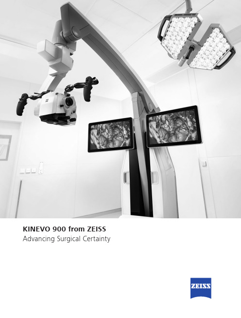
Advancing Surgical CertaintyKINEVO 900 – The Robotic Visualization System Just like you, we love challenging the status quo.The result? Over 100 innovations to perfect the already acclaimed surgical visualization platform. KINEVO® 900 from ZEISS is designedto deliver more functionalities than any surgical microscope today.ZEISS KINEVO 900 combines digital and optical visualization modalities, offers a unique Micro-Inspection Tool and will impress you with its Surgeon-Controlled Robotics. All to enable you to gain greater certainty in a virtually disruption-free workflow.Designed to meet real needs. To make a real difference!A lot more. And, a lot less too.When treating complex vascular conditions, you typically work at high magnification. Even the slightest vibrations can cause disruptions. And constant manual repositioning to better visualize structures or precisely approach deep-seated lesions can become extremely tedious. Not anymore! ZEISS KINEVO 900 delivers a lot more positioning precision with a lot less effort.PointLockSurgeon-Controlled Robotics adds a complete new level of ease to precise positioning. Imagine being able to focus and move around a structure to visualize the targeted anatomy – reducing any manual hassle. In addition, PointLock enables you to do a KeyHole movement to observe a larger area inside a cavity – a particular benefit in areas with narrow access. Simply put:Focus. Activate. Swivel.Active vibration dampingYou know the problems that can be created by the tiniest vibrations. The active damping provided by ZEISS KINEVO 900 minimizes collateral system vibrations, ensuring rock-solid stability. Enabling you to completely, and steadily, focus on what matters most:your treatment.Focus Activate Swivel5When you need it. Where you need it.The new navigation interface of ZEISS KINEVO 900 is designed to work in concert with your navigation device. When you require precise repositioning to reexamine previously visualized structures or when you need to align with a pre-mapped trajectory, making use of all six axes, the Robotic Visualization System ® delivers precise positioning at the push of a button. Putting you exactly where you need to be – when you need to be there.PositionMemoryWhen working on a tumor case, you may already have identified regions of concern where you want to protect the functional structure. After storing these in PositionMemory , you can come back and visualize them at the exact same magnification, working distance and focus – without losing time for manual repositioning. In a nutshell: Save. Move. Recall.Image-guided surgeryMinimize time-consuming efforts in approaching challenging neurosurgicalpathologies. Combine the Surgeon-Controlled Robotics of ZEISS KINEVO 900 with navigation interface to approach deep-seated pathologies in cranial surgery, brain stem or skull base tumor removals –right when you need it.Save Move RecallImage with Brainlab Microscope Navigation Software7New dimensions. Freedom of choice. Working through oculars at extreme angles can sometimes be a pain in the neck. Literally. With no way out, you might have to contend with uncomfortable working positions causing fatigue. Now, relief and revolutionary dimensions in visualization arein sight.The Digital Hybrid Visualization with integrated 4K technology of ZEISS KINEVO 900 welcomes you to a world of heads-up ocular-free surgery, giving you freedom of movement. And freedom of choice to use an optical setup, depending on the application need.Fully integrated 4K camera technologyDuring lateral lumbar or thoracic spine and posterior fossa approaches,ZEISS KINEVO 900’s integrated 4K visualization can be essential. It providesyou with multimodal visualization capabilities – the flexibility to decouple fromthe classic optical approach and to work with outstanding 4K picture qualityand clarity. Even when magnifying tiny details.What’s more… your assistant surgeon, OR staff and residents also benefit from the 4K visual clarity of ZEISS KINEVO 900. They share the same high-resolution, digital image to follow the procedure with comparable fidelity. Delivering indispensable education and training.9Critical challenge. Vital solution.Your challenge: When working from an external perspectiveof a surgical microscope, your visualization of the anatomy is limited to a straight line of sight – missing critical information behind tissue or corners. Efficient and effortless access to this comprehensive information is essential for treatment.Our solution: QEVO® from ZEISSThe unique, proprietary Micro-Inspection Tool from ZEISS complements intraoperative microsurgical visualization, enabling you to discover unexplored areas during the surgical intervention without additional footprint. You can look around corners and eliminate blind spots. And most importantly, you can gain greater insights – for better clinical decisions.To support your surgical workflow, ZEISS QEVO is engineered with an angled design – keeping your hands out of the lineof sight during insertion in the surgical field. And, it allowsfor an easy fit between the ZEISS KINEVO 900 and the situs, eliminating the need to reposition the head of the device. Greater insights, on demand.ZEISS QEVO enables you to inspect the perforator or examine the distal neck of the aneurysm to ensure the clip blades are fully extended.11Ease of use. Peace of mind.Surgical certainty is your imperative. Enabling you to achieve it is ours. That’s why, in the development of the Micro-Inspection Tool, we placed a high priority on its ease of use.ZEISS QEVO is truly integrated. You don’t have to plan foran additional device during surgery. Just plug it into your ZEISS KINEVO 900 for a seamless surgical workflow and to easily switch back and forth between views.ZEISS QEVO is fully autoclavable.So there’s no need forany additional draping. This is another attribute that makes ZEISS QEVO an indispensable tool – always available during surgery. On demand.ZEISS QEVO. Innovation in action.ZEISS KINEVO 900 can support discerning regions that are not directly visualized – avoiding unnecessary bone removal and retraction. During a Vestibular Schwannoma case, for instance, it can help identify the course of facial nerves. And, can support inspection of regions that are not directly visualized by a surgical microscope.1314For the fluorescence distribution: The IntensityMap enables you to conveniently identify relativefluorescence levels reached during the INFRARED800 observation period.For the speed of the flow: The Speed Mapindicates how fast the fluorescence intensityincreased during the observation period –indicating the speed of the blood flow.For the indicative time: The Delay Map (orSummary Map) provides quick information aboutthe time when the fluorescent signal appeared foreach image point in the map.1PZEISS BLUE 4001ZEISS YELLOW 5601Visualization of fluorescence-stained structures using BLUE 400 during surgery.Visualization of fluorescence-stained structures using YELLOW 560.For a complete picture: The Diagram Functionoutlines assessment of fluorescence intensityvariation over time and fast access to the keyindicators for further analysis.BeforeFor no compromises:After15Setting new benchmarks. Shaping a new future. When we envisioned the all-new Robotic Visualization System,we conceived a design that can deliver so much more withoutlosing its familiarity. With ZEISS KINEVO 900, we continue tolive our vision of supporting you in becoming one with yourvisualization system – of delivering purposeful innovations.ones that matter the most for you.The Robotic Visualization System: The first of its kind.Surgeon-Controlled RoboticsDelivering precise positioning with a lotless effort – with motors in all axes.ZEISS QEVO – The Micro-Inspection ToolComplementing intraoperative microsurgicalvisualization to discover unexplored areasduring surgical intervention. Gain greaterinsight. On demand.16Digital Hybrid VisualizationProviding an opportunity for ocular-free surgery, with the freedom to use a traditional optical setup – depending on the application need.Integrated Intraoperative Fluorescence –The Power of Four.The redesigned intraoperative fluorescence technologies from ZEISS offer you the Power of Four – so you always have the tools you need.17Digital connectivity. Transforming OR’s.ZEISS ConnectZEISS Observe Neurosurgery, in particular, is a technologically intensivesurgical discipline. This has pushed us toward the edge oftransformation: to develop leading digital technologiesenabling you to expand the boundaries of surgical care –to the next level.ZEISS KINEVO 900 offers full digital connectivity.Manage surgical data wherever you are: ZEISS Connect App1enables you to access your surgical data from your iOS device,and also delivers dedicated functionalities for efficient work-flows.Take teaching to new heights: ZEISS Observe App enablesyou to virtually broadcast your procedure in the OR. Yourstudents can follow the live surgery directly on mobile screensor immerse themselves in a rich VR Experience.Gain value with new digital services: ZEISS Smart Servicesenables faster support for you and your team with remoteconnectivity. Benefit from the increased system availabilitypowered by a secure connection to your ZEISS KINEVO 900.1 Available soon18Connecting simplicity and innovation.ZEISS SMARTDRAPEYour visualization needs are paramount to us. And, soare the needs of your team. That’s why we gave a specialfocus to the OR preparation process in the developmentof ZEISS KINEVO 900.Being an integral part of the optical path, the SMARTDRAPEwith VisionGuard® from ZEISS is designed together withZEISS KINEVO 900 so you and your team can have thebenefits of a vivid view, and effective patient protection.At the same time – the new innovations make the drapingprocess simply simple!• Innovative folding: to eliminate guesswork and complexity.• Intuitive attachment: for an effortless and simple self-locking mechanism.• Integrated RFID chip: for easy activation of AutoDrape®.Designed for ZEISS KINEVO 900.Support whenever you need it.ZEISS OPTIMEIf you rely on high system availability, consider our ZEISSOPTIME service agreements, which are designed to ensurethe readiness of our medical equipment when you need it.ZEISS OPTIME service agreements for ZEISS KINEVO 900now come with connectivity for ZEISS Smart Services.19Technical DataKINEVO ® 900 from ZEISS5°A x i s 6-25° / +135°A x i s 4±45°A x i s 5-28° / +20°A x i s 3n x 360°A x i s 1M o n i t o r R o t a t i o n : ±125°T i l t i n g : -20° / +5° (±3°)c a . 530 - 1635 m m820 m mm a x . c a .1760m m Technical DataRated Voltage 100 V – 240 VCurrent Consumption Max. 1.350 VARated Frequency 50 Hz – 60 HzElectrical Standard Complying with IEC 60601 1:2005+A1:2012Protection class I, degree of protection IP20Class 2 laser product as perapprox. 525 kg20QEVO® from ZEISS and QEVO ECUTechnical DataDirection of View45° upwardsShaft Diameter 3.6 mmShaft Length120.0 ± 1.0 mmTotal Diameter13.0 mmField of View 100° ± 5° wide angle viewIllumination20 – 35 lumen LEDWeight (without cable)250 gSterilization AutoclavableImage Resolution1920 x 1080 pixel full HDLength of Cable5000 mmOperation Temperature+10 to +40 °C (500/1000 s intermittent use)QEVO ECUDimensions Length = 265.0 ± 1 mm, height = 59.3 ±1 mm and depth = 212.2 ± 1 mmWeight 2.5 kgOperating Voltage24V (+/- 10%) ADCVideo Output DVI-D full HDCable length: 5 m21Technical DataVideoStereo video camera 3D HD, fully integrated, 2 x 3-chip HD, 1080p incl. 2nd HD 3D monitor 4K video camera, fully integrated 3-chip 4K, 2160p Stereo video camera 4K 3D, fully integrated, 2 x 3-chip 4K, 2160p, incl. 2nd HD 3D monitor Integrated HD video recording, withSmartRecording, low-Resolution recording, editing and streaming 2nd system monitor HD 2DAttachment for consumer (SLR) photo camera External 55" 4K 3D video monitor, with mobile cartIntraoperative FluorescenceBLUE 400INFRARED 800INFRARED 800 Compact INFRARED 800 with FLOW 800YELLOW 560Connectivity / Data Manage- mentDICOM module for image and video data transfer from / to PACS. Patient management by modality worklist management.Shared Network Data storage WLAN option, with WiFi Hotspot Navigation Interface Standard Navigation Interface ExtendedAccessories ZEISS QEVO and QEVO ECU12.5x magnetic wide field eyepieces with integrated eyecups Stereo co-observation tubeFoldable Tube f170 / f260, including the PROMAG function for additional 50 % magnification and integrated rotate functionTiltable binocular tube, swivel range 180°, focal length f = 170 mm14-function, wired foot control panel 14-function, wireless foot control panel 2-function foot switch Mouth switch3-step magnification changerApochromatic Optics Motorized focus; Varioskop ® with working distance 200 – 625 mmMotorized zoom; zoom ratio 1:6, magnification factor y = 0.4x – 2.4x10x magnetic wide field eyepieces with integrated eyecupsAutoFokus with 2 visible laser dots, automatic mode with magnetic brakesIllumination 2 x 300 W Xenon, with automatic lamp exchange Automatic Iris Control for adjusting the illumination to the field of view Individual light threshold settingFocus Light Link: working distance controlled light intensityManual adjustment of diameter of field of illuminationAdditional illumination beam to brighten up shadows, motorizedSystem OperationMultifunctional programmable handgrips Magnetic clutches for all system axes Central user interface with full-screen video XY robotic movement in 6 axes (variable speed)Active dampingManual and motorized PointLock function with variable speed PositionMemoryMotorized XY lateral movement with variable speedMultiVision System (HD), with shutter controlSystem Setup AutoBalanceAutoDrape – air evacuation system 1Park Position Drape PositionVideo Integrated 3-chip Full HD video camera, 1080p 24" HD video touchscreen on extendable arm, 16:9 aspect ratioIntegrated still image capturing both on HDD and USB-mediaConnectivity / Data Manage- ment Video-in for external HD video sources Remote diagnosis via internet / VPN Sterile DrapeZEISS SMARTDRAPE1Available with ZEISS SMARTDRAPE only.22Your needs. Our packages.Select a ZEISS KINEVO 900 built to fit your typical clinical use-cases. ZEISS KINEVO 900 comes with pre-defined packages giving you a head start in planning the most suitable configuration for your specific needs.Interested in digital visualization? Check out the digital package. That’s our commitment to cover you for tomorrow while keeping your present needs into focus.always included always included as INFRARED 800 only optional23S U R . 8733 R e v E P r i n t e d i n t h e U n i t e d S t a t e s . C Z -I V /2019 U n i t e d S t a t e s E d i t i o n . O n l y f o r s a l e i n s e l e c t e d c o u n t r i e s .T h e c o n t e n t s o f t h e b r o c h u r e m a y d i f f e r f r o m t h e c u r r e n t s t a t u s o f a p p r o v a l o f t h e p r o d u c t o r s e r v i c e o f f e r i n g i n y o u r c o u n t r y . P l e a s e c o n t a c t o u r r e g i o n a l r e p r e s e n t a t i v e s f o r m o r e i n f o r m a t i o n . S u b j e c t t o c h a n g e s i n d e s i g n a n d s c o p e o f d e l i v e r y a n d d u e t o o n g o i n g t e c h n i c a l d e v e l o p m e n t . R o b o t i c V i s u a l i z a t i o n S y s t e m , K I N E V O , Q E V O , F L O W , A u t o D r a p e , V a r i o s k o p a n d V i s i o n G u a r d a r e e i t h e r t r a d e m a r k s o r r e g i s t e r e d t r a d e m a r k s o f C a r l Z e i s s M e d i t e c A G .© C a r l Z e i s s M e d i t e c A G , 2019. A l l r i g h t s r e s e r v e d .View of the cerebellar tonsils and medulla. Image courtesy of Dr. Robert F. Spetzler, Barrow Neurological Institute, Phoenix, Arizona, USA. (Cover page)View onto cerebellum and lower cranial nerves. Image courtesy of Dr. Robert F. Spetzler, Barrow Neurological Institute, Phoenix, Arizona, USA. (Page 2) Front temporal area for STA-MCA bypass procedure. Image courtesy of Dr. Peter Nakaji, Barrow Neurological Institute, Phoenix, Arizona, USA (Page 2)View onto optic nerve and internal carotid artery. Image courtesy of Dr. Peter Nakaji, Barrow Neurological Institute, Phoenix, Arizona, USA (Page 4)Image-guided surgery. Image courtesy of BrainLab AG (Page 6 and 7)View onto spinal cord dura. Image courtesy of Dr. Robert F. Spetzler, Barrow Neurological Institute, Phoenix, Arizona, USA (Page 8 and 9)Small view of the cerebellum through the Retrosigmoid Approach. Image courtesy of Dr. Peter Nakaji, Barrow Neurological Institute, Phoenix, Arizona, USA (Page 10)Left mini-pterional approach for clipping an aneurysm. Image courtesy of Dr. Peter Nakaji, Barrow Neurological Institute, Phoenix, Arizona, USA (page 11)View onto corpus callosum and septum pellucidum. Image courtesy of Dr. Peter Nakaji, Barrow Neurological Institute, Phoenix, Arizona, USA (Page 12)Transnasal transspenoidal for re-exploration and excision of recurrent pituitary Macroadenoma with possible abdominal fat. Image courtesy of Dr. William White, Barrow Neurological Institute, Phoenix, Arizona, USA (Page 13)Right temporal Craniotomy for AVM. Image courtesy of Dr. Robert F. Spetzler, Barrow Neurological Institute, Phoenix, Arizona, USA (Page 14 and 15)Glioma surgery using BLUE 400. Image courtesy of Prof. Dr. Walter Stummer, University Clinic, Münster, Germany (Page 15)Left-temporal craniotomy for tumor resection with YELLOW 560. Image Courtesy of Dr. Peter Nakaji, Barrow Neurological Institute, Phoenix, Arizona, USA. (Page 15)Carl Zeiss Meditec AG Goeschwitzer Strasse 51–52 07745 Jena Germany/med /kinevoCarl Zeiss Meditec, Inc.5160 Hacienda Drive Dublin, CA 94568USA/med/us。
适合表面活性剂理论计算投稿的一些期刊-自己整理

1. Langmuir 影响因子4.186 (ACS)Langmuir is an interdisciplinary(多学科)journal publishing articles in the following subject categories:(1)Colloids: Surfactants and self-assembly, dispersions, emulsions, foams(2)Interfaces: Adsorption, reactions, films, forces.(3)Biological Interfaces: Bio-colloids, bio-molecular and bio-mimetic materials(生物仿生材料)(4)Materials: nano - structured and meso-structured materials(纳米和中间结构材料), polymers, gels, liquid crystals(5)Electrochemistry: Interfacial charge transfer (界面电子转移), charge transport, electro-catalysis(电催化作用), electro-kinetic phenomena(电动力学现象), bio-electrochemistry(生物电化学)(6)Devices and Applications: Sensors(传感器), fluidics(射流技术), patterning (仿生), catalysis(催化), photonic crystals(光电子晶体)期刊地址:/journal/langd52. JPC (A)影响因子2.946(ACS)The Journal of Physical Chemistry A (Isolated Molecules, Clusters, Radicals(自由基), and Ions; Environmental Chemistry, Geochemistry(地球化学), and Astrochemistry(天体化学); Theory) publishes studies on kinetics and dynamics; spectroscopy, photochemistry, and excited states(激发态); environmental and atmospheric chemistry, aerosol processes(气溶胶过程), geochemistry, and astrochemistry; and molecular structure, quantum chemistry, and general theory期刊地址:/journal/jpcafh3. JPC (B)影响因子3.696(ACS)The Journal of Physical Chemistry B (Biophysical Chemistry, Biomaterials, Liquids, and Soft Matter) publishes studies on biophysical chemistry and biomolecules; biomaterials, surfactants, and membranes(细胞膜); liquids; chemical and dynamical processes in solution; glasses, colloids, polymers, and soft matter期刊地址:/journal/jpcbfk4. JPC (C)影响因子4.805(ACS)The Journal of Physical Chemistry C (Energy Conversion and Storage, Optical and Electronic Devices, Interfaces, Nanomaterials, and Hard Matter) publishes studies on energy conversion and storage; energy and charge transport; surfaces, interfaces, porous materials, and catalysis; plasmonics(等离子体), optical materials, and hard matter; physical processes in nanomaterials and nanostructures.期刊地址:/journal/jpccck5. Journal of Colloid and Interface Science 影响因子3.070(Elsevier)The Journal of Colloid and Interface Science publishes original research findings and insights regarding the fundamental principles of colloid and interface science, and conceptually novel applications of these principles in chemistry, chemical engineering, physics, applied mathematics, materials science, polymer science, electrochemistry, geology, agronomy, biology, medicine, fluid dynamics, and related fields The Journal of Colloid and Interface Science emphasizes fundamental scientific innovation within the following categories:A. Colloidal Materials and NanomaterialsB. Surfactants and Soft MatterC. Adsorption, Catalysis and ElectrochemistryD. Interfacial Processes, Capillarity(毛细管作用)and WettingE. Biomaterials and NanomedicineF. Novel Phenomena and Techniques期刊地址:/journal-of-colloid-and-interface-science/ 6. Journal of Surfactants and Detergents 影响因子1.545(Springer)Journal of Surfactants and Detergents(洗涤剂), a journal of the American Oil Chemists Society (AOCS) publishes scientific contributions in the surfactants and detergents area. This includes the basic and applied science of petrochemical(石油化学)and oleochemical(油化学)surfactants, the development and performance of surfactants in all applications, as well as the development and manufacture of detergent ingredients(材料)and their formulation into finished products. Manuscripts involving performance, test method development, analysis, and the environmental fate of surfactants and detergent ingredients are welcome.期刊地址:/chemistry/journal/117437. Journal of Dispersion Science and Technology 影响因子0.628(Taylor & Francis Group content )Journal of Dispersion Science and Technology is an international journal covering fundamental and applied aspects of dispersions, emulsions, vesicles(囊泡), microemulsions, liquid crystals, particle suspensions(悬浮液)and sol-gel processes. Fundamental areas that are covered include new surfactants, polymers and indigenous stabilizers; surfactant and polymer association as well as phase equilibria (相平衡)in systems water and oil; surfactant and polymer films, monolayers and interfacial films; adsorption and desorption onto solid surfaces; stability and destabilization of dispersions, emulsions and particle suspensions; collodal templates and sol-gel processing. Industrial applications cover chemicals (surfactants, polymers, stabilizers, inhibitors), crude oils, food, pharmaceuticals, agriculture, nanotechnology, and soft condensed materials.期刊地址:/action/aboutThisJournal?show=aimsScope&journalCode =ldis208. Journal of Molecular Modeling (J MOL MODEL,JMM)影响因子1.797The Journal of Molecular Modeling was founded in 1995 as the first purely electronic journal in chemistry with the aim of publishing original articles on all aspects of molecular modeling. One reason for the electronic format was the ability to publish in full color at no extra cost and to be able to provide multimedia features or supplemental material electronically. From January 1st 2003 the Journal of Molecular Modeling is also published six times per year as a classical, but still full color, print journal. The electronic publication in advance of the printed issues continues as for the purely electronic journal. Electronic supplementary material will also be available from Springer's internet service as before. To our knowledge, the Journal of Molecular Modeling is the first scientific journal to make the move from purely electronic (with subsequent publication of the Molecular Modeling Annuals) to a more classical print format. We have decided to use the opportunity of the birth of the print edition of theJournal of Molecular Modeling to redefine the aims and scope of the journal to fit the fast-changing field of molecular modeling.The Journal of Molecular Modeling publishes all quality science that passes the critical review of expert reviewers and falls within the scope of the journal coverage, including:Life Science Modeling· Computer-aided molecular design· Rational drug design, de novo ligand design, receptor modeling and docking· Cheminformatics(化学信息学), data analysis, visualization and mining(采矿)· Computational medicinal chemistry· Homology modeling(同源建模)· Simulation of peptides, DNA and other biopolymers· Quantitative structure-activity relationships (QSAR)· Quantitative structure-property relationships (QSAR) and ADME-modeling· Modeling of biological reaction mechanisms·Combined experimental/computational studies in which calculations play a major roleMaterials Modeling· Classical or quantum mechanical modeling of materials· Modeling mechanical and physical properties· Computer-based structure determination of materials· Catalysis-modeling· Modeling zeolites(沸石), layered minerals(矿物)etc.· Modeling catalytic reaction mechanisms and computational catalysis optimization · Polymer modeling· Nanomaterials, fullerenes(富勒烯)and nanotubes· Modeling stationary phases in separation scienceNew Methods· New classical modeling techniques and parameter sets·New quantum mechanical techniques, including ab inito DFT and semiempiricalMO-methods, basis sets etc.· New hybrid QM/MM techniques· New computer-based methods for interpreting experimental data· New visualization techniques· New statistical methods for treating biopolymers· New software and new versions of existing software· New techniques for simulating environments or solventComputational Chemistry· Classical and quantum mechanical modeling of chemical structures and reactions · Molecular recognition· Modeling sensors· New desktop modeling software and techniques· Theories of chemical structure and reactions· Neural nets and genetic algorithms in chemistry期刊地址:/chemistry/journal/894。
Geometric Modeling

Geometric ModelingGeometric modeling serves as a cornerstone in various fields, including computer-aided design (CAD), computer graphics, and virtual reality. Its significance lies in its ability to represent and manipulate complex shapes and structures in a digital environment, facilitating visualization, analysis, and simulation tasks. Understanding the requirements and challenges of geometric modeling entails delving into its applications, methodologies, and implications across different domains. At its core, geometric modeling aims to accurately represent physical objects or phenomena in a virtual space. This representation involves defining the shape, size, and spatial relationships of geometric entities such as points, lines, curves, and surfaces. By employing mathematical principles and algorithms, geometric models can mimic real-world objects with varying degrees of fidelity, from simple geometric primitives to intricate organic shapes. In the realm of computer-aided design (CAD), geometric modeling plays a pivotal role in conceptualizing, designing, and refining mechanical and architectural structures. Engineers and architects rely on CAD software to create detailed 3D models oftheir designs, which can be further analyzed for structural integrity, manufacturability, and aesthetics. Geometric modeling enables precise control over geometric properties such as dimensions, angles, and curvature, empowering designers to iterate and optimize their designs iteratively. Moreover, geometric modeling extends its reach into computer graphics and animation, where it forms the foundation for generating lifelike images and visual effects. By manipulating geometric primitives and applying shading, texturing, and lighting techniques, artists and animators can craft compelling visual narratives that captivate audiences across various media platforms. From blockbuster films to video games, geometric modeling breathes life into virtual worlds, enabling immersive experiences that blur the line between reality and fantasy. Despite its widespread adoption and utility, geometric modeling poses several challenges and limitations that warrant attention. One such challenge is the trade-off between accuracy and efficiency in representing complex shapes. While high-fidelity models offer precise depictions of real-world objects, they often require substantial computational resources and storage space, which can impede real-time performanceand scalability. Balancing these competing demands requires innovative approaches to geometric representation and compression, such as level-of-detail techniques and hierarchical modeling structures. Furthermore, the interdisciplinary nature of geometric modeling necessitates interdisciplinary collaboration and expertise from diverse fields such as mathematics, computer science, and engineering. Developing robust geometric algorithms and software frameworks demands a deep understanding of geometric principles, numerical methods, and computational geometry. Interdisciplinary research initiatives and academic programs play a crucial role in nurturing the next generation of researchers and practitioners in geometric modeling, fostering innovation and knowledge exchange acrossdisciplinary boundaries. In conclusion, geometric modeling serves as a cornerstone in various domains, offering powerful tools and techniques for representing, analyzing, and manipulating complex shapes and structures in a digital environment. Its applications span from computer-aided design and computer graphics to virtual reality and simulation, empowering practitioners to tackle diverse challenges and unleash their creative potential. However, addressing the requirements and challenges of geometric modeling demands a multifaceted approach, encompassing technological innovation, interdisciplinary collaboration, and continuous learning and adaptation. By embracing these principles, we can unlock new frontiers in geometric modeling and pave the way for transformative advancements in science, engineering, and beyond.。
用说明方法介绍3d技术作文

用说明方法介绍3d技术作文英文回答:3D technology has become increasingly popular in recent years, revolutionizing the way we experience entertainment, design, and even medical procedures. The term "3D" standsfor three-dimensional, and it refers to the representationof an object with length, width, and depth. This technology has opened up a whole new world of possibilities, allowing us to immerse ourselves in virtual environments andinteract with objects in ways that were previously impossible.One of the most common applications of 3D technology is in the entertainment industry, particularly in the realm of movies and video games. 3D movies and games have theability to transport viewers into a different reality, allowing them to feel as though they are part of the action. For example, when watching a 3D movie, the audience canfeel as though they are right in the middle of the action,with objects and characters appearing to leap out of the screen. This creates a much more immersive and engaging experience compared to traditional 2D media.In addition to entertainment, 3D technology has alsohad a significant impact on the field of design. Architects and engineers, for example, can use 3D modeling software to create realistic representations of buildings andstructures before they are even built. This allows forbetter planning and visualization, as well as the abilityto identify and address potential issues before they become costly problems. Similarly, interior designers can use 3D technology to create virtual mock-ups of spaces, allowing clients to better understand and visualize the final result.Furthermore, 3D technology has also made significant advancements in the medical field. Surgeons can now use 3D imaging to better understand complex anatomical structures and plan for intricate procedures. This has led to improved surgical outcomes and reduced risk for patients. Additionally, 3D printing has enabled the creation of custom implants and prosthetics, tailored to the specificneeds of individual patients. This level of customization has greatly improved the quality of life for many individuals.Overall, 3D technology has transformed the way we experience and interact with the world around us. From entertainment to design to healthcare, the applications of 3D technology are vast and continue to expand. As the technology continues to evolve, we can expect to see even more innovative and exciting uses in the future.中文回答:3D技术近年来变得越来越受欢迎,彻底改变了我们体验娱乐、设计甚至医疗程序的方式。
3d modeling翻译
3d modeling翻译基本解释●3D Modeling:三维建模●音标:[ˈθriːdiːˈmɒdəlɪŋ]●名词(n) 意思:使用计算机软件创建三维物体的过程具体用法●名词(n):o意思:使用计算机软件创建三维物体的过程o同义词:3D design, 3D rendering, 3D graphics, 3D visualization, 3D draftingo反义词:2D drawing, 2D sketching, 2D illustration, 2D design, 2D graphicso例句:●3D modeling is an essential skill for architects who want tovisualize their designs before construction begins. (三维建模是建筑师在施工前想要可视化其设计时必备的技能。
)●Many video game developers use 3D modeling to createrealistic characters and environments for their games. (许多视频游戏开发者使用三维建模来为他们的游戏创建逼真的角色和环境。
)●The process of 3D modeling involves creating a mathematicalrepresentation of a three-dimensional object. (三维建模的过程涉及创建一个三维物体的数学表示。
)●3D modeling software like Blender and Maya are populartools among digital artists. (像Blender和Maya这样的三维建模软件是数字艺术家中流行的工具。
)●Engineers often rely on 3D modeling to simulate and test thefunctionality of their designs. (工程师通常依赖三维建模来模拟和测试其设计的功能。
大鼠脑三维标准图谱的构建及磁共振图像分析系统的研究及实现

AbstractNeuroscience is an important topic in nowadays, and rat is one of the broadest experimental animals. However, the huge amount of data collected by neuroscientists will become meaningless if we cannot give them a precise description of th eir locations. The Rat Brain Atlas is a powerful tool for such questions.This work is supported by the project of “3D reconstruction of rat brain atlas and MR image analyzing system”, which comes from Wuhan Institute of Physics & Mathematics the Chinese Academy of Sciences. The purpose of this project is to further implement the image analysis and visualization functions based on the former work “3D MR image processing system”.This thesis focuses on the following aspects of MR image visualization: preprocessing of MR and atlas images; 3D reconstruction and visualization; anatomical structures registration, localization and labeling.In the preprocessing part, the vectorial atlas slices are transformed into scalars. The brain of the rat is extracted from MR images with SNAKE model. Then, the two type slices are re-sampled with the same resolution.In the part of 3D reconstruction and visualization, we implemented volume rendering of rat brain based on ray-casting and texture mapping separately, surface rendering based on MC algorithm, and 3D arbitrary cutting view utilizing VTK.A manual registration method is introduced, f u rthermore, an automatic registration strategy based on PCA is implemented in the part of registration between atlas and MR image. In addition, the localization and labeling of ROI in 2D and 3D MR image are completed at last.Considering customers’ require, taking the engineering's principle of Object Oriented (OO) and stability and compatibility into account, this system is designed and implemented with Visual C++6.0 and VTK on windows platform. Practice proved that this system has a higher capability than our former MRI processing system in rat brain anatomic structure analysis.Key Words:MR Image, Rat Brain Atlas, ROI (Region of Interesting), 3D visualization, VTK, Image Registration, Localization and Labeling独创性声明本人声明所呈交的学位论文是我个人在导师指导下进行的研究工作及取得的研究成果。
BUILDINGINFORMATIONMODELING

Building Information ModelingIntroductionBuilding Information Modeling (BIM) is a process that involves the creation and management of digital representations of physical and functional characteristics of buildings. BIM provides a collaborative platform for architects, engineers, contractors, and other stakeholders involved in the construction industry to work together on a virtual model of a building, enabling better coordination, communication, and visualization throughout the entire lifecycle of the project.Benefits of BIMBIM offers several benefits compared to traditional 2D drafting and design methods:1.Improved collaboration: BIM allows differentdisciplines to work together on a shared model, reducing conflicts and improving coordination between teams. This collaboration leads to improved efficiency and reducederrors during construction.2.Visualization and simulation: BIM enables thecreation of realistic 3D models that can be visualized and simulated before construction begins. This allowsstakeholders to better understand the design, identifypotential issues, and make informed decisions before costly construction mistakes occur.3.Clash detection: BIM software can automatically detect clashes or conflicts between different elements of the design, such as MEP systems and structural components. This helps to identify and resolve clashes early in the design phase, reducing the need for costly rework during construction.4.Improved communication: BIM facilitates better communication between project teams through the use of a centralized database. All stakeholders have access to the most up-to-date information, reducing misunderstandings and improving decision-making.5.Efficient cost estimation: BIM software can generate accurate quantity takeoffs and cost estimates based on the 3D model, reducing the time and effort required for manual calculations. This helps to improve the accuracy of cost estimation and reduce the risk of cost overruns.BIM ProcessThe BIM process typically consists of the following key steps:1.Conceptualization and design: The first step in the BIM process is to create a conceptual design of the building using BIM software. This involves defining the basic architectural and structural elements of the building.2.Collaborative modeling: After the conceptual design is complete, different disciplines, such as architects, engineers, and contractors, collaborate on the model to addfurther details. This includes the addition of MEP systems, structural elements, and other building components.3.Clash detection and coordination: Once the modelis complete, it is checked for clashes and conflicts usingclash detection software. Any clashes or conflicts areresolved, and coordination between different disciplines is ensured to avoid errors during construction.4.Construction documentation: The BIM model isused to generate construction documentation, includingdetailed drawings and specifications. These documents are used by contractors during the construction phase.5.Construction and facility management: Duringthe construction phase, the BIM model is used to monitor the progress of the project and support decision-making.After construction, the BIM model serves as a valuableresource for facility management, allowing for efficientoperation and maintenance of the building.BIM ToolsThere are several BIM software tools available in the market that support the creation and management of BIM models. Some popular BIM tools include:•Autodesk Revit•ArchiCAD•Bentley MicroStation•Tekla StructuresThese tools provide a wide range of features and functionalities, including 3D modeling, clash detection, cost estimation, and construction documentation generation. The choice of the BIM tool depends on the specific requirements of the project and the preferences of the project team.ConclusionBuilding Information Modeling (BIM) has revolutionized the construction industry by improving collaboration, communication, and visualization throughout the entire project lifecycle. BIM enables stakeholders to work together on a shared model, detect and resolve clashes early, and generate accurate construction documentation. By embracing BIM and leveraging the power of BIM software tools, construction professionals can improve the efficiency, quality, and sustainability of their projects.。
- 1、下载文档前请自行甄别文档内容的完整性,平台不提供额外的编辑、内容补充、找答案等附加服务。
- 2、"仅部分预览"的文档,不可在线预览部分如存在完整性等问题,可反馈申请退款(可完整预览的文档不适用该条件!)。
- 3、如文档侵犯您的权益,请联系客服反馈,我们会尽快为您处理(人工客服工作时间:9:00-18:30)。
ModelingandVisualizationofBiologicalStructures
PrzemyslawPrusinkiewiczDepartmentofComputerScienceUniversityofCalgaryCalgary,Alberta,CanadaT2N1N4e-mail:pwp@cpsc.ucalgary.ca
FromProceedingofGraphicsInterface’93,pages128–137,May1993HeldinToronto,Ontario,19-21May1993
Fortechnicalreasons,theformattingofthispaperisslightlydifferentfromtheoriginalpublication.ModelingandVisualizationofBiologicalStructuresPrzemyslawPrusinkiewiczDepartmentofComputerScienceUniversityofCalgaryCalgary,Alberta,CanadaT2N1N4e-mail:pwp@cpsc.ucalgary.ca
ABSTRACTRapidprogressinthemodelingofbiologicalstructuresandsimulationoftheirdevelopmenthasoccurredoverthelastfewyears.Ithasbeencoupledwiththevisualizationofsimulationresults,whichhasleadtoabetterunderstandingofmorphogenesisandgivenrisetonewproceduraltech-niquesforrealisticimagesynthesis.Thispapercharacterizesselectedmodelsofmorphogenesiswithasignificantvisualcomponent.
KEYWORDS:developmentalmodelsinbiology,morpho-genesis,simulationandvisualizationofbiologicalphenom-ena,realisticimagesynthesis,reaction-diffusion,diffusion-limitedgrowth,cellularautomaton,L-system.
Howfarmathematicswillsufficetodescribe,andphysicstoexplain,thefabricofthebody,nomancanforsee.
D’ArcyThompson,OnGrowthandForm[40]
INTRODUCTIONInthelandmark1984paperPlants,fractals,andformallan-guages[37],Smithcoinedthetermdatabaseamplificationtodenotethesynthesisofcompleximagesfromsmalldatasets.Ageneralizationofthisnotion,calledemergence,becameacentralnotionofartificiallife.AccordingtoTaylor[39,page31],emergenceisaprocessinwhichacollectionofinteract-ingunitsacquiresqualitativelynewpropertiesthatcannotbereducedtoasimplesuperpositionofindividualcontributions.Morphogenesis,orthedevelopmentofcomplexformsandpatternsfoundinlivingorganisms,providesmanystrikingexamplesofemergence.Consequently,itsmodelsoftendis-playanastonishingcontrastbetweenthesimplicityoftherulesexpressingthebehaviorofindividualcomponents,andtheintricacyoftheresultingdevelopmentalprocesses,pat-terns,andforms.Simulationplaysanessentialroleinthestudyofmorphogen-esis.Thiswasanticipatedasearlyas1952byTuring,whowrote[42]:Thedifficultiesaresuchthatonecannothopetohaveanyveryembracingtheoryofsuchpro-cesses,beyondthestatementofequations.Itmightbepossible,however,totreatafewparticularcasesindetailwiththeaidofadigitalcomputer.Thismethodhastheadvantagethatisisnotsoneces-sarytomakesimplifyingassumptionsasitiswhendoingamoretheoreticaltypeofanalysis.Visualizationofsimulationresultsfacilitatestheirinterpreta-tion,andisusedasamethodforevaluatingmodels.Lackingaformalmeasureofwhatmakestwopatternsorforms(suchastrees)lookalike,werelyonvisualinspectionwhilecom-paringthemodelswiththereality.Forexample,Plate1showsaphotographandamodeloftheshellNaticaenzona,juxtaposedtofacilitatevisualevaluationofthemodel.Thenaturalandsyntheticpigmentationpatternsdifferindetails,yetweperceivethemasfairlysimilar.Thisobservationcon-tributestotheplausibilityofthemodel,althoughitdoesnotconstituteitsdefinitivevalidation.Photorealisticpresentationofthemodelsaidsintheircom-parisonswiththenaturalstructures,andmakesmodelsusefulforimagesynthesisapplications,suchascomputeranima-tion,landscapedesign,andcomputerart.Forexample,Plate2showsarenderingofthePelican’sFootshell(Aporrhaispespelecani),generatedbyamathematicalmodelofshellshape,andplacedinanartificialcontextthatblursthedis-tinctionbetweenbiologicalandarchitecturalforms.
Thispaperreviewsmathematicalmodelsofmorphogenesiscapableofproducingrealisticimagesofmodeledpatternsandforms.Ourmotivationistoexposetherelationshipsbetweenmodelsthatmayeventuallyleadtoabetterunderstandingofmorphogenesis,andtocollecttogetherthosesuitableforcomputerimagerypurposes.Modelsthatcapturebiologi-calformswithoutsimulatingdevelopmentalprocesses,suchas[3,46]arenotconsidered.Thepaperisconcludedbyareflectionontheroleofcomputerscienceinthestudyofbiologicalpattternsandforms.FEATURESOFMODELSOFMORPHOGENESISHistorically,thestudyofmorphogenesishasbeenapproachedfromtwodirections.Thefirstoneconsistsofviewingformasaderivativeofgrowth,andwasformulatedbyd’ArcyThompson[40,page79]:Itisobviousthattheformofanorganismisdeter-minedbyitsrateofgrowthinvariousdirections;hencerateofgrowthdeservestobestudiedasanecessarypreliminarytothetheoreticalstudyofform.TheseconddirectionfocusesontheflowofsubstancesthroughatissueandwasinitiatedbyTuring[42,page38]:Thesystemsconsideredconsistofmassesoftissueswhicharenotgrowing,butwithinwhichcertainsubstancesarereactingchemically,andthroughwhichtheyarediffusing.Thesesubstancesarecalledmorphogens,thewordbeingintendedtocon-veytheideaofaformproducer.Thedistinctionbetweenthesetwodirectionsiscapturedasthefirstcharacteristicofthemodelsofmorphogenesisonthelistgivenbelow.ThislistalsoincludesotherfeaturesthatIhavefoundusefulindescribingmodelsofbiologicaldevelopmentfromacomputerscientist’spointofview.1.Modelsmaybestructure-oriented,focusingonthecom-ponentsofthedevelopingstructure,orspace-oriented,capturingthewholespacethatembedsthisstructure.Amodelinthefirstcategorytypicallydescribeswhereeachcomponentofthestructureislocated.Amodelinthesecondcategorydescribeswhatislocatedat(orwhatisthestateof)eachpointofspace.2.Thedevelopingstructureandthespacethatembedsitmaybecontinuousordiscrete.Thestatecharacterizingeachpointorcellinspacemaybechosenfromacon-tinuousordiscretedomain.Themodelmayoperateincontinuousordiscretetime.3.Modelsmayhavedifferenttopologies,suchasanon-branchingfilament(sequenceofdiscretecomponents,ormodules),abranchingstructure,anetwork(graphwithcycles),a2Dsurface,ora3Dsolidobject.4.Themodelmayoccupyconstantspaceormayexpand(andcontract)overtime.Inthelattercase,theexpansionmaybelimitedtotheboundaryofthestructure,ormaytakeplaceintheinterioraswell.