Crude Polysaccharides Extracted From Several Medicinal Fungi Exhibit Hypoglycemic Functions in Mice
食用菌多糖的提取和纯化英语

食用菌多糖的提取和纯化英语Extraction and Purification of Edible Fungi Polysaccharides.Edible fungi, known for their nutritional and medicinal properties, have gained significant attention in recent years. Among their various bioactive components, polysaccharides stand out due to their potential health benefits. Extraction and purification of these polysaccharides is crucial for their effective utilization in food, pharmaceutical, and cosmetic industries.Extraction Methods.The extraction of polysaccharides from edible fungi typically involves two main steps: solvent extraction and isolation. Common solvents used for polysaccharide extraction include water, dilute acids, and alkaline solutions. Water extraction is the most widely used method due to its simplicity and effectiveness. However, for somefungi species, dilute acid or alkaline extraction may be necessary to disrupt the cell wall and release the polysaccharides.During the extraction process, temperature, time, and solvent-to-solid ratio are critical parameters. Generally, higher temperatures and longer extraction times enhance the yield of polysaccharides. However, excessive temperatures can lead to degradation of the polysaccharides, thus affecting their biological activities. Therefore, it is essential to optimize these parameters for each specific fungi species.Purification Methods.After extraction, the crude polysaccharide mixture often contains impurities such as proteins, lipids, and small molecules. Purification is necessary to obtain a pure polysaccharide fraction with high biological activity. Common purification methods include precipitation, chromatography, and dialysis.Precipitation is a simple and effective method to remove proteins and other impurities. By adjusting the pHor adding specific chemicals, the polysaccharides can be precipitated while the impurities remain in the supernatant. Chromatography, especially anion-exchange and gelfiltration chromatography, is widely used to further purify the polysaccharides. These methods allow for the separation of polysaccharides based on their charge and molecular size, respectively.Dialysis is another purification technique thatinvolves the diffusion of smaller molecules through a semi-permeable membrane. This method is particularly useful for removing small molecules and salts from the polysaccharide solution.Applications of Edible Fungi Polysaccharides.The purified polysaccharides from edible fungi exhibita range of biological activities, including antioxidant, antitumor, immunomodulatory, and hypoglycemic effects. These properties make them valuable ingredients infunctional foods, nutraceuticals, and pharmaceutical formulations.In functional foods, edible fungi polysaccharides can enhance the nutritional value and provide health benefits to consumers. For example, they can be added to beverages, yogurts, and cereals to improve their nutritional profile and functional properties.In the pharmaceutical industry, edible fungi polysaccharides are being investigated for their potential in treating various diseases such as cancer, diabetes, and immune disorders. The purified polysaccharides can be formulated into tablets, capsules, or injectable formulations for therapeutic use.Conclusion.The extraction and purification of polysaccharides from edible fungi is a crucial step in harnessing their numerous biological activities. By optimizing extraction conditions and employing suitable purification methods, it is possibleto obtain pure polysaccharides with high biologicalactivity. These polysaccharides find applications invarious industries, including food, pharmaceutical, and cosmetics, offering health benefits to consumers and therapeutic potential for treating various diseases.(Note: This article is a simplified overview of the extraction and purification of edible fungi polysaccharides. For a more detailed and comprehensive understanding, it is recommended to consult research articles and technical reports in this field.)。
3_5_二硝基水杨酸法测定铁皮石斛中多糖的含量
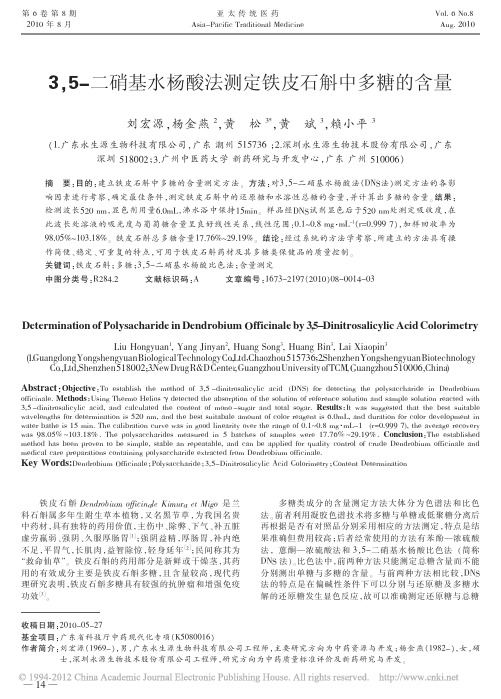
精 密 量 取 对 照 品 溶 液 、 蒸 馏 水 各 2.0mL 分 别 置 于 25mL 量瓶中, 分别加 DNS 试液 6.0mL, 于沸水浴中加热 15min,流水冷却至室温,加水稀释至刻度。 以显色剂—水、 葡萄糖显色液—水、 葡萄糖显色液—显色剂分别在波长 400~800nm 范围内分别进行扫描。 结果:葡萄糖显色液— 显 色 剂 最 大 吸 收 波 长 是 500nm, 但 由 于 DNS 显 色 剂 在 400~510nm 范 围 内有 较 大 的 吸 收 , 且 显 色 期 间 由 于 部 分 DNS 显色剂与待测液中还原糖相结 合 , 消耗 了 部 分 显 色 剂 ,从而造成测定吸光度时 ,作为空白参照的 溶 液 中 DNS 显色剂含量大于葡萄糖显色液中 DNS 试剂含量。 若选择 500nm 为检测波长,DNS 显色剂对测定结果干扰严重。 所 以最大吸收波长并非最佳工作波长,根据“吸收最大,干扰 最 小 ”原 则 ,从 图 1 中 可 以看 出 ,测 定 波 长 宜 选 择 在 520~ 540nm 范围内,本实验最终确定测定波长为 520nm。
此波长处溶液的吸光度与葡萄糖含量呈良好线性关系 ,线性范围:0.1~0.8 mg·mL-1(r=0.999 7),加样回收率为
98.05%~103.18%。 铁皮石斛总多糖含量17.76%~29.19%。 结论:经过系统的方法学考察,所建立的方法具有操
作简便、稳定、可重复的特点,可用于铁皮石斛药材及其多糖类保健品的质量控制。
中药覆盆子及其活性成分的提取与药用价值研究进展
中药覆盆子及其活性成分的提取与药用价值研究进展发布时间:2021-03-02T10:34:21.783Z 来源:《教学与研究》2020年11月第31期作者:李超越刘韩天林涛缪展鹏玛青[导读] 覆盆子为蔷薇科悬钩子属植物掌叶覆盆子(Rubus chingii)的干燥未成熟果实,主要生长在浙江、福建、江西等地,是一种医用价值很高的植物资源。
李超越刘韩天林涛缪展鹏玛青*浙江树人大学生物与环境工程学院,浙江省杭州市 310015[摘要] 覆盆子为蔷薇科悬钩子属植物掌叶覆盆子(Rubus chingii)的干燥未成熟果实,主要生长在浙江、福建、江西等地,是一种医用价值很高的植物资源。
覆盆子中含有覆盆子酸、鞣花酸、β-谷甾醇、黄酮类化合物以及三萜酸等多种化学活性成分,具有抗氧化、抗癌和抗心血管疾病等药理作用。
三萜酸类化合物是蔷薇科植物的特征性成分,也是覆盆子中的主要活性成分之一。
本文对覆盆子的基本特征、药用价值和药理作用进行了简单介绍,并对三萜酸类化合物的提取分离和生物活性研究进展进行了综述。
为设计合理的实验方案对覆盆子中三萜酸的提取工艺、含量分析和生物活性进行研究提供了有益的参考。
[关键词] 覆盆子,三萜酸,提取工艺,生物活性1.覆盆子简介覆盆子(Rubus chingii Hu)为蔷薇科(Rosaceae)悬钩子属(Rubus)掌叶覆盆子的干燥果实。
掌叶覆盆子又称华东覆盆子,属落叶灌木;叶掌状深裂,托叶条形;花单生于短枝顶端;聚合小核果,球形,红色,主要分布于浙江、福建、安徽、江西、江苏等华东各省[1-2]。
覆盆子为《中华人民共和国药典》2005年版一部收录的传统中药,主产浙江、福建,具有补肾、固精、缩尿等功效,可用于肾虚遗尿、小便频数、阳痿早泄、遗精滑精等;现代药理研究表明,覆盆子具有温肾助阳、抗诱变、抗氧化、改善记忆、延缓衰老、增强免疫活性、抑菌等药理作用[3]。
掌叶覆盆子作为药用植物已经有很长的历史,但对其系统的化学成分研究尚不充分。
刺梨果渣在动物生产中的应用研究进展

饲料营养与饲草772024.4·0 引言刺梨为蔷薇科蔷薇属,多分布于我国西南地区,其果实中富含丰富的维生素C 、黄酮、多酚等有效活性成分,具有抗疲劳、抗氧化、抗衰老、预防心血管疾病等药用价值和保健功能[1-3]。
畜禽生产中,可将刺梨果渣制备成天然替抗剂加入到饲料中,实现畜禽生产能力提升[4]。
近年贵州省刺梨产业发展迅速,截至2022年底,刺梨种植规模达1 400 km 2,鲜果产量达26.2万t [5]。
动物日粮中添加刺梨果渣(或发酵刺梨果渣)对饲料成本节约及生产性能提高具有一定的促进作用[4-6]。
本文对刺梨果渣在动物生产中的应用现状进行归纳,以期为有效利用提供参考。
1 刺梨果渣的营养成分刺梨果渣中水分含量最高,为76.70%,灰分含量0.86%,总糖含量4.67%,粗纤维含量11.20%,单宁含量0.156%[6]。
在每100 g 干物质中,含有粗蛋白6.38 g 、淀粉10.04 g 、粗脂肪1.40 g 、粗纤维40.67 g 、总膳食纤维60.40 g 、总酚2 838.36 mg 、总黄酮273.11 mg 、维生素C 366.02 mg 、磷1.09 g 、钙0.87 g 、铁148.68 mg 、锌23.70 mg [7]。
木仁,梁班妹.刺梨果渣在动物生产中的应用研究进展[J].现代畜牧科技,2024,107(4):77-79. doi :10.19369/ki. 2095-9737.2024.04.021. MU Ren ,LIANG Banmei .Research Progress on the Application of Rosa roxburghii Tratt Residue in Animal Production[J].Modern Animal Husbandry Science & Technology ,2024,107(4):77-79.刺梨果渣在动物生产中的应用研究进展木仁1,2*,梁班妹1(1. 黔南民族师范学院生物科学与农学院,贵州 都匀 558000; 2. 黔南州特色畜禽分子技术应用重点实验室,贵州 都匀 558000)摘要:在饲料禁抗和畜禽绿色健康养殖的趋势下,研究和开发新型植物源饲料原料和饲料添加剂备受关注。
在体外和从浒苔硫酸多糖的体内免疫调节活性

International Journal of Biological Macromolecules 49 (2011) 1051–1058Contents lists available at SciVerse ScienceDirectInternational Journal of BiologicalMacromoleculesj o u r n a l h o m e p a g e :w w w.e l s e v i e r.c o m /l o c a t e /i j b i o m acIn vitro and in vivo immunomodulatory activity of sulfated polysaccharides from Enteromorpha proliferaJin-Kyung Kim a ,Myoung Lae Cho b ,Supatra Karnjanapratum b ,Il-Shik Shin b ,Sang Guan You b ,∗a Department of Biomedical Science,Catholic University of Daegu,Gyeongsan,Gyeongbuk,712-702,KoreabDepartment of Marine Food Science and Technology,Gangneung-Wonju National University,120Gangneungdaehangno,Gangneung,Gangwon,210-702,Koreaa r t i c l ei n f oArticle history:Received 4March 2011Received in revised form 25August 2011Accepted 27August 2011Available online 2 September 2011Keywords:Enteromorpha prolifera Immunomodulation Sulfated polysaccharide Green seaweed Th-1cellsa b s t r a c tWater-soluble sulfated polysaccharides extracted from Enteromorpha prolifera and fractionated using ion-exchange chromatography (crude,F 1,F 2and F 3fractions)were investigated to determine their in vitro and in vivo immunomodulatory activities.The sulfated polysaccharides,especially the F 1and F 2frac-tions,stimulated a macrophage cell line,Raw 264.7,inducing considerable nitric oxide (NO)and various cytokine production via up-regulated mRNA expression.The in vivo experiment results show that the sulfated polysaccharides (the crude and F 2fractions)significantly increased Con A-induced splenocyte proliferation,revealing their potential comitogenic activity.In addition,IFN-␥and IL-2secretions were considerably increased by the F 2fraction without altering the release of IL-4and IL-5.This implies that the F 2fraction can activate T cells by up-regulating Th-1response and that Th-1cells might be the main target cells of the F 2fraction.These in vitro and in vivo results suggest that the sulfated polysaccharides are strong immunostimulators.© 2011 Published by Elsevier B.V.1.IntroductionSulfated polysaccharides are complex,heterogeneous and bioactive macromolecules in which some of the hydroxyl groups in sugar residues are substituted with sulfate groups.These anionic polysaccharides are ubiquitous in nature and occur in a wide variety of organisms,including mammals,invertebrates and flora [1,2].Seaweeds are the most important source of non-animal derived sulfated polysaccharides.Studies on sulfated polysac-charides from marine algae have been mainly carried out on brown algae (Phaeophyta),including fucoidans,ascophyllan,sar-gassan and glucuronoxylofucan,as well as red algae (Rhodophyta),including agar and carrageenan,because of their potent biologi-cal activities [3–7].Green algae belonging to Ulvaceae are a rich source of sulfated polysaccharides that are referred to as ulvans.Recently,ulvans have attracted increasing attention because they exhibit unique structural features containing rhamnose and uronic acid with major repeating disaccharide units of ␣-l -Rha p -(1→4)-d -Xyl and (→4)--d -Glc p A-(1→4)-␣-l -Rha p [8,9].They also possess various effective biological and pharmacological activities such as anticoagulant,anticancer,antiviral and anti-hyperlipidemia activ-ities [10–13].∗Corresponding author.Tel.:+82336402853;fax:+82336402340.E-mail address:umyousg@gwnu.ac.kr (S.G.You).In a recent study,ulvans from Ulva rigida stimulated macrophages to secrete nitric oxide (NO)and prostaglandin-2(PGE 2)by inducing an increase of inducible nitric oxide syn-thases (iNOS)and cyclooxygenase-2(COX-2)mRNA expression [6].Macrophages are known to be important components of host defense systems against bacterial infections and various types of invading cells,including cancer cells [14].Activated macrophages release cell factors such as NO,cytokines,reac-tive oxygen intermediates,and other substances to remove pathogens and inhibit cancer cell growth.In a more recent study,Lee et al.[15]report that ulvans from Codium fragile enhance the production of NO and various cytokines by acti-vating macrophages.The authors observed enhanced production of pro-inflammatory (interleukins [IL]-1,IL-6,IL-12and tumor necrosis factor ([TNF]-␣)and anti-inflammatory (IL-10)cytokines,suggesting that ulvans might possess potent immunostimulating activities by activating macrophages while preventing poten-tial detrimental inflammatory effects from excessive macrophage activation.In our previous work,sulfated polysaccharides from Enteromorpha prolifera ,one of the most popular green seaweeds in Asian countries,were extracted and fractionated into three fractions,F 1,F 2and F 3,using ion-exchange chromatography and their structures were characterized [16].These polysaccharides have no significant direct cytotoxicity to AGS and DLD-1cancer cells.However,a considerable amount of NO from macrophages is produced by these polysaccharides,especially in the F 2frac-tion.0141-8130/$–see front matter © 2011 Published by Elsevier B.V.doi:10.1016/j.ijbiomac.2011.08.0321052J.-K.Kim et al./International Journal of Biological Macromolecules49 (2011) 1051–1058Thus,in the present study,we further evaluated the immunomodulatory activity of these sulfated polysaccharides by investigating the production of other cytokines and their mRNA expressions through in vitro and in vivo studies.2.Materials and methods2.1.MaterialsGreen seaweed,E.prolifera,collected in the spring of2007from the coast of Wando,Chunnam,Korea,was purchased from a whole-saler.The raw material was washed with fresh water and air-dried at60◦C.The dried raw material was milled using a blender,sieved (<0.5mm),and stored at−20◦C.The crude polysaccharide was extracted from the milled biomass by using the method of Cho et al.[16].Briefly,5g dried biomass was rehydrated in100mL dis-tilled water at65◦C for2h and centrifuged at18,500×g for10min. Ethanol(99%)was added to the supernatant to obtain afinal ethanol concentration of30%.After centrifugation at18,500×g for10min, an additional150mL ethanol was added to the collected super-natant to obtain afinal ethanol concentration of70%(v/v),and the solution was stored at4◦C overnight.Crude polysaccharide was obtained byfiltering the ethanol solution with a nylon membrane (0.45m pore size;Whatman International Ltd.,Maidstone,UK), washing with ethanol(99%)and acetone,and then drying at room temperature overnight.Crude polysaccharide(100mg)dissolved in10mL distilled water was fractionated using ion-exchange chromatography on a DEAE Sepharose fastflow column(17-0709-01;GE Healthcare Bio-Science AB,Uppsala,Sweden).The column was washed with distilled water,and the bound polysaccharide was eluted with a solution of distilled water and different concentrations of NaCl increasing stepwise from0.5to1.0M.The three fractions obtained are referred to as F1,F2and F3.2.2.Macrophage proliferation assayRaw264.7cells(ATCC,Rockville,MD,USA)were seeded in a96-well microplate(1×10⁴cells/well,in a volume of100L) and incubated in RPMI-1640medium(Hyclone,Logan,UT,USA) containing10%FBS with100L sample polysaccharides at a con-centration of12.5,25,or50g/mL.The experiment was performed in triplicate.After cells were incubated at37◦C for72h,WST-1 reagent was used to determine the proliferation of macrophage cells as described previously[16].Absorbance at450nm was deter-mined by spectrophotometry.The absorbance(A)was translated into the macrophage proliferation ratio(%)=A t/A c×100,where A t and A c are the absorbances of the test and control groups,respec-tively.2.3.Endotoxin testThe concentration of endotoxin was determined by using the limulus amebocyte lysate(LAL)method obtained from E-TOXATE assay kit(Sigma,St.Louis,MO,USA).The experiment was per-formed according to the manufacturer’s instructions.In brief,the sterile endotoxin-free water was used to dissolve and dilute the test samples and endotoxin standard.The triplicate samples and stan-dard solutions were contained in a sterile glass tube,and100L E-TOXATE working reagent was added to each tube.The mixtures of samples and E-TOXATE reagent were incubated at37◦C for1h and then observed for evidence of gelation.The samples were interpreted as“+”(presence of a hard gel;i.e.,sample contains endotoxin)and“−”(absence of a hard gel;i.e.,sample does not contain endotoxin).2.4.NO production assayNO secretion from macrophages and the nitrite concentration was measured using the Griess reaction as described by Green et al.[17].Raw264.7cells were seeded in a96-well microplate (1×10⁵cells/well in a volume of100L)and incubated for24h at 37◦C.The cultured cells were treated with100L polysaccharide solution at12.5,25.0,or50g/mL or1g/mL lipopolysaccha-ride solution(LPS),which was used as a positive control,in the presence or absence of the arginine analogue,N(G)-monomethyl-l-arginine(l-NMMA)(500M),in triplicate.After incubating for 24h at37◦C,100L of cultured cell supernatant was mixed with an equal volume of Griess reagent(1%,[w/v]sulfanilamide and 0.1%[w/v]N-[1-naphthyl]ethylenediamine hydrochloride in2.5% [v/v]phosphoric acid)and left at room temperature for10min.The absorbance at540nm was measured using a microplate reader. The NO secretion from the Raw264.7cells was calculated with reference to a standard curve obtained with NaNO2(1–200M in culture medium).2.5.IL-1ˇproduction assayTo measure the concentration of IL-1,Raw264.7cells(1mL, 1×10⁵cells/well)were cultured in RPMI-1640medium containing 10%FBS with12.5,25.0,or50g/mL polysaccharide solution or LPS(1g/mL)as a positive control in a24-well microplate for72h. The supernatants were collected,and the IL-1concentration was measured with reference to the introduction of the examination agent box(R&D System,Inc.,Minneapolis,MN,USA).2.6.Reverse transcription polymerase chain reaction(RT-PCR)Raw264.7cells(1×10⁵cells/well)were treated in the presence of the sample solutions or LPS at37◦C for18h.The total RNA of Raw 264.7cells treated with LPS or samples was extracted using TRIzol reagent(Invitrogen,Carlsbad,CA,USA)according to the manufac-turer’s protocol and kept at−80◦C until use.The RNA concentration was measured by a spectrophotometer before the construction of cDNA with an oligo-(dT)20primer and Superscript III RT(Invitro-gen).PCR amplification was carried out using GoTaq Flexi DNA Polymerase(Promega,Madison,WI,USA)and specific primers. The primers were as follows:iNOS,5 -CCCTTCCGAAGTTTCTGGCA GCAGC-3 (forward)and5 -GGCTGTCAGAGCCTCGTGGCTTTGG-3 (reverse);IL-1,5 -ATGGCAACTATTCCTGAACTCAACT-3 (for-ward)and5 -CAGGACAGGTATAG ATTCTTTCCTTT-3 (reverse); TNF-␣,5 -ATGAGCACAGAAAGCATGATC-3 (forward)and5 -TACAGGCTTGTCACTCGAATT-3 (reverse);COX-2,5 -CCCCCACA GTCAAAGACACT-3 (forward)and5 -GAGTCCATGTTCCAGGAGGA-3 (reverse);IL-6,5 -TTCCTCTCTGCAAGAGACT-3 (forward) and5 -TGTATCTCTCTGAAGGAC T-3 (reverse);IL-10,5 -TACCTGGTAGAAGTGATGCC-3 (forward)and5 -CATCATGTATGCT-TCTATGC-3 (reverse);-actin,5 -TGGAATCCTGTGGCATCCAT GAAAC-3 (forward)and5 -TAAAACGCAGCTCAGTAACAGTCCG-3 (reverse).Reverse transcriptase amplification was conducted with initial denaturation at94◦C for3min;30cycles of denaturation (94◦C for30s),annealing(56◦C for40s)and extension(72◦C for 1min);and afinal extension step at72◦C for10min.PCR products were run on1%agarose gel and visualized by ethidium bromide staining.2.7.Experimental animalsSix-week-old female BALB/c mice(body weight,17–19g)were purchased from KOATECH(Pyeongtek,Korea).For adaption,the mice were housed in polycarbonate cages and fed a standard animalJ.-K.Kim et al./International Journal of Biological Macromolecules49 (2011) 1051–10581053 diet and water ad libitum under controlled temperature conditions22◦C(2◦C)with a12h light/dark cycle for a week before treatment.2.8.Experimental protocolThe mice were divided into5groups of6animals each.GroupI was the control and received physiologic solution(0.2mL,0.9%NaCl).Groups II and III received crude polysaccharide(5mg/kg,0.2mL and20mg/kg,0.2mL,respectively).Groups IV and Vreceived the F2fraction(5mg/kg,0.2mL and20mg/kg,0.2mL,respectively).All groups were treated for14consecutive days bygavage.2.9.Preparation of lymphocytes from the spleen and mesenteric lymph nodesSpleens and mesenteric lymph nodes(MLNs)were excised asep-tically and ground in RPMI-1640medium.Single cells from spleens and MLNs were collected,and contaminating red blood cells were removed by treatment with a RBC lysis buffer(eBioScience,San Diego,CA,USA).After two washes in RPMI-1640medium,the cells were resuspended in RPMI1640medium containing10%FBS.2.10.Lymphocyte proliferationProliferation was examined using the CellTiter96Aqueous One Solution Cell Proliferation Assay manual(Promega).A total of 1×10⁵cells in200L RPMI1640medium containing10%FBS were stimulated with2.5g/mL Con A or10g/mL LPS.The plates were then placed in a5%CO2incubator at37◦C for48or72h.The opti-cal density at490nm was measured using a microtiter plate reader. The experiment was performed in triplicate.2.11.Measurement of cytokine secretionThe cell culture supernatants used for the cytokine assay were collected after2×105splenocyte with2.5g/mL Con A were incu-bated for48h.The concentrations of mouse IL-2,interferon(IFN)-␥, IL-4and IL-5were evaluated using a Bio-Rad Multiplex bead array instrument and cytokine kit(Bio-Rad,Irvine,CA,USA)according to the manufacturer’s protocol.2.12.Lymphocyte phenotypesSingle cells from spleens and MLNs were counted and adjusted to1×105cells/mL.Splenocytes(200L)were then incubated with the appropriate directly labeled antibodies in PBS with1%BSA at4◦C for30min.The antibodies used werefluorescein isoth-iocyanate(FITC)-conjugated rat anti-mouse CD3,phycoerythrin (PE)-conjugated rat anti-mouse B220,FITC-conjugated rat anti-mouse CD4,PE-conjugated rat anti-mouse CD8and PE-conjugated rat anti-mouse PanNK antibodies.All antibodies and isotype control antibodies were purchased from eBioScience.After incubation,the cells were washed twice with1%BSA in PBS and subsequently ana-lyzed using a Guava Easycyte cytometer(Millipore,Billerica,MA, USA).2.13.NK cell activityAfixed number of carboxyfluorescein succinimidyl ester-labeled YAC-1target cells(1×10⁴)were mixed with2.5×104 splenocytes in a total volume of0.2mL in a microplate with round-bottomed wells.After the plate was incubated for4h and centrifuged,NK cell activity was measured using the Guava Cell Toxicity kit(Millipore)according to the manufacturer’s protocol.Macrophageproliferation(%ofcontrol)FCrude1F3F2ControlFig.1.Effect of various sulfated polysaccharides on Raw264.7cell proliferation. Raw264.7cells(1×10⁴cells/well)were incubated with different concentrations of polysaccharides for72h.Data are presented as means±standard deviation(n=3).2.14.Statistical analysisThe data are expressed as means±SEM.All values were eval-uated by one-way analysis of variance(ANOVA)with Bonferroni multiple comparison post tests using GraphPad Prism4.0software (GraphPad Software Inc.,San Diego,CA,USA).P values less than 0.05or0.01were considered statistically significant.3.Results and discussion3.1.Cell proliferationIn the preliminary study,the crude and fractionated polysac-charides(F1,F2and F3)mostly consisted of carbohydrates (50.8–62.5%),sulfates(14.5–18.8%)and uronic acid(13.8–16.6%) with protein contents ranging from1.0%to13.9%.Their monosac-charide compositions,including rhamnose(57.1–87.6%),glucose (3.6–39.1%)and xylose(2.4–8.8%),varied significantly[16].Among these polysaccharides,the F1and F2fractions induced consider-able amounts of NO in Raw264.7cells[16].Macrophage cells are essential for maintaining homeostasis regardless of varying external conditions and play a pivotal role in host defense against pathogens and any type of invading cells,including cancer cells [18].Activated macrophages release some immunomodulatory fac-tors,such as NO,IL-1,TNF-␣,reactive oxygen intermediates and other substances,to remove pathogens and inhibit the growth of invading cells.In particular,the released NO from macrophages is a highly reactive free radical and plays an important role in the reg-ulation of various immune responses.Therefore,Raw264.7cells are used to determine the immunomodulatory activities of var-ious compounds by investigating their cytokine production.The cytotoxic effects of the crude and fractionated polysaccharides at concentrations of12.5–50.0g/mL on Raw264.7cells are shown in Fig.1.When Raw264.7cells were cultured with the crude and frac-tionated polysaccharides at50g/mL,their proliferation increased slightly,suggesting that these polysaccharides do not exert cyto-toxic effects on Raw264.7cells over the range of concentrations. No endotoxins were detected when endotoxin contamination was tested within the crude and fractionated polysaccharides(data not shown).Therefore,the polysaccharides were not contaminated by endotoxins and the enhanced NO release of the polysaccharides by Raw264.7cells was not due to endotoxin compounds.1054J.-K.Kim et al./International Journal of Biological Macromolecules49 (2011) 1051–1058Fig.2.NO released from Raw264.7cells treated with various sulfated polysaccharides.(a)Raw264.7cells(1×10⁵cells/well)were incubated with50g/mL polysaccharides or polysaccharides with L-NMMA(positive control).Data are presented as means±standard deviation(n=3).x,y,z significant differences(p<0.05)between the concentrations of the crude polysaccharide and the fractions;a,b,c,d significant differences(p<0.05)between the crude polysaccharide and the fractions.(b)iNOS mRNA levels were determined by RT-PCR.Fig.3.IL-1released from Raw264.7cells treated with various sulfated polysaccharides.(a)Raw264.7cells(1×10⁵cells/well)were incubated with different concentrations of polysaccharide or1g/mL LPS(positive control)for18h.Data are presented as means±standard deviation(n=3).x,y,z significant differences(p<0.05)between the concentrations of the crude polysaccharide and the fractions;a,b,c significant differences(p<0.05)between the crude polysaccharide and the fractions at each concentration.(b)IL-1mRNA levels were determined by RT-PCR.J.-K.Kim et al./International Journal of Biological Macromolecules49 (2011) 1051–10581055Fig.4.mRNA levels of various cytokines released from Raw264.7cells treated with various sulfated polysaccharides.3.2.NO release and iNOS mRNA expressionTo determine whether the enhanced NO release by the polysac-charides was due to increased iNOS expression,Raw264.7cells were cultured with polysaccharides and an iNOS inhibitor,L-NMMA.As shown in Fig.2a,NO release by the polysaccharides was effectively blocked by the presence of L-NMMA(500M), especially in the F1and F2fractions.This result suggests that the polysaccharides might induce NO release via iNOS in Raw264.7 cells.Therefore,the mRNA levels of iNOS in polysaccharide-treated Raw264.7cells were investigated in further detail.Among the polysaccharides,the F1and F2fractions dramatically induced iNOS mRNA expression whereas only a slight induction was observed in Raw264.7cells treated with the F3fraction(Fig.2b).This sug-gests that the considerable NO production from Raw264.7cells is due to the enhanced iNOS mRNA expression after stimulation by the F1and F2fractions.Leiro et al.[6]observed a proportional relationship between NO release and the sulfate contents of a polysaccharide extracted from U.rigida.On the other hand,in a study of other polysaccharides,a relationship between molecular weight and NO production from macrophages was observed[19]. The authors suggest that higher molecular weight polysaccharides can induce greater NO production in Raw264.7cells.However,in the current study,it is not clear why the F1and F2fractions induced more NO than the F3fraction.It was difficult to discern a correla-tion between structures and biological activities because of their considerable compositional and structural heterogeneity.Further research is necessary to obtain a better understanding of theirfine structures and bioactivities.3.3.Cytokine production of Raw264.7cellsFig.3a shows the production of IL-1from Raw264.7cells treated with the crude and fractionated polysaccharides,which followed a tendency similar to that of NO release.The F1and F2fractions exhibited considerable levels of IL-1production from37.7to164.0pg/mL at12.5,25.0,and50.0g/mL,these are comparable to that in the positive control,LPS.However,the IL-1production induced by the F3fraction was relatively small com-pared with that of the other fractions.Furthermore,the F1and F2 fractions produced significantly increased IL-1mRNA expression, both greater than that of the F3fraction(Fig.3b).This suggests that the considerable IL-1production is due to enhanced IL-1mRNA expression by the stimulation from the F1and F2fractions.Similar trends were observed in the mRNA expressions of TNF-␣,IL-6and IL-10,and COX-2by the crude and fractionated polysaccharides at 12.5–50.0g/mL(Fig.4),the F1and F2fractions resulted in con-siderable mRNA expression of these cytokines,whereas relatively little mRNA expression was observed with the F3fraction.In gen-eral,pro-inflammatory mediators such as TNF-␣,IL-1,IL-6and PGE2are essential for host survival from injured and/orabnormalFig.5.Body(a)and spleen(b)weights of mice treated with the crude and F2frac-tions.1056J.-K.Kim et al./International Journal of Biological Macromolecules 49 (2011) 1051–1058Fig.6.Effect of the crude and F 2fractions on spleen and MLN cell proliferation in mice.(a)Spleen cell proliferation was measured with 2.5g/mL medium or Con A;(b)MLN cell proliferation was measured with 2.5g/mL medium or LPS.Significant differences from the control mice are indicated as follows:*p <0.05,**p <0.01.cells and tissues [14,20,21].These cytokines could be produced by the stimulation of immune cells,including T-cells,B-cells and macrophages.However,excessive secretion of pro-inflammatory cytokines by immoderate immune cell activation could produce harmful effects on the repair process of damaged cells and tissues by aggravating sites with severe inflammation.Therefore,anti-inflammatory cytokines such as IL-4,IL-10and IL-12also need to be released to prevent potential detrimental effects from excessive macrophage activation [22].Our current data demonstrate that the considerable expression of IL-10mRNA also occurred with the F 1and F 2fractions (Fig.4).This suggests that released IL-10might sup-press the excessive activation of Raw 264.7cells,thus avoiding the potential aggravation of the recovering sites.Therefore,the present data suggest that the sulfated polysaccharides might be immunos-timulating polysaccharides that can activate macrophages via the release of pro-inflammatory mediators while suppressing theirover-activation by producing anti-inflammatory cytokines.Such production of both pro-and anti-inflammatory mediators by Raw 264.7cells is also observed with a sulfated galactan from C.fragile ,which implies its considerable immunostimulation without the potential occurrence of severe inflammation [15].3.4.Body and spleen weights of miceThe in vivo polysaccharide experiments were performed with the crude and F 2fractions.The F 2fraction was selected for the in vivo experiment because of its considerable cytokine-releasing capacity in Raw 264.7cells and greater yield (32.0%)through the ion-exchange column than other fractions.Fig.5a shows the body weights of mice in each group before and after the administration of the crude and F 2fractions.The BALB/c mice of the control group gained 1.32g body weight during the course of the experiment.ATable 1Proportion of various immune cells (CD3+,B220+,CD4+,CD8+and NK cells)within the lymphocyte population of the spleen.GroupCD3+(%)B220+(%)CD4+(%)CD8+(%)NK cells (%)Control42.7±3.143.8±2.734.0±3.111.4±2.0 4.5±0.9Crude (5mg/kg)38.0±5.349.0±3.032.0±1.39.2±1.7 4.2±1.1Crude (20mg/kg)38.2±2.248.8±1.531.6±2.510.0±2.0 3.1±0.6F 2(5mg/kg)42.1±2.044.5±3.732.8±2.811.4±1.9 3.7±0.2F 2(20mg/kg)40.0±3.445.3±4.135.6±1.47.6±2.13.8±0.4J.-K.Kim et al./International Journal of Biological Macromolecules49 (2011) 1051–10581057Fig.7.Secretion levels of various cytokines;(a)IFN-␥,(b)IL-2,(c)IL-4and(d)IL-5.Significant differences from the control mice are indicated as follows:*p<0.05,**p<0.01.similar level of body weight gain was observed in the mice admin-istered the crude and F2fractions.In addition to body weight alteration,the relative spleen weights of mice in each group were also examined after thefinal polysaccharide administration.As shown in Fig.5b,no significant changes in relative spleen weight were observed among groups.Such small changes in body weight and relative spleen weight seem to imply that the polysaccharides are non-toxic to mice.3.5.Lymphocyte population in splenic lymphocytesThe proportions of various immune cells,including CD3+,B220+, CD4+,CD8+and NK cells,within the lymphocyte population of the spleen were determined for thefive groups of mice before and after polysaccharide treatment(Table1).The proportions of CD3+T cells, including CD4+and CD8+subsets,B220+B cells and NK cells among the groups of mice were not significantly altered by polysaccha-ride treatment.This suggests that the balance of the immune cell population in the spleen is not affected by the polysaccharides. 3.6.Mitogen response in splenic lymphocytesThe effects of the crude and F2fractions on splenic lymphocyte proliferation were observed using the T-cell mitogen,concanavalin A(ConA),or the B-cell mitogen,LPS.Little change in splenocyte pro-liferation was observed after the polysaccharide treatment in the absence of mitogens,which indicates that the polysaccharides are not directly mitogenic to mouse splenocytes(Fig.6).In the pres-ence of mitogens(Con A or LPS),the crude polysaccharides did not increase splenocyte proliferation,while the F2fraction resulted in a significant increase,suggesting that it has considerable comi-togenic activity.The comitogenic effect was only observed at low doses(5mg/kg)of F2administration.It is well known that spleno-cyte proliferation is an indicator of immunomodulation.Much in vivo and in vitro evidence demonstrates that polysaccharides exhibit immunomodulatory capacity by stimulating both cellular and humoral immunoresponses[23,24].Recently,Jiao et al.[14] also observed Con A-induced splenocyte proliferation by a sulfated polysaccharide from E.intestinalis.In a study of-carrageenan, Zhou et al.[4]observed a relationship between the immunomod-ulation and molecular weights of the sulfated polysaccharides, suggesting that low molecular weights(≤15kDa)might be benefi-cial for the immunostimulation of the polysaccharides.However,it remains unclear why the low molecular weight polysaccharides exhibit more enhanced splenocyte proliferation in vivo,further research is required to better understand their molecular structures and bioactivities in vivo.3.7.Cytokine production in splenic lymphocytesT cells are characterized as either CD8+or CD4+T cells on the basis of their functional capabilities and cytokine profiles.In addi-tion,there are two general subsets of CD4+T cells(Th-1and Th-2 cells)that produce different cytokines.The secretion of IFN-␥,IL-2 and TNF-␣leads to a Th-1type cellular response,while the secre-tion of IL-4and IL-5leads to a Th-2type cellular response.As shown in Fig.7a and b,the crude polysaccharide resulted in almost no difference in IFN-␥and IL-2production,whereas the F2fraction sig-nificantly increased the secretion of both cytokines.However,the levels of IL-4and IL-5secretion were not altered by the administra-tion of the crude and F2polysaccharides(Fig.7c and d).Therefore, these results suggest that the F2fraction can activate T cells by up-regulating Th-1response and that Th-1cells might be the main target cells of the F2fraction.It is reported that Th-2cytokine levels are higher than those of Th-1cytokines in various diseases such as cerebral infarction,allergy and asthma.Therefore,these results suggest that the F2fraction might be useful for improving Th-2dominant pathological disorders.The Th-1response is also。
虫草头孢菌粉粗多糖的理化性质和含量测定研究_邹峥嵘

取虫草菌粉粗多糖溶液2ml,加入浓硫酸2ml,静 置5min,再加入新配制的斐林试剂2ml,在沸水浴中反 应2min,观察反应中颜色的变化。
取虫草菌粉粗多糖溶液2ml,加入新配制的双缩脲 2ml(先加A 试剂2ml,再加B 试剂3~4 滴),在沸水浴 中反应2min,观察反应中颜色的变化。
粉直接制备而成,化学组分十分复杂,作用机制不明 确,质量控制的标准和方法也有待完善,从而极大地 影响了产品的使用和推广。虫草多糖作为虫草中含量最 高的生物活性物质,引起了人们对它的极大兴趣和广泛 关注。大量医学实验证实,虫草多糖可活化巨噬细胞, 刺激抗体产生,提高人体免疫能力[4-5]。此外,虫草多 糖的抗肿瘤活性也逐渐被人们公认[6]。
※基础研究
食品科学
2007, Vol. 28, No. 09 87
浓硫酸 天津市永大化学试剂开发中心;氢氧化钠、 硫酸铜、碳酸钡、葡萄糖 南昌鑫光精细化工厂。以 上试剂均为分析纯。 1.3 仪器
SHB-C型循环水真空泵、R201型旋转蒸发器 郑 州杜甫仪器厂;WFJ-7200型可见光光度计 尤尼柯(上 海)仪器有限公司;BS223电子天平 北京赛多利斯仪器 系统有限公司;DHG-9240A型电热恒温鼓风干燥箱 上 海精宏实验设备有限公司;HH-6型数显恒温水浴锅 国 华电器有限公司; SG250-H型超声波清洗器 上海冠特 超声仪器有限公司。 1.4 方法
1 材料与方法
1.1 材料 虫草菌粉(Cs-4) 深圳市汇康生物科技有限公司南昌
分公司。 1.2 试剂
乙醇、石油醚 天津市福晨化学试剂厂;乙酸乙 酯、苯酚 成都市科龙化工试剂厂;正丁醇、氯仿、
收稿日期:2007-08-10 作者简介:邹峥嵘(1970-),男,副教授,主要从事天然产物的研究和开发。
新疆芜菁多糖降血糖作用的研究

新疆芜菁多糖降血糖作用的研究新疆农业科学2011,48(3):471—479 XinjiangAgriculturalSciences新疆芜菁多糖降血糖作用的研究艾克拜尔江?阿巴斯,李冠,王静(新疆大学生命科学与技术学院,鸟鲁木齐830046)摘要:【目的】检测新疆芜菁多糖的组分结构与对血糖的作用.【方法】采用常规的水提法从芜菁中提取粗多糖,并用Sevag法对其进行脱蛋白,再通过回流,透析等步骤把粗多糖纯化获得芜菁精制多糖.用芜菁精制多糖进行小白鼠血糖实验.对芜菁精制多糖进行DEAE一52离子交换柱层析纯化,对第一个洗脱组分BRPS1再进行SephadexG一200凝胶柱层析纯度鉴定.此外,用红外光谱法分析BRPS1,并且对离子交换柱层析的7种洗脱组分分别进行了Gc—Ms分析.【结果】当芜菁精制多糖剂量为400mg/kg体重时,3d后的受试小鼠血糖值下降差异显着(P<0.05);6和9d后的血糖值下降差异极显着(分别是P<0.01和P<0.001);通过DEAE一52离子交换柱层析共分离出7种组分.对第一个洗脱组分BRPS1的SephadexG一200凝胶柱层析的结果表明,BRPS1是单一的多糖组分;其红外图谱结果表明BRPS1具有多糖的一般结构.对7种洗脱组分的GC—MS分析表明芜菁精制多糖含有6种单糖.【结论】芜菁多糖具有显着的降血糖效果;作为芜菁多糖主要成分的BRt~I为单纯的多糖,具有多糖的一般结构.关键词:芜菁;多糖;血糖测定;GC—MS中图分类号:S631.3;S609.9文献标识码:A文章编号:1001—4330(20l1)o3—0471一o9 HypoglycemicFunctionofP0IysaccharidesfromBrassicarapaL.ofXinjiangAikebaierjiangAbasi,LIGuan,WANGJing (CollegeofLifeScienceandTechnology,XinjiangUniversity,Urumqi8300 46,China)Abstract:【Objective]Toinvestigatethecompositionandstructureofpolysaecharidesfr omBrassicarapaL.ofXinjianganditseffectsonbloodsugarleve1.【Method】CrudepolysaccharidewasextractedfromBrassicarapaL. bycommonwaterextractionmethod,andthendeproteinizedbySevagmetho d.ThecrudepolysaccharidefromBrassicarapaL.wasfurtherpurifiedbyrefluxinganddialyzing,andthenpurifi edpolysaccharidewasobtained.BloodsugarleveltestWasperformedonwhitemicebyusingpurifiedpolysacc haride.DEAE一52ion—exchange columnchromatographywasperformedonpurifiedpolysaccharideandSeph adexG一200gelcolumnchromatography WasperformedonthefirstcomponentBRPS1forpurityanalysis.Andtheninf raredanalysiswasperformedonBRPS1;sevencomponentsobtainedbyion—exchangecolumnchromatogra phywereanalyzedbyGC—MSfor determiningitsmonosaccharidecomposition.【ResultIUnderthedoseof400mg/kgbw:after3daysthebloodsugar leveldecreasedrelativelyobviously(P<0.05);after6daysand9daysthebl oodsugarleveldecreasedquiteobviously(P<0.01andP<0.001respectively).Sevencomponentswere derivedbyDEAE一52ion—exchangecolumnchromatography.ResultofSephadexG一200gelcolunmchromatographyshowedthatBRPS1Waspure polysaccharide.InfraredanalysisindicatedthatBRPS1hadcommoncompos itionofpolysaccharides.Sixkindsof monosaccharideswereidentifiedinBrassicarapaL.purifiedpolysaccharide sbyGC—MS.【Conclusion】PolysaccharidesfromBr~siearapaL.ofXinjianghasobvioushypoglycemic effect;BRPS1,asthemaincomponentofBrassicarapaL.polysaccharides,ispurepolysaccharide,andithasthecom monstructureofpolysaccharides.Keywords:BrassicarapaL.;polysaccharide;bloodsugarchecking;GC—M S收稿日期:2011一O1—28基金项目:新疆大学植物生物技术重点实验室作者简介:艾克拜尔江?阿巴斯(1985一),男(维吾尔族),新疆人,硕士研究生,研究方向为植物生物技术,(E—mail)********************通讯作者:李冠(1949一),男.湖北人,教授,博士,博士生导师,研究方向为植物生理生化与分子生物学,(E—mail)******************472?新疆农业科学48卷0引言【研究意义】近年来,随着人们生活水平的不断提高,糖尿病的发病率13益趋高.糖尿病的主要症状之一是高血糖,如果糖尿病患者的血糖得不到及时控制,则会导致一系列的并发症,进而危及生命.在医疗上除了用胰岛素降低血糖之外,还常用一些西药,虽然这些西药药效迅速,但是它们具有副作用,而芜菁多糖是天然产物,研究其对血糖的作用具有非常重要的理论和应用价值.芜菁中含有糖类以及糖苷类这一事实使芜菁多糖的降血糖研究显得十分重要.【前人研究进展】新疆芜菁(BrassicarapaL.)又称恰玛古,隶属于十字花科芸苔族芸台属.芜菁由于对环境的适应性很强,产量较高,在新疆干旱缺水的南部种植非常广泛,而且携带及保存方便,汁多味甜可食…,深受当地维吾尔族等少数民族居民喜爱,并是有较长食用历史的传统蔬菜之一E23.芜菁除了具有很高的食用价值以外,还有重要的药用价值.维吾尔医学认为:”恰麻古儿”能够清除体内异常粘液质等异常体液,抑制细菌,真菌和寄生虫的作用,具有止咳,平喘,化痰,缓解食欲不振等功效,也具有抗衰老,抗辐射等方面的药理活性J.《中国中草药大全》,《本草纲目》,《维吾尔药志》,《中华人民共和国卫生部药品标准,维吾尔药分册》等药典对恰玛古的植物来源,药材性状(包括块根,叶,花和种子),药理作用,功能主治,配方加工等都有详细的记载.药典记载和新疆检测分析中心的检测报告表明,恰玛古无毒,含有丰富的芸苔素,亚油酸,亚麻酸,抗坏血酸,胡萝卜素和多种氨基酸,其营养丰富,具有性温排毒,促进消化,清肺,消肿,降血脂,抗癌等作用J.多糖是三大类重要的生物大分子之一,长期以来一直认为多糖类在生物体内仅仅具有作为结构组分与能量来源的作用.然而,目前逐渐发现多糖具有复杂多样的生物活性,如多糖具有免疫调节功能,可作为广谱免疫促进剂;作为药物可以治疗风湿病,慢性病毒性肝炎,癌症等;还具有抗病毒,抗感染,降血糖,降血脂,消除氧化自由基,促进核酸与蛋白质的生物合成等作用_5J.由于生物活性与药理作用上的多样性,近年来多糖的研究受到广泛的重视.2009年,周芳等_3J对阿克苏,伊犁,吐鲁番和和田等四个地区的芜菁的化学成分进行了研究,发现了其中有挥发油,氨基酸,蛋白质,糖及糖苷类,皂苷,鞣质,酚类,有机酸,生物碱,黄酮等多种成分.【本研究切入点】由于芜菁药理作用广泛,因而近年来越来越受到学术界的关注,但是其活性成分作用的研究仅仅局限于抗感染,抑菌等功能上.而且这些活性成分以小分子次生代谢物为主.至于芜菁多糖提取物对血糖的作用方面尚无报道.【拟解决的关键问题】研究利用常规水提法从新疆芜菁中提取多糖,用小白鼠进行血糖试验,并对多糖的单糖组分及结构进行分析,为芜菁多糖今后的大规模工业化生产提供了理论依据,分析新疆芜菁多糖的降血糖活性,为芜菁多糖在医药保健领域中的应用提供科学基础.1材料与方法1.1材料与化学试剂1.1.1材料新疆芜菁(BrassicarapaL.)来源于新疆焉耆县塔合其乡.1.1.2化学试剂四氧嘧啶,L一木糖,L一岩藻糖,D一葡萄糖醛酸,D一核糖,D一核糖醇,D一阿拉伯糖,D一半乳糖,D一半乳糖醛酸,L一阿拉伯糖(Sigma公司),D一果糖(北京化工厂),D一甘露糖(中国医药公司),L一鼠李糖(上海试剂二厂),D一葡萄糖(天津市化学试剂三厂),DEAE一52离子交换剂(Whatman产品),SephadexG一200(Pharmacia产品).其它试剂均为国产分析纯.1.1.3仪器型号Biofugestratos高速离心机(美国KENDRO公司),uV一1000型紫外/可见分光光度计(美国LabTech公司),722型光栅分光光度计(上海第三分析仪器厂),LAMBDAl7型紫外/可见分光光度计(美国PerkinElmer公司),ALPHA1—2LD型冷冻干燥器(德国Christ公司),Smart一606型微型血糖仪(美国台欣生物科技研发股份有限公司),EQUINOX55型红外光谱仪(德国BRUKER公司),QP2010型气相一质谱联用仪(日本SHIMADZU公司).1.2实验动物实验动物为昆明种小白鼠,购置于新疆维吾尔自治区实验动物研究中心,生产许可号为SCXK(新)2003—0002.饲喂小鼠饲料购置于新疆医科大学动物中心,生产许可证号为医动字第16—003.3期艾克拜尔江?阿巴斯等:新疆芜菁多糖降血糖作用的研究?473?1.3芜菁粗多糖的提取首先将新鲜芜菁块根削皮切成薄片,放在烘箱内在45℃下烘3d至干片,再粉碎成粉末.取1kg芜菁干粉,放置在5L的蒸馏水中,90℃下加热处理6h,收集上清液.残渣再用同样的方法处理2次.取每次收集的上清液,用30层纱布过滤,然后在20~C,5000r/rain的条件下离心30min,取上清液.用旋转蒸发仪在60~C下低压浓缩至变稠为止.往浓缩液上加4倍体积的95%乙醇过夜.在4~C,3000r/min下离心30min,弃上清.把残渣依次用无水乙醇,丙酮,乙醚洗涤多次,然后用冷冻干燥器干燥,得到芜菁粗多糖粉末.1.4粗多糖的初步纯化把芜菁粗多糖粉末置于蒸馏水中充分溶解,将多糖溶液与Sevag试剂(1倍体积的正丁醇加4倍体积的氯仿)相混合,体积比为芜菁多糖溶液:Sevag试剂=3:1.剧烈震荡30min,在20℃,5000r/min下离心30min,弃下层有机溶剂层以及中间白色变性蛋白层.上层多糖溶液重复用上述方法处理,直到中间无变性蛋白层,以及在260~280nm紫外检测无特征吸收峰为止.然后把多糖溶液醇析,离心,沉淀用95%乙醇在80℃下回流2h,除去黄酮,生物碱等醇溶性小分子杂质.再用石油醚在50℃下回流2h,除去脂溶性杂质.把多糖沉淀重新溶解在水中,用透析袋透析,直到电导率小于300gs[,用苯酚一硫酸法检测,在490nm附近无特征性吸收峰为止.将多糖溶液再通过醇析,离心,冻干等步骤获得白色精制芜菁多糖.1.5芜菁多糖降血糖活性的测试取昆明种小白鼠100只,雌雄各半,4~6周龄,体重1822g,在实验室环境下用小鼠标准饲料适应性的喂养3d后,禁食不禁水12h,从眼眶采血测空腹血糖值.然后分两组:取1O只为健康对照组,9O只为造模组.各组雌雄分别各半,对造模组的小白鼠腹腔注射四氧嘧啶300mg/kg体重,72h之后测其空腹血糖值,把其中血糖值在10mmol/L以上者筛选出来用于实验.将建模成功的小白鼠分为四组,即疾病对照组,及低,中,高剂量组.每组10只,将低剂量组用100mg/kg体重的芜菁精制多糖灌胃;中剂量组用200mg/kg体重的芜菁精制多糖灌胃;高剂量组用400mg/kg体重的芜菁精制多糖灌胃.健康对照组与疾病对照组用相同体积的蒸馏水灌胃.每天灌胃一次.每3d测一次空腹血糖值.1.6芜菁多糖的分级纯化与结构组分鉴定1.6.1DEAE一52离子交换柱层析取100g处理好的DEAE一52装入层析柱2.5×60(cm),其床体积为2.5×50(cm).用蒸馏水平衡24h.取0.5g精制多糖溶于适量蒸馏水,并上样.首先用蒸馏水洗脱,直到用苯酚一硫酸法检测不到被洗脱的多糖为止.然后,用相同方法依次用浓度为0.10,0.25,0.50和1.00mol/L的NaC1洗脱.其流速为1mL/min,每管收集10mL.用苯酚一硫酸法逐管检测,记录490nm处的OD值.以试管序号为横坐标,OD值为纵坐标绘制洗脱曲线.将最大峰处的洗脱液合并,经减压浓缩,透析,醇析,冻干,得到洗脱组分.1.6.2Sephadex一200凝胶柱层析取Sephadex一200干粉6g,用蒸馏水浸泡吸胀72h后装柱,其柱尺寸为1.5×100(em),床体积为1.5×90(em).用3倍床体积的蒸馏水平衡后,把离子交换柱层析的1号洗脱峰组分BRPS1上样,用0.1mol/L的NaC1溶液洗脱,流速为0.5mL/min,每管收集3mL,以试管序号为横坐标,OD值为纵坐标绘制洗脱曲线.将最大峰处的洗脱液合并,浓缩,透析,醇析,冻干,得到其洗脱组分.1.6.3红外光谱和GC—MS分析1.6.3.1红外光谱取DEAE一52离子交换柱层析的第一洗脱组分BRPS12mg,加入200mg经干燥的KBr晶体,研磨混合后压片,进行红外吸收光谱分析l_7J.1.6.3.2GC—MS分析分别称取BRPS1,BRPS2,BRPS3,BRPS4,BRPS5,BRPS6,BRPS7各5n1g,分别加入3mL2mol/L三氟乙酸,封口后置于电热鼓风干燥箱在110℃下水解2h,然后利用旋转蒸发法去除多余的三氟乙酸.往除去三氟乙酸的水解物中分别加入5mg盐酸羟胺,0.3mL吡啶,密封后在90~C水浴下振荡反应30min,取出冷却至室温,再加入0.45mL乙酸酐反应40min,反应产物直接进行GC—MS分析.再474?新疆农业科学48卷分别称取各标准单糖样品5mg,用相同的衍生化方法处理,然后直接进行GC—MS分析,确定其保留时间与质谱信号.把多糖的保留时间与标准单糖的保留时间相比,推测其单糖组分l8].色谱条件:RTX一5柱,程序升温:柱初温190℃,保留2min,以5~C/rain升至230℃,保留5min,汽化室温度:230~C,进样量1L,分流比30:1.质谱条件:源温200~C,接口250~C,扫描范围35—500am,电压0.7V.2结果与分析2.1芜菁多糖脱蛋白后纯度的鉴定将精制多糖配成水溶液,用紫外扫描仪在200~400am扫描.在260与280nm处没有特征性吸收峰,说明多糖样品中没有大量的蛋白质与核酸杂质存在.图1目0波长)图1芜菁精制多糖的紫外图谱Fig.1UVspectrumofpurifiedpolysaccharidefromBrassicarapaL.2.2芜菁多糖的降血糖活性造模之后小鼠的血糖值有了明显的提高,P<0.001;把造模组与同一天的健康对照组相比较,P<0.001,说明建模成功.建模成功后,疾病对照组的血糖值有了逐步的下降,但是与相同时间的健康对照组比较,P<0.001;而与疾病对照组本身刚建模后的血糖值比较,P>0.05,也就是说,没有统计学上的显着性.因此四氧嘧啶型糖尿病小鼠血糖的自动下降对实验的干扰完全可以排除.从实验结果可以观察到,各组与本身刚建模时的血糖值相比,在第3d 时,低与中剂量组的血糖没有明显的下降,然而高剂量组的血糖有了较显着的下降,即P<0.05;在第6d,低中剂量组的血糖仍然没有明显地下降,然而,高剂量组的血糖值下降得更明显,即P<0.01;在第9d,低剂量组的血糖值仍没有显着的下降,中剂量组的血糖值有显着的降低,P<0.05;而高剂量组的血糖值降低相当明显,P<0.001.把低中高剂量组与同一天的疾病对照组相比较,也得到同样的结果.上述结果说明,芜菁多糖在剂量在400mg/kg体重时,具有明显的降血糖作用.表1表1芜菁精制多糖对小白鼠血糖的作用(mmol/L)Table1TheeffectofBrassicarapaL.purifiedpolysaeeharideonthebloodsuga rlevelofwhitemice注:”*”代表与刚刚建模后的血糖值相比的显着性,表示P<0.05,**表示P<0.0l,***表示P<O.1301;”△”代表与同一天的疾病对照组的血糖值相比的显着性,△表示P<0.05,△△表示P<0.O1.△△△表示P<0.0013期艾克拜尔江?阿巴斯等:新疆芜菁多糖降血糖作用的研究?475?2.3芜菁多糖的分级纯化2.3.1DEAE一52离子交换柱层析通过离子交换柱层析,共分离了7种芜菁多糖洗脱峰.分别命名为BRPS1,BRPS2,BRPS3,BRPS4,BRPS5,BRPS6,BRPS7.其中BCPS1的含量最多,为主要的芜菁多糖组分,对其通过凝胶柱层析进行纯度鉴定.图2221口o1aaO255口751∞125试管号图2芜菁精制多糖的DEAE一52离子交换柱层析Fig.2ElutioncurveofpurifiedpolysaccharidefromBrassicarapaL.onDEAE 一52ion—-exchangecolumn2.3.2Sephadex—G200凝胶柱层析纯度鉴定通过凝胶柱层析,只分离出了一种组分,说明芜菁多糖组分BRPS1为纯净的单一组分多糖.图3oo0102030405060试管号图3芜菁精制多糖洗脱组分BRPS1的SephadexG一200凝胶柱层析Fig.3ElutioncurveofBRPS1(oneoftheelutedcomponentofBrassicarapaL. purifiedpolysaccharide)onSephadexG—-200gelcolumn2.3.3芜菁多糖组分BRPS1的红外光谱分析BRPS1的红外吸收图谱是典型多糖类吸收峰.在30002800cm.1的吸收峰是C—H的伸缩振动,是糖类的特征性吸收峰[.其中,在3600~3200cm有一组宽峰,这是多糖类分子间O—H氢键的伸缩振动;16501600cm-1的一组吸收峰为酰胺羰基的伸缩振动[m;1649cmI1的吸收峰是C=0非对称伸缩振动峰,又能表明多糖中含有糖醛酸[u;14401200cm.1的是羧基的伸缩振动.1075—1000cm是吡喃环的特征吸收峰[10].1100咖附近有两个特征性吸收峰,分别代表仲碳一OH的0一H变角振动和伯碳一OH的0一H变角振动.2929cm是一CH2的C—H伸缩振动_l.图40000000000476?新疆农业科学48卷焉董磊董卜_图4芜菁多糖组分BRPS1的红外吸收光谱Fig.4IRspectrumofBRPS12.3.4芜菁多糖的GC—MS分析通过与标准单糖衍生物的总离子流图相比较,推测了芜菁多糖的各组分有鼠李糖,木糖,阿拉伯糖,果糖,葡萄糖和核糖6种单糖组成.表2,图5表2芜菁精制多糖各组分的单糖组成Table2MonosaccharidecompositionofsevencomponentsofBrassicarapaL .purifiedpolysaccharideBRPSlBRPS2BRPS3BRPS4BRPS5BRPS6BRPs7鼠李糖Rhamnose木糖Xylose√阿拉伯糖Arabinose√菜堍Fructose葡萄糖Glucose√核糖Ribose√√√√3讨论植物多糖的提取主要采用水浸提法.在水浸提法中,需要使用各种有机溶剂进行多糖的提取.虽然水浸提法的成本低,但是耗时长,操作繁杂,其中的有些步骤需要重复多次.而用酶法提取植物多糖具有产率高,杂质容易去除,提取条件可控等优点_1.在多糖的脱蛋白方面,常用的方法有Sevag法,三氯乙酸法l引,ZnSO法,三氟三氯乙烷法_l.但多糖脱蛋白的适合性随所选植物材料的不同而有所不同.例如,罗莹等_l6_对大枣多糖脱蛋白时发现,无论是在多糖的得率方面,还是在蛋白质残留量方面,复合酶法(木瓜蛋ca酶+胰蛋白酶法)远优于Sevag法.朱越雄等l-1J对野生树舌多糖进行脱蛋白时,得出了单独利用有机溶剂的效率明显高于单独利用酶法和酶与有机溶剂结合法.研究单独利用Sevag法连续对芜菁粗多糖处理7~l3次,芜菁粗多糖脱蛋白后,通过紫外光谱仪发现几乎没有检测到蛋白质与核酸杂质的存在.说明Sevag法适合于芜菁多糖的脱蛋白.此外研究通过对芜菁多糖进行DEAE一52离子交换柱层析获得了7种洗脱组分,对芜菁多糖第一组分BRPS1进行SephadexG一200凝胶柱层析纯度检测得到了单一洗脱峰,从而证明了芜菁多糖第一组分BRPS1是单纯的多糖组分.芜菁多糖的单糖组分分析表明,芜菁多糖含有6种单糖,分别是鼠李糖,木糖,阿拉伯糖,果糖,葡萄糖和核糖.在多糖的降血糖作用方面,韩春然等口]研究的黑木耳多糖当剂量为400mg/kg体重时,30d后观察到血糖极明显地下降.徐茂红等_l8_做黄精多糖时发现,当剂量分别为330,660和1320mg/kg体重时,到了第15d时,才具有较明显的降血糖结果(P<0.05).而该研究的结果表明,当芜菁多糖的剂量为400mg/kg体重时,到第3d就有了较明显的作用(P<0.05),即芜菁多糖的降血糖活性比黑木耳以及黄精多糖快.因为芜菁精制多糖是天然来源的活性成分,而且芜菁是具有新疆地方特色以及民族特色的蔬菜,种植非常广泛,因此芜菁多糖作为辅助降血糖药物具有很非常广阔的应用开发前景.3期艾克拜尔江?阿巴斯等:新疆芜菁多糖降血糖作用的研究?糖类的一般结构.新疆芜菁多糖由6种单糖组成:鼠李糖,木糖,阿拉伯糖,果糖,葡萄糖和核糖.4.2新疆芜菁精制多糖具有明显的降血糖活性,当剂量为400mg/kg 体重时,3d后的受试小鼠血糖值下降差异显着(P<0.05);6和9d后的血糖值下降差异极显着(分别是P<0.01和P<0.001).3期艾克拜尔江?阿巴斯等:新疆芜菁多糖降血糖作用的研究?479? 参考文献:[1]孛福林.新疆”卡玛古”一芜菁甘蓝栽培[J].蔬菜,1999,(7):9.[2]张涛,田为真,米世伟,等.恰麻古尔提取物平喘止咳作用的初步探究[J].新疆医科大学,2009,32(9):1237—1239.[3]周芳,海力茜?陶尔大洪,郁文.维药恰麻古儿化学成分预实验[J].海峡药学,2009,21(6):100—102.[4]敬思群.恰玛古饮料工艺技术研究[J].食品与发酵工业,2008,34(10):162—165.[5]刘婷,周光明.多糖的提取和分析方法[J].化工时刊,2008,22(3):66—70.[6]贡汉坤.南瓜多糖的提取工艺及其分离纯化[J].食品与机械,2008,24(6):68—70.[7]马莉,唐健元,李祖伦,等.板蓝根多糖分离纯化及其性质的初步研究[C]//.中医药学术发展大会论文集:567—569.[8]何峰,杨英,周蓬蓬,等.链霉菌H03发酵液中具有抗菌活性多糖的分离纯化及其单糖组成分析[J].生命科学研究,2007,11(3):212—217.[9]周丽伟,陈彦,孙玉军.天葵子多糖的提取与鉴定[J].中国生化药物杂志,20O7,28(3):173—175.[10]刘荣,王向宏,李佳梅.柳蒿芽多糖的分离纯化及结构初步分析[J].食品科学,2009,30(13):81—83.[11]刘洪霞,田文莹,王志兵,等.粪鬼伞多糖的提纯与定性研究[J].长春工业大学(自然科学版),2009,30(2):126—129.[12]孙汉文,刘占锋.枸杞多糖的超声波辅助水提取与分级纯化[J].食品工业科技,2009,30(3):230—233.[13]韩春然,马永强,唐娟.黑木耳多糖的提取及降血糖作用[J].食品与生物技术,2006,25(5):1i1—114.[14]张强,牟雪姣,周正义,等.洋葱多糖的提取及其抗氧化活性研究[J].食品与发酵工业,2007,33(1):138—141.[15]杨泱,刘仲华,黄建安.茶多糖的提取,分离,纯化,组成研究概况[J].中国食物与营养,2009,(5):47—49.[16]罗莹,林勤保,赵国燕.大枣多糖脱蛋白方法的研究[J].食品工业科技,2007,28(8):126—128.[17]朱越雄,石亮,曹广力.野生树舌多糖的脱色和脱蛋白[J].氨基酸和生物资源.2005,27(3):1—3.[18]徐茂红,李卫平,公惠玲.黄精多糖对四氧嘧啶糖尿病模型小鼠糖脂代谢的影响[J].安徽医药,2009,13(3):263—265.自治区科协召开七届五次全委会议2010年2月18日自治区科协七届五次全委会在乌鲁木齐市举行,会议由自治区科协党组=记李冀东主持,自治区党委常委尔肯江?吐拉洪出席会议并讲话,他要求全疆各级科协组织找准在”十二五”开局之年服务全局工作主题主线的切入点和着力点,引导科技工作者以更多科研成果推进创新型新疆建设.自治区人大常委会副主任,自治区科协主席张国梁在工作报告中就今年自治区科协要做的10项重点工作安排作了部署,自治区科协副主席靳诺参加会议.会上表彰了新疆获全国优秀科技二[作者称号的个人和2010年度自治区科协系统先进集体和先进个人.新疆农科院副院长马祁研究员获全国优秀科技工作者称号(新疆10位),全疆共有45个学会获得先进集体,60位学会干部获先进个人,新疆农科院新疆农学会,新疆科技期干U编辑学会,新疆微生物学会,新疆植物保护学会获得2010年自治区科协学会先进集体,铁术尔?吐尔逊,李雪源,张琼,龙宣杞获得学会工作先进个人. 2011年是”民生建设年”,学会将不断创新工作形式,力争取得更大的成效.。
川牛膝不同组分体内抗凝血活性研究

川牛膝不同组分体内抗凝血活性研究孟宪群;梁珊珊;赵奕;王秋红;匡海学【摘要】目的:研究川牛膝不同浓度乙醇提取的粗多糖在体内的抗凝血活性.方法:用50%乙醇、20%乙醇、水为溶剂回流提取,乙醇醇沉,终浓度80%,沉淀出粗多糖,小鼠连续3 d灌胃给予高、中、低剂量川牛膝不同提取物沉淀的粗多糖,测定全凝血时间(CT)、出血时间(BT)、凝血酶原时间(PT)、凝血酶时间(TT)、活化的部分凝血活酶时间(APTT)和纤维蛋白原(FIB).结果:川牛膝水提粗多糖和20%乙醇提取粗多糖能显著延长CT、BT、APTT和TT,降低FIB值(P<0.05),而对PT无明显的影响.而50%乙醇提取粗多糖对CT、BT、PT、APTT、TT延长作用不明显,且FIB值无变化.结论:川牛膝水提取粗多糖具有显著的体内抗凝血活性.20%乙醇提取粗多糖对体内抗凝血活性不如水提取粗多糖.50%乙醇提取粗多糖在体内无明显抗凝血活性.多糖有可能是体内抗凝血的活性成分,并通过内源性凝血途径和共同凝血途径发挥抗凝血作用,但是否是抗凝血的活性成分还有待进一步研究.%Objective: To study the anticoagulant activity of the extraction from Radix Cyathulae by different ethanol concentrations. Methods: Solvent reflux extraction and alcohol precipitation were applied with water, 20% ethanol, and 50%ethanol, the final concentration was 80%, crude polysaccharides were precipitated. Different concentrations of the crude polysaccharides were administrated to the mice for three continuous days. Afterwards, whole blood coagulation time(CT), bleeding time (BT), prothrombin time(PT), thrombin time(TT), activated partial thromboplastin time (APTT), and fibrinogen (FIB) were tested. Results: The crude polysaccharides extracted by water and by 20% ethanol could significantly prolong CT, BT, APTT andTT time(P <0.05), they could reduce FIB value, but had no obvious effect on PT. However, the crude polysaccharides extracted by 50% ethanol had no significant effect on the prolongation of CT, BT, PT, APTT, and TT, it did not decrease the FIB value. Conclusion: The crude polysaccharides of Radix Cyathulae extracted by water has significant anticoagulant activity in vivo. Anticoagulant activity of 20% ethanol extract is secondary to water extract in vivo. 50% ethanol extract has no significant anticoagulant activity in vivo. Polysaccharides may be the active ingredients of anticoagulation in vivo, they play the anticoagulant role through the intrinsic coagulation pathway and the common coagulation pathway. However, whether polysaccharides are anticoagulant active ingredients needs to be further studied.【期刊名称】《中医药信息》【年(卷),期】2018(035)002【总页数】4页(P6-9)【关键词】川牛膝;粗多糖;抗凝血【作者】孟宪群;梁珊珊;赵奕;王秋红;匡海学【作者单位】黑龙江中医药大学, 北药基础与应用研究教育部重点实验室, 黑龙江省中药及天然药物药效物质基础研究重点实验室, 黑龙江哈尔滨 150040;黑龙江中医药大学, 北药基础与应用研究教育部重点实验室, 黑龙江省中药及天然药物药效物质基础研究重点实验室, 黑龙江哈尔滨 150040;黑龙江中医药大学, 北药基础与应用研究教育部重点实验室, 黑龙江省中药及天然药物药效物质基础研究重点实验室, 黑龙江哈尔滨 150040;广东药科大学中药学院, 广东广州 510006;黑龙江中医药大学, 北药基础与应用研究教育部重点实验室, 黑龙江省中药及天然药物药效物质基础研究重点实验室, 黑龙江哈尔滨 150040【正文语种】中文【中图分类】R285.5现今人们生活水平日益提高,饮食也不断的丰富,而丰富的饮食里含有大量高脂蛋白的物质,导致血栓患者不断加多[1-2]。
- 1、下载文档前请自行甄别文档内容的完整性,平台不提供额外的编辑、内容补充、找答案等附加服务。
- 2、"仅部分预览"的文档,不可在线预览部分如存在完整性等问题,可反馈申请退款(可完整预览的文档不适用该条件!)。
- 3、如文档侵犯您的权益,请联系客服反馈,我们会尽快为您处理(人工客服工作时间:9:00-18:30)。
Crude Polysaccharides Extracted From Several Medicinal Fungi Exhibit Hypoglycemic Functions in MiceAbstract:The hypoglycemic effects of crude polysaccharides extracted from five medicinal fungi were evaluated using alloxan-induced diabetic mice. High(1 g/kg) and low (0.5 g/kg) doses of polysaccharides extracted from Cordyceps taii and C. memorabilis caused significant (P<0.01) decreases in mice blood sugar levels. Polysaccharides from Ganoderma lucidum and C. militaris fruit bodies both exhibited weak hypoglycemic effects at significance levels of P<0.01 and P<0.05 for high and low doses,respectively. Polysaccharide extracted from mycelium of a Phellinus sp. was significantly (P<0.05) hypoglycemic at higher dose levels but lower doses had no effect.Key words:Cordyceps;Phellinus;Ganoderma;polysaccharides;hypoglycemic effectMedicinal fungi are a very important part of traditional Chinese herbal medicine. Extensive research on the structure and bioactivity of fungal components has indicated that fungal polysaccharides in particular are highly effective in maintaining general good health, and in the treatment of certain diseases such as hyperglycemia. For example, polysaccharides extracted from Ganoderma lucidum, Cordyceps spp, Coriulus versicolor, Auricularia polytricha, A. auricula, Tremella fuciformis, T. aurantialba, Hericium erinaceum, Grifola frondosa, Armillariella mellea and Agrocybe praecox increased the rate of insulin secretion and lowered blood glucose levels[1,2]. We now report hypoglycemic functions associated with crude polysaccharide preparations from fruit bodies of G. lucidum and Cordyceps militaris, and from vegetative mycelia of Cordyceps taii, Cordyceps memorabilis and Phellinus sp.1 Materials and Methods1.1Strains and test sampleAll fungal strains were provided by the Guangdong Institute of Microbiology (GIM). Phellinus sp. GIMYY0512 was isolated from a fruit body collected from the wild at Preah Vihear, Cambodia, and was identified as a new species of Phellinus through ITS sequence analysis (ITS sequence deposited in Genbank under accession No. EU826013); Cordyceps taii GIMYY0508 and Cordyceps memorabilis GIMYY0607 were isolated from fruit bodies collected from the wild at Henyang, Hunan Province and Dinghu, Guangdong Province, respectively. Fruit bodies of Ganoderma lucidum GIMYY0403 and Cordyceps militaris GIMYY0610 were cultivated in our laboratory.1.2Test miceSpecific pathogen-free (SPF) NIH mice (male and female, 18~22 g) were supplied by the Guangdong Medical and Laboratory Animal Center, Licence Nos. SCXK (Yue) 2003-002 and 2007A004.1.3Reagents and mediaAlloxan was from Acros Organics, New Jersey, USA, Lot:A011257301;metformin hydrochloride was from Shenzhen China Associated Pharmaceutical, Lot: 0610304;Glucose mono-reagent was from Shanghai Kexin Bio-technology Institute, Lot: 20070202.Liquid medium 1 (% w/v):glucose 5, peptone 1,KNO3 0.2,MgSO4 0.2,CaCl2 0.01, and (NH4) 3PO4·3H2O 0.2; pH adjusted to 7.0 with 1 mol/L NaOH.Liquid medium 2 (% w/v):wheat bran 3,glucose 3,peptone 0.3,yeast extract 0.1, K 2HPO4 0.1,and MgSO4 0.05; pH adjusted to 6.0 with 1 mol/L NaOH.1.4Cultivation of fungal myceliaLiquid medium 1 (200 mL) in 500 mL flasks was inoculated separately with 2%~5% (w/v) C. taii GIMYY0508 or C. memorabilis GIMYY0607 mycelium and incubated at 26 ℃for 5 d with shaking (120 r/min). Mycelia were harvested by filtration and dried at 50 ℃.Liquid medium 2 (200 mL) in 500 mL flasks was inoculated with 15%~20% (w/v) Phellinus sp. GIMYY0512 mycelium and incubated at 28 ℃for 7~10 d with shaking (120 r/min). Mycelia were harvested by filtration and dried at 50 ℃.1.5Preparation of crude polysaccharidesDry fruit bodies or mycelia were ground separately into a powder and suspended in 30 vol water. After extraction for 2 h at 100 ℃, the suspension was centrifuged (1 400 g, 10 min) and the solid residue re-suspended in 20 vols water and extracted again using the same procedure. Combined supernatants were concentrated 10-fold under vacuum, four volumes of 90% (v/v) ethanol were added, and the mixture was maintained at 4 ℃overnight to allow complete precipitation of the polysaccharides. The precipitate of crude polysaccharide was collected by centrifugation (1 400 g, 15 min) and dried at 50~60 ℃. The carbohydrate content was determined using the phenol-sulfuric acid method[3], and the polysaccharide yield (%) = weight of crudepolysaccharide /dry weight of fruit bodies or mycelia.1.6Preparation of diabetic miceMice were fed a normal diet for 3 days and then given only water for 24 hours. Alloxan (0.02 g/mL) was injected into the abdominal cavity at a dosage of 200 mg/kg. Blood sugar levels were determined 5 days later before the mice had been fed, and animals with a value >10.0 mmol/L were selected as diabetic mice.1.7Mice treatment groupsTen untreated mice were designated as the ‘normal’group. Diabetic mice were divided randomly into 12 groups (10 mice in each group) that included a negative control group (administered 10 mL/kg saline), a positive control group (administered 250 mg/kg metformin hydrochloride), and ten test groups administered either high (1 g/kg) or low (0.5 g/kg) doses of the different crude polysaccharide preparations. Stock solutions (prepared in saline) containing either 1.0 g/10 mL or 0.5 g/10 mL of crude polysaccharide were used to administer high and low doses, respectively. Treatments were administered through stomach filling (10 mL/kg) every 24 h for 7 days along with normal feed and water. After the final stomach filling, mice were fasted for 24 h and blood samples were taken from eye veins. Serum was separated and blood sugar values were determined using the glucose oxidase method according to the manufacturer s instructions.1.8Data analysisAll experimental data were subjected to statistical analysis by Excel software and the t-test.2 Results and analysis2.1Polysaccharide yieldsPolysaccharide yields from different fungi are shown in Table 1 (see the Chinese version). Highest polysaccharide yields (8.1%) were obtained from C. memorabilis mycelium whereas lowest yields (2.3%) were from G. lucidum fruit bodies.2.2Effect of crude polysaccharide preparations on diabetic miceThe effects of the different polysaccharide treatments on diabetic mice are summarized in Table 2 (see the Chinese version). All of the samples exhibited hypoglycemic functions that were more pronounced in the higher dosage groups. Crude polysaccharide extracted from mycelia of C. taii and C. memorabilis were mosteffective in lowering serum glucose levels. The impacts of both high and low dosages were significant at P<0.01, and glucose levels in both the higher dosage groups were similar to the level observed in the positive control group treated with metformin, a common medicine for diabetics. In comparison, relatively weak effects were observed with crude polysaccharide extracted from fruit bodies of G. lucidum and C. militaris; the impact of high and low doses was significant at P<0.01 and P<0.05, respectively. No significant effect compared with negative controls was observed with the test group given low doses of crude polysaccharide extracted from mycelium of Phellinus sp. although there was a significant impact (P<0.05) in the group administered higher doses of this material.3 DiscussionThe cytotoxin alloxan damages pancreatic β-cells and is widely used to induce experimental diabetes in animals in order to test different compounds for hypoglycemic functions. In the present study, lower dosages (0.5 g/kg) of crude polysaccharide extracted from G. lucidum lowered blood glucose levels by 9.3% in alloxan-induced diabetic mice compared with controls after one week of treatment. These findings are in agreement with previous reports by CHEN et al[4], and by ZHANG and FENG[5]. However, our finding that crude polysaccharide from C. militaris at doses of 1.0 g/kg lowered glucose levels by 11.3% did not comply with the results of ZHANG et al.[6], although this may have been due to differences in the fungal strain and/or the animal model use in their investigation.C. taii, which contains various chemical components in common with C. sinensis [7~9], has previously been reported to possess immuno modulatory functions [10,11],whereas C. memorabilis has been assigned nerve cell protecting properties [12,13], as well as antiseptic and cytotoxic activity[14]. However, as far as we are aware, this is the first report of hypoglycemic functions associated with crude polysaccharide material extracted from these two fungi. Both MCT and MCM exhibited good hypoglycemic effects when used at higher dosages (1 g/kg). Our data showed that blood glucose was lowered to similar levels observed in mice given metformin hydrochloride, and suggest that these materials are worthy of further investigation.References[1]DU M, ZHANG S. Mechanism of edible fungal polysaccharide on reducing blood sugar[J]. Journal of Microbiology,2007, 27(2):83-87. (in Chinese with English abstract)[2]SHUN YF. Pharmacological action and application of edible funguspolysacchaides[J]. Journal of Medicine Community,2006,4(1):29-31. (in Chinese)[3]TAO MH, PAN QL, ZHANG WM. Test method for polysaccharides in medicial fungi[J]. Edible Fungi of China,2005,24(5):37-38. (in Chinese)[4]CHEN WQ, HUANG JW, LUO LQ, et al. Studies on preventive effects of Ganoderma lucidum polysaccharide (GLP) against the formation of hyperlipidaemia in rats[J]. Chinese Journal of Gerontology,2005,25(8): 957-958. (in Chinese)[5]ZHANG LZ, FENG L. Researching the mechanism of GLP’s hypoglycemia [J]. Fujian Medical Journal,2004,26(3):137-140. (in Chinese with English abstract)[6]ZHANG GQ, HUANG YD, BIAN Y, et al. Hypoglycemic activity of the fungi Cordyceps militaris, Cordyceps sinensis, Tricholoma mongolicum and Omphalia lapidescens in streptozotocin-induced diabetic rats[J]. Appl Microbiol Biotechnol, 2006, 72(6): 1152-1156.[7]JIAO YC, LIANG ZQ, LIU AY. Studies on the anamorph of Cordyceps taii Ⅱpolysaccharide[J]. Southwest China Journal of Agricultural Sciences,1993,6(1):27-32. (in Chinese with English abstract)[8]LIU JL, LIU RY. Enhancement of Cordyceps taii polysaccharide on cellular immunological function in vitro[J]. Chinese Pharmaceutical Journal,2001,36(11):738-741. (in Chinese with English abstract)[9]LIU JL, FEI Y. Enhancement of Cordyceps taii polysaccharide and Cordyceps pruinosa poly-saccharide on cellular immune function in vitro[J]. Immunological Journal,2001,17(3):189-191. (in Chinese with English abstract)[10]LI XM, LI YF. The cultivation and component analysis of Cordyceps taii mycelia[J]. Journal of Henan Agricultural Sciences, 2003,(12):44-46. (in Chinese with English abstract)[11]GUO XY, GUO LL, CHEN F. Comparing of chemical components in Cordyceps taii and Cordyceps sinensis[J]. Journal of Chinese Medicinal Materials, 1995,18(8):403-404. (in Chinese)[12]CHENG YX, SCHNEIDER B, RIESE U, et al. Farinosones A-C, neurotrophic alkaloidal meta-bolites from the entomogenous deuteromycete Paecilomyces farinosus[J]. J Nat Prod, 2004, 67(11): 1854-1858.[13]CHENG YX, SCHNEIDER B, RIESE U, et al. (+)-N-deoxymillitarinone A, a neuritogenic pyridone alkaloid from the insect pathogenic fungus Paecilomycesfarinosus[J]. J Nat Prod, 2006, 69(3): 436-438.[14]LANG G, BLUNT JW, CUMMINGS NJ, et al. Paecilosetin, a new bioactive fungal metabolite from a New Zealand isolate of Paecilomyces farinosus[J]. J Nat Prod, 2005, 68(5): 810-811.67。