医学专业英语-Chapter-5-Respiratory-System
医学专业英语上册(第五章)chapter 5 Cardiovascular system
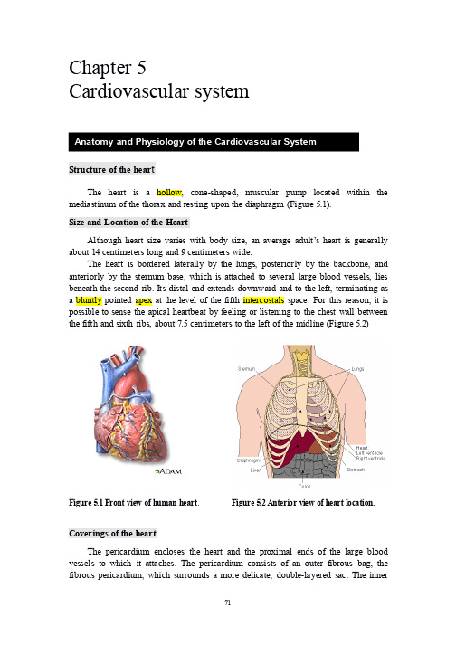
The wall of the heart is composed of three distinct layers: an outer epicardium, a middle myocardium, and an inner endocardium (Figure 5.4):
The epicardium, which corresponds to the visceral pericardium, is protective. It is a serous membrane that consists of connective tissue covered by epithelium, and it includes blood capillaries, lymph capillaries, and nerve fibers. The deeper portion of the epicardium often contains fat, particularly along paths of coronary arteries and cardiac veins that provide blood flow through the myocardium.
基础医学英语课文翻译

【Chapter 1】The connection can be so close that no movement is possible, as is the case in the skull. Other kinds of joints permit movement: either back and forth in one plane—as with the hinge joint of the elbow— or movement around a single axis—as with the pivot joint that permits the head to rotate.颅骨不能运动,是由於骨与骨之间的连接太过紧密.但其他的关节可允许活动,如一个平面上的前後屈身运动,如肘关节;或是绕轴心旋转运动,如枢轴点允许头部转动.The ends of these muscles are attached to different bones by connective tissue bands so that when the muscle contracts, one bone moves in relation to the other. This makes it possible to move the whole body, as when walking, or to move jus one part of the body, as when bending a finger.结缔组织是肌肉末端附着於不同的骨面上,所以当肌肉收缩时,两骨彼此靠近而产生运动.这也就使整个人体可以运动起来,如走路,运动躯体某个部位,如弯曲手指.The heart is a muscle that is divided into two nearly identical halve: one half receives blood from the lungs and sends it to the rest of the body, the other half sends blood that has traveled through the body back to the lungs.心脏是一块被分为几乎对等两半的肌肉.一办吸收来自肺部的血液,并把血液运送到机体的其余部位,另一半使流经全身的血液回流入肺.The trachea divides to enter each of the two lungs and then divides more than 20 times to form a very large number of small air spaces. Oxygen from the air enters the blood through capillaries in the walls of these air spaces, and the blood releases carbon dioxide into the air spaces to be exhaled.气管分成左右支气管,各连结左右肺,左右之气管在分支20多次,在终端形成大量为小的肺泡.从空气摄取的氧气流经这些肺泡壁内的毛喜血管流入血液.血液在经肺泡把释放出的二氧化碳排出体外.The urinary system maintains normal levels of water and of certain small molecules such as sodium and potassium in the body. It does this by passing blood through the kidneys, two efficient filtering organs that get rid of any excess of various molecules and conserve those molecules that are in short supply.泌尿系统维持水分及体内某些小分子物质,如钠`钾的正常水平.身体是通过让肾过滤血液来做到这一点的.肾是两个有效的过滤器官,他滤出各种多余的小分子物质,保留那些供应不足的小分子物质.A major gland is the pituitary, which is located under the brain in the middle of the head. It produces at least eight hormones, which affect growth, kidney function, and development of the sex organs.脑垂体是一个主要的腺体,他位於头中部脑下方.他至少分泌八种激素,这些激素对人体生长,肾功能及性器官发育有影响.The female productive system is responsible for producing and transporting ova( the female sex cells), eliminating ova from the body when they are not fertilized by sperm, nourishing and provid- ing a place for growth of an embryo when an ovum is fertilizedby sperm, and nourishing a newborn child.女性生殖系统产生,输送卵子(女性性细胞),将未受精的卵子排出体外,而当精,卵结合时,女性生殖系统培养,提供胚胎生长场所,并孕育新生儿.【Chapter 2】A symptom is something a patient can de-tect, such as fever, bleeding, or pain. A sign is something a doctor can detect, such as a swollen blood vessel or an enlarged internal body organ.症状是病人自己就能察觉到的,比如,高烧,流血,或是疼痛.而徵兆则是医生所能够观察到的,比如,血管扩张或是体内器官肿大.The skin and mucous membranes covering the body or lining its openings offer considerable resis-tance to invasion by bacteria and other infectious organisms. If these physical barriers are injured or burned, infection resistance drops. In minor cases, only boils or pimples may develop. In major cases, however m large areas of the body might become infected.覆盖在体表或者器官开口处的皮肤和黏膜能在很大程度上抵抗细菌或其他感染体的入侵.如果这些屏障遭到了损坏或损伤,身体对感染的抵抗力就会下降.在一些病情较轻的病例中,疥子和小脓胞可能会发生.在病情较重的病例中,身体的大面积区域则可能会被感染.Breathing passages are especially vulnerable to infection. Fortunately, they are lined with mu-cus-secreting cells that trap tiny organisms and dust particles. Also, minute hairs called cilia line the breathing passages, wave like a field of wheat, and gently sweep matter out of the respiratory tract.呼吸通道尤其容易受到感染,幸运的是,呼吸道内附盖满了能分泌黏液的细胞,他们能捕捉微小的有机体和尘粒.另外,被叫做纤毛的细小毛发也覆盖了呼吸道,他们像微风下麦田里的小麦一样舞动着,轻轻地将异物扫出呼吸道.In addition, foreign mater in the breathing passages can often be ejected by nose blowing, coughing, sneezing, and throat clearing.除此之外,呼吸道内的异物还常常因为擤鼻涕`咳嗽`打喷嚏和清喉咙而被弹出.Unless the abscess breaks and allows the pus to drain, the infection is likely to spread.如果脓块不破裂,里面的脓不排除掉,感染很可能会扩大.1.Each antibody is made of a heavy chain of chemical subunits, or amino acids, anda light chain of them. The light chain has special sites where the amino acidscan link with their com-plements on the antigen molecule.每一个抗体由一条化学亚单位(及氨基酸)的重链和一条轻链所构成.这条轻链上有特别的部位,在那里,氨基酸能使其补体和抗原分子相连.2.In some cases, through the process of opsonization, antibodies “butter” thesurface of some antigens and make them “tastier” to phagocytes, which engulf the antigens.在某些情况下,通过调理素作用的过程,抗体在抗原表面涂抹上一些”奶油”,让吞噬细胞更喜欢吞噬他们.3.Sometimes an antibody hooks to bacterial antigen but needs an intermediate, orcomplement, to actually destroy the bacterium, As the antibody-antigen complex circulates in the blood, the complex “fixes” complement to it.在另一些情况下,抗体和一个细菌抗原合上以後,却需要一个中间体,或补体来实施对该细菌的消灭.於是,当抗体和抗原的结合体随血液循环时,该结合体会有一个补体附体.4.During the first day or so , antibodies against the infection cannot be found inthe blood. But this is only because the basic cells involved in antibody production have been triggered by the presence of antigen to multiply themselves.在第一天左右,血液中没有发现对付传染病的抗体,但是,这只是因为涉及抗体制造的基本细胞已被当前的抗原存在所触发而准备开始繁殖.【Chapter 3】The fleshy belly is attached to one bone while the tendon passes over a joint to become firmly attached to the adjoining bone.肌腱跨过关节牢固连接相邻的两块骨头,而腹肌则与骨头紧密相接.Shortening of the fleshy part of the muscle produces movement at the joint by pulling on the tendon. The tendon itself does not change in length.腹肌收缩拉动肌腱使关节运动,而肌腱本身的长度是不变的.The many bundles surrounded by the fibrous connective tissue fascia form the fleshy belly of the muscle.许多纤维束又被纤维结缔组织筋膜所包绕,最後形成肌肉的肌腹部份.The relation of the muscle bundles to the tendons is that the muscle bundles ate surrounded and held together by the fibrous connective tissue that is continuous with the fibrous connective tissue of the tendonous part of the muscle.肌束和肌腱之间的关系是:肌束被纤维结缔组织包绕并连接在一起,纤维结缔组织又与肌键部份的结缔组织相延续.The nerve fibers separate within a muscle with a terminal branch of the nerve going to each muscle fiber.在一块肌肉中神经纤维可分枝出许多神经末梢,分配到每块肌纤维中.【Chapter 4】Flat bones are generally thin and composed of two more or less parallel plates of compact bone enclosing a layer of spongy bone.扁骨一般较薄,由两层大致平行的骨密质骨板围绕一层松质骨构成.Bones undergoing either intramembranous or endochondral ossification are continually remodeled from he time that initial calcification occurs until the final structure appears.自最初的钙化发生开始,骨通过膜内骨化或软骨内骨化而不断地得以重塑,直至最後结构的形成.And still others, espe-cially the sex hormones, aid osteoblastic activity and thuspromote the growth of new bone. The sex hormones act as a double-edged sword. They aid in the growth of new bone, but they also bring about the degeneration of all the cartilage cells in epiphyseal plates.还有其他激素,特别是性激素,协助成骨细胞活动因而促进骨生长.性激素作用具有两面性,他能促进骨生长,但也使骺板所有软骨细胞退化.There are two principal effects of aging on the skeletal sys-tem. The first effect is the loss of calcium from bones.衰老对骨骼系统有两个主要作用.第一个作用是骨钙丧失.The second principal effect of aging on the skeletal system is a decrease in the rate of protein formation that results in a decreased ability to produce the organic portion of bone matrix.衰老对骨骼系统的第二个主要影响,是蛋白质合成速度降低至使产生骨基质的有机成分的能力下降.【Chapter 5】The cardiac sphincter relaxes and contracts to move food from the esophagus into the stomach, whereas the py-loric sphincter allows food to leave the stomach when it has sufficiently digested.贲门括约肌的舒张与收缩使食物由食管入胃,而幽门括约肌却使食物在充分消化後出胃. These substances help transform food present in the stomach into a semifluid substance called chime. The pyloric sphincter allows food to pass into the small intestine only after it has been transformed into chime.这些物质(盐酸)协助将胃内现存的食物转变成为称为食糜的半流质物质.幽门括约肌只有在食物完全变为食糜後才将其排入小肠.【Chapter 6】Air enters the body through the nose and passes through the nasal cavity, which is lined with a mucous membrane and fine hairs(cilia) to help filter out foreign bodies, as well as to warm and moisten the air.空气通过鼻进入人体内.在通过鼻腔时,其内排列的黏膜和纤毛过滤了异物,同时使进入的空气温暖而湿润Paranasal sinuses are hollow, air-containing spaces within the skull that communi-cate with the nasal cavity.副鼻窦位於头颅骨内,中空含气,并与鼻腔相通.They, too, have a mucous membrane lining and function to provide the lubricating fluid mucus, as well as to lighten the bones of the skull and help produce sound.副鼻窦也有黏膜衬里,其功能是提供润滑黏液,减轻头颅骨负荷,以及协同发声.It is in the hypopharyngeal region that the pharynx, serving as a common passageway for food from the mouth and air from the nose, divides into two branches, the larynx(voice box) and the esopha-gus.下咽部是来自於嘴的食物和来自鼻的空气之共同通道,他在这里又分为两支,喉(声音盒)和食管.A special deterrent to this event is provided for by a flap of cartilage attached the root of the tongue that acts like a lid over the larynx.这一起着特殊阻滞作用的物体是一层连着舌根的软骨结构,它像块盖子盖过喉.The measure of how easily the lungs expand under pressure is compliance.肺器之所以能在压力下轻松自如地展开,其方法就是因势利导,顺其自然.Breathing is regulated unconsciously by center in the brainstem. These centers adjust the rate and rhythm of breathing according o changes in the composition of the blood, especially the concen-tration of carbon dioxide.脑干里呼吸中心在不知不觉中控制和调节了呼吸.这些中心根据血液里的成分,特别是二氧化碳的浓度来调节呼吸的速率和节奏.If too much carbon dioxide is exhaled by hyper-ventilation, body fluids tend to become more alkaline, a condition termed alkalosis. If too little car-bon dioxide is exhaled as a result of hypoventilation, body fluids tend to become more acid, a condi-tion termed acidosis.如果因为换气过度而二氧化碳呼出过多,身体体液就容易变的偏硷性,一种被称为硷中毒的状态.然而,如果由於换气不足,二氧化碳呼出过少,身体体液就容易变的偏酸性,一种被称为酸中毒的状态.Lining the trachea and bronchial tree are cells that secrete mucus, which traps pollutants and bacteria. Also in the bronchi are cells containing tiny cilia, that project into the blanket of mucus and with constant wavelike motions push the mucus up out of the airways.第一,气管和支气管树铺满能分泌黏液的细胞,它们能捕捉污染物质和细菌.第二,支气管里还有长有细小纤毛的细胞,它们深入遍布的黏液层,不停地通过波浪般的动作把黏液向上清扫出呼吸道.【Chapter 7】There are three major types of blood vessels, . , veins, and capillaries.血管分为三大类,即动脉、静脉、毛细血管The largest artery, the aorta, is about 1 inch in diameter and has the thickest wall.主动脉是最大的动脉,管腔直径约为1英寸,血管壁最厚The capillary boundaries are the most important center of activity of the entire circulatory system.毛细血管网是整各循环系统的最重要活动中心Most veins are equipped with one-way valves that permit the blood to flow in only one direction.They are most numerous in the veins of the extremities.大多数静脉具有单向瓣膜,使血液朝着一个方向流动.在四肢的静脉中,这样的瓣膜最多The pulmonary arteries carry blood low in oxygen from the right ventricle, while the pulmonary veins carry blood high in oxygen from the lungs into the left atrium.肺动脉携带右心室出来的、含氧量低的血液;而肺静脉将含氧量高的血液从肺携带到左心房Blood returning from tissues other than the lungs enters the heart by way of the venae cavae: the superior vena cava and the inferior vena cava.从组织(肺组织除外)而来的血液经腔静脉,即上腔静脉与下腔静脉,回到心脏When the atria contract, blood in the right atrium is forced through the tricuspid valve into the right ventricle.当心房收缩时,右心房的血液则通过三尖瓣进入右心室Atrial contractions force blood from the left atrium through the mitral valve, also called bicuspid valve, into the left ventricle.心房收缩将血液从左心房挤压通过二尖瓣,进入左心室When the ventricles contract, blood in the left ventricle is forced through the aortic semilunar valve into the aorta, the body’s largest artery, for distribution to the tissues.当心室收缩时,左心室的血液被挤压通过主动脉瓣,进入主动脉(机体内的最大动脉),然後分配到机体的各个组织【Chapter 8】Oxygen from the lungs and nutrients from the digestive tract are absorbed into blood for transport to the tissues.血液吸收肺部来的氧和消化道来的营养物质,并输送到组织At the same time, carbon dioxide and other waste products of cellular metabolism are absorbed from the tissues for transport to the organs of elimination.同时,组织的细胞代谢产生的二氧化碳和其他废物,送到排泄器官The blood also transports hormones from endocrine glands to their target organs.血液还将内分泌腺产生的激素输送到它们的靶器官。
医学专业英语词汇大全
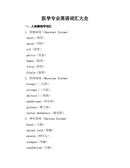
医学专业英语词汇大全一、人体解剖学词汇1. 骨骼系统(Skeletal System)skull(颅骨)spine(脊柱)rib(肋骨)pelvis(骨盆)femur(股骨)tibia(胫骨)fibula(腓骨)2. 肌肉系统(Muscular System)biceps(二头肌)triceps(三头肌)deltoid(三角肌)quadriceps(四头肌)gluteus(臀大肌)rectus abdominis(腹直肌)3. 神经系统(Nervous System)brain(大脑)spinal cord(脊髓)neuron(神经元)synapse(突触)cerebellum(小脑)hypothalamus(下丘脑)4. 循环系统(Circulatory System) heart(心脏)artery(动脉)vein(静脉)capillary(毛细血管)blood(血液)plasma(血浆)5. 呼吸系统(Respiratory System) lung(肺)trachea(气管)bronchus(支气管)alveoli(肺泡)diaphragm(膈肌)二、临床医学词汇1. 疾病与症状diabetes(糖尿病)hypertension(高血压)asthma(哮喘)fever(发热)headache(头痛)nausea(恶心)2. 检查与诊断physical examination(体格检查)Xray(X光检查)CT scan(CT扫描)MRI(磁共振成像)biopsy(活检)diagnosis(诊断)3. 治疗与药物medication(药物治疗)surgery(手术治疗)vaccination(疫苗接种)antibiotic(抗生素)painkiller(止痛药)insulin(胰岛素)三、医学分支词汇1. 内科学(Internal Medicine) cardiology(心脏病学)gastroenterology(消化病学) nephrology(肾脏病学)endocrinology(内分泌学)hematology(血液病学)2. 外科学(Surgery)general surgery(普通外科) orthopedic surgery(骨科)neurosurgery(神经外科)plastic surgery(整形外科)cardiac surgery(心脏外科)3. 妇产科(Obstetrics and Gynecology)pregnancy(妊娠)childbirth(分娩)contraception(避孕)menopause(更年期)cervical cancer(宫颈癌)4. 儿科学(Pediatrics)immunization(免疫)growth chart(生长曲线)developmental milestones(发育里程碑)asthma in children(儿童哮喘)childhood obesity(儿童肥胖)本词汇大全旨在帮助医学专业人员和爱好者更好地掌握医学英语,提高专业英语水平。
护理专业英语翻译

01
02
喉,恰当的称为“语音盒”,位于气管的上方,起着把气管与咽连接起来的作用。
1
气管,是一根12cm长的软骨,是食道前从喉下降到细支气管的圆柱形管道。它的直径大约2cm,同其它呼吸管道一样,气管表面覆盖着纤毛上皮细胞。 气管使空气进入肺部,在这一过程中通过它粘膜层的纤毛过滤温暖湿润。 在第五胸椎的水平,气管被划分为支气管,右边比左边的更短,粗,直。随着支气管被再细分为越来越小的结构,它逐渐丢失了软骨和纤维组织直到最后仅留下极薄层的平滑肌和弹性纤维组成的细支气管。
02
石膏使骨头不能移动所以可以减少疼痛并使骨头愈合的更好。当一个石膏被放上的时候,一个袜子状的东西被放在你受伤的胳膊或腿上。然后一个柔软纯棉材质被覆盖垫在你的皮肤上。然后温玻璃纤维或煅石膏被覆盖在上面,当它被放上的时候就会开始感觉到温热。在5-10分钟内它会变硬并固定。在石膏被运用之后,它需要被干燥。煅石膏需要在24~28小时后才能干。一个干燥的煅石膏是无味,白色且发亮的。一个湿石膏是灰色的,冰凉的且有霉味的。以下是应用石膏护理的规则:
大多数的头痛并不是由严重的中枢神经系统问题引起的。跟随者头痛的疼痛可因跳痛或戳痛不同而不同,比如偏头痛,严重的疼痛停几天又发作几天的间歇性,比如群集性疼痛。头痛经常由鼻窦,头皮,或头部周围的肌肉的问题引起。
神经护理是一个有吸引力的领域。在这个领域里护士有利用他一切观察能力的机会。他是帮助诊断制定患者治疗方案的重要信息来源。 通过乐观,护理能力和把病人当人来对待,护士可以帮助患者及其家庭缓解许多困难。当认识到一个人的行为和个性深受大脑器质性病变影响,就会较少的倾向于认为患者是一个不合作的坏脾气病人。相反的,他成为一个需要帮助和理解的人。他可能无法控制他的反应,护士肯定了解这一点。
respiratory system医学专业英语 呼吸系统 课文翻译
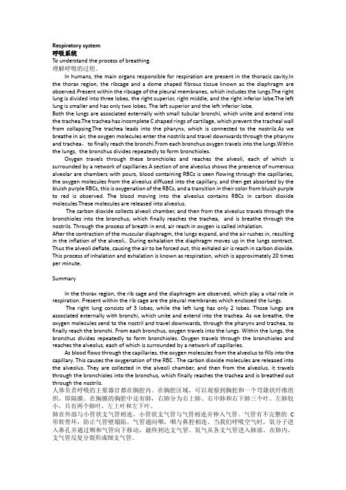
Respiratory system呼吸系统To understand the process of breathing.理解呼吸的过程。
In humans, the main organs responsible for respiration are present in the thoracic cavity.In the thorax region, the ribcage and a dome shaped fibrous tissue known as the diaphragm are observed.Present within the ribcage of the pleural membranes, which includes the lungs.The right lung is divided into three lobes, the right superior, right middle, and the right inferior lobe.The left lung is smaller and has only two lobes, The left superior and the left inferior lobe.Both the lungs are associated externally with small tubular bronchi, which unite and extend into the trachea.The trachea has incomplete C shaped rings of cartilage, which prevent the tracheal wall from collapsing.The trachea leads into the pharynx, which is connected to the nostrils.As we breathe in air, the oxygen molecules enter the nostrils and travel downwards through the pharynx and trachea,to finally reach the bronchi.From each bronchus oxygen travels into the lungs.Within the lungs, the bronchus divides repeatedly to form bronchioles.Oxygen travels through these bronchioles and reaches the alveoli, each of which is surrounded by a network of capillaries.A section of one alveolus shows the presence of numerous alveolar are chambers with pours, blood containing RBCs is seen flowing through the capillaries, the oxygen molecules from the alveolus diffused into the capillary, and then get absorbed by the bluish purple RBCs, this is oxygenation of the RBCs, and a transition in their color from bluish purple to red is observed. The blood moving into the alveolus contains RBCs in carbon dioxide molecules.These molecules are released into alveolus.The carbon dioxide collects alveoli chamber, and then from the alveolus travels through the bronchioles into the bronchus, which finally reaches the trachea, and is breathe through the nostrils. Through the process of breath in end, air reach in oxygen is called inhalation.After the contraction of the muscular diaphragm, the lungs expand, and the air rushes in, resulting in the inflation of the alveoli,. During exhalation the diaphragm moves up in the lungs contract. Thus the alveoli deflate, causing the air to be forced out, this exhaled air is reach in carbon dioxide. This process of inhalation and exhalation is known as respiration, which is approximately 20 times per minute.SummaryIn the thorax region, the rib cage and the diaphragm are observed, which play a vital role in respiration. Present within the rib cage are the pleural membranes which enclosed the lungs.The right lung consists of 3 lobes,while the left lung has only 2 lobes. Those lungs are associated externally with bronchi, which unite and extend into the trachea. As we breathe, the oxygen molecules send to the nostril and travel downwards, through the pharynx and trachea, to finally reach the bronchi. From each bronchus, oxygen travels into the lungs. Within the lungs, the bronchus divides repeatedly to form bronchioles. Oxygen travels through the bronchioles and reaches the alveolus, each of which is surrounded by a network of capillaries.As blood flows through the capillaries, the oxygen molecules from the alveolus to fills into the capillary. This causes the oxygenation of the RBC . The carbon dioxide molecules are released into the alveolus. They are collected in the alveoli chamber, and then from the alveolus,it travels through the bronchioles i nto the bronchus, which finally reaches the trachea and is breathed out through the nostrils.人体负责呼吸的主要器官都在胸腔内。
95第五章呼吸系统【Respiration】PPT课件
一、概 述 二、肺通气 三、呼吸气体的交换及运输 四、呼吸的调节
1
整体概述
概述一
点击此处输入
相关文本内容
概述二
点击此处输入
相关文本内容
概述三
点击此处输入
相关文本内容
2
呼吸
第一节 呼吸的过程和呼吸器官
一、呼吸全过程
机体同外界环境之间的气体交换过程,称为呼吸 ( respiration ),它是由以下三个环节组成: 示意图
27
残气量 补呼气量
潮气量
补吸气量
功能残气量 肺总容量
呼吸
五、肺通气量:
每分通气量——每分钟进或出肺的气体总量。 每分通气量=潮气量 X 呼吸频率
肺泡通气量=(潮气量-生理无效腔)X 呼吸频率
解剖无效腔
28
肺泡无效腔
呼吸
第三节 气体交换及运输
一、气体交换 二、气体运输
29
呼吸
气体交换原理:
30
20
呼吸
哺乳动物的呼吸式有3种类型: ①胸式呼吸(thoracic breathing):
吸气时以肋间外肌收缩为主,胸壁起伏明显;
②腹式呼吸(abdominal breathing):
吸气时以隔肌收缩为主,腹部起伏明显;
③胸腹式呼吸(混合式呼吸) (combined breathing):
吸气时肋间外肌与膈肌都参与的,胸壁和腹壁的 运动都比较明显。
15
呼吸
胸膜腔——
胸膜有两层, 即紧贴于肺表面的 脏层和紧贴于胸廓 内壁的壁层。两层 胸膜形成一个密闭 的、潜在的腔隙。
16
呼吸
胸膜腔内只有少量的浆液,没有气体:
系统解剖 呼吸系统 英文版.Respiratory system
Membranes and ligaments of larynx
Quadrangular membrane 方形膜
Between epiglottic, thyroid and arytenoid cartilages
vestibular ligament 前庭韧带 Lower
free border of quadrangular membrane
上鼻道 下鼻道 中鼻道
Nasal cavity鼻腔 鼻腔
Remove the middle nasal conchae Ethmoidal bulla 筛泡 Semilunar hiatus 半月裂孔 Ethmoidal infundibulum 筛漏斗
Nasal cavity鼻腔 鼻腔
Mucous membrane of nasal cavity 鼻腔粘膜
Epiglottic cartilage 会厌软骨
Leaf-shaped elastic cartilage Attached by its stalk to the thyroid cartilage
Laryngeal joints
Cricothyroid joint 环甲关节 Cricoarytenoid joint 环杓关节
Thyroarytenoid lateral cricoarytenoid posterior cricoarytenoid Cricothyroid
Muscles of larynx
Cricothyroid 环甲肌 环甲肌---tense the vocal ligament
Cricothyroid
Respiratory region 呼吸区
The lower part of the nasal cavity Lined with respiratory mucous membrane Its function is to warm, moisten, and clean the inspired air
(完整版)医学专业英语翻译与答案
Chapter 1Passage 1 Human BodyIn this passage you will learn:1. Classification of organ systems2. Structure and function of each organ system3. Associated medical termsTo understand the human body it is necessary to understand how its parts are put together and how they function. The study of the body's structure is called anatomy; thestudy of the body's function is known as physiology. Other studies of human body include biology, cytology, embryology, histology, endocrinology, hematology, immunology, psychology etc.了解人体各部分的组成及其功能,对于认识人体是必需的。
研究人体结构的科学叫解剖学;研究人体功能的科学叫生理学。
其他研究人体的科学包括生物学、细胞学、胚胎学、组织学、内分泌学、血液学、遗传学、免疫学、心理学等等。
Anatomists find it useful to divide the human body into ten systems, that is, the skeletal system, the muscular system, the circulatory system, the respiratory system, the digestive system, the urinary system, the endocrine system, the nervous system, the reproductive system and the skin. The principal parts of each of these systems are described in this article.解剖学家发现把整个人体分成骨骼、肌肉、循环、呼吸、消化、泌尿、内分泌、神经、生殖系统以及感觉器官的做法是很有帮助的。
- 1、下载文档前请自行甄别文档内容的完整性,平台不提供额外的编辑、内容补充、找答案等附加服务。
- 2、"仅部分预览"的文档,不可在线预览部分如存在完整性等问题,可反馈申请退款(可完整预览的文档不适用该条件!)。
- 3、如文档侵犯您的权益,请联系客服反馈,我们会尽快为您处理(人工客服工作时间:9:00-18:30)。
Chapter 5Respiratory SystemIn this passage you will learn:● The organs of the respiratory system● The structure and function of these organs● The mechanism of breathing● The gas transport and cleaning systemAll living animals must take in oxygen and get rid of carbon dioxide. In the vertebrates——animals with backbones ——that get their oxygen from the air, both tasks are performed by special gas-exchange organs called lungs. The lungs provide a place where oxygen can reach the blood and carbon dioxide can be removed from it. They are equipped with tubes and a bellows system for drawing in air from the outside, while the pulmonary veins and arteries circulate blood through from inside. The lungs also have a cleaning system that traps, ejects, or destroys irritants and other harmful substances that travel in with the air.In the simpler cold-blooded amphibians and reptiles, the lungs are two balloon-like sacs. In active animals that require large amounts of oxygen, especially the warm-blooded birds and mammals , the lungs are a spongy labyrinth of sacs that supply an enormous surface area for the transfer of gases. In the adult human the total lung surface, if flattened out, would be larger than a badminton court, about 100 square yards (83 square meters).Air enters the body through the nose and passes through the nasal cavity, which is lined with a mucous membrane and fine hairs (cilia) to help filter out foreign bodies, as well as to warm andmoisten the air. Paranasal sinuses are hollow, air-containing spaces within the skull that communicate with the nasal cavity. They, too, have a mucous membrane lining and function to provide the lubricating fluid mucus. as well as to lighten the bones of the skull and help produce sound.After passing through the nasal cavity, the air next reaches the pharynx (throat). There arethree divisions of the pharynx. The nasopharynx is the first division, and it is nearest to the nasalcavities. It contains the adenoids, which are masses of lymphatic tissue. The adenoids (also known as the pharyngeal tonsils) are more prominent in children, and if enlarged, they can obstruct air passageways . Below the nasopharynx and closer to the mouth is the second division of the pharynx, the oropharynx. The palatine tonsils, two rounded massed of lymphatic tissue, are located in the oropharynx. The third division of the pharynx is the hypopharynx (also called the laryngopharynx). It is in the hypopharyngeal region that the pharynx, serving as a common passageway for food from the mouth and air from the nose, divides into two branches, the larynx (voice box) and the esophagus.The esophagus leads into the stomach and carries food to be digested. The larynx contains the vocal cords and is surrounded by pieces of cartilage for support. Sounds are produced as air is expelled past the vocal cords, and the cords vibrate. The tension of the vocal cords determines the high or low pitch of the voice.Since food entering from the mouth and air entering from the nose mix in the pharynx, what prevents the passing of food or drink into the larynx and respiratory system after it has been swallowed? Even with a small quantity of solid or liquid matter finding its way into the air passages, breathing could be seriously blocked.A special deterrent to this event is provided for by a flap of cartilage attached to the root of the tongue that acts like a lid over the larynx. This flap of cartilage is called the epiglottis. The epiglottis lies over the entrance to the larynx. In the act of swallowing, when food and liquid move through the throat, the epiglottis closes off the larynx, so that these things cannot enter.On its way to the lungs, air passes from the larynx to the trachea (windpipe), a vertical tubeabout 41 inches long and 1 inch in diameter. The trachea is kept open by 16-202C-shaped rings of cartilage separated by fibrous connective tissue that stiffens the front and sides of the tube.In the region of the mediastinum, the trachea divides into two branches called bronchi. Each bronchus leads to a separate lung and divides and subdivides into smaller and finer tubes, somewhat like the branches of a tree (see Figure 6-1) .DiaphragmFigure 6-1The smallest of the bronchial branches are called bronchioles. At the end of the bronchioles are clusters of air sacs called alveoli. Each alveolus is made of a one-cell layer of epithelium. The very thin wall allows for the exchange of gases between the alveolus and the capillaries that surround and come in close contact with it. The blood that flows through the capillaries accepts the oxygen from the alveolus and deposits carbon dioxide into the alveolus to be exhaled. Oxygen is combined with a hemoglobin in erythrocytes and carried to all parts of the body. Each lung is enveloped in a double-folded membrane called the pleura. The outer layer of the pleura, nearest the ribs, is the parietal pleura, and the inner layer, closest to the lung, is the visceral pleura. The pleura is moistened with a serous secretion that facilitates the movements of the lungs within the thorax.The two lungs are not quite mirror images of each other. The right lung, which is the slightly larger of the two, is divided into three lobes, or divisions, and the left lung is divided into two lobes. It is possible for one lobe of the lung to be removed without damage to the rest, which can continue to function normally; The uppermost part of the lung is called the apex, and the lower area is the base. The hilum of the lung is the midline region where blood vessels, nerves, and bronchialtubes enter and exit the organ.The lungs extend from the collarbone to the diaphragm in the thoracic cavity. The diaphragm is a muscular partition that separates the thoracic from the abdominal cavity and aids in the process of breathing. The diaphragm contracts and descends with each inhalation (inspiration) .The downward movement of the diaphragm enlarges the area in the thoracic cavity and reduces the internal air pressure, so that air flows into the lungs to equalize the pressure. When the lungs are full, the diaphragm relaxes and elevates, making the area in the thoracic cavity smaller, and thus increasing the air pressure in the thorax. Air then is expelled out of the lungs to equalize the pressure; this is called exhalation (expiration) . Other parts are also involved in the process. The cycle of respiration really begins when the phrenic nerve stimulates the diaphragm to contract and flatten. Also, the intercostal muscles between the ribs aid in inspiration by pulling the ribs up and out. The measure of how easily the lungs expand under pressure is compliance.Breathing is regulated unconsciously by center in the brainstem. These centers adjust the rate and rhythm of breathing according to changes in the composition of the blood, especially the concentration of carbon dioxide.Gas Transport.Oxygen is carried in the blood bound to hemoglobin in red blood cells. The oxygen is released to the cells as needed. Carbon dioxide is carried in several ways, but is mostly converted to an acid called carbonic acid. The amount of carbon dioxide that is exhaled is important in regulating the acidity or alkalinity of the blood. If too much carbon dioxide is exhaled by hyperventilation, body fluids tend to become more alkaline, a condition termed alkalosis. If too little carbon dioxide is exhaled as a result of hypoventilation, body fluids tend to become more acid, a condition termed acidosis.The cleaning system of the lungs has four main components. Lining the trachea and bronchial tree are cells that secrete mucus, which traps pollutants and bacteria. Also in the bronchi are cells containing tiny cilia, that project into the blanket of mucus and with constant wavelike motions push the mucus up out of the airways. Irritating chemicals, stagnant and excessive mucus, and large bitsof foreign matter are forcibly ejected as sputum from the bronchi by a cough. This third important cleaning device —— like breathing, under partial voluntary control —— is a rapid muscle contraction and bronchial-tube constriction that generates a wind force far stronger than a tornado. Small harmful substances that make their way into the alveoli are destroyed by the fourth line of defense, the macrophages. These are patrolling cells that "swallow up" foreign particles or destroy them with enzymes.New Words and PhrasesExercisesA. Discuss the following topics:1. Imagine you were lecturing in front of rural health workers on the topic ofrespiratory system, draw a picture to illustrate the whole system.2. Describe their structures and functions.3. How can epiglottis prevent the passing food and drink into the respiratory system?4. What role does the diaphragm play in the process of breathing?5. What are the four components of the cleaning system? How do they function?B. Fill in the blanks with the words given below and change their forms if necessary.alveolus capillary diaphragmhemoglobinthoraciccavitygas exchangeparietalpleuravisceralpleuramediastinumpleural spaceThe lungs are two cone-shaped, spongy organs consisting of alveoli, blood vessels, elastic tissue and nerves. Each of the two lungs consists of smaller divisions called lobes; the left lung hastwo lobes, while the right lung is divided into three lobes. In the lungs, ( 1 ) are surrounded by a network of tiny blood vessels called capillaries; oxygen from the lungs passes into these ( 2 ) for distribution to tissue cells, while carbon dioxide from the blood passes into the lungs to be expelled by exhalation. Once absorbed into blood cells, oxygen becomes attached to ( 3 ) and is released to tissue cells as needed. Thus, the primary function of thelungs is to bring airinto close contact with blood, which allows ( 4 ) to occur.The lungs are surrounded by a membrane called the ( 5 ) . The space that the lungs occupy within the chest is called the ( 6 ) , which is lined by a membrane called the ( 7 ). The parietal and visceral pleurae lie very close to each other; the small space between theses membranes, called the ( 8 ) , is filled with a fluid that prevents friction when the two membranes slide against each other during respiration. In the central portion of the thoracic cavity (in the area between the lungs) is a space called the ( 9 ) , which contains the heart. A group of smooth muscles called the ( 10 ) separates the lower portion of the thoracic cavity from the abdomen.C. Match Column I with Column n.Column I Column IIbronchiole[ 1 ]any of the small subdivision of the bronchicompliance[ 2 ]the peak portion of the lungshypoventilation[ 3 ]a condition in which there is too much carbon dioxidein the bloodintercostal[ 4 ]between the ribsparanasal sinuses[ 5 ]air-conditioning cavities in the bones near the nose acidosis[ 6 ]a condition in which there is too much carbon dioxidein the bloodapex[ 7 ]weak , inadequate exchange of gaspharynx[ 8 ]rounded masses of lymph tissue in the oropharynx ( palatinemeans roof of the mouth) .alkalosis[ 9 ]in agreement withpalatine tonsils[ 10 ]throat; composed of the nasopharynx, oropharynx, andlaryngopharynx.D. Define the following terms of the respiratory system in line with the text youhave just, learned (making use of the vocabulary provided in the brackets if necessary), for exam- ple:Oxygenfood, metabolism) 1. mirror image as if, left side, vice versa)2. adenoids ynx)3. alveolus respiration)4. bronchus mediastinum)5. epiglottis larynx, prevent, trachea)6. hyperventilation(extreme, rapid,deep, result in, alkalosis)7. diaphragm (dome-shaped, muscle,move, increase, decrease, space,thoracic cavity)8. pharynx ( common, passageway,esophagus, food, air, larynx)racic cavity)coughing, clearing the throat, res-piratory tract)E. Translate the following into English.1.副鼻窦2.口咽3.脊椎动物4.肺泡5.二氧化碳6.肺换气不足7.横膈膜神经8.双重折叠的9.威慑物10.润滑液11.滞痰12.食管13.纵隔14.哺乳动物15.碱中毒16.迷宫17.污染物质18.脑干19.上皮;上皮细胞20.刺激物Passage Two Respiratory Disorders and DiseasesIn this passage you will learn:● Various disorders and diseases of the respiratory system● Their definitions, causes and treatment●Detailed description of the signs and symptoms of chronic obstructive pulmonary disease● Medical terms pertaining to the diseases of the respiratory system● The respiratory system is subject to a wide variety of disorders and diseases. The most frequent attacks come from common cold and flu viruses. Other diseases that affect the lungs include bacterial infections such as pneumonia and tuberculosis. The lungs are especially vulnerable to allergic dis- eases such as asthma. There are more serious diseases such as respiratory distress syndrome, em-physema, chronic obstructive pulmonary diseases (COPD), lung cancer, etc.Influenza and pneumonia.Influenza is a viral disease of the respiratory tract. Different strains of the influenza virus have caused serious epidemics through history. Pneumonia is caused by several different microorganisms. The name represents any inflammation of the lungs caused by in-fection, so an alternate term for pneumonia is pneumonitis. Streptococcal pneumonia usually in- volves one or more lobes of the lung and described as lobar pneumonia.Other agents of pneumonialocalize in the bronchial tubes, causing bronchopneumonia.Pleurisy is severe chest pain accompanying each deep breath in a person with an inflamed pleura, the twin membranes around each lung and lining the chest cavity. Pleurisy can attend pneu-monia or result from direct infection of the pleura.Tuberculosis (TB)has increased in recent years along with the rise of AIDS and the appear-ance of resistance to antibiotics in the organism that causes the disease. The name of the disease comes from the small lesions, or tubercles, that appear with the infection. The symptoms of TB in-clude fever, weight loss, weakness, cough, and as a result of damage to blood vessels in the lungs, hemoptysis, i. e. the coughing up of phlegm (sputum) containing blood. Accumulation of exudatein the alveoli may result in solidification or consolidation of lung tissue. The tuberculin test is used to reveal tuberculosis infection, PPD (purified protein derivative) is the form of tuberculin commonly used.Asthma. Attacks of asthma result from narrowing of the bronchial tubes. The constriction, a-long with edema, swelling of the bronchial linings, and accumulation of mucus results in wheezing, extreme dyspnea and cyanosis. Although the cause of asthma is uncertain, foreign particles such as pollen or certain environmental pollutants are believed to be the culprits, which stimulate the smooth muscle of the bronchial tree to releases histamine causing the muscle to contract. The bronchial air- ways are consequently restricted. Treatment of asthma includes removal of allergens, administration of bronchodilators to widen the airways, and administration of steroids.Respiratory distress syndrome is a disorder of some prematurely bom infants. The alveoli of afflicted babies are lined with a protein material, limiting the amount of oxygen their blood can re-ceive. The disease is often fatal. Mechanical ventilators can be used to help infants breathe until their lungs become more mature. As a result of some accidents anddiseases, such as polio, the res-piratory center or nerves carrying its impulses may be paralyzed. Treatment may involve cutting a hole through the windpipe and passing a tube attached to a mechanical respirator through the hole. In other cases, the patient may be placed on a heart and lung machine that maintains respiration and heartbeat.Acute pulmonary edema results when fluid quickly accumulates in the lungs and fills the alveoli. The fluid buildup is caused by heart trouble that, in turn, produces back pressure in the pulmonary veins and the left atrium of the heart to which they carry oxygen-rich blood from the lungs. A person suffering acute pulmonary edema is suddenly breathless and turns blue because of oxygen-poor blood. The condition is treated with oxygen, digitalis to strengthen heart action, and diuretics to speed fluid removal by the kidneys.Pneumothorax occurs when air gets into the chest between the pleural lining. The lung then cannot fully expand and breathing becomes difficult. As a result, the lung may even collapse. Pneumothorax may result from a wound in the chest, such as a knife wound, or after a sudden tear in the lung. Infection of the pleural space by gas-producing microbes can also cause pneumothorax. Physicians treat pneumothorax by removing the gas by suction, surgically repairing the chest or lung, or prescribing antibiotics when an infection is present.Pneumoconiosis (black lung) means "dust disease." It can strike miners and industrial work-ers who inhale damaging amounts of dust over a long period of time. One of the most serious is sili-cosis, which results from inhaling quartz dust. Another, anthracosilicosis, arises from inhalation of coal and quartz dust. Pneumoconiosis often occurs in combination with other diseases, such as bron-chitis , emphysema, or tuberculosis. There is no treatment for it, but the disease can be prevented by minimizing dust inhalation.Emphysema.This is a chronic disease associated with overexpansion and destruction of the alveoli. Common causes are exposure to cigarette smoke and other forms of pollution, as well as chronic infection. Emphysema is the main disorder included under the heading of chronic obstructive pulmonary disease (COPD), which will be discussed in detail soon.Chronic obstructive pulmonary disease (COPD) is a rather broad term used to describe sim-ple chronic bronchitis, chronic obstructive bronchitis, asthmatic bronchitis and emphysema, for it is convenient to describe various combinations of these disorders that may coexist, for instance, pa-tients often have chronic obstructive bronchitis as well as emphysema.Unfortunately, chronic bronchitis has been used variably to refer to a simple smoker's cough or, as in the British literature, to severe COPD. In this discussion, chronic bronchitis will be con-sidered "simple," "obstructive," or "asthmatic" to reduce ambiguity. It is useful clinically to dif-ferentiate between the extremely common simple chronic bronchitis and the less common but often devastating form of chronic obstructive bronchitis.Simple chronic bronchitis, a syndrome characterized primarily by a chronic productive cough, is the result of low-grade exposure to bronchial irritants in an individual without hyperreac-tive airways. This syndrome is associated with enhanced mucous secretion, reduced ciliary activity, and impaired resistance to bronchial infection. Simple chronic bronchitis is defined in clinical terms: (1) excessive production of mucus; (2) presence of symptoms, largely cough, on most days for at least three months annually during two or more successive years; (3) exclusion of bronchiecta-sis, tuberculosis, or other causes of these symptoms. The term does not describe the underlying pro-cess , which may vary widely. The patient population ranges from those who are asymptomatic except for a morning "cigarette cough" productive of mucus in small amounts (simple chronic bronchitis) to patients with a severe disabling condition manifested by increased resistance to airflow, hypoxia, and often hypercatnia (chronic obstructive bronchitis) .Chronic obstructive bronchitis,which develops in a relatively small proportion of individuals with simple chronic bronchitis, results in irreversible narrowing of airways. Because the obstruction is in bronchioles and bronchi 2 mm or less in diameter, the term small airways disease has been used.Brochospasm. Exposure to bronchial irritants in individuals with hyperreactive or "twitchy"airways can lead to bronchospasm (i.e. , bronchial smooth muscle constriction), frequently accom-panied by excessive mucous production and edema of bronchial walls. Recurrent episodes of symp-tomatic bronchospasm are called asthma. The present discussion must consider bronchospasm, since a degree of reversible airways obstruction often accompanies other reactions to inhaled noxious a-gents. In fact, episodic airways obstruction is common in individuals with chronic bronchitis. This combination, called asthmatic bronchitis, may closely resemble classic asthma. The term chronic asthmatic bron'chitis is applied in patients with persistent airways obstruction, a chronic productive cough, and a major problem of episodic bronchospasm.Emphysema, another lung response to noxious stimuli, is characterized by abnormal, perma-nent enlargement of airspaces distal to the terminal bronchioles, accompanied by destruction of their walls, and without obvious fibrosis. The alterations of emphysema cause reduction in lung elastic re-coil, which permits excessive airway collapse upon expiration and leads to irreversible airflow ob-struction .These definitions are not mutually exclusive; there is considerable crossover between the em-physematous (type A) and bronchial (type B) signs and symptoms. For example, most individuals with emphysema also have a chronic productive cough. It may be difficult to determine the relative importance of emphysema and chronic obstructive bronchitis, with obliteration of small airways. Ac-cordingly, a general term such as chronic obstructive pulmonary disease (COPD)has been used to describe this clinical syndrome.New Words and Phrases(注:可编辑下载,若有不当之处,请指正,谢谢!)请预览后下载!。