caspase1抗体说明书
Caspase活性检测说明书

Caspase活性检测(基于p NA标记底物的比色法)在caspase 的底物多肽上标记发色团如:p NA,可用于检测凋亡细胞中的caspase活性及筛选caspases的激活剂或抑制剂。
本操作步骤主要针对使用酶标仪进行读数而设计。
其中使用的细胞抽提物、底物、抑制剂及p NA的用量为每个样本140ul。
若您希望设计针对分光光度计读数的实验,建议对每种溶液体积进行进一步优化。
所需溶液、试剂及设备• Caspase底物• Caspase抑制剂• 细胞裂解缓冲液:50mM HEPES, pH7.4, 100mM NaCl, 0.1%CHAPS, 1mM DTT, 100uM EDTA • 检测缓冲液:50mM HEPES, pH7.4, 100mM NaCl, 0.1%CHAPS, 10mM DTT, 100uM EDTA, 10%Glycerol• PBS:将8g NaCl、200mg KCl和1.44g Na2HPO4溶于800ml蒸馏水;用HCl调至pH7.4;定容至1L• 酶标板和酶标仪其它试剂(推荐)• 纯化的Caspase(阳性对照)• p NA细胞抽提物的制备以适合的凋亡诱导剂对细胞进行诱导(参考前述操作方法),同时做阴性对照,包括未处理细胞、用诱导剂的无诱导活性类似物处理细胞(若可得到),或一个凋亡诱导处理“零时间”样品。
应保证有足够细胞进行重复实验,包含或不含抑制剂的对照以检测蛋白浓度。
一般来说,我们下面推荐的裂解前细胞数量可以产生足够蛋白浓度供酶标仪检测;然而不同大小、体积的细胞及蛋白浓度可能需要增加铺板密度。
当裂解2×10⁷/ml细胞是,产生的蛋白浓度约为1-3mg/ml,体积10ul,含约10-30ug蛋白。
依据其细胞系,最少应有106个细胞,体积50ul的裂解缓冲液进行处理。
• 细胞计数,离心收集细胞。
以PBS缓冲液洗涤一次。
若细胞已被caspase抑制剂处理过,建议多洗几次以防止后继实验中可能造成的不利影响。
Caspase 1 活性检测试剂盒(比色法)
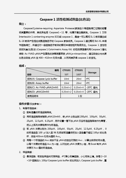
Caspase 1活性检测试剂盒(比色法)简介:Caspase(Cysteine-requiring Aspartate Protease)家族在介导细胞凋亡过程的起着极其重要的作用,其成员包括Caspase1~11等,均属于蛋白酶家族。
Caspase 1又称Interkeukin 1 conberting enzyme (ICE)或caspase-1,是唯一可以剪切IL-1前体蛋白或IL-18前体产生相应成熟细胞因子的Caspase 家族成员。
Caspase 1通过剪切Bcl-XL 来调节细胞凋亡,并通过对一些细胞因子前体的剪切来调控相关免疫反应。
Caspase 1活性检测试剂盒(比色法) (Caspase 1 Colorimetric Assay Kit) 的检测原理是利用Caspase 1催化底物 Ac-YVAD-p NA)产生黄色的游离硝基苯胺p NA(p -nitroaniline),通过测定分光光度比色法测定p NA 在400~410nm 处吸光值,从而间接获得caspase 1的活性。
组成:操作步骤(仅供参考):1、 制备标准曲线:① 配制适量的标准品稀释液。
② 用标准品稀释液稀释p NA(10mM),使p NA 分别达到200μM 、100μM 、50μM 、25μM 、12.5μM 、6.25μM ,另外设置一管不加p NA 仅含标准品稀释液作为零管,把以上系列浓度物质作为标准品。
③ 取p NA 浓度分别为200μM 、100μM 、50μM 、25μM 、12.5μM 、6.25μM 、0的标准品各100 μl 加入至96孔板或取适量体积加入至容量不超过100μl 的比色杯,测定405nm 处吸光值即A 405。
④ 用每一个标准品的A 405减去不含p NA 的空白对照的A 405,计算出实际的吸光值。
以每个浓度的标准品A 405为x 轴,以对应的p NA 浓度为y 轴,用Excel 制作p NA 浓度对A 405值的标准曲线。
抗超氧化物歧化酶1抗体说明书
Product nameAnti-Superoxide Dismutase 1 antibody DescriptionRabbit polyclonal to Superoxide Dismutase 1Host speciesRabbit Tested applicationsSuitable for: IHC-P, WB Species reactivityReacts with: Human ImmunogenSynthetic peptide corresponding to Rat Superoxide Dismutase 1.General notes The Life Science industry has been in the grips of a reproducibility crisis for a number of years.Abcam is leading the way in addressing this with our range of recombinant monoclonal antibodiesand knockout edited cell lines for gold-standard validation. Please check that this product meetsyour needs before purchasing.If you have any questions, special requirements or concerns, please send us an inquiry and/orcontact our Support team ahead of purchase. Recommended alternatives for this product can befound below, along with publications, customer reviews and Q&AsFormLiquid Storage instructionsShipped at 4°C. Upon delivery aliquot and store at -20°C. Avoid freeze / thaw cycles.Storage bufferPreservative: 0.1% Sodium azide Constituents: PBS, 50% Glycerol (glycerin, glycerine)PurityImmunogen affinity purified Purification notesThis antibody was purified on an antigen coupled sepharose column.ClonalityPolyclonal Isotype IgGThe Abpromise guarantee Product datasheetAnti-Superoxide Dismutase 1 antibody ab134995 References 2 ImagesOverviewPropertiesApplicationsOur Abpromise guarantee covers the use of ab13499 in the following tested applications.The application notes include recommended starting dilutions; optimal dilutions/concentrations should be determined by the end user.Function Destroys radicals which are normally produced within the cells and which are toxic to biologicalsystems.Involvement in disease Defects in SOD1 are the cause of amyotrophic lateral sclerosis type 1 (ALS1) [MIM:105400].ALS1 is a familial form of amyotrophic lateral sclerosis, a neurodegenerative disorder affectingupper and lower motor neurons and resulting in fatal paralysis. Sensory abnormalities are absent.Death usually occurs within 2 to 5 years. The etiology of amyotrophic lateral sclerosis is likely tobe multifactorial, involving both genetic and environmental factors. The disease is inherited in 5-10% of cases leading to familial forms.Sequence similarities Belongs to the Cu-Zn superoxide dismutase family.Post-translational modifications Unlike wild-type protein, the pathogenic variants ALS1 Arg-38, Arg-47, Arg-86 and Ala-94 are polyubiquitinated by RNF19A leading to their proteasomal degradation. The pathogenic variants ALS1 Arg-86 and Ala-94 are ubiquitinated by MARCH5 leading to their proteasomal degradation.The ditryptophan cross-link at Trp-33 is reponsible for the non-disulfide-linked homodimerization. Such modification might only occur in extreme conditions and additional experimental evidence is required.Cellular localization Cytoplasm. The pathogenic variants ALS1 Arg-86 and Ala-94 gradually aggregates andaccumulates in mitochondria.Western blot - Anti-Superoxide Dismutase 1 antibody (ab13499)Anti-Superoxide Dismutase 1 antibody (ab13499) at 1 µg/ml + Cell lysates prepared from mixed human cell lines (A431, A549,HCT116, HeLa, HEK293, HepG2, HL-60, HUVEC, Jurkat, MCF7, PC3 and T98G)Predicted band size: 18 kDaApplication Abreviews NotesIHC-P Use a concentration of 2 µg/ml. Perform heat mediated antigenretrieval before commencing with IHC staining protocol.WB Use a concentration of 1 µg/ml. Detects a band of approximately19, 23 kDa (predicted molecular weight: 18 kDa).TargetImagesImmunohistochemistry (Formalin/PFA-fixed paraffin-embedded sections) - Anti-Superoxide Dismutase 1 antibody (ab13499)Ab13499 staining Human normal colon. Staining is localized to the nucleus.Left panel: with primary antibody at 2 ug/ml. Right panel: isotype control.Sections were stained using an automated system DAKO Autostainer Plus , at room temperature. Sections were rehydrated and antigen retrieved with the Dako 3-in-1 AR buffer citrate pH 6.0 in a DAKO PT Link. Slides were peroxidase blocked in 3% H2O2 in methanol for 10 minutes. They were then blocked with Dako Protein block for 10 minutes (containing casein 0.25% in PBS) then incubated with primary antibody for 20 minutes and detected with Dako Envision Flex amplification kit for 30 minutes. Colorimetric detection was completed with Diaminobenzidine for 5 minutes. Slides were counterstained with Haematoxylin and coverslipped under DePeX. Please note that for manual staining we recommend to optimize the primary antibody concentration and incubation time (overnight incubation), and amplification may be required.Please note: A ll products are "FOR RESEA RCH USE ONLY. NOT FOR USE IN DIA GNOSTIC PROCEDURES"Our Abpromise to you: Quality guaranteed and expert technical supportReplacement or refund for products not performing as stated on the datasheetValid for 12 months from date of deliveryResponse to your inquiry within 24 hoursWe provide support in Chinese, English, French, German, Japanese and SpanishExtensive multi-media technical resources to help youWe investigate all quality concerns to ensure our products perform to the highest standardsIf the product does not perform as described on this datasheet, we will offer a refund or replacement. For full details of the Abpromise, please visit https:///abpromise or contact our technical team.Terms and conditionsGuarantee only valid for products bought direct from Abcam or one of our authorized distributors。
细胞焦亡Caspase-1-induced pyroptotic cell

• 一般含有:含CARD的凋亡相关斑点样蛋白 (apoptosis-associated speck-like protein containing CARD, ASC), caspase蛋白酶及 病原识别受体( Pathogen recognition receptors, PRRs)
焦亡在细菌感染 疾病中的作用
焦亡是重要的先天免疫因素, 能够清除胞内病原体 •细胞崩解,入侵病原暴露于 胞外环境,被中性粒细胞吞噬 吸收后通过ROS信号途径清除 •破坏了病原菌赖以生存繁殖 的微环境,清除受感染的细胞
焦亡在HIV感染中的作用
• Gilad Doitshd等体外培养人扁桃体组织,结果 显示> 95%死亡细胞不是有效感染细胞,这些 “旁观者”的死亡是由HIV流产感染造成的
• Gilad Doitshd进一步研究认为通过caspase-3介 导的凋亡途径死亡的细胞只占很小一部分,大 多数是通过caspase-1介导的细胞焦亡途径死亡
• Gilad Doitshd进一步研究认为DNA识别受体 IFI16识别非完全逆转录产物启动了焦亡程序
焦亡在HIV感染中的作用
小结
• 细胞程序性死亡(PCD)是个体生长发育及维 持组织稳态的必要组成部分
Caspase-1激活与调节-炎性小体
炎性小体分类 • NLR家族:NLRP1, NLRP3, NLRP6, NLRP12,
NLRC4 • RIG-I家族:RIG-I • ALR家族:AIM-2和IFI-6
Caspase-1激活与调节-炎性小体
• 炎性小体通过病原识别受体识别不同刺激, • 炎性小体识别外来刺激之后,可通过其
caspass1通路调控机制
caspass1通路调控机制
Caspase 1 是一种重要的炎症相关蛋白酶,参与调节炎症反应和免疫应答。
Caspase 1的活性受到多种机制的调控。
1. 转录调控:Caspase 1基因的表达受到多个转录因子的调控,如NF-κB、AP-1等,它们能够结合在Caspase 1基因的启动子区域上,促进或抑制Caspase 1的转录。
2. 翻译后修饰:Caspase 1的活性可以通过翻译后修饰来调控。
例如,Caspase 1的自我断裂(autocleavage)是其激活的重要步骤之一,这一过程受到其他蛋白酶的调控,如Caspase 8、Caspase 11等。
3. 组蛋白修饰:组蛋白修饰也参与了Caspase 1的调控。
研究表明,组蛋白去乙酰化修饰酶(HDACs)和组蛋白甲基转移酶(HMTs)可以调控Caspase 1特定位点上的甲基化、乙酰化等修饰,从而影响Caspase 1的活性和功能。
4.反式调控:一些信号通路分子可以与Caspase 1发生反式调控,调节Caspase 1的活性与功能。
例如,NLRP3炎症小体信号通路被认为是Caspase 1激活的一个重要启动机制。
总的来说,Caspase 1的活性和表达受到多种机制的调控,包括转录调控、翻译后修饰、组蛋白修饰和反式调控等。
这些调控机制的研究有助于深入了解炎症反应和免疫应答的调控机制,并有望为相关疾病的治疗提供新的治疗策略。
Caspase-1调控单核—巨噬细胞分化的效应及机制

Caspase-1调控单核—巨噬细胞分化的效应及机制巨噬细胞是单核吞噬细胞系统中的重要成员,在天然免疫和获得性免疫中都发挥着重要作用。
体内巨噬细胞的来源包括单核细胞起源的巨噬细胞、原位增殖而生成的巨噬细胞以及卵黄囊巨噬细胞来源的巨噬细胞,其中单核细胞起源的巨噬细胞是组织巨噬细胞的主要来源。
在机体稳态或发生炎症的时候,单核细胞都能在微环境中一些因子的调控下向巨噬细胞分化。
单核-巨噬细胞分化过程的紊乱与多种疾病的发生发展密切相关,如自身免疫疾病、自身炎症性疾病、代谢性疾病以及肿瘤等。
因此,对于单核-巨噬细胞分化过程相关调控及分子机制的研究一直以来都是免疫学领域的热点。
Caspase-1是caspases家族中与炎症密切相关的一个成员,对于巨噬细胞的功能具有关键的调控作用。
Caspase-1缺失的巨噬细胞释放炎症因子IL-1p和IL-18的能力显著下降;同时caspase-1还能调控巨噬细胞由M2型到M1型的极化。
此外,caspase-1也能参与细胞分化程序的控制。
Caspase-1能够通过调控IL-1p的加工、释放而控制脂肪细胞和Th17细胞的分化。
但是,对于caspase-1在单核-巨噬细胞分化中的功能我们还知之甚少。
基于caspase-1广泛的生物学功能及其在单核细胞、巨噬细胞的核心地位,caspase-1在单核-巨噬细胞的分化过程中也具有重要的调控功能。
在本研究中,我们通过构建体外诱导人单核细胞白血病细胞株(THP-1、U937)或人外周血单核细胞(PBMCs)向巨噬细胞分化的模型,深入探讨了 caspase-1对于单核-巨噬细胞分化过程的调控效应以及所涉及的分子机理,主要研究内容和结果如下:1.我们运用PMA成功诱导THP-1和U937细胞向巨噬细胞分化,并通过分析此过程中细胞形态、表面标志分子(markers)及吞噬功能的变化情况而将单核-巨噬细胞的分化过程分为早期和晚期两个阶段。
2.我们发现caspase-1在THP-1和U937细胞中呈组成型的高表达;然而随着分化的进行其活性不断升高,并伴随有IL-1β和IL-18的释放。
Caspase1活性检测试剂盒比色法

本试剂盒用酶标仪检测或容量不超过 100µL 的分光光度检测杯检测时,除标准曲线外可以 检测 20 个样品。
试剂准备 1. 裂解液融化后混匀并置于冰浴上备用。 2. 检测缓冲液融化后混匀并置于冰浴上备用。
气泡。随后再加入 10µL Ac-YVAD-pNA(2mM)。
3. 加入 Ac-YVAD-pNA (2mM)后混匀,注意避免在混匀时产生气泡。37°C 孵育 60-120min。发
ห้องสมุดไป่ตู้
现颜色变化比较明显时即可测定 A405。如果颜色变化不明显,可以适当延长孵育时间,甚至可
以孵育过夜。
4. 样品的 A405 减去空白对照的 A405,即为样品中 caspase 1 催化产生的 pNA 产生的吸光度
二. 样品的收集: 1. 对于悬浮细胞:将没有诱导凋亡的对照品和诱导凋亡的样品,在 4°C 下,600g 离心 5min 后, 收集细胞,小心吸除上清,同时确保尽量没有细胞被吸除,PBS 洗涤一次。同前吸尽上清后,2×106 细胞加入 100µL 裂解液(建议加入 1%蛋白酶抑制剂(CC088)),重悬沉淀,冰浴裂解 15~20min。 下接步骤 4。 2. 对于贴壁细胞:将没有诱导凋亡的对照品和诱导凋亡的样品,吸取细胞培养液,备用。用胰 酶消化贴壁细胞,并收集至备用的细胞培养液中。在 4°C 下,600g 离心 5min 后,收集细胞,
1
小心吸除上清,同时确保尽量没有细胞被吸除,PBS 洗涤一次。同前吸尽上清后,2×106 细胞加
入 100µL 裂解液(建议加入 1%蛋白酶抑制剂(CC088)),重悬沉淀,冰浴裂解 15~20min。
人半胱氨酸蛋白酶-1(caspase-1)说明书

控制在 5 分钟内,如标本数量多,推荐使用排枪加样。 4. 请每次测定的同时做标准曲线,最好做复孔。如标本中待测物质含量过高(样本 OD 值
计算 以标准物的浓度为横坐标,OD 值为纵坐标,在坐标纸上绘出标准曲线,根据样品的
OD 值由标准曲线查出相应的浓度;再乘以稀释倍数;或用标准物的浓度与 OD 值计算出标
准曲线的直线回归方程式,将样品的 OD 值代入方程式,计算出样品浓度,再乘以稀释倍数,
即为样品的实际浓度。
注意事项 1.试剂盒从冷藏环境中取出应在室温平衡 15-30 分钟后方可使用,酶标包被板开封后如未
试剂盒组成 1 30 倍浓缩洗涤液 2 酶标试剂 3 酶标包被板 4 样品稀释液 5 显色剂 A 液 6 显色剂 B 液
20ml×1 瓶 6ml×1 瓶 12 孔×8 条 6ml×1 瓶 6ml×1 瓶 6ml×1/瓶
7 终止液
6ml×1 瓶
8 标准品(320 pmol/L) 0.5ml×1 瓶
9 标准品稀释液
2. 加样:分别设空白孔(空白对照孔不加样品及酶标试剂,其余各步操作相同)、标准孔、
待测样品孔。在酶标包被板上标准品准确加样 50µl,待测样品孔中先加样品稀释液 40µl, 然后再加待测样品 10µl(样品最终稀释度为 5 倍)。加样将样品加于酶标板孔底部,尽 量不触及孔壁,轻轻晃动混匀。 3. 温育:用封板膜封板后置 37℃温育 30 分钟。 4. 配液:将 30 倍浓缩洗涤液用蒸馏水 30 倍稀释后备用 5. 洗涤:小心揭掉封板膜,弃去液体,甩干,每孔加满洗涤液,静置 30 秒后弃去,如此 重复 5 次,拍干。 6. 加酶:每孔加入酶标试剂 50µl,空白孔除外。 7. 温育:操作同 3。 8. 洗涤:操作同 5。 9. 显色:每孔先加入显色剂 A50µl,再加入显色剂 B50µl,轻轻震荡混匀,37℃避光显色 10 分钟. 10. 终止:每孔加终止液 50µl,终止反应(此时蓝色立转黄色)。 11. 测定:以空白空调零,450nm 波长依序测量各孔的吸光度(OD 值)。 测定应在加终止 液后 15 分钟以内进行。 操作程序总结:
- 1、下载文档前请自行甄别文档内容的完整性,平台不提供额外的编辑、内容补充、找答案等附加服务。
- 2、"仅部分预览"的文档,不可在线预览部分如存在完整性等问题,可反馈申请退款(可完整预览的文档不适用该条件!)。
- 3、如文档侵犯您的权益,请联系客服反馈,我们会尽快为您处理(人工客服工作时间:9:00-18:30)。
SANTA CRUZ BIOTECHNOLOGY,INC.caspase-1(14F468):sc-56036Santa Cruz Biotechnology,Inc. 1.800.457.3801831.457.3800fax 831.457.3801Europe +BACKGROUNDCaspase-1,originally designated ICE (for IL-1converting enzyme),is a member of the group of caspases with large prodomains.Caspase-1promotes matura-tion of interleukin IL-1βand interleukin18(IL-18)by proteolytic cleavage of precursor forms into biologically active pro-inflamatory cytokines.Active caspase-1,a (p20/p10)2tetramer,is necessary and sufficient for cleavage of precursor IL-1as well as for induction of apoptosis in some cell lines.The highly conserved family of caspases mediate many of the morphological and biochemical features of apoptosis,including structural dismantling of cell bodies and nuclei,fragmentation of genomic DNA,destruction of regulatory proteins and propagation of other pro-apoptotic molecules.The human cas-pase-1gene maps to chromosome 2q14and encodes a cytoplasmic protein expressed in liver,heart,skeletal muscle kidney and testis.caspase-1is implicated in inflammation,septic shock,and other situations such as wound healing and the growth of certain leukemias.REFERENCES1.Alnemri,E.S.,et al.1995.Cloning and expression of four novel isoforms of human interleukin-1βconverting enzyme with different apoptotic activities.J.Biol.Chem.270:4312-4317.2.Gu,Y.,et al.1995.Interleukin-1βconverting enzyme requires oligomer-ization for activity of processed forms in vivo .Embo J.14:1923-1931.3.Tatsuta,T.,et al.2000.The prodomain of caspase-1enhances Fas-mediated apoptosis through facilitation of caspase-8activation.J.Biol.Chem.275:14248-14254.4.Wang,J.,et al.2000.Role of caspases in apoptosis,development,and cytokine maturation revealed by homozygous gene deficiencies.J.Cell.Sci.113:753-757.5.Eldadah,B.A.and Faden,A.I.2000.Caspase pathways,neuonal apopto-sis,and CNS injury.J.Neurotrauma 10:811-829.6.SWISS-PROT/TrEMBL (P29466).World Wide Web URL:http://www.expasy.ch/sprot/sprot-top.htmlCHROMOSOMAL LOCATIONGenetic locus:CASP1(human)mapping to 11q23;Casp1(mouse)mapping to 9A1.SOURCEcaspase-1(14F468)is a mouse monoclonal antibody raised against amino acids 371-390of caspase-1of human origin .PRODUCTEach vial contains 100µg IgG 1in 1.0ml of PBS with <0.1%sodium azide and 0.1%gelatin.STORAGEStore at 4°C,**DO NOT FREEZE**.Stable for one year from the date of shipment.Non-hazardous.No MSDS required.APPLICATIONScaspase-1(14F468)is recommended for detection of caspase-1of mouse,rat and human origin by Western Blotting (starting dilution 1:200,dilution range 1:100-1:1000),immunoprecipitation [1-2µg per 100-500µg of total protein (1ml of cell lysate)],immunofluorescence (starting dilution 1:50,dilution range 1:50-1:500)and immunohistochemistry (including paraffin-embedded sec-tions)(starting dilution 1:50,dilution range 1:50-1:500).Suitable for use as control antibody for caspase-1siRNA (h):sc-29235,caspase-1siRNA (m):sc-29922,caspase-1siRNA (r):sc-61878,caspase-1shRNA Plasmid (h):sc-29235-SH,caspase-1shRNA Plasmid (m):sc-29922-SH,caspase-1shRNA Plasmid (r):sc-61878-SH,caspase-1shRNA (h)Lentiviral Particles:sc-29235-V,caspase-1shRNA (m)Lentiviral Particles:sc-29922-V and caspase-1shRNA (r)Lentiviral Particles:sc-61878-V.Molecular Weight of caspase-1:45kDa.Positive Controls:HL-60whole cell lysate:sc-2209,HL-60+LPS cell lysate:sc-24704or Jurkat whole cell lysate:sc-2204.RECOMMENDED SECONDARY REAGENTSTo ensure optimal results,the following support (secondary)reagents are recommended:1)Western Blotting:use goat anti-mouse IgG-HRP:sc-2005(dilution range:1:2000-1:32,000)or Cruz Marker™compatible goat anti-mouse IgG-HRP:sc-2031(dilution range:1:2000-1:5000),Cruz Marker™Molecular Weight Standards:sc-2035,TBS Blotto A Blocking Reagent:sc-2333and Western Blotting Luminol Reagent:sc-2048.2)Immunopre-cipitation:use Protein A/G PLUS-Agarose:sc-2003(0.5ml agarose/2.0ml).3)Immunofluorescence:use goat anti-mouse IgG-FITC:sc-2010(dilution range:1:100-1:400)or goat anti-mouse IgG-TR:sc-2781(dilution range:1:100-1:400)with UltraCruz™Mounting Medium:sc-24941.4)Immuno-histochemistry:use ImmunoCruz™:sc-2050or ABC:sc-2017mouse IgG Staining Systems.DATARESEARCH USEFor research use only,not for use in diagnostic procedures.caspase-1(14F468):sc-56036.Western blot analysis of caspase-1expression in HL-60(A ),LPS-treated HL-60(B )and THP-1(C )whole cell lysates.----ABCWestern blot analysis of caspase-1cleavage in untreated (A )and PMA (sc-3576)and LPS (sc-3535)treated (B )THP-1whole cell lysates.Antibody tested is caspase-1(14F468):sc-56036(A ,B ).Note cleavage of caspase-1in lane B .––––AB<。