Western Blot Protocol
WesternBlotProtocols

Western Blot Protocols1. Transfer proteins from gel to membrane (nitrocellulose of PVDF): Gloves should be worn when handling gels or blot membranes.∙ A. Pour enough transfer buffer (or CAPS buffer) into a container so that half of the assembled cassette w ill be 0.5 cm below the surface of the buffer.∙ B. After the gel has finished running, carefully remove the front glass plate and remove the stacking g el. Remove a small portion of the upper left-hand corner of the running gel to mark its orientation during the transfer pr ocess.∙ C. Wet the membrane w ith transfer buffer and carefully lay it across the exposed running gel, making sure that air bubbles between the gel and membr ane are excluded. In the same fashion, add two layers of pre-wetted filterpaper to the membrane. Pre-wet one of the cassette sponges and add to the stack. Top with the outer half of thecassette itself and carefully turn the entire stack of glass plate, gel, membrane, filter paper, sponge and cassetteover. Place the stack in the container w ith transfer buffer.∙ D. Carefully remove the second glass plate from the gel. Using the same process, make sure that no air bubbles have for med between the gel and the membrane.∙ E. Keeping the stack generously wet, top the gel w ith two layers of pre-wet filter paper, wet sponge and the remaining half of the cassette unit. Snap the cassette unit together firmly.∙ F. Place the cassette into the tr ansfer unit so that the current w ill flow through the gel onto the membrane. This would be w ith the gel closer to the black (-) cathode and the membrane closer to the red (+) anode.∙G. Add transfer (or CAPS) buffer to the transfer unit according to manufacturer's directions. Transfer over night witha setting of 20V / 40 mA or for 3 hours at 70V/160 mA. The transfer process needs to take place under coldtemperatures to prevent the gel fr om sticking to the membrane. This can be accomplished either by using a w ater cooling core in the transfer unit or placing the entir e unit at 4°C. Please follow the manufacturer'srecommendations.∙H. At the end of the transfer per iod, disconnect the power connections and disassemble the cassette. All of the Coomassie Blue-dyed bands should now be on the membrane leaving the gel fairly clear.2. Staining the Blot (Optional):∙ A. Br iefly w ith water, rinse any bits of gel off the blot then incubate it in ponceau S solution for 1 minute. Rinse in water until the background is reduced.∙ B. Place the membr ane on a transparency sheet and photocopy the blot for a permanent transfer record.∙ C. Rinse the blot several times in TBS-Tween to remove the stain.3. Blocking the Blot:∙ A. Gently shake the blot for 2 hours in 5% non-fat dry milk/TBS/Tween at room temperature or at 4°C on a rocker.∙ B. Either pour off blocking buffer or transfer the blot to a sealing bag for probing.4. Probing the Blot:∙ A. Dilute the pr imary antibody in 5% non-fat dry milk/TBS. Add to the blot, seal the container and gently rock for 2 hours at room temperatur e or over night at 4° C. If using a bag for the incubation, eliminate all air bubbles beforesealing.∙ B. Remove the pr imary antibody and wash the blot 3 x 5 minutes in TBS/Tween. In some instances, the primary antibody can be saved for additional blot procedures.∙ C. Dilute the secondary antibody in 5% non-fat dry milk/TBS. Add to the blot, seal and incubate as before.∙ D. Remove the secondary antibody and w ash the blot 3 x 10 minutes in TBS/Tween.5. Detection / Chemiluminescence:∙ A. Cut two transparency sheets to fit into the film cassette.∙ B. Mix oxidizing and luminol reagent (from Dupont NEN) in a 1:1 ratio.∙ C. Pour onto a piece of parafilm and lay the blot on the pool of solution for 1 minute.∙ D. Insert the blot into the cassette and top w ith the remaining sheet of transparency, making sure that all bubbles are eliminated.∙ E. Insert film and close cassette. Start w ith a 1 minute exposure and adjust from there. The signal w ill decay over time. If no bands are found, the blot can be rinsed and stored in PBS/Tw een overnight for repeated blocking andprobing.6. Str ipping and Reprobing Blots : Works well w ith chemiluminescence and radioactive detection, but is not useful in colorimetr ic detection due to the production of an insoluble reaction product formed as a results of that detection method.∙ A. Br iefly r inse the blot in water.∙ B. Wash the blot 2 x 10 minutes in TBS/Tween.∙ C. Re-block in 5% milk/TBS/Tween for one hour at room temperature.∙ D. Add stripping buffer to the blot, seal the container and incubate for 30 minutes at 50° C. Occasionally agitate the container for best results.∙ E. Wash the blot 2 x 30 minutes followed by 2 x 10 minutes in TBS/Tw een.∙ F. If desired (optional), can incubate the blot in the detection reagent and expose to film to ensure that no residual detection antibody remains. In order to check for the presence of primary antibody, the blot would have to beincubated with secondary antibody and detection reagent to ver ify complete removal.∙G. Block the blot for 2 hours in 5% milk/TBS/Tw een at room temperature before probing w ith the desired primary antibody again.。
Western Blotting Protocol-中山大学(参考)

Western Bloting (张雪雁)操作步骤:1.做胶:下层胶7.5ml一块,以水或乙醇隔离空气。
待下层胶凝固后做上层胶,每块胶配置3ml,灌胶、插梳子,赶走气泡。
2.蛋白变性:在胶凝固过程中,变性蛋白质,蛋白质的上样量按事先所测弄浓度计算准确,分装到EP管或,按5ul/100ul样品的浓度加β—巯基乙醇,然后以PCR仪95℃变性10分钟。
3.上样、电泳:1)上层胶为安全凝固(大约15分钟)后,两只手同时向上用力取下梳子。
以蒸馏水仔细清洗各泳道内残留得碎胶。
2)将SDS—PAGE胶转移到电泳槽中,如果只做一块胶,对面以玻璃板平衡。
1х电泳缓冲液充满电泳槽后,以玻璃棒将电泳槽中的气泡尽量赶除。
3)先以少量加有溴酚蓝1х上样缓冲液标记各泳道。
4)变性过的各管蛋白质样品以加有溴酚蓝1х上样缓冲液补齐至50ul。
蛋白Marker也补齐至50ul。
5)以一定顺序将蛋白样品和Marker,用微量移液器加至泳道。
上样过程中,手要稳,避免将泳道划破,样品自枪头打出时要缓慢,以免样品自泳道飘出。
剩余未加样的泳道以50ul上样缓冲液平衡。
6)加样完毕,红对红,黑对黑将电泳槽和电泳仪接好,打开电源,电压调至60-100V之间。
一般来说样品在上层胶时电压可稍高,进入下层胶后较低的电压容易将个分子量蛋白粉理清楚。
溴酚蓝自胶内跑出后电泳结束。
4.电转:1)准备PVDF膜及滤纸。
PVDF大小8.2х5~5.5,滤纸按电转海绵大小剪,一块胶准备四张滤纸。
2)将PDVF膜置于干净的容器中,倒入适量甲醇浸泡1~3分钟,然后浸泡于电转浸泡液中。
3)小心取出两层玻璃之间的胶,切去上层胶。
在电转液中完成如下:电转孔板黑色面铺放海绵一张,依次铺放滤纸两张,胶一块,PVDF膜一张,滤纸再两张,海绵再一张,然后小心扣好孔板,将其黑色面对黑色面扣入电转架,将电转架放入电泳槽。
电泳槽内置小冰盒一个,整个电泳槽置于大冰盒中。
4)接好电源,按蛋白分子量大小调整电压。
Western blot protocol
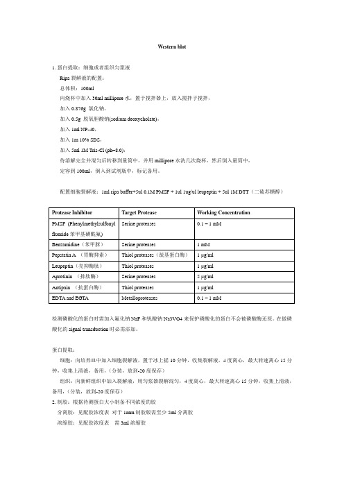
Western blot1.蛋白提取:细胞或者组织匀浆液Ripa裂解液的配置:总体积:100ml向烧杯中加入30ml millipore水,置于搅拌器上,放入搅拌子搅拌,加入0.876g 氯化钠,加入0.5g 脱氧胆酸钠(sodium deoxycholate),加入1ml NP-40,加入1m 10% SDS,加入5ml 1M Tris-Cl (ph=8.0),待溶解完全并混匀后转移到量筒中,并用millipore水洗几次烧杯,然后倒入量筒中,定容到100ml,倒入到试剂瓶中,标记备用。
配置细胞裂解液:1ml ripa buffer+5ul 0.1M PMSF + 1ul 1ug/ul leupeptin + 5ul 1M DTT(二硫苏糖醇)Protease Inhibitor Target Protease Working ConcentrationSerine proteases 0.1 – 1 mMPMSF (Phenylmethylsulfonylfluoride苯甲基磺酰氟)Benzamidine(苯甲脒)Serine proteases 1 mMPepstatin A (胃酶抑素)Thiol proteases(巯基蛋白酶) 1 μg/mlLeupeptin(亮抑酶肽)Thiol proteases 1 μg/mlAprotinin (抑肽酶)Serine proteases 5 μg/mlAntipain (抗蛋白酶)Thiol proteases 1 μg/mlEDTA and EGTA Metalloproteases 0.1 – 1 mM检测磷酸化的蛋白时需加入氟化钠NaF和钒酸钠Na3VO4来保护磷酸化的蛋白不会被磷酸酶还原。
在做磷酸化的signal transduction时必需添加。
蛋白提取:细胞:向培养皿中加入细胞裂解液,置于冰上摇10分钟,收集裂解液,4度离心,最大转速离心15分钟,收集上清液,备用,(分装,放到-20度保存)组织:向新鲜组织中加入裂解液,用匀浆器裂解混匀,4度离心,最大转速离心15分钟,收集上清液,备用,(分装,放到-20度保存)2.制胶:根据待测蛋白大小制备不同浓度的胶分离胶:见配胶浓度表对于1mm制胶版需至少5ml分离胶浓缩胶:见配胶浓度表需3ml浓缩胶H2O 30% acrylamide(丙烯10%APS TEMED 1.5M Tris(8.8) 1.0M Tris(6.8) 酰胺)2.3ml 1.3ml 50ul 5ul 1.3ml8%分离胶(5ml)2.1ml 0.5ml 30ul 3ul 0.38ml5%浓缩胶(3ml)3.玻璃板用酒精棉球擦一遍,晾干,短板向外夹好在绿色架上。
蛋白印记WesternBlotprotocol

蛋白印记WesternBlotprotocolWestern blot for PAR-2一、试剂的配制1. PMSF(100 mM)的配制:0.1742 g PMSF → 10 ml异丙醇(-20℃保存)2. 裂解液的准备:购自碧云天公司,分强,中,弱三种(三个小瓶,各50 ml).在提取总蛋白之前3-5min,将分装裂解液(强)融化置于冰上,每个肌条300-500ul裂解液不要超过450ul,(1ml加入100 mM的PMSF10 μl(使PMSF 的终浓度为1 mM),置于冰上。
(-20℃保存)3.2X Sample Buffer(要用才配或配好后置于-80℃)1倍DTT加上9倍的2X Laemmli Buffer(1). DTT (Di-dithiothreitol) 1M1.542 g DTT →10 ml的10 mM Sodium acetate(醋酸钠)(pH=5.2)中10 mM Sodium acetate(醋酸钠):0.082 g 无水醋酸钠→ 100 ml dH2O(2). 2X Laemmli Buffer (100 ml) (置于-20°C,经常使用时置于4°C)Glycerol (甘油)20 mlβ-mercaptoethanol (β-me,β-巯基乙醇) 5 ml (恶臭,注意安全)20﹪SDS (十二烷基硫酸钠)10 mlBromophenol Blue(溴酚蓝)20 mg1.5M Tris-Cl(三羟甲基氨基甲烷)pH=6.820 ml (90.855 g Tris→420 ml dH2O,浓盐酸滴定至pH=6.8,定容至500 ml ) dH2O 45 ml4.30% A+B溶液29.2 g Acrilamide (丙烯酰胺)0.8 g Bis-acrilamide (甲叉双丙烯酰胺)Add dH2O dilute to 100 mL and filter 避光4°C储存注意:该两种药品有毒,配药时注意安全PS. Acrylamide 及Bisacrylamide 是neurotoxin会穿过皮肤,配药时要穿实验衣及戴口罩。
Western blot protocol

Western blot 步骤(1)蛋白样品制备离心收集菌体。
加入200μl SDS-PAGE loding buffer重悬菌液,100°C煮6min,然后上样。
(2) SDS-PAGE电泳上样蛋白量为15μl。
(3) 转膜1、制备足够转移缓冲液以用于平衡凝胶和膜以及润湿滤纸。
2、从玻璃板上取下凝胶,去除所有浓缩胶。
3、将凝胶浸入转移缓冲液中10~15 分钟。
4、滤纸在转移缓冲液中至少浸泡10分钟。
5、准备PVDF膜:剪取一张PVDF膜以及4张转膜滤纸(膜与滤纸的面积应等于或略大于凝胶)。
PVDF膜在甲醇中润湿膜10 秒;接着小心将膜放入双蒸水水中浸泡2 分钟;然后小心地把膜放入转移缓冲液中平衡至少10 分钟。
6、转膜(并标记MARKER)打开转膜装置依次铺上第一层滤纸,第二层滤纸,胶,PVDF膜,第三层滤纸,第四层滤纸,每次都用15ml离心管擀出气泡。
膜两边的滤纸不能相互接触,接触后会发生短路。
接线,盖上盖子,开电源跑00:30~2:00h。
如果要看转移后的效果,可转膜完成后用丽春红染液对膜进行染色,具体的操作步骤如下:1、将膜置于培养盒中。
2、加适当体积的丽春红S,在脱色摇床中染色5分钟,观察转印效果。
3、去除染液(可重复使用),双蒸水洗膜两次,每次5 分钟,这时如果膜上有转印的蛋白时,可以看见数条蛋白条带及泳道痕迹。
(4) 封闭:将膜(如膜干应先用甲醇湿润)用PBST稍漂洗2次,每次5min。
浸泡于封闭液(BLOTTO+T)中于37℃2小时(三维摇床50/分)。
(5) 洗膜:在培养皿中用PBST洗两遍,每遍约5min。
稀释一抗:用预先配制的BLOTTO+T来6倍稀释一抗。
配制一抗(稀释6倍):0.33ml血浆+0.67ml BLOTTO+T。
(6) 膜与一抗孵育:把膜转至另一干净培养皿,加进一抗,于室温用三维摇床摇4℃过夜。
(7) 稀释二抗:取出培养皿,把膜移至另一培养皿,加进预制的PBST约15ml,洗4次,每次5min。
Western blot protocol
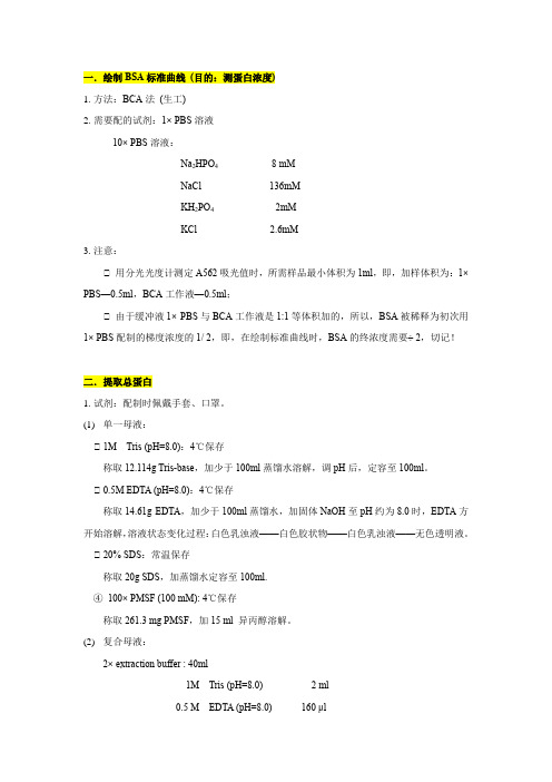
一.绘制BSA标准曲线(目的:测蛋白浓度)1.方法:BCA法(生工)2.需要配的试剂:1× PBS溶液10× PBS溶液:Na2HPO48 mMNaCl 136mMKH2PO4 2mMKCl 2.6mM3.注意:① 用分光光度计测定A562吸光值时,所需样品最小体积为1ml,即,加样体积为:1× PBS—0.5ml,BCA工作液—0.5ml;① 由于缓冲液1× PBS与BCA工作液是1:1等体积加的,所以,BSA被稀释为初次用1× PBS配制的梯度浓度的1/ 2,即,在绘制标准曲线时,BSA的终浓度需要÷ 2,切记!二.提取总蛋白1.试剂:配制时佩戴手套、口罩。
(1)单一母液:① 1M Tris (pH=8.0):4℃保存称取12.114g Tris-base,加少于100ml蒸馏水溶解,调pH后,定容至100ml。
① 0.5M EDTA (pH=8.0):4℃保存称取14.61g EDTA,加少于100ml蒸馏水,加固体NaOH至pH约为8.0时,EDTA方开始溶解,溶液状态变化过程:白色乳浊液——白色胶状物——白色乳浊液——无色透明液。
① 20% SDS:常温保存称取20g SDS,加蒸馏水定容至100ml.④100× PMSF (100 mM): 4℃保存称取261.3 mg PMSF,加15 ml 异丙醇溶解。
(2)复合母液:2× extraction buffer : 40ml1M Tris (pH=8.0) 2 ml0.5 M EDTA (pH=8.0) 160 μl20% SDS 4 mlWater 33.84 ml2.取样:取细胞浓度为2×10⁷的藻液1ml于1.5ml EP管中,厌氧箱中离心1-2min,将沉淀物于液氮中速冻后置于-80①保存;3.提取总蛋白:3.1 根据样品数量计算所需的提取试剂体积:1个样品——1ml 1× extraction buffer3.2 配制1× extraction buffer:试剂:① 2× extraction buffer① 100× PMSF3.3 提取:将-80①保存的样品取出,置于冰上,加入步骤2.2配制的1× extraction buffer 1ml, 涡旋至EP管底部无沉淀黏着后,4①,12,000 rpm离心10 min,用1ml移液枪小心吸取上清于另一干净的1.5 ml EP管中,置于冰上。
Western Blot Protocol实验操作步骤

Western Blot Protocol实验操作步骤一、提取抗原蛋白将提取RNA途中留存的样品,加入150μl100%酒精充分混匀,静置 5min(RT),2000×g,4℃离心5min,吸取上清至新管中,加入750μl异丙醇,混匀,静置10min(RT),120 00×g,4℃离心 10min,弃上清,加入1ml0.3mol/L盐酸胍/95%酒精重悬沉淀,用加样器打散沉淀,混旋20~30秒,静置20min(RT),7500×g,4℃离心5min,弃上清,重新加入1ml0.3mol/L盐酸胍/95%酒精两次,重悬沉淀,离心后弃上清,加入 100%无水乙醇1 ml,混旋1min,静置20min(RT),7500×g,4℃离心5min,弃上清,真空干燥5min,50μl1% SDS溶解沉淀,用50℃热水助溶,直至全部溶解。
10000×g,离心10min,去除沉渣。
二、蛋白定量1. 取PBS、各样品10ul,加DW990ul2.取DW匀浆缓冲液、样品稀释液、系列牛血清白蛋白标准浓度0.5ml。
3.向各管加入2.5mlD试剂(A50ml+B0.5ml+C0.5mlA-2%Na2CO3、 0.1NNaOH,B-0.5%CuSO4,C-1%酒石酸钠)混匀,静置10分钟。
4.迅速加入酚试剂0.25ml,混匀37℃水浴30分钟。
5.721分光光度计650nm,比色,S0标准管调零。
6.最后稀释为 4ug/ul,加载样缓冲液后,终浓度为2ug/ul,上样量为30~80ug/泳道。
三、电泳1.makegel.11%分离胶 12ml 两块胶4%积层胶6ml两块胶DW4.36 ml DW堵漏3.66ml3M Tris3.0 ml 0.5M Tris1.5 ml30%Arc4.4 ml 30%Arc0.5 ml0.78 ml10% SDS0.24 ml 10%SDS60 ul10%APS0.12 ml 10%APS20ul30 ulTEMED10ul TEMED5ul6ul2.上样前样品处理:样品100℃、3分钟、冷却,900×g、离心30S。
Western Blot protocol

Western Blot protocol一、试剂准备1.蛋白裂解液配方:2. 1.5 mol/L Tris.HCL(PH 8.8)O 800 mlTris base (MW 121.1) 181.7g dd H2溶解之后用浓盐酸调PH至8.8(一般加几滴即可,可以在调之前先测溶解液的PH,然后再估计浓盐酸的使用量),然后定容至1L。
高压灭菌后保存。
3. 1 mol/L Tris.HCL(PH 6.8)Tris base (MW 121.1) 30.9 g dd HO 200 ml2溶解之后用浓盐酸调PH至6.8(,可以在调之前先测溶解液的PH,然后再估计浓盐酸的使用量。
一般)然后定容至250 ml,经高压灭菌后使用。
4.10% AP (过硫酸铵):O 溶解后分装,-20℃保存。
0.1 g过硫酸铵 + 1.0 ml Dd H2O加100 g SDS加热至68℃助溶,然后用浓盐酸调节PH 5.10% SDS:用900 ml的H2至7.2,最后定容至1L。
6.分离胶( 12% )[常用]:7.浓缩胶( 5% ):8. 5 × Running Buffer(储存液):9. 1 × Running Buffer (工作液):200 ml的5 × Running Buffer + 800 ml的ddO。
H210.转膜缓冲液:3.03 g Tris base + 14.4g甘氨酸溶解在少量的蒸馏水中,完全溶解后加100ml的甲醇,再用蒸馏水定容至1L。
11.10 × TBS ( 储存液) :24.2 g Tris base + 80 g NaCl 溶解,用Hcl调PH至7.6,最后定容至1L。
O。
12.1 × TBS (工作液):100 ml的10 × TBS + 900 ml的dd H213.TBST:含0.1% 吐温—20的1 × TBS。
- 1、下载文档前请自行甄别文档内容的完整性,平台不提供额外的编辑、内容补充、找答案等附加服务。
- 2、"仅部分预览"的文档,不可在线预览部分如存在完整性等问题,可反馈申请退款(可完整预览的文档不适用该条件!)。
- 3、如文档侵犯您的权益,请联系客服反馈,我们会尽快为您处理(人工客服工作时间:9:00-18:30)。
Western Blot Protocol
1.SDS-PAGE试剂的配制
1)40%丙烯酰胺溶液(Acrylamide)
2)10%过硫酸铵溶液(APS)
3)TEMED
4)分离胶缓冲液4X Buffer (pH 8.8)
5)浓缩胶缓冲液4X Buffer (pH 6.8)
6)10X电泳缓冲液(Running buffer)
2.制胶
1)10%分离胶的制备
ddH2O 4.92ml 9.98ml
Tris-HCl缓冲液(pH 8.8,1.5mol/l) 2.5ml 5ml
40%丙烯酰胺 2.48ml 4.96ml
10% APS 100μl 200μl
TEMED 4μl 8μl
10%分离胶10ml 20ml
2)灌注分离胶
在加完TEMED后,速在两玻璃板间灌注丙烯酰胺溶液,预留1.5cm灌注浓缩胶(约在夹子高度稍低),应排除凝胶底部玻璃与板间的气泡。
用移液枪在丙烯酰胺溶液上层覆盖无水乙醇约4ml(至溢出),凝胶静置室温30min。
30min后,分离胶聚合凝固,倾倒出上层覆盖的无水乙醇,并用滤纸吸尽残留的液体。
3)5%浓缩胶的制备
ddH2O 2.5ml 5ml
Tris-HCl缓冲液(pH 8.8,1.5mol/l)1ml 2ml
40%丙烯酰胺0.46ml 0.92ml
10% APS 40μl 80μl
TEMED 2μl 4μl
浓缩胶4ml 8ml
4)灌注浓缩胶
在加完TEMED后,迅速在已经聚合的分离胶上直接灌注浓缩胶至溢出,并立即插入梳子,避免产生气泡。
凝胶静置30min。
浓缩胶聚合凝固,小心拔出梳子,立即用ddH2O洗涤加样槽,以去除未聚合的丙烯酰胺。
3.加样
1)蛋白样品变性
在浓缩胶聚合的同时,将蛋白抽提液和加样缓冲液在沸水中加热5分钟,使蛋白变性。
2)灌注电泳缓冲液
电泳槽中加满电泳缓冲液(ph8.3的Tris-Gly)。
3)蛋白加样
用移液枪加样槽中加入样品和分子量蛋白标准品(Markers)。
所有加样槽均加样,空白孔用加样缓冲液代替,第一个孔加Markers5μl,其余孔加样品30μl。
4.电泳
电泳装置与电源相连,凝胶上所加电压80V,当染料前沿进入分离胶后,把电压提至150V,继续电泳至溴酚蓝到达分离胶的底部,关闭电源。
5.转膜
1)电泳结束前,裁一张硝酸纤维素膜(nitrocellulose, NC),大小应与凝胶大小吻合,
约2 ~ 2.5cm。
2)在一个托盘中将6张滤纸、海绵及硝酸纤维素膜浸于20%甲醇中泡5~10min,再转
移至转膜缓冲液中泡5min,切去右上角做标记。
3)转膜缓冲液的配制
甘氨酸14.4g
Tris碱3g
甲醇200ml
ddH2O To 1L
转膜缓冲液1L
4)转膜
a)在塑料夹上放置用转移缓冲液浸泡过的海绵及滤纸,逐张叠放,精确对齐,然
后用一个玻璃移液管做滚筒以挤出气泡。
b)把硝酸纤维素膜放在滤纸上,精确对齐,滤纸与膜间不能留有气泡。
c)从电泳槽上撤下放置SDS-PAGE凝胶的玻璃,把凝胶转移至去离子水中漂洗一
下,然后平放于硝酸纤维素膜上并对齐,排除所有气泡。
d)最后3张滤纸放在凝胶上方,各层精确对齐并排除气泡。
5)将夹层物中滤膜面正对阳极,接通电源,电流250mA,150min左右。
6)断开电源并拔下电泳槽上的插头,拆卸转移装置,逐一揭去各层,取出纤维素膜,
置于1X TBST。
6.Western blot 鉴定
1)封闭:把硝酸纤维素膜转移至含封闭液(含5%BSA的1X TBST)的托盘中,平放在
摇床上,于室温孵育1h或4°C孵育过夜。
2)一抗杂交孵育:回收封闭液,加入按一定比率稀释过的一抗溶液,4°C孵育过夜。
3)回收一抗稀释液,用1XTBST洗涤滤膜3次,每次15min。
4)二抗杂交孵育:加入按一定比率稀释过的二抗溶液,室温孵育1小时。
5)回收二抗稀释液,用1XTBST洗涤滤膜3次,每次15min。
6)扫描仪扫描。
7.蜕膜
1)滤膜至于1X TBST中,放置于4°C冰箱保存。
2)蜕膜:将滤膜至于1X蜕膜液(shipping buffer)中,于55°C孵育20min。
3)弃蜕膜液,用1X TBST洗涤滤膜3次,每次15min。
4)重复5%BSA封闭、一抗孵育过夜、二抗孵育1h的步骤。