Basics_of_Confocal_Microscopy LSM880——【蔡司高级应用】
成年SD大鼠心肌细胞分离及其胞内钙离子动态变化测定
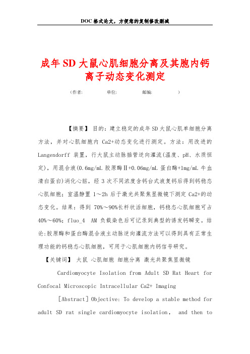
成年SD大鼠心肌细胞分离及其胞内钙离子动态变化测定(作者:___________单位: ___________邮编: ___________)【摘要】目的:建立稳定的成年SD大鼠心肌单细胞分离方法,并对心肌细胞内Ca2+动态变化进行测定。
方法:用改进的Langendorff装置,行大鼠主动脉插管逆向灌流(温度、pH、水质恒定),用混合液(0.6mg/mL胶原酶Ⅱ+0.06mg/mL蛋白酶+1mg/mL牛血清白蛋白)消化心脏,经3次不同浓度含钙台式液复钙后得到钙稳态心肌细胞;室温静置1~2h后于激光共聚焦显微镜下测定Ca2+的动态变化。
结果:得到70%~90%长杆状活细胞,钙稳态心肌细胞可占40%~60%;fluo_4 AM负载染色后可记录到典型的诱发钙瞬变。
结论:胶原酶和蛋白酶混合液主动脉逆向灌流方法可以得到具有正常生理功能的钙稳态心肌细胞,可用于心肌细胞内钙信号研究。
【关键词】大鼠心肌细胞细胞分离激光共聚焦显微镜Cardiomyocyte Isolation from Adult SD Rat Heart for Confocal Microscopic Intracellular Ca2+ Imaging[Abstract]Objective: To develop a stable method for adultSD rat single cardiomyocyte isolation,and then to determine the intracellular Ca2+ signalling by confocal imaging. Methods: Rat heart was digested by aorta retrograde perfusion with mixture [collagenaseⅡ(0.6 mg/mL), pronase(0.06 mg/mL)and bovine serum albumin(1 mg/mL)]using a modified Langendorff system. The temperature, pH and water quality should be properly controlled in the process of digested rat heart. Three times of re_calcification with different concentration of Ca2+ Tyrode solution were used to procure calcium homeostasis ventricular cardiomyocytes. Single cell was used for confocal microscopic Ca2+ imaging after storage at room temperature for 1~2 hours. Results: There were about 70%~90% rod_shaped fresh viable cells, in which 40%~60% were calcium homeostasis cells. In single calcium homeostasis cardiomyocytes calcium transients could be evoked by a stimulator and recorded with an LSM510 confocal microscopic system after incubation with fluo_4 AM. Conclusion: Aorta retrograde perfusion with collagenaseⅡand pronase is a proper method to procure single calcium homeostasis cardiomyocytes from adult SD rat with normal physiological features, and fit for confocal microscopic Ca2+ imaging.[Key Words]rat; cardiomyocyte; cell isolation; laser scanning confocal microscopy随着心脏疾病研究的不断发展,具备正常生理功能的单个心肌细胞已成为研究心脏代谢、功能、病理生理机制的重要基础,心肌细胞内钙离子浓度([Ca2+]i)变化是一系列心脏疾病的诱因及病理基础。
pGL3-Basic_Vector质粒图谱及其说明

Promega Corporation ·2800 Woods Hollow Road ·Madison, WI 53711-5399 USA·Fax 608-277-2516 ·1.Description ..........................................................................................................22.Product Components and Storage Conditions ............................................23.pGL3 Vector Maps and Sequence Reference Points . (2)A.pGL3-Basic Vector................................................................................................3B.pGL3-Enhancer Vector........................................................................................4C.pGL3-Promoter Vector........................................................................................5D.pGL3-Control Vector...........................................................................................64.Cloning Methods .. (7)A.Cloning Strategies.................................................................................................7B.Preparation of pGL3 Vectors and Insert DNA for Cloning...........................8C.Transformation Protocols for pGL3 Vectors....................................................8D.Isolation of Plasmid DNA (8)5.Transfection of Mammalian Cells ..................................................................96.Assay of Luciferase Activity ............................................................................97.Sequencing of Luciferase Reporter Vectors .. (11)8.Appendix mon Structural Elements of the pGL3 LuciferaseReporter Vectors ................................................................................................12B.Advantages of the pGL3 Vectors.....................................................................13C.The pGL3 Vectors luc + Gene............................................................................14D.Mapping Genetic Elements Located Within DNA position of Buffers and Solutions............................................................16F.References............................................................................................................17G.pGL3-Basic Vector Restriction Sites.................................................................18H.pGL3-Enhancer Vector Restriction Sites.........................................................20I.pGL3-Promoter Vector Restriction Sites.........................................................23J.pGL3-Control Vector Restriction Sites............................................................26K.Related Products. (28)pGL3 Luciferase Reporter VectorsAll technical literature is available on the Internet at: /tbs/ Please visit the web site to verify that you are using the most current version of this Technical Bulletin. Please contact Promega Technical Services if you have questions on useofthissystem.E-mail:********************1.DescriptionThe pGL3 Luciferase Reporter Vectors(a–c)provide a basis for the quantitativeanalysis of factors that potentially regulate mammalian gene expression. Thesefactors may be cis-acting, such as promoters and enhancers, or trans-acting,such as various DNA-binding factors. The backbone of the pGL3 LuciferaseReporter Vectors is designed for increased expression, and contains a modifiedcoding region for firefly (Photinus pyralis) luciferase that has been optimized formonitoring transcriptional activity in transfected eukaryotic cells. The assay ofthis genetic reporter is rapid, sensitive and quantitative. In addition, theseLuciferase Reporter Vectors contain numerous features aiding in the structuralcharacterization of the putative regulatory sequences under investigation.2.Product Components and Storage ConditionsProduct Size Cat.# pGL3-Control Vector20μg E1741 pGL3-Basic Vector20μg E1751 pGL3-Promoter Vector20μg E1761 pGL3-Enhancer Vector20μg E1771 Information on related products, including the Luciferase Assay System, is provided inSections 4–7 and 8.K.Storage Conditions:Store the pGL3 Luciferase Reporter Vectors at –20°C.3.pGL3 Vector Maps and Sequence Reference PointsThe listings of restriction sites for the pGL3 Luciferase Reporter Vectors areprovided in Section VIII.G–J.Note: The specific transcriptional characteristics of the pGL3 Vectors willvary for different cell types. This may be particularly true for COS cells,which contain the SV40 large T antigen. The SV40 large T antigen promotesreplication from the SV40 origin, which is found in the promoter of thepGL3-Promoter and pGL3-Control Vectors. The combination of large T antigenand SV40 origin will result in a higher copy number of these vectors in COScells, which in turn may result in increased expression of the reporter genecompared to other cell and vector combinations.Promega Corporation·2800 Woods Hollow Road ·Madison, WI 53711-5399 USA·3.A. pGL3-Basic VectorThe pGL3-Basic Vector lacks eukaryotic promoter and enhancer sequences,allowing maximum flexibility in cloning putative regulatory sequences.Expression of luciferase activity in cells transfected with this plasmid depends on insertion and proper orientation of a functional promoter upstream from luc +. Potential enhancer elements can also be inserted upstream of the promoter or in the BamHI or SalI sites downstream of the luc + gene.Figure 1. pGL3-Basic Vector circle map. Additional description: luc +, cDNAencoding the modified firefly luciferase; Amp r , gene conferring ampicillin resistance in E. coli ; f1 ori, origin of replication derived from filamentous phage; ori, origin of replication in E. coli.Arrows within luc + and the Amp r gene indicate the direction of transcription; the arrow in the f1 ori indicates the direction of ssDNA strand synthesis.pGL3-Basic Vector Sequence Reference Points:Promoter (none)Enhancer(none)Multiple cloning region 1–58Luciferase gene (luc +)88–1740GLprimer2 binding site 89–111SV40 late poly(A) signal 1772–1993RVprimer4 binding site2080–2061ColE1-derived plasmid replication origin 2318β-lactamase gene (Amp r )3080–3940f1 origin4072–4527upstream poly(A) signal 4658–4811RVprimer3 binding site4760–4779Promega Corporation ·2800 Woods Hollow Road ·Madison, WI 53711-5399 USA·Fax 608-277-2516 ·(for 0746V A 08_4AThe pGL3-Enhancer Vector contains an SV40 enhancer located downstream of luc + and the poly(A) signal. This aids in the verification of functional promoter elements because the presence of an enhancer will often result in transcription of luc + at higher levels.Figure 2. The pGL3-Enhancer Vector circle map. Additional description: luc +,cDNA encoding the modified firefly luciferase; Amp r , gene conferring ampicillin resistance in E. coli ; f1 ori, origin of replication derived from filamentous phage; ori,origin of plasmid replication in E. coli . Arrows within luc + and the Amp r gene indicate the direction of transcription; the arrow in f1 ori indicates the direction of ssDNA strand synthesis.pGL3-Enhancer Vector Sequence Reference Points:Promoter(none)Multiple cloning region 1–58Luciferase gene (luc +)88–1740GLprimer2 binding site 89–111SV40 late poly(A) signal 1772–1993Enhancer2013–2249RVprimer4 binding site2307–2326ColE1-derived plasmid replication origin 2564β-lactamase gene (Amp r )3329–4186f1 origin4318–4773upstream poly(A) signal 4904–5057RVprimer3 binding site5006–5025Promega Corporation ·2800 Woods Hollow Road ·Madison, WI 53711-5399 USA·0745V A 08_4AThe pGL3-Promoter Vector contains an SV40 promoter upstream of theluciferase gene. DNA fragments containing putative enhancer elements can be inserted either upstream or downstream of the promoter-luc + transcriptional unit.Figure 3. The pGL3-Promoter Vector circle map. Additional description: luc +,cDNA encoding the modified firefly luciferase; Amp r , gene conferring ampicillin resistance in E. coli ; f1 ori, origin of replication derived from filamentous phage; ori,origin of plasmid replication in E. coli . Arrows within luc + and the Amp r gene indicate the direction of transcription; the arrow in f1 ori indicates the direction of ssDNA strand synthesis.pGL3-Promoter Vector Sequence Reference Points:Enhancer(none)Multiple cloning region 1–41Promoter48–250GLprimer2 binding region 281–303Luciferase gene (luc +)280–1932SV40 late poly(A) signal 1964–2185RVprimer4 binding region2253–2272ColE1-derived plasmid replication origin 2510β-lactamase gene (Amp r )3272–4132f1 origin4264–4719Upstream poly(A) signal 4850–5003RVprimer3 binding region4952–4971Promega Corporation ·2800 Woods Hollow Road ·Madison, WI 53711-5399 USA·Fax 608-277-2516 ·51115212832360748V A 08_4AThe pGL3-Control Vector contains SV40 promoter and enhancer sequences,resulting in strong expression of luc + in many types of mammalian cells. This plasmid is useful in monitoring transfection efficiency, in general, and is a convenient internal standard for promoter and enhancer activities expressed by pGL3 recombinants.Figure 4. pGL3-Control Vector circle map. Additional description: luc +, cDNA encoding the modified firefly luciferase; Amp r , gene conferring ampicillin resistance in E. coli ; f1 ori, origin of replication derived from filamentous phage; ori, origin of plasmid replication in E. coli . Arrows within luc + and the Amp r gene indicate the direction of transcription; the arrow in f1 ori indicates the direction of ssDNA strand synthesis.pGL3-Control Vector Sequence Reference Points:Multiple cloning region 1–41Promoter48–250Luciferase gene (luc +)280–1932GLprimer2 binding site 281–303SV40 late poly(A) signal 1964–2185Enhancer2205–2441RVprimer4 binding site2499–2518ColE1-derived plasmid replication origin 2756β-lactamase gene (Amp r )3518–4378f1 origin4510–4965upstream poly(A) signal 5096–5249RVprimer3 binding site5198–5217Promega Corporation ·2800 Woods Hollow Road ·Madison, WI 53711-5399 USA·(for 0747V A 08_4AFigure 5. pGL3 Vector multiple cloning regions. Shown are the upstream and downstream cloning sites and the locations of the sequencing primers (GLprimer2,RVprimer3 and RVprimer4). The large primer arrows indicate the direction ofsequencing. The positions of the promoter (in the pGL3-Promoter and pGL3-Control Vectors) and the enhancer (in the pGL3-Enhancer and pGL3-Control Vectors) are shown as insertions into the sequence of the pGL3-Basic Vector. (Note that the promoter replaces four bases [AAGT] of the pGL3-Basic Vector.) The sequence shown is of the DNA strand generated from the f1 ori.4.Cloning Methods4.A. Cloning StrategiesThe restriction sites for XhoI and SalI have compatible ends, as do BglII and BamHI. Therefore, cloning into the XhoI or BglII sites upstream of luc +, or the downstream SalI or BamHI sites, allows easy interchange of DNA insertsbetween upstream and downstream positions relative to the luciferase reporter gene. Thus, positional effects of a putative genetic element may be readily tested. Cloning fragments into a single site will generally yield both possible orientations relative to the reporter gene, making these effects also readily testable.The other upstream restriction sites may be used for cloning. However, note that some of the sites are required for generating nested deletions (see Section 8.D). Specifically, the KpnI or SacI site is needed to generate a 3´ overhang upstream of the insert.Promega Corporation ·2800 Woods Hollow Road ·Madison, WI 53711-5399 USA·Fax 608-277-2516 ·CATTCCGGTACTGTTGGTAAAGCCACCATGGAAGACGCCAAAAACATAAAG . . . (1892bp) . . . GGATCCGTCGACRVprimer35′GGTACCGAGCTCTTACGCGTGCTAGCCCGGGCTCGAGATCTGCGATCTAAGTAAGCTTGG . . .KpnI Acc65ISacIMluINheIXmaI SmaIXhoIBglIIHindIIISV40Enhancerluc+ Coding Region ′BamHI SalI0756M A 08_4A4.B.Preparation of pGL3 Vectors and Insert DNA for CloningThe fragment and vector DNA should be digested with restriction enzymesthat will generate compatible ends for cloning. In some cases, the ends of theDNA fragment may require modification, either by using synthetic linkers, bya PCR amplification using primers containing sites for appropriate restrictionenzymes, or by filling in the restriction site overhangs. It may be advantageousto treat the vector DNA with calf intestinal alkaline phosphatase (CIAP; Cat.#M2825) or TSAP Thermosensitive Alkaline Phosphatase (Cat.# M9910) toremove 5´ phosphate groups, thus preventing reclosure of the vector on itselfwithout an insert. Sufficient DNA should be prepared to perform controlreactions for digestion, ligation and transformation steps.To ensure capture of the correct insert DNA, the desired restriction fragmentcan be purified by electrophoresis on an acrylamide or agarose gel and thenrecovered from the gel by one of several methods, such as using the Wizard®PCR Preps DNA Purification System Technical Bulletin#TB118. Alternatively,unfractionated restriction fragments can be cloned into the target plasmid, andthe desired recombinant then can be identified by gel electrophoresis ofplasmid DNA.Protocols for restriction digestion, alkaline phosphatase treatment, linkerligation and transformation of competent cells can be found in MolecularCloning, A Laboratory Manual(1).4.C. Transformation Protocols for pGL3 VectorsBecause the Luciferase Reporter Vectors are supplied as modified DNA, E. colihosts may be either restriction + or restriction –. The use of a rec A host such asJM109 is preferred because this prevents undesirable recombination betweenthe insert and the host chromosomal DNA. A strain that has an F´ episome isrequired for ssDNA production.Grow JM109 on minimal plates (M-9) supplemented with 1.0mM thiamine-HCl prior to preparation of competent cells and transformation. This selectsfor the presence of the F´ episome.4.D. Isolation of Plasmid DNAThe Wizard®Plus SV Minipreps DNA Purification System (Cat.# A1340,A1470) may be used for small-scale preparation of plasmid DNA for screeningclones. DNA suitable for transfection may be purified using the PureYield™Plasmid Midipreps System (Cat.# A2492, A2495).Promega Corporation·2800 Woods Hollow Road ·Madison, WI 53711-5399 USA·5.Transfection of Mammalian CellsTransfection of DNA into eukaryotic cells may be mediated by cationic lipid compounds (2), calcium phosphate (3,4), DEAE-dextran (3,5), or electroporation (4). Transfection systems based on cationic lipids (TransFast™ Transfection Reagent, Transfectam®Reagent and Tfx™ Reagents) and calcium phosphate (Profection®Mammalian Transfection System) are available from Promega. Formore information on these transfection reagents, please request the TransFast™Transfection Reagent Technical Bulletin(#TB260), the Transfectam®Reagent Technical Bulletin(#TB116), the Tfx™-Reagents Technical Bulletin(#TB216) or the ProFection®Mammalian Transfection System Technical Manual(#TM012). All of thesedocuments are available on our web site at: /tbs/6.Assay of Luciferase ActivityExperimental strategies using firefly luciferase may involve the analysis of afew samples per day or as many as several thousand samples per hour, and equipment used to measure luminescence may vary from inexpensive, single-sample luminometers to high-end CCD luminometers. To support this widerange of applications, we have developed three luciferase assays with different,but complementary, characteristics: Luciferase Assay System (Cat.# E1500),Bright-Glo™ Luciferase Assay System (Cat.# E2610), Steady-Glo®LuciferaseAssay System (Cat.# E2510), and ONE-Glo™ Luciferase Assay System (Cat.#E6110). Reagent choice depends on the relative importance of experimentalformat, assay sensitivity, and luminescence duration.Table 1. Characteristics of Promega Luciferase Assay Reagents.LuciferaseBright-Glo™Steady-Glo®Assay ONE-Glo™Reagent Reagent Reagent ReagentFormat NH or H NH or H NH NH or HProcess continuous batch bench scale batch orcontinuous Number of Steps1141Sensitivity highest lower higher highSignal Half-Life~30 minutes~5 hours~12 minutes~50 minutes Precision High High High HighestCell Lysis Time~2 minutes~5 minutes NA~3 minutesmaximum maximumNH = nonhomogeneous (first create a lysate); H = homogeneous; NA = not applicablePromega Corporation·2800 Woods Hollow Road ·Madison, WI 53711-5399 USA·Fax 608-277-2516 ·6.Assay of Luciferase Activity (continued)The Luciferase Assay System has long been the standard reagent for routine laboratory analysis. Before using this reagent, cells from which the luciferase is to be measured must be washed and lysed. This reagent was optimized for high sensitivity in nonhomogeneous, single-sample measurements. The Luciferase Assay System requires a luminometer fitted with injectors to efficiently measure luminescence in 96-well plates.The Bright-Glo™, Steady-Glo®and ONE-Glo™ Reagents were developed to perform assay reactions within multiwell plates and in the presence of complete cell culture medium: no cell preparation steps such as washing or lysing are required before the luminescence reaction is initiated. All of these are single-step reagents, requiring only addition of the reagent before measuring luminescence. This makes them ideal reagents for efficient and precise quantitation in 96-, 384- and 1536-well plates.The Bright-Glo™ and Steady-Glo®Reagents are complementary in their characteristics based on the inverse relationship between luminescence duration and assay sensitivity (6). Generally, as the half-life of the luminescence increases, assay sensitivity decreases. The Steady-Glo®Reagent provides long luminescence duration (changing only about 10% per hour); however, toachieve this long luminescence duration, the assay sensitivity must be reduced. This reagent was designed for experiments in which many microplates are processed as a batch.In contrast, the Bright-Glo™ Reagent provides high assay sensitivity with shorter luminescence duration (<10% decrease per 5 minutes). This reagent is designed for general research applications and for experiments using robotics for continuous sample processing. Furthermore, as a result of increased sample capacity, the Bright-Glo™ Reagent provides greater assay sensitivity than the Luciferase Assay Reagent in most applications (6).The ONE-Glo™ Reagent provides the ultimate performance for luciferase assays. It features a high-sensitivity assay with extended duration. TheONE-Glo™ Reagent also demonstrates more robust performance and provides reagent handling enhancements.The Luciferase Assay System, Bright-Glo™ Reagent, Steady-Glo®Reagent and ONE-Glo™ Reagent provide the highest standards in assay quantitation, sensitivity and convenience. Since these reagents are based on the same underlying design principles, different reagents can be used as experimental needs change. For more information, request the Luciferase Assay System Technical Bulletin#TB281, the Steady-Glo®Luciferase Assay System Technical Manual#TM051, the Bright-Glo™ Luciferase Assay System Technical Manual#TM052, or the ONE-Glo™ Luciferase Assay System Technical Manual#TM292.When studying promoter functionalities, it is often desirable to include asecond reporter (e.g., Renilla luciferase) as an internal control for normalization. Plasmids derived from pGL3 or pGL4 vectors can be co-transfected with Renilla luciferase vectors, such as phRL-TK, and assayed using the Dual-Luciferase®Reporter Assay System (Cat.# E1910) or the Dual-Glo™ Luciferase AssaySystem (Cat.# E2920).Table 2. Characteristics of Promega Dual-Luciferase Assays.Dual-Luciferase®Dual-Glo™Assay AssayFormat NH HProcess bench scale batchNumber of Steps52Sensitivity higher lowerSignal Half-Life—firefly~9 minutes~2 hoursSignal Half-Life—Renilla~2 minutes~2 hoursPrecision High HighCell Lysis Time~10 minutes~15 minutesmaximum maximumNH = nonhomogeneous (first create a lysate); H = homogeneous7.Sequencing of Luciferase Reporter VectorsYou may desire to sequence the DNA inserted into the Luciferase Reporter Vectors. Two examples of such applications are to determine the exact positionof generated deletions and to confirm production of a site-specific mutation.Three primers are available for sequencing the pGL3 Vectors: RVprimer3 (Reporter Vector Primer 3) for sequencing clockwise across the upstreamcloning sites, RVprimer4 for sequencing counterclockwise across the BamHIand SalI cloning sites downstream of luc+, and GLprimer2 for sequencing counterclockwise upstream of luc+.RVprimer35´-CTAGCAAAATAGGCTGTCCC-3´RVprimer45´-GACGATAGTCATGCCCCGCG-3´GLprimer25´-CTTTATGTTTTTGGCGTCTTCCA-3´RVprimer3 is especially useful for identifying positions of nested deletions.Note:All three primers can be used for dsDNA sequencing, but onlyRVprimer4 and GLprimer2 also may be used for ssDNA sequencing.Promega Corporation·2800 Woods Hollow Road ·Madison, WI 53711-5399 USA·Fax 608-277-2516 ·8.Appendix8.A. Common Structural Elements of the pGL3 Luciferase Reporter VectorsExcept for the inclusion of promoters and enhancers, the four pGL3 Luciferase Reporter Vectors are structurally identical. Each plasmid’s distinguishing features are summarized in Section 3. The pGL3 Vectors each contain a high-copy-number prokaryotic origin of replication for maintenance in E. coli , an ampicillin-resistance gene for selection, and a filamentous phage origin of replication (f1 ori) for single-stranded DNA (ssDNA) production. Restriction sites for insertion of DNA fragments are located upstream and downstream of the luciferase gene. Two of the upstream sites (XhoI and BglII) yield cohesive ends compatible with the downstream sites (SalI and BamHI, respectively),allowing the interchange of the DNA insert for rapid analysis of positional effects.Figure 6. Comparison of luciferase activities expressed in HeLa cells transfected with the pGL2-Control and pGL3-Control Reporter Vectors. The expression level of luc + is dramatically higher with the pGL3-Control Vectors. In repeatedexperiments with several cell lines, we observed 20- to 100-fold higher luciferase activity from cells transfected with pGL3-Control. Luciferase activity was measured with a Turner Designs luminometer. (Absolute light values and relative expressionprofiles may vary between different cell types.)1,4001,2001,0008006004002000Improved Expression Level with the pGL3-Control VectorConstruct TransfectedpGL3-ControlVectorpGL2-ControlVectorA v e r a g e R e l a t i v e L i g h t U n i t sFigure 7. A representative experiment comparing luciferase activities expressed in HeLa cells transfected with the pGL2 and pGL3 Vector series. The increase in luciferase expression observed with these new vectors provides greater sensitivity,while maintaining relatively low background luciferase expression.8.B. Advantages of the pGL3 VectorsThe pGL3 Reporter Vectors contain a modified firefly luciferase cDNAdesignated luc + and a redesigned vector backbone. These changes were made to increase luciferase expression, improve in vivo vector stability, and provide greater flexibility in performing genetic manipulations. The modified reporter vectors have resulted in luciferase expression levels dramatically higher than those obtained with pGL2 Reporter Vectors (Figure 6), while maintaining relatively low background luciferase expression (Figure 7).The substantial increase in the expression of luciferase observed with the pGL3Vectors provides greater sensitivity. It may now be possible to obtainmeasurable luciferase expression in cell types that are difficult to transfect or when studying weak promoter elements. Users of the pGL2 and pGL3 Vectors should be aware, however, that absolute light unit values and relative expression profiles vary between different cell types (7). Therefore, it is important to include the appropriate control vectors in all experiments.Further refinements have been made since the pGL3 Vectors became available.Our newest series of luciferase reporter vectors, the pGL4 Luciferase Vectors,provide additional features and benefits as compared to the pGL3 Vectors. For more information, see the pGL4 Luciferase Reporter Vectors Technical Manual #TM259 available at:/tbs/Promega Corporation ·2800 Woods Hollow Road ·Madison, WI 53711-5399 USA·Fax 608-277-2516 ·80604020Construct TransfectedControl Basic Enhancer PromoterA v e r a g e R e l a t i v e L i g h t U n i t sA v e r a g e R e l a t i v e L i g h t U n i t sConstruct TransfectedControl Basic Enhancer Promoter 08.C. The pGL3 Vectors luc+ GeneModifications that distinguish the luc+ gene from the native luciferase genegenerally fall into four categories: i) the C-terminal tripeptide has beenremoved to eliminate peroxisome targeting of the expressed protein; ii) codon usage was improved for expression in plant and animal cells; iii) two potential sites of N-glycosylation were removed; and iv) several DNA sequence changes were made to disrupt extended palindromes, remove internal restriction sites, and eliminate consensus sequences recognized by genetic regulatory binding proteins, thus helping to ensure that the reporter gene itself is unaffected by spurious host transcriptional signals. (For a detailed description of themodifications to the luc+ gene, see reference 8.)Four major modifications were made to the vector backbone: i) the SV40 early poly(A) signal has been replaced with the SV40 late poly(A) signal to increase the efficiency of transcription termination and polyadenylation of theluciferase transcripts (9); ii) a synthetic poly(A) and transcriptional pause site (10,11) have been placed upstream of the multiple cloning site to terminatespurious transcription, which may initiate within the vector backbone; iii) the small T intron has been removed to prevent reduced reporter gene expression due to cryptic RNA splicing (12,13); and iv) a Kozak consensus sequence (14) has been inserted to increase the efficiency of translation initiation of theluciferase gene (7; Table 3).There is a newer luciferase gene available, luc2. The luc2gene not only shares the same features as luc+, but the sequence was codon-optimized forexpression in mammalian cells. For further information about the luc2genepresent in the pGL4 Luciferase Vectors, see Technical Manual #TM259available at: /tbs/Table 3. Changes Made to the pGL3 Vectors.Changes Made Purpose of Modification Reference Modifications made to Changes eliminate peroxisome(8)the luciferase gene targeting of expressed protein,(luc to luc+).eliminate consensus bindingsequences for various geneticregulatory proteins, improvecodon usage for mammalianand plant cells, and provideconvenient restriction sites.A unique NcoI site created Ability to create N-terminalat 5´ end of luc+ gene. NcoI gene fusions with luc+sites removed from SV40 using unique NcoI site.enhancer and promoterregions.Intron from SV40 small Intron from SV40 small T (12,13)T antigen removed. antigen can reduceexpression when placed 3´of certain genes due tocryptic splicing.Poly(A) site for back-Avoids possible recombination (9,10)ground reduction changed between two SV40 poly(A)from SV40 early site to a sequences in thesynthetic poly(A) and same plasmid.transcriptional pause site.Poly(A) signal for luc+ Late SV40 poly(A) signal is(7)changed from early to more efficient than early SV40late SV40 poly(A) signal.poly(A).Kozak consensus Provides optimal (14) sequence created translation efficiency.immediately 5´ of theluc+ gene.Unique XbaI site User convenience; facilitatescreated just downstream subcloning of the luc+ gene.of the luc+ gene.SmaI site moved to User convenience; blunt-ended insertsinternal position in can now be cleaved on either sidemultiple cloning region.by restriction endonucleases.Promega Corporation·2800 Woods Hollow Road ·Madison, WI 53711-5399 USA·Fax 608-277-2516 ·。
SP5 confocal microscope 用户手册说明书

PCICPlant Cell Imaging Center(BTI, room 104B)CONFOCAL LEICA SP5 user manualStartup procedure:-Remove the dust cover from the microscope-Turn on the mercury lamp (A) if you need to see the fluorescence through the eye pieces. Check that its shutter is on the “open” position, and that the intensity is notadjusted beyond the second setting.-On the main switch board (B), switch on PC/microscope (button Nº1). Wait for 15 seconds. (The computer will turn on automatically).-Switch on the Scanner powe r button (Nº2, on the main switch board). Wait for 15 seconds. Check that the fan is working properly (C).-Switch on the Laser power button (Nº3, on the main switch board). It may be already on , in which case leave it that way.-Turn the laser key (Nº4, on the main switch board) from OFF to ON. -Fill out the log book .-On PC: login as “your first name” and password is PCICuser!STARTING THE LAS AF SOFTWARE:-After login, wait for about 45 seconds (important).-After 45seconds, double click on the LAS AF icon on the desktop -On the starting screen, check that the configuration is OK:“MACHINE” should be picked (If you need the resonant scanner, mark the box “Activate the resonant scanner”). Then click OK.-When the window “Microscope stand ” p ops up, say NO unless you are doing advanced techniques (such as Multiple Field,mosaic, etc.).In the configuration tab (1), select the laser window (2). Turn on the laser(s) you need (3). By checking the box next to laser means starting up the laser.Adjust the Argon laser to approximately 30 % (4) if you use it (You can increase the power if you are doing bleaching, or if your signal is really too weak for example).If lasers were turned off by the previous user recently, make sure the Argon laser was left off for at least 2 hours before you turn it back on. HeNeLasers have to rest at least one hour before being back on. The 405 nm laser needs to rest only about 15 minutes. The DPSS 561nm can be turned back on immediately.SLIDE OBSERVATION THROUGH THE EYEPIECES:First select the 10 X objective to identifyyour sample, and check if you are in focuswhen the stage moves to its preset Z=0 position(more details on next page).Although it is possible to change theobjectives manually, our SP5 is equipped withan automated objective turret. To use thisfunction, go back in the “Acquire” tab andselect the appropriate objective (see thesupplementary information for details onavailable objectives, and correct ring settings).Avoid switching manually the objectives,as you may inadvertently rotate the objectivering, or even worse, the objective itself.Be careful not to put oil on waterobjectives! Use ONLY Leica OIL 11-513-859for oil immersion.Lower the stage by pressing the “Down” Z axis button of the stage knob (D). Place your sample under the microscope. Be very gentle when you place your slide on the stage : it is very delicate. Press the “Up” button of the stage knob until it stops automatically.Look down the eyepiece and turn the X/Y or Z axis wheels of the stage knob to identify your sample (you can adjust the speed of movement (X/Y and Z) on the right).Fine focus along the Z axis by turning the knob on the side of the microscope stand. Check the Z position on the XYZ Screen (see below).If you are too far from the preset Z=0, you must reset this parameter so that you can safety switch to higher magnification objective. Click on the floppy icon of the touch pad (1), make sure the “on focus” icon is selected (2) and that your sample is on focus , then press “Set” (3). Now you can safely switch objectives automatically. You don’t need to reset this parameter for each objective.You can visualize current settings by pressing the microscope icon of the touch pad.Status ScreenIllumination typeObjective Filter cubeLight ScreenLight intensityField and aperture settingsXYZ ScreenStage positionTRANSMITTED LIGHT:To use transmitted light (TL),lever (behind the microscope stand) is directed towards the eyepieces.Note: Adjusting the microscope foran optimal brightfield image(Köhler illumination) is more complicated than for fluorescence. Although we regularly check thatthe settings are optimal, you mayneed further readjustment. Pleaseask for help the first few times.By pressing the buttons on theleft side of the microscopestand, you can switch to TL.You can also pick betweenvarious TL modes: brightfield,polarized and DIC.The last button allows you tovisualize fluorescence and DICtogether (when available).mark will appear on the touch pad.On the left side of the stand,underneath the stage, push the TL/ILbutton until “TL”appears on thetouch pad.Look through the eyepieces andadjust the brightness with the INTbuttons. You can adjust the shapeand size of the illuminated areawith the “FD” button. ArrayFine adjustment of the rotation of theWollaston prism for DIC imaging isperformed by rotating the thumbscrew locatedright above the objectives.FLUORESCENCE:By pressing the buttons on the right side of themicroscope stand, you can switch to fluorescencemode. You can choose the appropriate filter cubewith the “cube”button. Its name will appear on thetouch pad. Filter cubes available: “A” = UV“I3” = Green LP“GFP” = Green BP“N21”= Red“N3” = Red BPSee supplementary information for more details.The last button is the fluorescence shutter.On the left side of the stand, underneath the stage, push the TL/IL button until “fluo” appears on thetouch pad. Look through the eyepieces and adjust the brightness with the INT buttons. You canadjust the shape and size of the illuminated area with the “FD” button.CONFOCAL ACQUISITIONADJUSTING THE PARAMETERS FOR PMT1, PMT3, PMT4 and PMT5Go to the Acquire Tab of the LAS AF software. You can expand any menu on the left part of the settings screen by clicking on the black arrows .In the first panel, pick the desired acquisition mode (1). Standard mode is XYZ.If you want to use preset adjustments or have already saved yours, load them from the scrolling list (2). Otherwise activate the UV or Visible icons , and adjust the required laser lines (3). Usually between 5 and 50 % depending on the line and your sample. Then activate and adjust the required PMTs by checking the “Active” box (4). Use first the PMTs located just under the spectral band you want to detect.You can download the emission spectra of numerous dyes to help you adjust your settings (5). This has no impact on your image collection, and the display of any spectra from the list will not affect the data collection.Make sure the detection wavelengths don’t cover any laser line! Move the cursor slowly to prevent damage to the mechanical part.If you want to use transmitted light , reposition the transmitted light lever (see page 3) towards the confocal position. Click on “Additional channels” in the acquisition tab, then select SCAN -BF or SCAN-DIC (1). Then activate the transmitted light PMT by checking the “Active” box .Start preview scanning by clicking on LIVE (bottom of the leftscreen).In the image window (right screen), activate the multi-panel view (1).Click on the “Quick-LUT ” icon to obtain a false color optimization screen (2). Click on the panel of the first fluorophore.Gain adjustment:Increase “Smart gain” until just a few single blue dots appear (saturated pixels=value 255). Offset adjustment:Reduce the “Smart offset” level until very few green dots appear (Green pixels= no signal=0).Repeat with the other channels. Check the settings by moving along the Z axis (you can use the button bar). Save the settings.NOTE: For proper gain adjustment, you may want to increase your smart gain value up to 900-1000V. A smart gain value lower than 400V means that you can lower the laser line intensity, and readjust your smart gain. A smart gain between 1100-1250 would suggest increasing the laser line intensity. REMEMBER: Enhancing the laser line intensity will bleach your fluorophore faster, but increasing the gain does not affect your sample. Therefore, in order to protect your sample, it is better to keep the laser line intensity as low as possible, and increase the gain instead. To adjust the TL channel : exit the Q-LUT mode and adjust gain and offset in the standard mode. In the XY panel (left screen), set the image format, scan speed and zoom factor (3). Faster scans require zooming. You can use thearrows to move the imaged area . You can also zoom specifically on an area by activating the “zoom” box (3), then drawing a rectangle around your region of interest (the rectangle drawing tool will appear on the right screen once you select the “zoom” option). Adjust the averaging to improve the image definition (4): - For live imaging: use line averaging - For fixed samples: line or frame averaging - For weakly fluorescent samples, you can also use the accumulating function. NOTE : if you change the scan speed or the zoom factor, or if you switch objectives, you may need to readjust gain and offset.New HYD Detector (old PMT2)ADJUSTING THE PARAMETERS FOR PMT2 (HyD Detector) : If your sample is very dim, you may want to use HyD detector to collect your signal.Do not use high laser with HyD detector: use as low laser as possible (1%-max 15%)PMT2 is HYD detector which provides:Large Dynamic rangeImproved cell viabilityHigh speed ImagingSingle photon countingLow dark noiseExquisite contrastGain adjustment: can go from 0-500Increase “Smart gain” until just a few single blue dots appear (saturated pixels=value 255).Offset adjustment:Sets automatically in HyDLeica HyD has 3 modes of collection:1)Standard mode (most commonly used)2)BrightR (rarely used, non-linear gain applied to structure)3)Photon Counting Mode (good for quantitative imaging; works best with dim sample) ---------------------------------------------------------------------------------------------------------------------------Z stackExpand the Z-stack panel and activate the“live” viewing mode.Move along the Z axis with the appropriatewheel of the button bar.Click the red arrow next to the “Begin”field to mark the start position.Then define the end position by rotating thewheel and clicking the “end” arrow.Define the number of slices by eitherentering the number of steps, or by choosing adefined z-step size.Sequential scanTo avoid crosstalk between fluorophores, you can perform a sequential scan.Click on the “Sequential ” icon (1).Define the number of successive sequential scans by clicking on the “-” and “+” icons (2). Decide when the scanning modes should be switched :- For live imaging: use between lines. This mode does not provide the full range of scanning options. - For fixed samples: use between frames or stacks.For each individual scan , make sure only one laser line will be activated (3), and adjust accordingly the corresponding PMT (4). Adjust separately scan 1, then scan 2, etc. You can control the settings by using the “Live ” function.ACQUIRING AND SAVING DATAWhen you are satisfied with your settings, click on “Capture image ” to acquire one image, or “Start ” if you want to acquire an image sequence (such as a Z-stack).Save your files regularly by going into the “experiments” sub-tab. Place the mouse arrow over your file and right click to display the menu and save the images. Saving the images in the “.lif ” format (LEICA image format) will allow you to import acquisition parameters during future experiments, as well as having an access to detailed information regarding the parameters used for the acquisition of the data (see below).You can also export single images or the entire experiment as Tiff , rename the file , etc.Save your files in D:/users/your full name . Your old files (from 2008) are all saved under d:\images\your lab folder\your folderNOTE: do not go beyond 2 Giga for a single experiment.EXPERIMENTSOpen an experiment.Right-click on the image name in the file list or theimage window itself. Open Properties and click on“Apply settings” at the bottom of the window.TRANSFER YOUR DATA TO THE SERVE RFor this you have to map a network drive:MAP A NETWORK DRIVE (do it after you login as you on confocal PC) Double Click on MY COMPUTERTop Menu Bar –TOOLS – MAP A NETWORK DRIVEZ\\10.236.156.203\PCIC\Users Data\Check Reconnect at LogonClick Connect Using a Different User NameUser Name: PCIC_userPassword: enter password (contact PCIC if you forgot the password.)OKFinishNew window showing the Users Data folder on the Workstation will appearCREATE A SHORTUCT ON YOUR DESKTOP:My ComputerScroll to bottom on screenYou will see your mapped network driveRight Click on it (rename it if you want for eg. “Data transfer to Server”)Create ShortcutOn Desktop(it creates an icon “shortcut to users data”or an icon “Data transfer to Server”)When you need to transfer your data to the Workstation – use the shortcut on your desktop (which is as before “shortcut to users”)Please note it may take a few minutes before the two computers are able to communicate.Copy (or drag) your files from the D: drive to this folder.TO RETRIEVE YOUR DATA FROM THE SERVER VIA YOUR COMPUTEROn a PC: Go to “My Computer” and right click. Choose "map network drive"In the window that comes up type the following:Drive: ZFolder: \\10.236.156.203\PCIC\Users Data\Check Reconnect at LogonClick Connect Using a Different User NameUser Name: PCIC_userPassword: enter password (contact PCIC if you forgot the password.)OKFinishNew window showing the Users Data folder on the Workstation will appearWe will regularly erase files from the server and the confocal computer, so don't wait to transfer your files to your own computer...On a Mac:From the Finder select "Connect to server" from the GO menu (or press command K)Type smb: \\10.236.156.203\PCIC\Users DataIn the address box / ConnectIn the dialog box that appears, leave confocal in the workgroup or domain box.Enter user name and password (contact PCIC if you forgot it).You can then download an entire folder.If it still doesn't --Apple MenuSystem PreferencesSharingServices tab -- make sure personal file sharing is checkedFirewall tab -- make sure personal file sharing is checkedthen try againIfitstilldoesn'twork,contactElaine(****************). TO PROCESS YOUR FILESThe workstation 1 hosts an offline version of the LAS AF software, as well as Image J and Image-Pro Plus version 6.3. Image-Pro Plus is a powerful and customizable image processing and analysis software.The workstation 2 has Photoshop and two free softwares (LAS AF Lite and Image J) allowing limited manipulations of your Leica files.It is extremely important that you keep an intact copy of all your original files:-Some journals request the access to the unmodified, original data before publication-If you open and save a .lif file in LAS AF Lite or the offline full version of LAS AF, you may not be able to open this file and reapply its settings on the confocal machine (depends on the confocal version and the offline and the Lite versions).-On your own computer:To install LAS AF Lite or Image J on your computer, follow the instructions on the PCIC website(look under software).TROUBLESHOOT:.-I am not sure if the fluorescence comes from my fluorophoreMore info on how to perform a lambda scan can be provided upon request. Contact PCIC.-I turned on everything according to the manual, but nothing is workingCheck that the microscope control box is on (E, page 1)-The fluorescence is too dim, although I adjusted gain and contrast3 factors can affect the brightness: the laser line power (see supplementary information for details), the laser intensity (change the % in the laser configuration tab, but keep it as low as possible) and the pinhole size (should be set to 1 Airy). If you increase the pinhole size, you will have a brighter image, but it will affect the definition of your image). Also, more info on how to use the Enhanced Data Transfer Mode or frame accumulation for dim samples can be provided upon request. Contact PCIC. -The fluorescence of my sample is bleachingYou can reduce the number of frame/line averaging, or increase the scan speed (it will decrease the definition of your image). Also, more info on how to use the Enhanced Data Transfer Mode for dim samples can be provided upon request. Contact PCIC.-I am using live samples and some components are moving too rapidlyMore info on how to use the bidirectional scan or the resonant scanner for high-speed imaging can be provided upon request. Contact PCIC.Need some help?Technical help can be provided by Mamta Srivastava from 9 am to 1 pm.For any technical question or request, call 254-4436 or come between 9 am and 1 pm at the PCIC, or send an E-mail to ****************SUPPLEMENTARY INFORMATION:Filter cubes:Configurationof the lasers:Relative Argon laser power: Source: /Lasers.htmlObjectives:Max speed of the bi-directional resonant scanner: about 25 frames / second (512/512 pixels)SHUTDOWN PROCEDURE:First, check the Oracle calendar on the PCIC workstation No. 2.If someone booked within one hour after you (DO NOT UNCHECK THE BOX NEXT TO LASER)-Save your images and leave the objective at 10X (do not rotate the objective manually).-Exit the program.-Transfer your data to the serve r (see above)-Log off and fill out the logbook-Lower the stage and clean objectives with fresh lens tissue(NO Kimwipes!)-clear up the desk and the working areaIf the next booking is later today (>1h but <4h later)-Save your images and leave the objective at 10X (do not rotate the objective manually).-In the laser configuration tab (see page 2), uncheck all lasers except the Argon. Slide down the argon power to 0%.-Exit the program.-Transfer your data to the serve r (see above)-Log off and fill out the logbook-Turn off the mercury lamp (see page 1, A)- Lower the stage and clean objectives with fresh lens tissue(NO Kimwipes!)-Put the dust cover on the microscope-clear up the desk and the working areaIf no one booked after you (last user of the day or >4h later)-Save your images and leave the objective at 10X (do not rotate the objective manually).-Lower the stage-In the laser configuration tab (see page 2), uncheck all lasers.-Exit the program.-Transfer your data to the serve r (see above)-Turn off the computer via the “Start” menu of Windows-Turn off the mercury lamp (A)-Turn the laser key (Nº4, on the main switch board) from ON to OFF-Keep the Laser power button (Nº3, on the main switch board) ON for at least 15 minutes-Switch off the Scanner powe r button (Nº2, on the main switch board).-Switch off the PC/microscope button(Nº1).-Log off and Fill out the logbook-Clean objectives with fresh lens tissue(NO Kimwipes!)-Clear up the desk and the working area-Put the dust cover on the microscope-15 minutes after shut down, turn off the laser power button (Nº3).Important: after hours, make sure the next person is coming (contact by e-mail or phone). If nobody comes, perform a total shutdown.。
ABB ACS880产品使用Modbus RTU配置指南

G UIDE FOR CONFIGURING THE ACS880 FOR M ODBUS RTU USING CSA2.8/3.0P ROFILEScope:The ACS880 does not support the CSA profile that the ACS600/ACS800 supported on Modbus. This guide will show how to use Transparent mode in the ACS880 to create the CSA control and status words to mimic the CSA profile.Explanation:The ACS880 should be first be configured for standard Modbus RTU operation and the parameters below need to be set to enable the embedded Modbus and to tell the drive that the start/stop and speed references will come from communications if Modbus is being used for control and reference.20.01 EXT1 command = Embedded Fieldbus (If using Modbus for control)22.11 Speed ref source = EFB ref1 (If using Modbus for control)58.01 Protocol enable = Modbus RTU58.03 Node Address = Node address 1 (247)58.04 Baud Rate = Network Baud rate58.05 Parity = Parity type and stop bits58.06 Communication Control = Refresh settings to save EFB parameters58.25 Control Profile = TransparentWhen the profile is set to Transparent, the drive does NOT do any data conversion on the CW or SW. Speed Reference and Actual values are treated separately and can be handled/scaled in their usual way. The raw control word sent from the PLC will come into the drive and be visible in par 06.05 EFB Transparent Control Word. The bit structure is below:What we will do is point our drive control parameters (like enable and start) to the corresponding bits in this CW. For example:20.12 Run Enable 1 Source = 6.05.120.03 Ext1 In1 Source = 6.05.319.11 Ext1/Ext2 Selection = 6.05.531.11 Fault Reset Selection = 6.05.8Now, on to the status word. The ACS880 allows us to build a status word bit by bit using par 6.50. We will use this parameter as the source of our CSA status word:58.30 EFB Status Word Transparent Source = 6.50The status word is in par 06.05 EFB Transparent Control Word. The bit structure is below:Parameters 6.60 to 6.75 are used to assign each bit to match the functionality of the CSA status word: 6.60 User status word 1 bit 0 sel = 6.11.1 (RDY_RUN)6.61 User status word 1 bit 1 sel = 6.11.4 (OFF_2_STA)6.62 User status word 1 bit 2 sel = False6.63 User status word 1 bit 3 sel = 6.11.2 (RDY_REF)6.64 User status word 1 bit 4 sel = False6.65 User status word 1 bit 5 sel = 6.11.9 (REMOTE)6.66 User status word 1 bit 6 sel = False6.67 User status word 1 bit 7 sel = 6.11.8 (AT_SETPOINT)6.68 User status word 1 bit 8 sel = 6.11.3 (TRIPPED)6.69 User status word 1 bit 9 sel = 6.11.7 (ALARM)6.70 User status word 1 bit 10 sel = 6.11.10 (ABOVE_LIMIT)The Modbus register addresses for control and status word remain the same as they were and are shown below:Documents or other reference material:ACS880 primary control program Firmware manual 3AUA0000085967 ACS800 Standard Control Program 7.X 3AFE64527592。
MIPI_DSI_Specification_v1b_8320061508

MIPI Alliance Standard for Display Serial InterfaceV1.0MIPI Board approved 5 April 2006* Caution to Implementers *This document is a MIPI Specification formally approved by the MIPI Alliance Board of Directors per the process defined in the MIPI Alliance Bylaws. However, the Display Working Group has identified certain technical issues in this approved version of the specification that are pending further review and which may require revisions of or corrections to this document in the near future. Such revisions, if any, will be handled via the formal specification revision process as defined in the Bylaws.A Release Notes document has been prepared by the Display Working Group and is available to all members. The intent of the Release Notes is to provide a list of known technical issues under further discussion with the working group. This may not be an exhaustive list; its purpose is to simply catalog known issues as of this release date. Implementers of this specification should be aware of these facts, and take them into consideration as they work with the specification.Release Notes for the Display Serial Interface Specification can be found at the following direct, permanent link:https:///members/file.asp?id=4844MIPI Alliance Standard for Display Serial InterfaceVersion 1.00a – 19 April 2006MIPI Board Approved 5-Apr-2006Further technical changes to DSI are expected as work continues in the Display Working GroupNOTICE OF DISCLAIMER12The material contained herein is not a license, either expressly or impliedly, to any IPR owned or controlled 3by any of the authors or developers of this material or MIPI. The material contained herein is provided on 4an “AS IS” basis and to the maximum extent permitted by applicable law, this material is provided AS IS 5AND WITH ALL FAULTS, and the authors and developers of this material and MIPI hereby disclaim all 6other warranties and conditions, either express, implied or statutory, including, but not limited to, any (if7any) implied warranties, duties or conditions of merchantability, of fitness for a particular purpose, of8accuracy or completeness of responses, of results, of workmanlike effort, of lack of viruses, and of lack of 9negligence.10ALSO, THERE IS NO WARRANTY OF CONDITION OF TITLE, QUIET ENJOYMENT, QUIET11POSSESSION, CORRESPONDENCE TO DESCRIPTION OR NON-INFRINGEMENT WITH REGARD 12TO THIS MATERIAL OR THE CONTENTS OF THIS DOCUMENT. IN NO EVENT WILL ANY13AUTHOR OR DEVELOPER OF THIS MATERIAL OR THE CONTENTS OF THIS DOCUMENT OR 14MIPI BE LIABLE TO ANY OTHER PARTY FOR THE COST OF PROCURING SUBSTITUTE15GOODS OR SERVICES, LOST PROFITS, LOSS OF USE, LOSS OF DATA, OR ANY INCIDENTAL, 16CONSEQUENTIAL, DIRECT, INDIRECT, OR SPECIAL DAMAGES WHETHER UNDER17CONTRACT, TORT, WARRANTY, OR OTHERWISE, ARISING IN ANY WAY OUT OF THIS OR18ANY OTHER AGREEMENT, SPECIFICATION OR DOCUMENT RELATING TO THIS MATERIAL, WHETHER OR NOT SUCH PARTY HAD ADVANCE NOTICE OF THE POSSIBILITY OF SUCH1920DAMAGES.21Without limiting the generality of this Disclaimer stated above, the user of the contents of this Document is further notified that MIPI: (a) does not evaluate, test or verify the accuracy, soundness or credibility of the2223contents of this Document; (b) does not monitor or enforce compliance with the contents of this Document;24and (c) does not certify, test, or in any manner investigate products or services or any claims of compliance 25with the contents of this Document. The use or implementation of the contents of this Document may26involve or require the use of intellectual property rights ("IPR") including (but not limited to) patents,27patent applications, or copyrights owned by one or more parties, whether or not Members of MIPI. MIPI does not make any search or investigation for IPR, nor does MIPI require or request the disclosure of any2829IPR or claims of IPR as respects the contents of this Document or otherwise.30Questions pertaining to this document, or the terms or conditions of its provision, should be addressed to: 31MIPI Alliance, Inc.32c/o IEEE-ISTO33445 Hoes Lane34Piscataway, NJ 0885435Attn: Board SecretaryContents3637Version 1.00 – 13 April 2006 (i)381Overview (8)391.1Scope (8)401.2Purpose (8)412Terminology (Informational) (9)422.1Definitions (9)432.2Abbreviations (10)442.3Acronyms (10)453References (Informational) (13)463.1DBI and DBI-2 (Display Bus Interface Standards for Parallel Signaling) (13)473.2DPI and DPI-2 (Display Pixel Interface Standards for Parallel Signaling) (13)3.3DCS (Display Command Set) (14)48493.4CSI-2 (Camera Serial Interface 2) (14)503.5D-PHY (MIPI Alliance Standard for Physical Layer) (14)514DSI Introduction (15)524.1DSI Layer Definitions (16)534.2Command and Video Modes (17)4.2.1Command Mode (17)54554.2.2Video Mode Operation (17)564.2.3Virtual Channel Capability (18)5DSI Physical Layer (19)57585.1Data Flow Control (19)595.2Bidirectionality and Low Power Signaling Policy (19)605.3Command Mode Interfaces (20)615.4Video Mode Interfaces (20)625.5Bidirectional Control Mechanism (20)5.6.1Clock Requirements (21)64655.6.2Clock Power and Timing (22)666Multi-Lane Distribution and Merging (23)676.1Multi-Lane Interoperability and Lane-number Mismatch (24)686.1.1Clock Considerations with Multi-Lane (25)696.1.2Bi-directionality and Multi-Lane Capability (25)706.1.3SoT and EoT in Multi-Lane Configurations (25)717Low-Level Protocol Errors and Contention (28)727.1Low-Level Protocol Errors (28)737.1.1SoT Error (28)747.1.2SoT Sync Error (29)757.1.3EoT Sync Error (29)7.1.4Escape Mode Entry Command Error (30)76777.1.5LP Transmission Sync Error (30)787.1.6False Control Error (31)797.2Contention Detection and Recovery (31)807.2.1Contention Detection in LP Mode (32)817.2.2Contention Recovery Using Timers (32)7.3Additional Timers (34)82837.3.1Turnaround Acknowledge Timeout (TA_TO) (34)847.3.2Peripheral Reset Timeout (PR_TO) (35)7.4Acknowledge and Error Reporting Mechanism (35)85868DSI Protocol (37)878.1Multiple Packets per Transmission (37)888.2Packet Composition (37)898.3Endian Policy (38)908.4General Packet Structure (38)8.4.2Short Packet Format (40)92938.5Common Packet Elements (40)948.5.1Data Identifier Byte (40)958.5.2Error Correction Code (41)968.6Interleaved Data Streams (41)978.6.1Interleaved Data Streams and Bi-directionality (42)988.7Processor to Peripheral Direction (Processor-Sourced) Packet Data Types (42)998.8Processor-to-Peripheral Transactions – Detailed Format Description (43)1008.8.1Sync Event (H Start, H End, V Start, V End), Data Type = xx 0001 (x1h) (43)1018.8.2Color Mode On Command, Data Type = 00 0010 (02h) (44)1028.8.3Color Mode Off Command, Data Type = 01 0010 (12h) (44)1038.8.4Shutdown Peripheral Command, Data Type = 10 0010 (22h) (44)8.8.5Turn On Peripheral Command, Data Type = 11 0010 (32h) (44)1041058.8.6Generic Short WRITE Packet, 0 to 7 Parameters, Data Type = xx x011 (x3h and xBh) (44)1068.8.7Generic READ Request, 0 to 7 Parameters, Data Type = xx x100 (x4h and xCh) (44)1078.8.8DCS Commands (45)1088.8.9Set Maximum Return Packet Size, Data Type = 11 0111 (37h) (46)1098.8.10Null Packet (Long), Data Type = 00 1001 (09h) (46)8.8.11Blanking Packet (Long), Data Type = 01 1001 (19h) (46)1101118.8.12Generic Non-Image Data (Long), Data Type = 10 1001 (29h) (47)1128.8.13Packed Pixel Stream, 16-bit Format, Long packet, Data Type 00 1110 (0Eh) (47)8.8.14Packed Pixel Stream, 18-bit Format, Long packet, Data type = 01 1110 (1Eh) (48)1131148.8.15Pixel Stream, 18-bit Format in Three Bytes, Long packet, Data Type = 10 1110 (2Eh) (49)1158.8.16Packed Pixel Stream, 24-bit Format, Long packet, Data Type = 11 1110 (3Eh) (50)1168.8.17DO NOT USE and Reserved Data Types (50)1178.9Peripheral-to-Processor (Reverse Direction) LP Transmissions (51)1188.9.1Packet Structure for Peripheral-to-Processor LP Transmissions (51)1198.9.2System Requirements for ECC and Checksum and Packet Format (51)1208.9.3Appropriate Responses to Commands and ACK Requests (52)1218.9.4Format of Acknowledge with Error Report and Read Response Data Types (53)1228.9.5Error-Reporting Format (53)8.10Peripheral-to-Processor Transactions – Detailed Format Description (54)1231248.10.1Acknowledge with Error Report, Data Type 00 0010 (02h) (55)1258.10.2Generic Short Read Response with Optional ECC, Data Type 01 0xxx (10h – 17h) (55)8.10.3Generic Long Read Response with Optional ECC and Checksum, Data Type = 01 1010 126127(1Ah) 551288.10.4DCS Long Read Response with Optional ECC and Checksum, Data Type 01 1100 (1Ch)..56 1298.10.5DCS Short Read Response with Optional ECC, Data Type 10 0xxx (20h – 27h) (56)1308.10.6Multiple-packet Transmission and Error Reporting (56)1318.10.7Clearing Error Bits (56)1328.11Video Mode Interface Timing (56)1338.11.1Traffic Sequences (57)1348.11.2Non-Burst Mode with Sync Pulses (58)1358.11.3Non-Burst Mode with Sync Events (58)1368.11.4Burst Mode (59)1378.11.5Parameters (60)1388.12TE Signaling in DSI (61)1399Error-Correcting Code (ECC) and Checksum (63)1409.1Hamming Code for Packet Header Error Detection/Correction (63)1419.2Hamming-modified Code for DSI (63)9.3ECC Generation on the Transmitter and Byte-Padding (67)1421439.4Applying ECC and Byte-Padding on the Receiver (67)9.5Checksum Generation for Long Packet Payloads (68)14414510Compliance, Interoperability, and Optional Capabilities (70)14610.1Display Resolutions (70)14710.2Pixel Formats (71)14810.3Number of Lanes (71)14910.4Maximum Lane Frequency (71)15010.5Bidirectional Communication (71)15110.6ECC and Checksum Capabilities (72)15210.7Display Architecture (72)15310.8Multiple Peripheral Support (72)154Annex A (Informative) Contention Detection and Recovery Mechanisms (73)A.1PHY Detected Contention (73)155156A.1.1Protocol Response to PHY Detected Faults (73)MIPI Alliance Standard for Display Serial Interface 1571 Overview158The Display Serial Interface (DSI) specification defines protocols between a host processor and peripheral 159160devices that adhere to MIPI Alliance specifications for mobile device interfaces. The DSI specification 161builds on existing standards by adopting pixel formats and command set defined in MIPI Alliance 162standards for DBI-2 [2], DPI-2 [3], and DCS [1].1.1 Scope163Interface protocols as well as a description of signal timing relationships are within the scope of this 164165specification.166Electrical specifications and physical specifications are out of scope for this document. In addition, legacy interfaces such as DPI-2 and DBI-2 are also out of scope for this specification. Furthermore, device usage 167168of auxiliary buses such as I2C or SPI, while not precluded by this specification, are also not within its 169scope.1.2 Purpose170171The Display Serial Interface specification defines a standard high-speed serial interface between a 172peripheral, such as an active-matrix display module, and a host processor in a mobile device. By 173standardizing this interface, components may be developed that provide higher performance, lower power, 174less EMI and fewer pins than current devices, while maintaining compatibility across products from 175multiple vendors.2 Terminology (Informational)176177The MIPI Alliance has adopted Section 13.1 of the IEEE Standards Style Manual, which dictates use of the 178words “shall”, “should”, “may”, and “can” in the development of documentation, as follows:179The word shall is used to indicate mandatory requirements strictly to be followed in order to conform to the standard and from which no deviation is permitted (shall equals is required to).180181The use of the word must is deprecated and shall not be used when stating mandatory requirements; must is 182used only to describe unavoidable situations.183The use of the word will is deprecated and shall not be used when stating mandatory requirements; will is 184only used in statements of fact.185The word should is used to indicate that among several possibilities one is recommended as particularly 186suitable, without mentioning or excluding others; or that a certain course of action is preferred but not 187necessarily required; or that (in the negative form) a certain course of action is deprecated but not 188prohibited (should equals is recommended that).189The word may is used to indicate a course of action permissible within the limits of the standard (may 190equals is permitted).191The word can is used for statements of possibility and capability, whether material, physical, or causal (can 192equals is able to).193All sections are normative, unless they are explicitly indicated to be informative.2.1 Definitions194195Forward Direction: The signal direction is defined relative to the direction of the high-speed serial clock. 196Transmission from the side sending the clock to the side receiving the clock is the forward direction.197Half duplex: Bidirectional data transmission over a Lane allowing both transmission and reception but 198only in one direction at a time.199HS Transmission: Sending one or more packets in the forward direction in HS Mode. A HS Transmission 200is delimited before and after packet transmission by LP-11 states.201Host Processor: Hardware and software that provides the core functionality of a mobile device.Lane: Consists of two complementary Lane Modules communicating via two-line, point-to-point Lane 202203Interconnects. A Lane is used for either Data or Clock signal transmission.204Lane Interconnect: Two-line point-to-point interconnect used for both differential high-speed signaling 205and low-power single ended signaling.206Lane Module: Module at each side of the Lane for driving and/or receiving signals on the Lane.207Link: A complete connection between two devices containing one Clock Lane and at least one Data Lane. 208LP Transmission: Sending one or more packets in either direction in LP Mode or Escape Mode. A LP 209Transmission is delimited before and after packet transmission by LP-11 states.Packet: A group of two or more bytes organized in a specified way to transfer data across the interface. All 210211packets have a minimum specified set of components. The byte is the fundamental unit of data from which 212packets are made.213Payload: Application data only – with all Link synchronization, header, ECC and checksum and other 214protocol-related information removed. This is the “core” of transmissions between host processor and 215peripheral.216PHY: The set of Lane Modules on one side of a Link.217PHY Configuration: A set of Lanes that represent a possible Link. A PHY configuration consists of a 218minimum of two Lanes: one Clock Lane and one or more Data Lanes.219Reverse Direction: Reverse direction is the opposite of the forward direction. See the description for 220Forward Direction.221Transmission: Refers to either HS or LP Transmission. See the HS Transmission and LP Transmission 222definitions for descriptions of the different transmission modes.223Virtual Channel: Multiple independent data streams for up to four peripherals are supported by this 224specification. The data stream for each peripheral is a Virtual Channel. These data streams may be 225interleaved and sent as sequential packets, with each packet dedicated to a particular peripheral or channel. 226Packet protocol includes information that directs each packet to its intended peripheral.227Word Count: Number of bytes.2.2 Abbreviations228229e.g. Forexample2.3 Acronyms230231AM Active matrix (display technology)232ProtocolAIP ApplicationIndependent233ASP Application Specific Protocol234BLLP Blanking or Low Power intervalPixel235perBPP Bits236Turn-AroundBTA Bus237InterfaceCSI CameraSerial238DBI Display Bus InterfaceDI Data239Identifier240DMA Direct Memory Access241DPI Display Pixel InterfaceDSIDisplay Serial Interface242 DT Data Type243 ECC Error-Correcting Code 244 EMI Electro Magnetic interference 245 EoTEnd of Transmission246 ESD Electrostatic Discharge 247 FpsFrames per second248 HS High Speed 249 ISTOIndustry Standards and Technology Organization250 LLP Low-Level Protocol 251 LP Low Power 252 LPI Low Power Interval 253 LPS Low Power State (state of serial data line when not transferring high-speed serial data) 254 LSBLeast Significant Bit255 Mbps Megabits per second256 MIPI Mobile Industry Processor Interface 257 MSBMost Significant Bit258 PE Packet End 259 PF Packet Footer 260 PH Packet Header 261 PHY Physical Layer 262 PI Packet Identifier 263 PPI PHY-Protocol Interface 264 PS Packet Start 265 PT Packet Type 266 PWB Printed Wired Board267 QCIFQuarter-size CIF (resolution 176x144 pixels or 144x176 pixels)268 QVGA Quarter-size Video Graphics Array (resolution 320x240 pixels or 240x320 pixels)269RAM Random Access Memory270271RGB Color presentation (Red, Green, Blue)272SLVS Scalable Low Voltage Signaling273SoT Start of Transmission274SVGA Super Video Graphics Array (resolution 800x600 pixels or 600x800 pixels) 275VGA Video Graphics Array (resolution 640x480 pixels or 480x640 pixels)VSA Vertical276ActiveSync277WVGA Wide VGA (resolution 800x480 pixels or 480x800 pixels)278CountWC Word3 References (Informational)279280[1] MIPI Alliance Standard for Display Command Set, version 1.00, April 2006281[2] MIPI Alliance Standard for Display Bus Interface, version 2.00, November 2005[3] MIPI Alliance Standard for Display Parallel Interface, version 2.00, September 2005282283[4] MIPI Alliance Standard for D-PHY, version 0.65, November 2005284Design and Analysis of Fault Tolerant Digital System by Barry W. Johnson285Error Correcting Codes: Hamming Distance by Don Johnson paper286Intel 8206 error detection and correction unit datasheet287National DP8400-2 Expandable Error Checker/Corrector datasheetMuch of DSI is based on existing MIPI Alliance standards as well as several MIPI Alliance standards in 288289simultaneous development. In the Application Layer, DSI duplicates pixel formats used in MIPI Alliance 290Standard for Display Parallel Interface [3] when it is in Video Mode operation. For display modules with a 291display controller and frame buffer, DSI shares a common command set with MIPI Alliance Standard for 292Display Bus Interface [2]. The command set is documented in MIPI Alliance Standard for Display 293Command Set [1].3.1 DBI and DBI-2 (Display Bus Interface Standards for Parallel Signaling)294295DBI and DBI-2 are MIPI Alliance specifications for parallel interfaces to display modules having display 296controllers and frame buffers. For systems based on these specifications, the host processor loads images to 297the on-panel frame buffer through the display processor. Once loaded, the display controller manages all 298display refresh functions on the display module without further intervention from the host processor. Image 299updates require the host processor to write new data into the frame buffer.300DBI and DBI-2 specify a parallel interface; that is, data is sent to the peripheral over an 8-, 9- or 16-bit-301wide parallel data bus, with additional control signals.302The DSI specification supports a Command Mode of operation. Like the parallel DBI, a DSI-compliant 303interface sends commands and parameters to the display. However, all information in DSI is first serialized 304before transmission to the display module. At the display, serial information is transformed back to parallel 305data and control signals for the on-panel display controller. Similarly, the display module can return status 306information and requested memory data to the host processor, using the same serial data path.3.2 DPI and DPI-2 (Display Pixel Interface Standards for Parallel Signaling)307DPI and DPI-2 are MIPI Alliance specifications for parallel interfaces to display modules without on-panel 308309display controller or frame buffer. These display modules rely on a steady flow of pixel data from host 310processor to the display, to maintain an image without flicker or other visual artifacts. MIPI Alliance 311specifications document several pixel formats for Active Matrix (AM) display modules.312Like DBI and DBI-2, DPI and DPI-2 are specifications for parallel interfaces. The data path may be 16-, 31318-, or 24-bits wide, depending on pixel format(s) supported by the display module. This specification 314refers to DPI mode of operation as Video Mode.Some display modules that use Video Mode in normal operation also make use of a simplified form of 315316Command Mode, when in low-power state. These display modules can shut down the streaming video 317interface and continue to refresh the screen from a small local frame buffer, at reduced resolution and pixel318depth. The local frame buffer shall be loaded, prior to interface shutdown, with image content to be319displayed when in low-power operation. These display modules can switch mode in response to power-320control commands.3.3 DCS (Display Command Set)321322DCS is a specification for the command set used by DSI and DBI-2 specifications. Commands are sent 323from the host processor to the display module. On the display module, a display controller receives andinterprets commands, then takes appropriate action. Commands fall into four broad categories: read 324325register, write register, read memory and write memory. A command may be accompanied by multiple 326parameters.3.4 CSI-2 (Camera Serial Interface 2)327CSI-2 is a MIPI Alliance standard for serial interface between a camera module and host processor. It is 328329based on the same physical layer technology and low-level protocols as DSI. Some significant differencesare:330331•CSI-2 uses unidirectional high-speed Link, whereas DSI is half-duplex bidirectional Link332•CSI-2 makes use of a secondary channel, based on I2C, for control and status functions333CSI-2 data direction is from peripheral (Camera Module) to host processor, while DSI’s primary data334direction is from host processor to peripheral (Display Module).3.5 D-PHY (MIPI Alliance Standard for Physical Layer)335MIPI Alliance Standard for D-PHY [4] provides the physical layer definition for DSI. The functionality 336337specified by the D-PHY standard covers all electrical and timing aspects, as well as low-level protocols, 338signaling, and message transmissions in various operating modes.4 DSI Introduction339340DSI specifies the interface between a host processor and a peripheral such as a display module. It builds on 341existing MIPI Alliance standards by adopting pixel formats and command set specified in DPI-2, DBI-2 342and DCS standards.343Figure 1 shows a simplified DSI interface. From a conceptual viewpoint, a DSI-compliant interface 344performs the same functions as interfaces based on DBI-2 and DPI-2 standards or similar parallel display 345interfaces. It sends pixels or commands to the peripheral, and can read back status or pixel information 346from the peripheral. The main difference is that DSI serializes all pixel data, commands, and events that, in 347traditional or legacy interfaces, are normally conveyed to and from the peripheral on a parallel data bus 348with additional control signals.349From a system or software point of view, the serialization and deserialization operations should be 350transparent. The most visible, and unavoidable, consequence of transformation to serial data and back to 351parallel is increased latency for transactions that require a response from the peripheral. For example, 352reading a pixel from the frame buffer on a display module will have a higher latency using DSI than DBI. 353Another fundamental difference is the host processor’s inability during a read transaction to throttle the 354rate, or size, of returned data.355356Figure 1 DSI Transmitter and Receiver Interface4.1 DSI Layer Definitions357Application Processor Peripheral358Figure 2 DSI Layers359360A conceptual view of DSI organizes the interface into several functional layers. A description of the layers 361follows and is also shown in Figure 2.362PHY Layer: The PHY Layer specifies transmission medium (electrical conductors), the input/output 363circuitry and the clocking mechanism that captures “ones” and “zeroes” from the serial bit stream. This part 364of the specification documents the characteristics of the transmission medium, electrical parameters for 365signaling and the timing relationship between clock and Data Lanes.366The mechanism for signaling Start of Transmission (SoT) and End of Transmission (EoT) is specified, as 367well as other “out of band” information that can be conveyed between transmitting and receiving PHYs. 368Bit-level and byte-level synchronization mechanisms are included as part of the PHY. Note that the 369electrical basis for DSI (SLVS) has two distinct modes of operation, each with its own set of electrical 370parameters.371The PHY layer is described in MIPI Alliance Standard for D-PHY [4].372Lane Management Layer: DSI is Lane-scalable for increased performance. The number of data signals 373may be 1, 2, 3, or 4 depending on the bandwidth requirements of the application. The transmitter side of the 374interface distributes the outgoing data stream to one or more Lanes (“distributor” function). On the receiving end, the interface collects bytes from the Lanes and merges them together into a recombined data 375376stream that restores the original stream sequence (“merger” function).Protocol Layer: At the lowest level, DSI protocol specifies the sequence and value of bits and bytes 377378traversing the interface. It specifies how bytes are organized into defined groups called packets. The 379protocol defines required headers for each packet, and how header information is generated and interpreted.The transmitting side of the interface appends header and error-checking information to data being 380381transmitted. On the receiving side, the header is stripped off and interpreted by corresponding logic in the 382receiver. Error-checking information may be used to test the integrity of incoming data. DSI protocol also383documents how packets may be tagged for interleaving multiple command or data streams to separate384destinations using a single DSI.385Application Layer: This layer describes higher-level encoding and interpretation of data contained in the386data stream. Depending on the display subsystem architecture, it may consist of pixels having a prescribed387format, or of commands that are interpreted by the display controller inside a display module. The DSI 388specification describes the mapping of pixel values, commands and command parameters to bytes in the389packet assembly. See MIPI Alliance Standard for Display Command Set [1].4.2 Command and Video Modes390391DSI-compliant peripherals support either of two basic modes of operation: Command Mode and Video392Mode. Which mode is used depends on the architecture and capabilities of the peripheral. The mode393definitions reflect the primary intended use of DSI for display interconnect, but are not intended to restrict 394DSI from operating in other applications.Typically, a peripheral is capable of Command Mode operation or Video Mode operation. Some Video 395396Mode displays also include a simplified form of Command Mode operation in which the display may 397refresh its screen from a reduced-size, or partial, frame buffer, and the interface (DSI) to the host processor398may be shut down to reduce power consumption.Mode3994.2.1 Command400Command Mode refers to operation in which transactions primarily take the form of sending commands401and data to a peripheral, such as a display module, that incorporates a display controller. The display 402controller may include local registers and a frame buffer. Systems using Command Mode write to, and readfrom, the registers and frame buffer memory. The host processor indirectly controls activity at the 403404peripheral by sending commands, parameters and data to the display controller. The host processor can also 405read display module status information or the contents of the frame memory. Command Mode operationrequires a bidirectional interface.406407Operation4.2.2 VideoMode408Video Mode refers to operation in which transfers from the host processor to the peripheral take the form of409a real-time pixel stream. In normal operation, the display module relies on the host processor to provide410image data at sufficient bandwidth to avoid flicker or other visible artifacts in the displayed image. Video 411information should only be transmitted using High Speed Mode.412Some Video Mode architectures may include a simple timing controller and partial frame buffer, used to413maintain a partial-screen or lower-resolution image in standby or low-power mode. This permits the 414interface to be shut down to reduce power consumption.415To reduce complexity and cost, systems that only operate in Video Mode may use a unidirectional data416path.。
LSM880

ZEISS LSM 880 with Airyscan Revolutionize Your Confocal ImagingProduct Information Version 1.1ConfocalAiryscanClick here to view this videoYour samples tend either to be very small, move very fast or bleach very quickly. Or do all of that at once. To get unbiased data from live cells or other weakly labelled samples, there's no such thing as too much sensitivity, resolution or speed. Each photon of emission light is precious.Now you can use multicolor samples with any label and get image quality like you’ve never seen before. With Airyscan you are always able to select the optimal acquisition strategy for your sample: Simply decide whether you want to gain 1.7x higher resolution in all three dimensions – resulting in a 5x smaller confocal volume. Or push the sensitivity beyond the limits of all conventional confocals. Or use the increase in signal-to-noise ratio to speed up your image acquisition. The choice is yours.Revolutionize Your Confocal Imaging with AiryscanHeLa cells stained for Actin (green), Adapter Protein AP-3 (magenta) and Septin A (red). Courtesy of S. Traikov, BIOTEC, TU Dresden, Germany› In Brief › The Advantages ›The Applications› The System› Technology and Details › ServiceAiryscanAiryscan ConfocalConfocal Click here to view this videoAiryscan: Enter a New World of Confocal PerformanceImagine a true confocal that could give you high sensitivity, improved resolution in x, y and z, and high speed – all that in a single system.With Airyscan you will be increasing the resolution of your imaging far beyond the resolution of a classic confocal point scanning microscope. You can resolve 140 nm laterally and 400 nm axially, at 488 nm – without sacrificing sensitivity or speed. In the optional Virtual Pinhole Mode, you can de-cide even after the acquisition, which pinhole size best suits your application.Perform Quantitative ImagingScientific results depend on unbiased data. LSM 880 guarantees gentle imaging of your sample through homogeneous illumination, combining linear scanning with a sensitive detection infra-structure.Working with identical pixel times and continuous scanner monitoring, you can perform quantitative imaging at all speeds and scan modes.Get robust and reliable results, even from your most demanding single molecule imaging and analysis.Increase Your ProductivitySave time on investigations into localization and interaction of proteins that require multiple fluores-cent labels. LSM 880 collects all these signals in one go, with high speed and high sensitivity.You perform simultaneous spectral detection in a single scan with the highest number of descanned or non-descanned channels – featuring GaAsP technology, too.LSM 880 lets you take full advantage of large fields of view and the highest speed of any linear scanning confocal – up to 13 fps.Simpler. More Intelligent. More Integrated.Heterochromatin protein 1 (HP-1) fused to GFP and expressed in the nucleus of a human Hep G2 cell.Left panel shows the distribution of HP-1 between Euchromatin and denser Heterochromatin areas.Right panel represents a brightness map demonstrating dimerization of HP-1 within heterochomatic regions.Sample: courtesy of P. Hemmerich, Leibniz-Institute for Age Research (FLI), Jena, GermanyMonolayer of human intestinal stem cells. F-actin (phalloidin, green), alpha-fodrin (red). Courtesy of O. Kovbasnjuk, Johns Hopkins University School of Medicine, Baltimore, USAMitochondria, RK 13 TOMM20 labeled with Cerulean 3; comparing confocal GaAsP and Airyscan detection. Sample: courtesy of M. Davidson, The Florida State University, Tallahassee, USA› In Brief› The Advantages › The Applications › The System› Technology and Details › ServiceResolutionConfocalAiryscanSpeedSNRSensitivity (SNR) 24.0%Resolutionin Z 24.7%Resolution in XY 15.1%Other 11.0%Maximum Frame Rate 25.3%Airyscan – higher ResolutionAiryscan – higher SNRAiryscan – higher Speed0.5 µm 5 µm0.5 µmConfocal Confocal 5 µmConfocalClick here to view this videoClick here to view this videoYour Insight into the Technology Behind ItA survey among 250 researchers working with confocal microscopes revealed that their imaging would benefit most from an increase in sensitivity, resolution and speed.Airyscan extends exactly those parameters for your experiments and even allows combinations, such as increased resolution and signal-to-noise-ratio (SNR) at a given speed.› In Brief› The Advantages › The Applications › The System› Technology and Details › ServiceHEp2 cells, movement of GFP labelled kinetochores. Courtesy of V. Döring, Leibniz-Institute for Age Research (FLI), Jena, Germany10 µmConfocal Airyscan123454Click here to view this videoYour Insight into the Technology Behind ItThe Airyscan PrincipleThe beam path of every microscope transforms the emission signal of even an infinitesimally small spot into a complex three-dimensional structure called an Airy disk or Airy pattern. This transformation is known as point spread function (PSF).A classic confocal microscope illuminates one spot on your sample to detect the emitted fluorescence signal. Out-of-focus emission light is rejected at a pinhole, the size of which determines how much of the Airy disk reaches the detector. You can increase the resolution by making the pinhole smaller, but signal-to-noise drops significantly since less valuable emission light is passing through. With Airyscan ZEISS introduces a new concept. Instead of throwing light away at the pinhole, a 32 channel area detector collects all light of an Airy disk simultaneously. Each detector element functions as a single, very small pinhole. Knowing the beam path and the spatial distribution of each Airy disk enables a very light efficient imaging: you can now use all of the photons that your objective collected. It's up to you whether you use the additional information from your sample to get better signal-to-noise, resolution or speed. In the optional Virtual Pinhole Mode, you can decide even after the acquisition, which pinhole size best suits your application.1. Mirror2. Emission filters3. Zoom optics4. Airy disk5. Airyscan detector› In Brief› The Advantages › The Applications › The System› Technology and Details › Service12 34567891011Click here to view this videoYour Insight into the Technology Behind ItBeam path of LSM 880 with AiryscanEmission light travels through the Twin Gate maindichroic beam splitter with its very efficient lasersuppression to deliver supreme contrast.Then, at the secondary beam splitter, all emissionlight either travels via the recycling loop to theinternal spectral detection unit (Quasar) with up to34 channels. Or, light is sent to the revolutionaryAiryscan detector with GaAsP technology.1. Excitation laser lines2. Twin Gate main beam splitters3. Galvo scanning mirrors4. Objective5. Pinhole and pinhole optics6. Secondary beam splitters7. Recycling loop8. Quasar detection unit9. Emission filters10. Zoom optics11. Airyscan detectorAfrican green monkey kidney. TOMM20 (Alexa 568, red), Tubulin(Alexa 488, green) and nucleus (DAPI, blue). Sample: courtesy ofM. Davidson, The Florida State University, Tallahassee, USA› In Brief› The Advantages› The Applications› The System› Technology and Details › Service10 µmClick here to view this videoYour Insight into the Technology Behind ItFast and Linear Scanning – Your Powerful CombinationTo fully resolve the movement of labeled proteins in dynamic cellular and subcellular processes you often need to image at around 10 frames per second. Now, with LSM 880, you can achieve up to 13 frames per second at 512 x 512 pixels.While you're performing unidirectional or bidirectional scanning, LSM 880 is constantly monitoring and calibrating the scanner position. This guarantees a stable field of view and equal pixel integration times over the whole field of view. Linear scanning is an essential prerequisite for your quantitative and correlative imaging. It gives you a constant signal-to-noise level and uniform exposure to the illuminating laser through-out the scanned area, including manipulated regions of interest. Unlike traditional sine scanning confocals, LSM 880 uses more than 80% of the scanning time for data acquisition. That means you will enjoy a 29 % improvement of signal-to-noise ratio because of longer pixel integration times at a defined frame rate. You can't always influence the orientation of your structure of interest as regards to the detection optics, but with LSM 880 you can always adapt the scanfield and rotate it freely to best suit your sample.Arabidopsis root with GFP tagged MBD (microtubule binding domain). Sample: courtesy of O. Samajova, Faculty of Science, Palacky University Olomouc, Poland› In Brief› The Advantages › The Applications › The System› Technology and Details › ServiceTypical Sensitivity of DetectorsSchematic beam path of LSM 880 with AiryscanYour Insight into the Technology Behind ItParallel Acquisition of Multiple ChannelsIt takes multiple labels to analyze interactions between different cellular or subcellular structures, but you can achieve the highest timing precision and speed up your imaging time by recording their intensities simul-taneously. LSM 880 lets you acquire the entire spectrum – and all your labels – in just one scan with 32 channels, 512 x 512 pixels at 5 frames per second.Set up 10 channels for multichannel spectral imaging and then add the transmission detector. You can now image all fluorescent dyes and the additional oblique contrast in a single scan. This protects your sample and also saves you time.Especially for your demanding multiphoton experiments, you will profit from having this fundamental capability: up to 12 non-descanned detectors can be read out in parallel.GaAsP detectors have proven to be the best choice for confocal imaging of weak fluorescent signals. In photon counting mode you can use them for single molecule techniques such as fluorescence correlation spectro-scopy (FCS) and cross correlation spectroscopy.Benefit from the Most Spectral Confocal Investigations into localization and interaction of proteins often require multiple fluorescent labels. Now you can save time and collect all these signals in one go, with high speed and high sensitivity. LSM 880 lets you perform spectral detection with any number of descanned or non-descanned channels in a single scan.HeLa cells stained for Actin (green), Adapter Protein AP-3 (magenta) and Septin A (red). Courtesy of S. Traikov, BIOTEC, TU Dresden, GermanyTypical spectral quantum efficiency (QE) of PMT and GaAsP detectors› In Brief› The Advantages › The Applications › The System› Technology and Details › Service1. Repetitive Manipulation Experiments2. Multiposition Z-stack acquisition with individual heights1243Experiment Designer: Your Smart Automation Module for Enhanced ProductivitySometimes your applications require complex acqui-sition strategies. Especially for statistical analysis, repetitive imaging of a large number of samples with different imaging set ups comes into play. Experiment Designer is an easy-to-use module for ZEN imaging software that sets up your imaging for multiple positions, using the large number of imaging modalities of LSM 880. It is complemented by a number of hardware and software options so your sample always stays in focus, even during the most demanding long-term time lapse experiments.Expand Your Possibilities1. Repetitive manipulation experiments2. Multiposition Z-stack acquisition with individual heights3. Screening of multiple samples4. Heterogeneous time lapse imagingWith the ZEN software module Experiment Designer you can set up complex imaging routines consisting of freely defined and repeatable experiment blocks with multi-position tile scans of multichannel Z-stacks.› In Brief› The Advantages › The Applications › The System› Technology and Details › ServiceMultiphoton MicroscopyMultiphoton microscopy lets you acquire optical sections of deep tissue layers. This imaging method makes use of three basic principles:• The longer the wavelength of light, the less it is scattered when entering tissue. Therefore, excitation light in the near-infrared range penetrates deeper into biological samples than visible light can.• Light of a wavelength between 600 to 1300 nm experiences the lowest absorption in tissue, making it nearly transparent in this spectral range.• A fluorescent dye with an excitation maximum at 500 nm can be excited with one photon of the same wavelength. Or with two photons of the doubled wavelength – 1000 nm – that arrive simultaneously.A powerful pulsed laser at wavelengths of 700 to 1300 nm makes sure that enough photons arrive simultaneously to excite the fluorescent dye. Outside the focal plane, the laser intensity drops exponentially and produces no excitation. Anothe effect allows you to visualize non-stained tissue structures when excitation light of very high intensity interacts with tissue by doubling its frequency.Expand Your Possibilities› In Brief› The Advantages › The Applications › The System› Technology and Details › ServiceU2OS cell (human Osteosarcoma cell). CEP152, a centriolar protein(labeled with Alexa 647 conjugated antibodies); ELYRA 3D-PALM acquisition with 10 ms / frame. Courtesy of T. Klein and M. Sauer, University of Würzburg, GermanyClick here to view this videoSuperresolution MicroscopyLSM 880 is the only confocal laser scanning microscope that can be combined with three complementary superresolution techniques in one system, delivering true multimodal imaging of your samples. Your LSM 880 with Airyscan can deliver resolutions down to 140 nm laterally and 400 nm axially even in thicker and denser samples. Acquire even smaller structures by combining it with ELYRA, the dedicated superresolution system for structured illumination (SR-SIM) and photoactivated localization microscopy (PALM). SR-SIM works best with thinner, less scattering samples and delivers large fields of view with a resolution down to 120 nml aterally and 350 nm axially. PALM uses photo-switchable fluorescent molecules, recording them over time and then superimposing these data. This lets you achieve resolutions down to 20 nm laterally and 50 nm axially.Expand Your Possibilities› In Brief› The Advantages › The Applications › The System› Technology and Details › ServiceClearing MethodsTissue clearing opens up a new dimension of optical penetration depth into biological samples such as tissue sections, mouse brains, embryos, organs, spheroids or biopsies.With Axio Examiner and special objectives such as Clr Plan-Neofluar 20x / 1.0 Corr n d =1.45 or Clr Plan- Apochromat 20x/1.0 Corr n d =1.38, you can look deep into tissue that has been treated with a respective clearing agent such as Focus Clear or Scale inlcuding modified versions thereof. Thus the tissue gets almost transparent and the objectives provide the matching refractive index to the immersion medium. Image up to six times deeper than with a multiphoton microscope and up to 60 times deeper than with a conventional laser scanning microscope.Get ready to be impressed by the quality of structural information you retrieve from the deepest layers: expect a big push forward, especially in basic neurobiological research and mapping of neuron networks.Expand Your PossibilitiesMaximum intensity projection, brain of 7-week old YFP-H mouse, fixed and cleared with Scale clearing technique (Hama et al, Nat Neurosci. 2011). Courtesy of H. Hama, F. Ishidate, A. Miyawaki, RIKEN BSI, Wako, Japan› In Brief› The Advantages › The Applications › The System› Technology and Details › ServiceThe upright fixed stage microscope Axio Examiner.Z1 gives you ample specimen space and room for micromanipulation. This stable stand is ideally suited for multiphoton experiments with LSM 880. Combine the system with Airyscan or incubation for your demanding experiments with living specimens.Use BiG.2 with its two GaAsP detectors for FCS, photon counting experiments and FLIM on your LSM 880. Combine Axio Observer.Z1 with integrated incubation modules, e.g. Incubator XL, to get the best tool for long term live cell imaging with stable temperature conditions.The upright research microscope Axio Imager.Z2 can be combined with LSM 880, Airyscan and incubation, too.BiG.2 works perfectly as a non-descanned detector, also providing a highly sensitive direct coupled detector for FLIM.As your needs grow, LSM 880 grows with you, forming the basis for a number of enhancements. Like every system from ZEISS, open interfaces and a modular architecture guarantee the seamless interaction of all components now and in the future. These include:Expand Your PossibilitiesAiryscan can be added to any of the LSM 880 system configu-rations, including the version with BiG.2 detector.› In Brief› The Advantages › The Applications › The System› Technology and Details › Service2 µmThe two channel GaAsP NDD with flexible filter settings completes the ensemble of non-descanned detectors for Axio Examiner.Z1.You can add a choice of cameras from the Axiocam series to LSM 880 for widefield imaging experiments – also in combination with LSM imaging.With the highly sensitive and stable GaAsP detectors of LSM 880 you easily perform Fluorescence Correlation Spectroscopy (FCS). You get information about the kinetics of single molecules.Förster Resonance Energy Transfer (FRET) and Fluorescence Recovery After Photobleaching (FRAP) are methods to study molecule interaction and motion.With the Number&Brightness analysis tool you evaluate the connection between the intensity in your sample and the number of molecules responsible for this signal intensity.As your needs grow, LSM 880 grows with you, forming the basis for a number of enhancements.Like every system from ZEISS, open interfaces and a modular architecture guarantee the seamless interaction of all components now and in the future. These include:Expand Your PossibilitiesWith Autocorr objectives and ZEN imaging software it's easy to adjust your microscope optics to your sample. You get crisp contrast and better signal to noise – even in your most challenging samples.› In Brief› The Advantages › The Applications › The System› Technology and Details › ServiceTailored Precisely to Your Applications› The Advantages› The Applications› The System› Technology and Details› ServiceTailored Precisely to Your Applications› The Advantages› The Applications› The System› Technology and Details› Service2 µm2 µmConfocal Airyscan ConfocalAiryscanConfocal AiryscanClick here to view this videoZEISS LSM 880 at WorkHuman RPE cells, ZO1 (tight junction marker) in blue, photoreceptor outer segments stained with FITC in green, EEA1 (endosomal marker) in red. Courtesy of S. Almewadar, CRTD, TU Dresden, GermanyFixed tumor cells, tubulin labelled with Alexa 555, Airyscan SR mode. Sample: courtesy of P. O`Toole and P. Pryor, University of York, UKSkin tissue from pig labelled with Ethyleneblue. The unfixed sample was excited with 1100 nm using an OPO (optical para-metric oscillator). Fluorescent lifetime measurement was per-formed using the detector module BiG.2 connected to the TCSPC electronics from Becker&Hickl. The color coded image shows the variation in lifetime within different types of skin cells.Fluorescent 1 µm ringbeads imaged at 488 nm› In Brief › The Advantages › The Applications › The System› Technology and Details › ServiceConfocal AiryscanConfocal Airyscan 10 µm10 µm Click here to view this videoClick here to view this videoFixed tumor cells, tubulin labelled with Alexa 555, Airyscan SR mode. Sample: courtesy of P. O`Toole and P. Pryor, University of York, UKZEISS LSM 880 at WorkOligodendrocyte, CNPase-antibody staining. Courtesy of C. Dornblut, Leibniz Institute for Age Research (FLI), Jena, GermanySlice of mouse brain, CNPase-antibody staining, imaged with 10x objective. Courtesy of C. Dornblut, Leibniz Institute for Age Research (FLI), Jena, Germany› In Brief › The Advantages › The Applications › The System› Technology and Details › ServiceConfocal Confocal Airyscan Airyscan AiryscanConfocalZEISS LSM 880 at WorkIMR90 human diploid lung fibroblasts. DNA has been stained with DAPI, the telomeric G strand (leading strand) in green with a Peptide Nucleic Acid probe and Alexa 488 and the telomeric C strand (lagging strand) in red with a Peptide Nucleic Acid probe and Alexa 546. Prior to their harvest the cells have been treated with siRNAs targeting RTEL1. RTEL1 is a helicase that is essential for telomere replication, and lack of the protein leads to stalled forks at telomeres and telomere breakage. This can be seen by individual telomeres that appear as more than one dot, as highlighted in the images. Airyscan resolves multiple telomere dots, thereby allowing an accurate quantification of telomere replication problems. Sample: Courtesy of J. Karlseder, Molecular and Cell Biology Laboratory; J. Fitzpatrick, Waitt Advanced Biophotonics Core, Salk Institute for Biological Studies, La Jolla, USA.› In Brief › The Advantages › The Applications › The System› Technology and Details › Service51234ZEISS LSM 880: Your Flexible Choice of Components1 Microscope• Inverted stand: Axio Observer• Upright stand: Axio Examiner, Axio Imager • Port for coupling of ELYRA• Camera port• Manual or motorized stages• Incubation solutions• Fast Z piezo inserts• Definite Focus2 Objectives• C-APOCHROMAT• Plan-APOCHROMAT• W Plan-APOCHROMAT, Clr Plan-APOCHROMAT, Clr Plan-NEOFLUAR• LCI Plan-APOCHROMAT 3 Illumination• UV laser: 355 nm, 405 nm• VIS laser: 440 nm, 458 nm, 488 nm, 514 nm,543 nm, 561 nm, 594 nm, 633 nm• NIR laser for multiphoton imaging: Ti:Sa4 Detection• 3 or 34 descanned spectral channels(GaAsP and/or multialkali PMT)• Airyscan detector• 2 additional GaAsP channels (BiG.2)• Up to 6 non-descanned GaAsP detectors• Up to 12 non-descanned GaAsPor PMT detectors total• Transmitted light detector (T-PMT)5 Software• ZEN (black edition), recommended modules:Tiles & Positions, Experiment Designer,FRAP, FRET, RICS, FCS, Deconvolution,3D VisArt› In Brief› The Advantages› The Applications› The System› Technology and Details › ServiceZEISS LSM 880: System Overview› In Brief› The Advantages› The Applications› The System› Technology and Details› ServiceTechnical Specifications› The Advantages› The Applications› The System› Technology and Details› ServiceTechnical SpecificationsTechnical Specifications› The Advantages› The Applications› The System› Technology and Details› ServiceTechnical Specifications› The Advantages› The Applications› The System› Technology and Details› ServiceTechnical Specifications› The Advantages› The Applications› The System› Technology and Details› ServiceTechnical Specifications› The Advantages› The Applications› The System› Technology and Details› ServiceTechnical Specifications› The Advantages› The Applications› The System› Technology and Details› ServiceTechnical Specifications› In Brief› The Advantages› The Applications› The System› Technology and Details› ServiceTechnical Specifications› The Advantages› The Applications› The System› Technology and Details› ServiceBecause the ZEISS microscope system is one of your most important tools, we make sure it is always ready to perform. What’s more, we’ll see to it that you are employing all the options that get the best from your microscope. You can choose from a range of service products, each delivered by highly qualified ZEISS specialists who will support you long beyond the purchase of your system. Our aim is to enable you to experience those special moments that inspire your work.Repair. Maintain. Optimize.Attain maximum uptime with your microscope. A ZEISS Protect Service Agreement lets you budget for operating costs, all the while reducing costly downtime and achieving the best results through the improved performance of your system. Choose from service agreements designed to give you a range of options and control levels. We’ll work with you to select the service program that addresses your system needs and usage requirements, in line with your organization’s standard practices.Our service on-demand also brings you distinct advantages. ZEISS service staff will analyze issues at hand and resolve them – whether using remote maintenance software or working on site. Enhance Your Microscope System.Your ZEISS microscope system is designed for a variety of updates: open interfaces allow you to maintain a high technological level at all times. As a result you’ll work more efficiently now, while extending the productive lifetime of your microscope as new update possibilities come on stream.Profit from the optimized performance of your microscope system with services from ZEISS – now and for years to come.Count on Service in the True Sense of the Word>> /microservice31› In Brief › The Advantages › The Applications › The System› Technology and Details › ServiceE N _41_011_082 | C Z 05-2015 | D e s i g n , s c o p e o f d e l i v e r y , a n d t e c h n i c a l p r o g r e s s s u b j e c t t o c h a n g e w i t h o u t n o t i c e . | © C a r l Z e i s s M i c r o s c o p y G m b HCarl Zeiss Microscopy GmbH 07745 Jena, Germany microscopy@ /lsm880。
细胞生物学英语

细胞生物学英语Cell Biology英语Cell Biology is a branch of biology that focuses on the study of cells, their structure, functions, and interactions with other cells. It plays a crucial role in understanding the fundamental principles of life and forms the basis for various medical and scientific advancements. In this article, we will explore the key concepts in cell biology and their importance in various fields of research.1. Introduction to Cell Biology1.1 Cell StructureEvery living organism is made up of one or more cells. Cells are the basic building blocks of life, and each cell is enclosed by a cell membrane, which separates the internal components from the external environment. The cell membrane maintains the integrity of the cell and controls the movement of molecules in and out of the cell.1.2 Cell TypesThere are two main types of cells in organisms: prokaryotic cells and eukaryotic cells. Prokaryotic cells, found in bacteria and archaea, lack a nucleus and membrane-bound organelles. On the other hand, eukaryotic cells, found in plants, animals, and fungi, have a nucleus and various specialized organelles, such as mitochondria and endoplasmic reticulum.2. Cell Functions2.1 Energy ProductionMitochondria are the powerhouses of the cell, responsible for generating energy in the form of ATP through cellular respiration. This process involves the breakdown of glucose and other nutrients to release energy. Energy produced by mitochondria is essential for the cell's metabolic activities.2.2 Protein SynthesisProteins are essential molecules involved in various cellular processes. Ribosomes, located in the cytoplasm and endoplasmic reticulum, synthesize proteins using information encoded in the DNA. This process, called translation, plays a vital role in cell growth, repair, and regulation.2.3 Cell SignalingCells communicate with each other through a complex network of signaling molecules. Signaling pathways, such as the receptor-ligand interaction, transmit signals inside the cell and coordinate responses, such as cell division, differentiation, and apoptosis. Dysregulation of cell signaling can lead to various diseases, including cancer.3. Techniques in Cell BiologyAdvancements in technology have revolutionized the field of cell biology, enabling scientists to study cells and their components in greater detail. Some widely used techniques include:3.1 MicroscopyMicroscopes are essential tools in cell biology, allowing scientists to visualize cells and subcellular structures. Light microscopy, electron microscopy, and confocal microscopy are commonly used techniques to observe cells at various levels of resolution.3.2 Cell CultureCell culture involves the growth and maintenance of cells outside their natural environment. It provides a controlled environment for studying cell behavior, development, and response to external stimuli. Cell culture has been instrumental in drug development and regenerative medicine research.3.3 Molecular Biology TechniquesTechniques like polymerase chain reaction (PCR), gel electrophoresis, and DNA sequencing have revolutionized the field of molecular biology. These techniques enable researchers to study cellular components, such as DNA, RNA, and proteins, and understand their roles in cellular processes.4. Applications of Cell Biology4.1 Medical ResearchCell biology has a tremendous impact on medical research and healthcare. It provides insights into the cellular mechanisms of diseases, facilitating the development of novel therapies and drugs. For example, understanding cancer at a cellular level has led to the development of targeted therapies that specifically target cancer cells while sparing normal cells.4.2 Tissue EngineeringCell biology plays a significant role in tissue engineering, a field that aims to repair or replace damaged tissues and organs. By utilizing the knowledge of cell behavior and interactions, scientists can grow artificial tissues and organs in the laboratory for transplantation or drug testing purposes.4.3 BiotechnologyCell biology is crucial in biotechnology, where living cells are used to produce valuable products. Genetic engineering techniques, such as recombinant DNA technology, allow scientists to modify cells to produce proteins, enzymes, and other useful compounds. This has applications in medicine, agriculture, and industrial processes.In conclusion, cell biology is a fascinating field that provides insights into the fundamental units of life. Through studying cells and their functions, researchers gain a deeper understanding of diseases, develop novel treatments, and make advancements in various scientific disciplines. As technology continues to advance, the future of cell biology holds even more exciting possibilities for the improvement of human health and the betterment of society.。
激光共聚焦显微镜的样本

激光共聚焦显微镜的样本激光共聚焦显微镜(Laser Scanning Confocal Microscopy,简称LSM)是一种高分辨率的光学显微镜技术,使用激光束扫描样本而不使用传统的白光光源。
这种显微镜技术常用于生物学、医学和材料科学中。
以下将介绍几个常见的激光共聚焦显微镜样本。
第一个样本是细胞样本。
细胞是生物界最基本的单位,通过激光共聚焦显微镜观察细胞能够提供高分辨率的细胞内部结构信息。
例如,可以观察到细胞核、细胞质、细胞骨架等结构,同时还可以研究细胞内各种生物分子的动态过程,如细胞分裂、细胞凋亡等。
第二个样本是组织样本。
组织是由大量细胞组成的结构,通常包含多种细胞类型和细胞之间的间质。
使用激光共聚焦显微镜观察组织样本可以显示细胞在组织中的分布、空间排列以及细胞与细胞之间的相互作用。
例如,可以研究组织中细胞的分化过程、组织损伤后的修复过程等。
第三个样本是活体样本。
激光共聚焦显微镜可以实时观察活体样本的结构和功能。
通过使用特定的染料,可以观察到活体样本中特定细胞类型或生物分子的分布和活动。
例如,可以观察到神经元在活体大脑中的连接和传导过程,研究神经系统的功能。
此外,还可以观察到血管、淋巴管等在活体组织中的分布和动力学过程,研究血液循环和淋巴系统的功能。
第四个样本是材料样本。
激光共聚焦显微镜可以用于研究材料中的微观结构和表面形貌。
例如,可以观察金属材料、陶瓷材料、高分子材料等的晶体结构、纳米颗粒的分布和形态等。
此外,还可以观察材料的表面形貌,如表面粗糙度、孔隙结构等,为材料科学研究提供重要的信息。
总之,激光共聚焦显微镜可以应用于各种样本的观察和研究,包括细胞样本、组织样本、活体样本和材料样本。
这种高分辨率的显微镜技术为科学研究和工程实践提供了强大的工具。
- 1、下载文档前请自行甄别文档内容的完整性,平台不提供额外的编辑、内容补充、找答案等附加服务。
- 2、"仅部分预览"的文档,不可在线预览部分如存在完整性等问题,可反馈申请退款(可完整预览的文档不适用该条件!)。
- 3、如文档侵犯您的权益,请联系客服反馈,我们会尽快为您处理(人工客服工作时间:9:00-18:30)。
Carl Zeiss Microscope
Yan Runchuan
2017/3/14
4. 3D + Time Series
3D立体成像+时间序列
果蝇胚胎,Jupiter-GFP 标记微管 间隔15分钟拍摄一张,共拍摄11.5小时 每个时间点拍摄72层z-stack,图像中不同的颜色代表不同的z轴深度
33DD可立视体化成及像动画
3D可视化及动画
Stereo cilia in vestibular hair cells. Sample courtesy of Drs. Matt Avenarius, Jocelyn Krey and Peter Barr-Gillespie, OHSU.
3. 2D+Time-Series
视野拼接: 227um x 125um
Carl Zeiss Microscope
Yan Runchuan
2017/3/14
6. Unmixing
荧光光谱拆分—去除自发荧光
光谱拆分前
去除自发荧光
光谱拆分后
Carl Zeiss Microscope
斑马鱼胚胎(GFP) 拟南芥 (GFP)
Yan Runchuan
ZEISS
Plan-NEOFLUAR
40x /1,3 Oil
Emission
ZEISS
Plan-NEOFLUAR
40x /1,3 Oil
Confocal
Problem:
Detecting in-focus information together with outof-focus fluorescence signals in wide-field microscopy
Yan Runchuan
2017/3/14
1. Multichannel
多通道荧光成像
Topographic relationship of targets
Carl Zeiss Microscope
Yan Runchuan
2017/3/14
2. 3D Image
3D立体成像
How is a X/Y/Z Stack produced?
2017/3/14
7. Fluorescence Recovery After Photobleaching(FRAP)
Yan Runchuan
2
ZEISS —— The Pioneer of Optics
蔡司,光学领域的领先者
Carl Zeiss Microscope
Yan Runchuan
2018/8/7 2017/3/14
ZEISS —— The Pioneer of Optics
蔡司,光学领域的领先者
蔡司公司研发工程师诺贝尔奖获得者 1911 Allvar Gullstrand 1925 Richard A. Zsigmondy 1953 Frits Zernike 1967 Manfrde Eigen 2014 Eric Betzig
Sample courtesy of Balazs Erdi, Max F. Perutz Laboratories, University of Vienna, Vienna Biocenter
Carl Zeiss Microscope
Yan Runchuan
2017/3/14
5. Tile
大图拼接
The Basics: Confocal Laser Scanning Microscopy
Yan Runchuan
02/05/2018
Agenda
1 Confocal Application 2 Confocal Principle 3 Innovative Beam Path Technology 4 Confocal Imaging: Images and Z-Stacks 5 Scanning Strategies 6 Resolution: Point Spread Function
Yan Runchuan
Confocal
2017/3/14
Principle of Confocal
激光共聚焦基本原理
Excitation
ZEISS
Plan-NEOFLUAR
40x /1,3 Oil
Emission
ZEISS
Plan-NEOFLUAR
40x /1,3 Oil
Wide Field
Excitation
Z-Drive
Carl Zeiss Microscope
Yan Runchuan
2017/3/14
2. 3D Image
3D立体成像
How is a X/Y/Z Stack produced?
Z-Drive
Carl Zeiss Microscope
Yan Runchuan
2017/3/14
应2. 用3D实Im例age
Yan Runchuan
5
Why we need Confocal Microscopy?
我们为何需要激光共聚焦?
Carl Zeiss Microscope
Yan Runchuan
2017/3/14
Importance of Confocal 激光共聚焦的必要性
Widefield
Carl Zeiss 于1846年创建Carl Zeiss品牌, 成为光学仪器制造领域鼻祖
Ernst Abbe
蔡司创始人之一, 现代光学成像 理论奠基人
Carl Zeiss Microscope
Yan Runchuan
2017/3/14
Agenda
1 Confocal Application 2 Confocal Principle 3 Innovative Beam Path Technology 4 Confocal Imaging: Images and Z-Stacks 5 Scanning Strategies 6 Resolution: Point Spread Function
Carl Zeiss Microscope
Yan Runchuan
2017/3/14
Application of Confocal
激光共聚焦应用
Imaging
3D Imaging
Regions / Tiles
Time Lapse
Spectral Unmixing FRET / FRAP
Carl Zeiss Microscope