经食管超声心动图对左心耳形态的分析
实时三维经食管超声心动图评估左心耳结构和功能的研究进展

实时三维经食管超声心动图评估左心耳结构和功能的研究进展侯玲丽任建丽摘要左心耳是左房在胚胎发育过程中残留的肌性结构,具有特殊的组织结构和血流动力学等特点,其功能和解剖结构有较大的变异性,与心房颤动患者血栓形成、复发等关系密切。
实时三维经食管超声心动图(RT-3D TEE )能够多角度显示左心耳的立体解剖结构,并对左心耳图像实施任意平面的切割,完整准确地评估左心耳各项参数。
本文就RT-3D TEE 评估左心耳结构和功能的研究进展进行综述。
关键词超声心动描记术,经食管,三维,实时;左心耳;结构;功能[中图法分类号]R445.1[文献标识码]AResearch progress of real-time three-dimensional transesophageal echocardiography in evaluating structure and function ofleft atrial appendageHOU Lingli ,REN JianliDepartment of Ultrasound ,the Second Affiliated Hospital of Chongqing Medical University ,Institute of Ultrasound Imaging ,Chongqing Medical University ,Chongqing 400010,ChinaABSTRACT Left atrial appendage (LAA )originated from the left atrial remnant in the embryonic period.It has uniquetissue structure ,physiological function and hemodynamic characteristics.Its anatomical morphology and structure have greatvariability and are closely related to thrombosis and recurrence in patients with atrial fibrillation.Real-time three-dimensional transesophageal echocardiography can display the three-dimensional anatomical structure of LAA from multiple angles ,and has the function of arbitrary plane cutting of LAA images ,and completely and accurately evaluate the parameters of LAA.This article reviewsresearch progress of real-time three-dimensional transesophageal echocardiography in evaluating structure and function of LAA.KEY WORDSEchocardiography ,transesophageal ,three-dimensional ,real-time ;Left atrial appendage ;Structure ;Function·综述·基金项目:国家自然科学基金面上项目(81873901);重庆市自然科学基金重点项目(cstc2019jcyj-zdxmX0020)作者单位:400010重庆市,重庆医科大学第二附属医院超声科重庆医科大学超声影像学研究所通讯作者:任建丽,Email :**********************左心耳紧邻左房前侧壁,是左房向前下延伸形成的弯曲狭窄、边缘有数个齿状切迹的立体肌性管型盲端结构,紧邻左室、肺静脉、二尖瓣等结构,受周围结构影响较大。
经食管超声心动图评估左心耳形态在左心耳介入封堵中的价值

经食管超声心动图评估左心耳形态在左心耳介入封堵中的价值崔晶晶;宋红宁;谭团团;张兰;周青【摘要】Percutaneous left atrial appendage transcatheterocclusion(PLAATO) has become a new field of interventional cardiology as a new technology,while transesophageal echocardiography plays an important role in the screening patients and choosing the appropriate occluder. The left atrial wall is thin and rich in blood vessels. So it is important to assess the morphological characteristics of left atrial appendage to reduce the number of occluder release recovery,shorten the operative time,avoid tissue damage effectively and reduce postoperative complications before PLAATO. This article aims to introduce the value of transesophageal echocardiography in assessment of left atrial appendage shape in percutaneous left atrial appendage transcatheter occlusion.%经皮左心耳封堵术作为一种新术式已进入介入心脏病学治疗新领域,而经食管超声心动图在经导管左心耳封堵术前筛选患者、选择合适的封堵器型号等方面均发挥很重要的作用.由于左心耳内壁较薄,血管丰富,因此在封堵术前应用影像学技术准确评估左心耳形态特征,对减少封堵器释放回收次数、缩短手术时间、有效避免组织损伤及减少术后并发症均有重要意义.本文就经食管超声心动图评价左心耳形态在经皮左心耳封堵术中的价值进行综述.【期刊名称】《临床超声医学杂志》【年(卷),期】2017(019)006【总页数】4页(P408-411)【关键词】超声心动描记术,经食道;左心耳封堵,经皮;心房颤动【作者】崔晶晶;宋红宁;谭团团;张兰;周青【作者单位】430060 武汉市,武汉大学人民医院超声影像科;430060 武汉市,武汉大学人民医院超声影像科;430060 武汉市,武汉大学人民医院超声影像科;430060 武汉市,武汉大学人民医院超声影像科;430060 武汉市,武汉大学人民医院超声影像科【正文语种】中文【中图分类】R540.45心房颤动(以下简称房颤)是临床常见的心律失常,房颤患者5年内脑梗死发生率高达20%,且超过90%的心源性血栓来自左心耳[1]。
经食管超声心动图评估不同类型房颤患者左心耳结构和收缩功能的研究

Doi:10.13621/j.1001—5949.2018.10.0871
经食 管超 声 心 动 图评 估 不 同类 型 房 颤 患者 左 心 耳 结 构 和 收 缩 功 能 的 研 究
other pa r ametem were measured by conventional transthoracic echocardiography. The LAA entrance of maximum width and the depth,the peak flow velocity of filling and emptying were measured by TEE at the range of 0 —1 80。.At the same time.the spontaneous echo con—
ZHOU Li,YE Jingfing,HUANG Xuan,LI Qing,CAO Wei,WANG Fang,NA Lisha.Department of Cardiac Functions Examination, General Hospital o f Ningxia Medical University,Yinchuan 750004,China
Corresponding author:NA Ⅱ,Emai1:1ishana2003@ 163.cor n
[Abstract] Objective To use the Transesophageal echocardiography(TEE)to analyze the structure and systolic function of left atrial appendage(LAA)in patients with different types of atrial f ibr illation(AF).Methods 144 patients were examined by TEE and divided into the persistent AF group(80 cases),the paroxysmal AF group(39 cases)and the control group(25 cases due to other rea- sons for TEE).The left atrial diameter(LAD)、Left ventrieular end diastolic diameter(LVEDD)、lef t ventricular EF value(EF% )and
经食管实时三维超声心动图评估心房颤动患者左心耳结构及其与血栓形成的相关性

张恒 温赐 祥 朱文燕 高晓梅
【摘要 】 目的 应 用经 食管 实 时三 维超 声心 动 图 (RT-3D TEE)评 估 心房颤 动 患者左 心耳 (LAA)形态及 排空分数 ,同时分析LAA血栓形成 的独立 危险因素 。方法 选择2015年 1月至2017年 1月 在 广 东 省 珠 海 市 人 民医 院行 RT-3D TEE检 查 的90例 患 者 。其 中53例 心 房 颤 动 患 者 (AF组 ) ,37例 非 心房 颤 动 患 者 (N 组 )。53 ̄tJAF组 患 者 中 , l1例 患 者 A内血 栓 (血栓 组 ) ,13例 患 者LAA自发 显 影 (自发显影组 ),29例患者LAA未见异常 (未见异常组)。采用RT-3D TEE测量并计算所有患者LAA 分 叶 数 、排 空 分 数 、 开 口宽 度 指 数 (LAA—WI) 、长 度 指 数 (LAA.LI)、 开 口面 积 指 数 (LAA.oi) 、 最大容 积指数 (LAA.Vlmax)、最小容 积指数 (LAA.VImin)、射 血分数 (LAA.EF)、排 空血 流速度 (LAA.v)、左 心房 最大容积 指数 (LA VImax)。采用 方差分析 比较 血栓组 、 自发显影组 、未见异常 组 及NAF组 患 者 LAA分 叶 数 ,进 一 步 组 间 两 两 比较 采 用LSD.f检 验 ; 采 用 f检 验 分 别 比较 AF组 和 NAF 组患者 二维面积法 、三维面积法和 三维容积法 测量 的LAA排 空分数 ;采用f检验 比较血栓组或 自发显 影组与 未见异常组 患者年龄 、LAA—wI、LAA.Ll、LAA.OI、LAA.LA—Vlmax及LAA分 叶数 差异 ;采 用Logistic回归 分析分析LAA血栓 形成 的独 立危险 因素 。 结 果 血栓 组 、 自发显影 组 、未 见异 常 组 、NAF组患 者LAA平 均 分 叶数分 别为 (3.57±0.77)、 (3.28±O.99)、 (2.57±0.68)、 (2.76±1.13)叶。血栓 组患者LAA平均分 叶数较 自发显影组、未见 异 常 组 、NAF组 患 者 增 多 ,且 与NAF组 比较 差 异 有 统 计 学 意 义 (t=2.294,P< 0.05);而 其 余 任 意 两 组 间 差 异 均 无 统 计 学 意 义 。AF组 患 者 二 维 面 积 法 、三 维 面 积 法 和 三 维 容 积 法 测 量 的LAA排 空分 数 均 低 于 NAF组 患者 , 且 差 异 均 有 统计 学 意 义 (t=8.671、 7.082、 1O.432,P均 < 0.05) 。血 栓 组 或 自发 显 影 组 患者 LAA.W I、LAA.OI、 LAA—Vlmax、 LAA—VImin、LA—VImax及 LAA分 叶 数 均 大 于 未 见 异 常 组 患 者 L (18.27±2.14)mm/mz vs (12.76± 1.93)mm/m , (3.45±0.46)cm2/m2 vs (2.64±0.37) cm2/m , (6.63±0.73)ml/m2 VS (4.72±0.48)ml/m2, (4.22±0.53)ml/n ̄VS (2.51±0.22)ml/m2, (4.57±0.32) ml/m vs (4.21±0.28)ml/m , (3.62±0.11)叶 VS (2.57±0.08) 叶 ] ,LAA—EF、LAA.v均 小 于 未 见 异常组患者 [(34.12±2.31)% VS (48.09±2.74)%, (29.11±1.08)cm/s VS (48.18±2.11)crn/s], 且 差 异 均 有 统 计 学 意 义 (t=9.849、 7.107、 11.000、 14.787、4.367、 40.471、 l9.814、42.417, P均 <0.001);而年 龄、LAA.LI差异均无统计 学意义 。Logistic回归分 析结果表 明,LAA排空 分数 是 LAA血 栓 或 自发 显 影 形 成 的独 立 危 险 因 素 (OR=2.323,95%CI. 1.471.2.821) 。结 论 RT-3D TEE评 估L A复杂结构 可行且准确 性更高 。LAA分叶数增多 、排空功 能减低与L A血栓形 成相关 ,且LAA 排 空 分 数 降低 是 LAA血 栓 形 成 的独 立 危 险 因素 。
经食道三维超声心动图在左心耳封堵术中的临床应用

论著㊃临床研究 d o i :10.3969/j.i s s n .1671-8348.2022.12.011网络首发 h t t ps ://k n s .c n k i .n e t /k c m s /d e t a i l /50.1097.R.20220121.1357.006.h t m l (2022-01-22)经食道三维超声心动图在左心耳封堵术中的临床应用*张容亮1,陶四明2,陆永萍1ә,汤跃跃1,陈建福1,王子龙1(云南大学附属医院:1.超声科;2.心内科,昆明650021) [摘要] 目的 评价经食道三维超声心动图(3D -T E E )在非瓣膜性心房颤动(N V A F )患者左心耳封堵术(L A A C )中的应用价值㊂方法 选取2017年7月至2020年10月在该院就诊并行L A A C 的N V A F 患者110例,应用3D -T E E 行术前评估,观察左心耳形态大小㊁分叶及其内部梳状肌情况,测量左心耳口径及封堵器可植入的有效深度;术中实时监测,引导房间隔穿刺,跟踪监测导丝㊁输送鞘㊁封堵器的行进,即刻评估封堵效果;术后随访,观察封堵器位置㊁周边残余漏及封堵器表面附器血栓情况㊂结果 110例左心耳W a t c h m a n 封堵术患者中,107例(97.3%)手术成功;2例左心耳开口大㊁深度浅,术中封堵器放置后稳定性差,放弃手术;1例术前3D -T E E 提示左心耳内血栓,暂停手术计划㊂术前3D -T E E 与术中数字减影血管造影技术测量左心耳口径无明显差异(P >0.05),且均与所选封堵器型号呈正相关(r =0.931㊁0.922,P <0.05)㊂术后随访时间1个月至3年,107例患者封堵器位置正常,无明显残余漏㊂结论 3D -T E E 对于经导管左心耳W a t c h m a n 封堵术的术前评估㊁术中监测引导和即刻疗效评估㊁术后随访均具有重要的指导作用㊂[关键词] 经食道三维超声心动图;左心耳;心房颤动;W a t c h m a n 封堵术[中图法分类号] R 654.2[文献标识码] A[文章编号] 1671-8348(2022)12-2031-06C l i n i c a l a p p l i c a t i o n o f t h e t r a n s e s o p h a ge a l t h r e e -d i m e n s i o n a l e c h o c a r d i o g r a p h y i n l ef t a t r i a l a p p e n d a ge c l o s u r e *Z HA N G R o n g l i a n g 1,T A O S i m i n g 2,L U Y o n g p i n g 1ә,T A N G Y u e y u e 1,C H E N J i a n f u 1,WA N G Z i l o n g1(1.D e p a r t m e n t o f U l t r a s o u n d ;2.D e p a r t m e n t o f C a r d i o l o g y ,A f f i l i a t e d H o s p i t a l o fY u n n a n U n i v e r s i t y ,K u n m i n g ,Y u n n a n 650021,C h i n a ) [A b s t r a c t ] O b je c t i v e T o e v a l u a t e t h e a p p l i c a t i o n v a l u e of t h r e e -d i m e n s i o n a l t r a n s e s o p h ag e a l e ch o c a r -di o g r a p h y (3D -T E E )i n l e f t a t r i a l a p p e n d a ge c l o s u r e (L A A C )i n p a t i e n t s w i t h n o n v a l v u l a r a t r i a lf i b r i l l a t i o n (N V A F ).M e t h o d s A t o t a l o f 110p a t i e n t s w i t h N V A F w h o w e r e t r e a t e d w i t h L A A C i n t h i s h o s pi t a l f r o m J u l y 2017t o O c t o b e r 2020w e r e s e l e c t e d .T h e 3D -T E E w a s u s e d f o r p r e o p e r a t i v e e v a l u a t i o n ,t h e s h a pe ,s i z e ,l o b u l a t i o n a n d i n t e r n a l c o m b m u s c l e of l e f t a t r i a l a p p e n d ag e (L A A )w e r e o b s e r v e d ,a n d th e o p e ni n g di a m e t e r o f L A A a n d e f f e c t i v e d e p t h o f o c c l u d e r i m p l a n t a t i o n w e r e m e a s u r e d .T h e g u i d i n g a t r i a l s e p t a l pu n c t u r e ,t r a c k -i n g t h e p r o g r e s s o f g u i d e w i r e ,d e l i v e r y s h e a t h a n d o c c l u d e r w e r e i n c l u d e d i n t h e i n t r a o pe r a t i v e r e a l -t i m e m o n i -t o r i n g .T h e s e a l i n g ef f e c t i v e n e s s w a s e v a l u a t e d i mm e d i a t e l y .T h e o c c l u d e r p o s i t i o n ,p e r i p h e r a l r e s i d u a l l e a k a ge a n d t h r o m b u s o n t h e s u rf a c e o f t h e o c c l u d e r w e r e o b s e r v e d d u r i ng th e p o s t o p e r a ti v e f o l l o w -u p.R e s u l t s A t o -t a l o f 107(97.3%)o f 110p a t i e n t s w e r e s u c c e s s f u l l y un d e r w e n t L A A c l o s u r e w i t h W a t c h m a n ,t w o p a t i e n t s g a v e u p t h e o p e r a t i o n d u e t o p o o r s t a b i l i t y o f o c c l u d e r s a s a r e s u l t o f l a r g e o p e n i n g a n d s h a l l o w d e pt h o f L A A ,a n d o n e p a t i e n t s u s p e n d e d t h e o p e r a t i o n p l a n d u e t o p r e o p e r a t i v e p r o m p t i n g of t h r o m b u s i n L A A v i a 3D -T E E .T h e r e w a s n o s ig n i f i c a n t d i f f e r e n c e i n th e o p e ni n g d i a m e t e r o f L A A m e a s u r e d b y p r e o pe r a t i v e 3D -T E E a n d i n -t r a o p e r a t i v e d i g i t a l s u b t r a c t i o n a n g i o g r a p h y (P >0.05),a n d b o t h w e r e p o s i t i v e l y co r r e l a t e d w i t h t h e s e l e c t e d t y p e o f o c c l u d e r (r =0.931,0.922,P <0.05).T h e p o s t o p e r a t i v e f o l l o w -u p p e r i o d r a n ge df r o m o n e m o n t h t o t h r e e y e a r s .A l l 107c a s e s h a d t h e n o r m a l o c c l u d e r p o s i t i o n ,a n d t h e r e w a s r n o o b v i o u s e s i d u a l l e a k a ge .C o n c l u s i o n 3D -1302重庆医学2022年6月第51卷第12期*基金项目:国家自然科学基金项目(81660084);云南省高校超声分子影像医学工程研究中心(云南省高等学校工程研究中心建设)(2021-03-31);云南省科技厅-昆明医科大学应用基础研究联合专项[2019F E 001(-093)]㊂ 作者简介:张容亮(1976 ),主治医师,硕士,主要从事心血管超声研究㊂ ә 通信作者,E -m a i l :l u y o n g p@163.c o m ㊂T E E p l a y s a n i m p o r t a n t r o l e i n t h e p r e o p e r a t i v e e v a l u a t i o n,i n t r a o p e r a t i v e m o n i t o r i n g a n d g u i d a n c e,i mm e d i a t e e f f i c a c y e v a l u a t i o n a n d p o s t o p e r a t i v e f o l l o w-u p o f t r a n s c a t h e t e r L A A c l o s u r e w i t h W a t c h m a n.[K e y w o r d s]t h r e e-d i m e n s i o n a l t r a n s e s o p h a g e a l e c h o c a r d i o g r a p h y;l e f t a t r i a l a p p e n d a g e;a t r i a l f i b r i l l a-t i o n;W a t c h m a n o c c l u s i o n非瓣膜性心房颤动(n o n-v a l v u l a r a t r i a l f i b r i l l a-t i o n,N V A F)是临床常见的心律失常疾病,老年人群中患病率高[1]㊂2014年‘中国心血管病报告“指出,中国30~85岁人群的N V A F患病率为0.77%,据此估算中国N V A F患者有800~1000万例[2]㊂血栓栓塞并发症是N V A F致死㊁致残的主要原因㊂而临床上,部分依从性差的老年患者和不宜长期服用抗凝药物的患者如何预防心源性血栓是较为棘手的难题[3]㊂左心耳因其特殊的解剖结构和生理特点,成为心源性血栓的好发部位[4-5]㊂左心耳封堵术(l e f t a t r i a l a p-p e n d a g e c l o s u r e,L A A C)是近年来全球预防N V A F 患者卒中的治疗新趋势,能有效降低患者的致死㊁致残率,同时减少出血的风险[6]㊂由于左心耳解剖结构变异性大,封堵术前的影像学精准评估尤其关键,左心耳紧邻食管,经食管高频超声探头可很好地显示左心耳的细致结构[7]㊂本研究利用经食道三维超声心动图(t h r e e-d i m e n s i o n a l t r a n s e s o p h a g e a l e c h o c a r d i o-g r a p h y,3D-T E E)进行左心耳W a t c h m a n封堵术的术前评估㊁术中跟踪监测引导及评估㊁术后随访复查,探讨3D-T E E在L A A C中的临床应用价值㊂1资料与方法1.1一般资料选取2017年7月至2020年10月在本院确诊为N V A F拟行经导管L A A C的患者110例,其中男70例,女40例,平均年龄(70.37ʃ9.09)岁㊂纳入的所有N V A F患者C H A2D S2-V A S c评分[对房颤患者进行卒中风险评估的评分;C:心力衰竭计1分;H:高血压计1分;A2:年龄ȡ75岁计2分;D:糖尿病计1分; S:血栓栓塞㊁卒中或短暂性脑缺血发作计2分;V:血管性疾病(心肌梗死㊁外周动脉血管病或主动脉瓣疾病)计1分;A:年龄65~<75岁计1分;S:性别,女性计1分]ȡ2分,同时具有下列情况之一:(1)不适合长期口服抗凝药物;(2)服用华法林,国际标准化比值(I N R)达标的基础上仍发生卒中或栓塞事件;(3) H A S-B L E D评分(主要是针对心房颤动患者的出血风险来进行量化评分,具体包括高血压1分,肾和肝功能异常1~2分,卒中1分,出血1分,I N R易变性1分,年龄>65岁1分,有药物或酒精使用各1分,合计最高为9分)ȡ3分;(4)可长期服用阿司匹林或氯吡格雷;(5)年龄>18岁(推荐>65岁)㊂排除标准:(1)左心房前后径大于65mm㊁经胸及经食道超声发现心内及下腔静脉近心端血栓或疑似血栓者;(2)存在风湿性心脏瓣膜病㊁二尖瓣狭窄(瓣口面积小于1.5 c m2)或机械瓣换瓣术后者;(3)经胸或经食道超声检查显示心底部或后壁存在10mm以上心包积液,且原因未明者;(4)合并其他心脏疾病,需要接受择期心外科手术者;(5)急性心肌梗死3个月内者;(6)左心室射血分数(L V E F)<30%者;(7)左心耳形态不适合者(左心耳口部最大径<12mm或>30mm,或左心耳有效深度小于拟选封堵器型号的2/3);(8)预计生存期1年者,低卒中风险(C H A2D S2-V A S c评分0或1分)或低出血风险(H A S-B L E D评分3分以下)者;(9)需华法林抗凝治疗的除N V A F外其他疾病患者;(10)未控制的纽约心功能分级Ⅳ的心力衰竭患者㊂本研究经医院医学伦理委员会批准㊂1.2方法1.2.1仪器设备P h i l i p s E P I Q7C心血管超声诊断系统㊁C X50实时三维移动超声诊断系统,X7-2t探头,频率2~7 MH z,配备3D软件㊂封堵器选择美国波士顿科技的W a t c h m a n封堵器㊂1.2.2方法1.2.2.1术前针对性评估对入选手术患者于术前1d或手术当天行3D-T E E检查,连接心电图,插入食道探头至食道中段水平,快速轻柔完成各个切面㊁各个角度扫查,留存二维动态及三维容积动态影像,拔出食道探头完成检查,嘱患者卧床休息10m i n;调取影像进行细致观察和测量分析㊂术前检查需明确:心腔内有无血栓,若有血栓,则详细观察部位㊁回声特征,测量大小;针对性观察左心耳形态㊁内部小叶分布及梳状肌情况,并以二维或三维X-P l a n e切面分别于0ʎ㊁45ʎ㊁90ʎ㊁135ʎ4个角度依次测量左心耳口径及有效深度,左心耳口径测量为左心耳壁回旋支起始水平横断面至左上肺静脉嵴下方1.5~2.0c m处的间距,左心耳有效深度测量以口径连线中心为起点,沿心耳长轴方向测量至较大小叶底部;再结合左心耳形态㊁小叶分布及梳状肌情况综合分析,初步选择W a t c h m a n封堵器型号㊂1.2.2.2术中实时监测引导根据麻醉方式选择食道超声监测的方式,全身麻醉患者常规选择食道超声全程跟踪监测;局部麻醉加镇静患者,术前食道超声针对性评估(通常选择手术当天进行);术中根据手术需要选择放入食道探头的2302重庆医学2022年6月第51卷第12期时机,一般情况下只需于左心耳封堵器到位后,放食道探头即刻评估封堵效果㊂全身麻醉患者全程食道超声监测:术中放入食道探头重新观察和测量左心耳的所有参数,结合X 射线数字减影血管造影技术(d i g i t a l s u b t r a c t i o n a n g i o gr a -p h y,D S A ),共同决定选择W a t c h m a n 封堵器型号;术中启动3D -T E E 的X -P l a n e 模式,运用双平面同步实时显示引导监测房间隔穿刺,首先根据T E E 90ʎ切面显示左心耳的位置和轴向选择合适的穿刺部位,左心耳前位选择房间隔后下位穿刺,中位选择房间隔中下位穿刺,后位选择房间隔前下位穿刺㊂临床上发现绝大多数左心耳为前位,选房间隔后下部位穿刺㊂通常情况下,T E E 在90ʎ~100ʎ从上㊁下腔静脉切面清晰显示房间隔上㊁下位置,在45ʎ~50ʎ从主动脉短轴切面显示房间隔前㊁后位置㊂导丝㊁输送鞘过房间隔入左心房后,3D -T E E 全程跟踪导丝㊁输送鞘及封堵器位置,大致判断输送鞘与左心耳的同轴性㊂1.2.2.3 术后即刻评估封堵伞展开,在输送鞘管撤离前,二维或三维X -P l a n e 模式分别于0ʎ㊁45ʎ㊁90ʎ㊁135ʎ多角度连续探查封堵器位置是否适当(是否露肩及露肩程度)㊁封堵器周边有无残余漏,冻结定帧测量封堵伞直径并计算压缩比,然后进行牵拉试验判断封堵器的稳定性㊂若满足封堵器释放P A S S 原则:(1)位置理想,即封堵器放置于左心耳口部或稍远的位置(po s i t i o n ,P );(2)锚定稳固,即固定锚已嵌入左心耳壁/牵拉确认器械稳定(a n c h o r ,A );(3)封堵效果好,即左心耳所有分叶都被封堵住,周边残余漏小于5mm (s e a l ,S );(4)压缩比适当,器械相对原尺寸压缩10%~25%(s i z e ,S);再结合X 射线D S A 造影,共同评判手术效果理想,撤离封堵器输送鞘,完全释放封堵器,L A A C 完成㊂最后撤离食道探头前,3D -T E E 还需观察心脏其他情况,包括心包积液㊁室壁运动㊁二尖瓣口及左上肺静脉血流等情况㊂1.2.2.4 术后随访复查手术患者分别于术后1个月㊁3个月㊁半年㊁1年㊁2年㊁3年定期复查3D -T E E ,观察封堵器位置是否正常㊁周边有无残余漏㊁有无附器血栓㊁左心房内血流及房间隔的穿刺通道闭合状况㊂1.3 统计学处理应用S P S S 23.0统计软件分析数据,正态分布计量资料以x ʃs 表示,两组间比较采用独立样本t 检验,相关性分析采用P e a r s o n 相关性分析㊂以P <0.05为差异有统计学意义㊂2 结 果2.1 手术结果110例手术患者,封堵器成功植入107例(97.3%)㊂未成功患者中2例为大开口的浅心耳,植入封堵伞后露肩较多,稳定性不够,遂放弃封堵器植入;另1例为术前3D -T E E 检查提示左心耳血栓,暂停手术计划㊂107例封堵器成功植入患者中,一站式手术(导管射频消融术+W a t c h m a n 封堵术)87例(81.3%),单纯W a t c h m a n 封堵术20例(18.7%)㊂2.2 术前3D -T E E 检查结果术前3D -T E E 显示:左心耳形态及分叶各异,分叶数1~5个不等,其中1例为早分叶的双叶,类似裤衩 状;左心耳内部梳状肌发育程度各异;左心耳开口平面绝大多数呈近椭圆形,少数呈近圆形;107例患者术前食道超声测量左心耳口径分别为:0ʎ切面(21.62ʃ2.84)mm ㊁45ʎ切面(19.93ʃ2.85)mm ㊁90ʎ切面(20.06ʃ2.71)mm ㊁135ʎ切面(22.24ʃ3.01)mm ,3D -T E E 重建左心耳口部平面测量平均口径为(22.26ʃ2.97)mm ,术中D S A 测量左心耳口径为(22.27ʃ2.83)mm ,选取的W a t c h m a n 封堵器型号为21~33mm 不等,平均(27.96ʃ3.34)mm ㊂T E E135ʎ切面和3D -T E E 重建左心耳口部平面测量值最关键,见图1㊁2㊂术前3D -T E E 测量左心耳口径及术中D S A 测量左心耳口径均与所选封堵器型号呈正相关(r =0.931㊁0.922,P <0.05),见图3㊁4㊂此两种方法测量左心耳口径比较,差异无统计学意义(P >0.05)㊂ A :0ʎ切面;B :45ʎ切面;C :90ʎ切面;D :135ʎ切面;L A A :左心耳㊂图1 术前T E E 多角度左心耳测量2.3 术中3D -T E E 监测引导结果术中3D -T E E 全程跟踪导丝㊁输送鞘及封堵器位置,结合X 射线造影监测引导,107例患者成功植入封堵器,100%满足封堵器释放P A S S 原则㊂释放后封堵器压缩比为(17.1ʃ6.3)%㊂85例(79.4%)无残余漏,17例(15.9%)细丝状残余漏宽约1mm ,5例3302重庆医学2022年6月第51卷第12期(4.7%)残余漏宽2~4mm ㊂93例(86.9%)封堵器位置佳,9例(8.4%)轻度露肩1~5mm ,均远远小于封堵器型号的1/3;5例(4.7%)封堵器植入位置稍深,封堵器表面位于回旋支水平下方1~2mm ,亦属于合适位置㊂仅2例(1.9%)患者术后新增出现少量心包积液(3~6mm ),出院时经胸部超声心动图复查心包积液均已吸收㊂见图5~7㊂a :45ʎ切面;b :135ʎ切面;c :三维重建显示左心耳口部平面;L A A :左心耳㊂图2 术前3D -T E E 左心耳口部平面三维重建及测量2.4 术后3D -T E E 随访复查结果1例(0.9%)术后半年复查发现附器血栓(1.1c mˑ0.6c mˑ0.5c m ),位于W a t c h m a n 封堵器表面,见图8㊂其余106例(99.1%)封堵器植入成功患者,术后定期随访复查,均无明显残余漏,封堵器位置正常,未发现附器血栓㊂图3 3D -T E E测量左心耳口径与封堵伞大小的相关性分析图4 D S A测量左心耳口径与封堵伞大小的相关性分析A :鞘管位于左上肺静脉内;B :猪尾管位于左心耳内;C :3D -TE E 显示左心耳内猪尾管;L A A :左心耳㊂图5 术中3D -T E E实时跟踪监测A :0ʎ切面;B :45ʎ切面和135ʎ切面(X -P l a n e 模式);C :90ʎ切面㊂图6 术中3D -T E E 多角度评估封堵效果4302重庆医学2022年6月第51卷第12期A :3D -T E E 显示封堵效果;B :3D -T E E 显示封堵器骨架结构㊂图7 术后即刻3D -T EEA :45ʎ切面;B :90ʎ切面;C :45ʎ切面三维显示;T H :血栓㊂图8 术后半年复查3D -T E E 发现附器血栓3 讨 论左心耳是从左心房伸出的耳状小囊,属于左心房的一部分,也是心源性血栓的主要来源部位[8]㊂临床上通过封堵左心耳来降低N V A F 患者心源性血栓栓塞引发致残或死亡的风险,同时,可消除患者对长期口服抗凝药物治疗的依赖性,为患者提供治疗新选择[9]㊂经导管L A A C 自2001年面世以来,经大量临床研究证实,该方法具有操作安全㊁可替代药物治疗预防血栓及减少出血风险等优点,成为了近年来全球预防N V A F 患者卒中的治疗新趋势[10]㊂目前国内常用的塞式封堵器W a t c h m a n 由美国波士顿科学研发[11-12],2006年在欧洲获批上市,2014年3月在我国正式上市,W a t c h m a n 封堵器型号有5种,分别为21㊁24㊁27㊁30㊁33mm ,推荐对应范围16~31mm 的左心耳口部最大径[13]㊂常规二维食道超声135ʎ切面测量的左心耳口径为最大径,部分患者受自身体型㊁心脏及左心耳解剖位置的限制,二维食道超声无法清晰获得135ʎ㊁0ʎ切面时,需借助3D -T E E 的X -P l a n e 模式,在45ʎ㊁90ʎ切面的基础上获得垂直正交的135ʎ㊁0ʎ切面来进行观察和测量[14]㊂3D -T E E 指导监测经导管左心耳W a t c h m a n 封堵术成功的关键点:(1)封堵器型号的选择是否适当,合适的封堵器型号选择不仅取决于左心耳口径和有效深度,还要结合其形态㊁分叶及内部梳状肌况综合评定;(2)封堵器能否与左心耳同轴性取决于房间隔穿刺的部位;(3)封堵器的稳定性不仅取决于合适的压缩比(合适的型号),也取决于封堵器着陆的位置[15-17]㊂本研究通过3D -T E E 细致观察左心耳形态及分叶㊁封堵器着陆面形态大小㊁有效深度及左心耳内部梳状肌分布情况进行综合评估,3D -T E E 测量左心耳口径与D S A 造影相互印证,共同指导封堵器型号选择,提高一次封堵器释放成功率,减少回收或半回收造成的手术并发症发生风险㊂3D -T E E 测量左心耳口径与封堵器型号相关性高(r =0.931,P <0.05),可作为左心耳封堵器型号选择的重要参考标准,D S A 测量左心耳口径与封堵器型号相关性亦高(r =0.922,P <0.05),此两种方法测量结果无明显差异㊂与D S A 造影相比,3D -T E E 的优势在于不需借助含碘造影剂,对于合并肾功能不全的患者则是手术成为可能的保证;此外,可全程跟踪监测手术器械位置,多角度多平面实时观察,监测指导手术安全行进㊂3D -T E E 是术者在经导管L A A C 中的第3只眼睛,可起到 保驾护航 的作用,术前的针对性评估和术后的随访复查也是必不可少的影像学手段㊂参考文献[1]何奔,江立生.浅谈‘中国左心耳封堵预防心房颤动卒中专家共识(2019)“对我国左心耳封堵技术发展的影响[J ].中华心血管病杂志,2021,49(3):212-216.[2]王逸轩,董倩.超声心动图在非瓣膜性心房颤动5302重庆医学2022年6月第51卷第12期患者经皮左心耳封堵术中的应用进展[J].临床超声医学杂志,2020,22(3):212-215.[3]F I G I N I F,MA Z Z O N E P,R E G A Z Z O L I D,e ta l.L e f t a t r i a l a p p e n d a g e c l o s u r e:a s i n g l e c e n t e re x p e r i e n c e a n d c o m p a r i s o n of t w o c o n t e m p o r a-r y d e v i c e s[J].C a t h e t e r C a r d i o v a s c I n t e r v, 2017,89(4):763-772.[4]WA I S H,K Y U K,G A L U P O M J,e t a l.A s-s e s s m e n t o f l e f t a t r i a l a p p e n d a g e f u n c t i o n b y t r a n s t h o r a c i c p u l s e d d o p p l e r e c h o c a r d i o g r a p h y: c o m p a r i n g a g a i n s t t r a n s e s o p h a g e a l i n t e r r o g a-t i o n a n d p r e d i c t i n g e c h o c a r d i o g r a p h i c r i s k f a c-t o r s f o r s t r o k e[J].E c h o c a r d i o g r a p h y,2017,34(10):1478-1485.[5]WY R E M B A K J,C AM P B E L L K B,S T E I N B ER G B A,e t a l.I n c i d e n c e a n d p r e d i c t o r s o f l e f t a t r i a l a p p e n d a g e t h r o m b u s i n p a t i e n t s t r e a t e dw i t h n o n v i t a m i n K o r a l a n t i c o a g u l a n t s v e r s u sw a r f a r i n b e f o r e c a t h e t e r a b l a t i o n f o r a t r i a l f i-b r i l l a t i o n[J].A m J C a r d i o l,2017,119(7):1017-1022.[6]董利,马小静,何亚峰,等.经食管超声在经皮左心耳W a t c h m a n封堵术围手术期的应用价值[J].中华超声影像学杂志,2015,24(2):109-112.[7]郁怡,王群山,虞崚崴,等.经食管实时三维超声心动图联合双源C T在左心耳封堵术及其随访中的应用价值[J].中国超声医学杂志,2020,36(4):373-376.[8]K U R O K I K,D O S H I S K,WH A N G W,e t a l.F o l l o w-u p i m a g i n g a f t e r l e f t a t r i a l a p p e n d a g e c l o s u r e[J].H e a r t R h y t h m,2020,17(11):1848-1855.[9]历志,王辉山,陶登顺,等.左心耳封堵治疗心房颤动疗效评价:随机对照试验M e t a分析[J].临床军医杂志,2020,48(6):621-625. [10]夏林莺,刘毅,陶凌.W a t c h m a n封堵器左心耳封堵术后器械相关血栓的发生率及临床转归的M e t a分析[J].中国介入心脏病学杂志,2020,28(3):153-158.[11]MÖB I U S-W I N K L E R S,S A N D R I M,MA N GN E R N,e t a l.T h e WA T C HMA N l e f t a t r i a la p p e n d a g e c l o s u r e d e v i c e f o r a t r i a l f ib r i l l a t i o n[J].J V i s E x p,2012(60):3671.[12]WA N G D D,E N G M,K U P S K Y D,e t a l.A p p l i-c a t i o n o f3-D i m e n s i o n a l c o m p u t ed t o m o g r a p h i c i m a ge g u i d a n c e t o WA T C HMA N i m p l a n t a t i o n a n d i m p a c t o n e a r l y o p e r a t o r l e a r n i n g c u r v e: s i n g l e-c e n t e r e x p e r i e n c e[J].J A C C C a r d i o v a s cI n t e r v,2016,9(22):2329-2340.[13]兰贝蒂,杜亚娟,谢学刚,等.压缩比偏大W A-T C HM A N左心耳封堵器的疗效与安全性[J].心脏杂志,2020,32(3):257-261.[14]A L-K A S S O U B,T Z I K A S A,S T O C K F,e t a l.Ac o m p a r i s o n o f t w o-d i me n s i o n a l a n d r e a l-t i m e3Dt r a n s o e s o p h a g e a l e c h o c a r d i o g r a p h y a n d a n g i o g r a-p h y f o r a s s e s s i n g t h e l e f t a t r i a l a p p e n d a g e a n a t o-m y f o r s i z i n g a l e f t a t r i a l a p p e n d a g e o c c l u s i o n s y s-t e m:i m p a c t o f v o l u m e l o a d i n g[J].E u r o I n t e r v e n-t i o n,2017,12(17):2083-2091.[15]曹剑峰,周微微,刘楠楠,等.实时三维经食管超声心动图在左心耳W a t c h m a n封堵术中临床应用研究[J].临床军医杂志,2020,48(10):1218-1220,1224.[16]K U B O S,M I Z U T A N I Y,M E E M O O K K,e t a l.I n c i d e n c e,c h a r a c t e r i s t i c s,a n d c l i n i c a l c o u r s e o fd e v i c e-r e l a t e d t h r o m b u s a f t e r w a t c h m a n l e f ta t r i a l a p p e n d a g e o c c l u s i o n d e v i c e i m p l a n t a t i o ni n a t r i a l f i b r i l l a t i o n p a t i e n t s[J].J A C C C l i nE l e c t r o p h y s i o l,2017,3(12):1380-1386.[17]L I U P,L I U R,Z H A N G Y,e t a l.T h e v a l u e o f3Dp r i n t i n g m o d e l s o f l e f t a t r i a l a p p e n d a g e u s i n g r e a l-t i m e3D t r a n s e s o p h a g e a l e c h o c a r d i o g r a p h i c d a t a i n l e f t a t r i a l a p p e n d a g e o c c l u s i o n:a p p l i c a t i o n s t o w a r d a n e r a o f t r u l y p e r s o n a l i z e d m e d i c i n e[J].C a r d i o l o-g y,2016,135(4):255-261.(收稿日期:2021-11-19修回日期:2022-02-23)(上接第2030页)c a u s ed b y K le b s i e l l a p n e u m o n i a e:f i r s t T a n z a n i-a n e x p e r i e n c e[J].P a n A f r M e d J,2020,36: 191.[14]L I N Y T,C H E N G Y H,C HU A N G C,e t a l.M o l e c u l a r a n d c l i n i c a l c h a r a c t e r i z a t i o n o f m u l-t i d r u g-r e s i s t a n t a n d h y p e r v i r u l e n t K l e b s i e l l ap n e u m o n i a e s t r a i n s f r o m l i v e r a b s c e s s i n T a i-w a n[J].A n t i m i c r o b A g e n t s C h e m o t h e r,2020, 64(5):e00120-00174.[15]S E R R A I N O C,E L I A C,B R A C C O C,e t a l.C h a r a c-t e r i s t i c s a n d m a n a g e m e n t o f p y o g e n i c l i v e r a b-s c e s s:a E u r o p e a n e x p e r i e n c e[J].M e d i c i n e(B a l t i m o r e),2018,97(19):e0628.[16]WA N G Y,WA N G X,D I Y.S u r g e r y c o m b i n e dw i t h a n t i b i o t i c s f o r t h e t r e a t m e n t o f e n d o g e-n o u s e n d o p h t h a l m i t i s c a u s e d b y l i v e r a b s c e s s[J].B M C I n f e c t D i s,2020,20(1):661.(收稿日期:2021-11-26修回日期:2022-03-03)6302重庆医学2022年6月第51卷第12期。
经食道超声与左心声学造影对心房颤动患者诊断左心耳血栓的价值分析

经食道超声与左心声学造影对心房颤动患者诊断左心耳血栓的价值分析彭宏超,刘俊兰,邢佳侬心房颤动(房颤),是心房无序激动及无效收缩的一种房性节律。
全球中约每1000人中就有5人罹患房颤,其发病率约为0.3%~1.1%。
随着目前人口老龄化趋势的发展,房颤的发病率也逐年升高。
血栓栓塞是房颤患者最常见的并发症,若不及时治疗,可导致心脏功能恶化,导致心力衰竭,从而影响患者生命[2,3]。
左心耳为房颤患者血栓的主要来源,早期诊断左心耳血栓形成,给予及时有效的治疗,对降低患者死亡率具有重要意义。
目前临床主要采用的筛查方法为经食道超声检测和左心声学造影检测,其中经食道超声检测认为是诊断左心耳血栓的“金标准”。
近年来,随着影像技术的不断发展,左心声学造影迅速发展并逐渐应用于临床,但其具有一定的限制。
因此,本研究中随机选取我院90例择期行射频消融术治疗的房颤患者为研究对象,给予经食道超声检测与左心声学造影,并对患者左心耳血栓结果及其诊断左心耳血栓的价值进行探讨。
1.1 研究对象随机选取2020年1月至2021年8月于唐山中心医院择期行射频消融术治疗的房颤患者90例为研究对象。
所有患者术前均给予经食道超声检测及左心声学造影检测,根据结果分为左心耳血栓组(n=11)及非左心耳血栓组(n=79)。
纳入标准:所有研究对象均符合关于房颤的相关诊断标准;行经食道超声检测及左心声学造影检测;临床资料完整,无相关精神功能障碍,配合相关研究;患者及其家属均同意并签订知情同意书。
排除标准:合并严重心、脑、肝肾等脏器疾病者;风湿性心脏瓣膜病或其他瓣膜性心脏病、卵圆孔未闭等患者;感染性疾病;合并自身免疫性疾病;凝血功能障碍者。
本研究经我院伦理委员会审批。
经食道超声检测以超声平面可见左心耳团块影,边缘清晰,且密度明显不同为阳性现象;左心声学造影检测以注射造影剂后,左心耳出现充盈缺损,缺损密度与周围组织明显不同认为阳性现象。
1.2 方法经食管超声检测:检测前10 h禁食水,检测起始时给予患者10 ml盐酸奥布卡因凝胶5 min,患者取左侧卧位,超声探头频率为5 MHz,经食管插管30 cm并记录心电图,食管超声常规切面扫查,重点检测左心耳,左心房和左心室,观察上述部位是否存在团块或雾状回声。
经食管超声心动图组织多普勒评价左心耳功能

关 键 词
组 织 多普 勒 经 食 管 超 声 心 动 图 左 心 耳
Ev a l ua t i ng Le f t At r i a l App e nd a g e Fu nc t i o n wi t h Ti s s u e Do ppl e r I ma gi n g o n Tr a n s e s 0 pha g e a l Ec h 0 c a r di 0 g r a ph y
Li u Yo n gt a i , Ti a n Zhua n g, Fa n Li g a ng, e t a l
De p a r t me n t o f Me d i c a l Ca r d i o l o g y,PUM C a n d CM AS,Be i j i n g 1 0 0 7 3 0 Ch i n a
维普资讯
~
3 5 2 一
中国 超 声 医 学 杂 志 2 0 0 7年
第 2 3卷第 5期
C h i n e s e J Ul t r a s o u n d Me d Vo l 2 3 No 5 2 0 0 7
经食 管超 声 心 动 图组织 多普勒 评 价 左心 耳 功 能
( LAA )i n s i nu s r h y t h m a n d a t r i a l f i b r i l l a t i o n, a n d t o a s s e s s t h e v a l u e o f TDI a n d p u l s e d Do p p l e r i n e v a l u a t i n g t h e l e f t
经食管超声心动图诊断先天性左心耳缺如1例

·病例报道·患者女,65岁,因反复心悸20年,加重6个月入院。
既往有心房颤动(以下简称房颤)、心房扑动病史20年,高脂血症病史15d ,无高血压、冠心病、糖尿病病史,无经皮左心耳封堵和左心耳切除手术史。
体格检查:体温36.5℃,脉搏92次/min ,呼吸20次/min ,血压112/83mmHg (1mmHg=0.133kPa )。
入院后心电图提示:不纯型心房扑动;CHA 2DS 2-VASc 评分2分。
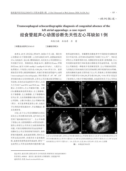
患者拟接受经导管心脏射频消融术,术前行常规经食管超声心动图(transesophageal echocardiography ,TEE )检查:0°、45°、90°、135°多切面扫查示左房内壁光滑,正常左心耳区域未探及明确左心耳结构,双房内未见血栓回声(图1);左房大小为62.7mm×45.4mm×54.8mm 。
TEE 提示:左房增大;左心耳缺如可能。
心脏CTA 横断面和容积再现示:右上肺静脉、右下肺静脉、左上肺静脉、左下肺静脉内径均正常,左房及肺静脉区域未见明显左心耳结构。
心脏CTA 提示:左心耳缺如可能(图2)。
术后患者恢复窦性心律,出院后予以利伐沙班抗凝治疗,并定期随访,暂未发现异常。
讨论:由于左心耳是房颤触发点的认识及左心耳封堵术的发展,近年来左心耳受到了越来越多的关注[1]。
左心耳来源于原始心房,在胚胎期第3~4周发育成形。
窦性心律者左心耳具有主动收缩和舒张功能,可探及规则的特征性血流频谱[2]。
房颤时左心耳正常的舒缩节律消失、整体舒缩功能减低,血流速度缓慢,同时左心耳形态复杂多样,内部具有丰富的梳状肌,血液容易淤积进而形成血栓,研究[3-4]也表明左心耳形态结构和功能参数与血栓形成密切相关。
非瓣膜性房颤患者卒中风险较非房颤患者至少升高5倍,且约90%的血栓栓子来源于左心耳[5]。
TEE 是评估左心耳最常用的方法,房颤患者在电复律、射频消融、左心耳封堵术前常需行TEE 检查以排除是否有血栓形成。
- 1、下载文档前请自行甄别文档内容的完整性,平台不提供额外的编辑、内容补充、找答案等附加服务。
- 2、"仅部分预览"的文档,不可在线预览部分如存在完整性等问题,可反馈申请退款(可完整预览的文档不适用该条件!)。
- 3、如文档侵犯您的权益,请联系客服反馈,我们会尽快为您处理(人工客服工作时间:9:00-18:30)。
经食管超声心动图对左心耳形态的分析吴晓霞 张凤羽 孟越之 马东星作者单位:100039 北京市,中国武警总医院超声科(吴晓霞、张凤羽),心内科(孟越之、马东星)通信作者:马东星 E-mail:madongxing2004@126.com摘 要目的 探讨经食管超声心动图(TEE)在左心耳(LAA)大小、分叶及形态分析中的应用价值。
方法 135例患者行TEE检查,男79例,女56例,年龄(53.7±19.1)岁,由2位观察者对LAA开口最大径、深度、分叶、形态进行单盲测量。
结果 2位观察者测量LAA开口最大径分别为(18.8±3.6)mm、(19.8±3.4)mm,深度为(28.2±5.6)mm、(29.2±5.2)mm,均具有统计学差异,经Bland-Altman分析TEE观察者之间一致性较好。
128例可观察LAA分叶,1叶47例、2叶63例、三叶13例、多叶5例,分别占36.7%、49.2%、10.2%、3.9%。
127例LAA中鸡翅型44例,仙人掌型31例,风向袋型29例,菜花型23例,分别占34.7%、24.4%、22.8%、18.1%。
结论 二维多平面TEE对LAA的大小、分叶及解剖形态的分析具有准确度及可重复性。
关键词经食管超声心动图 左心耳 心房颤动Assessment of the Morphology of the Left Atrial Appendage by Transesophageal EchocardiographyWu Xiaoxia,Zhang Fengyu,Meng Yuezhi,et al.Department of Ultrasound,the General Hospital of Chinese People′s Armed Police Force,Beijing 100039,ChinaAbstract:Objective To evaluate the application value of transesophageal echocardiography in the analysis of thesize dimension,lobes number and morphology classification of left atrial appendage(LAA).Methods All 135pa-tients were performed with TEE examination.TEE Images ware analyzed by two experienced echocardiogram expertsseparately to measure the max diameter of LAA orifice,the depth of LAA,the lobes number and to classify the mor-phology of LAA into 4types:chicken wing,cactus,windsock and cauliflower.Results The two separate observers′measurement results of the maximum diameter of LAA orifice were(18.8±3.6)mm and(19.8±3.4)mm,and ofthe depth of LAA were(28.2±5.6)mm and(29.2±5.2)mm,respectively,both with a statistical difference.Thetwo observers′measurement results exhibited a high level of agreement at Bland-Altman plot analysis.In 128patients,a 2-lobe LAA was most common(49.2%),followed by 1lobe(36.7%),3lobes(10.2%),and more than 3lobes(3.9%).In 127patients,the distribution of LAA morphologies was as follow:chicken wing type 44cases(34.7%),cactus type 31cases(24.4%),windsock type 29cases(22.8%)and cauliflower type 23cases(18.1%).Conclusions The 2DTEE is an accurate and reliable technique with reproducibility to analyze the size dimension,lobes numberand morphology classification of LAA.Key words:Transesophageal echocardiography,Left atrial appendage,Atrial fibrillation 心房颤动(房颤)是最常见的心律失常之一,其最主要的危害是因血栓形成导致动脉系统栓塞。
非瓣膜性房颤患者中90%的血栓来源于左心耳(left atri-al appendage,LAA)[1]。
经食管超声心动图(transesophageal echocardio-graphy,TEE)可以清晰显示LAA内部结构,区分血栓与梳状肌,二维多平面TEE的扫查可以有助了解LAA复杂的形态[2]。
Di Biase等[3]经磁共振及CT成像提出的LAA分型方法,将LAA外形分为鸡翅状、仙人掌状、风袋状和菜花状4种类型。
本研究着眼于应用二维多平面TEE对LAA的大小、分叶及解剖形态进行分析。
资料与方法1.研究对象2014年12月至2015年11月在我院就诊并接受TEE检查患者135例,男79例,女56例,年龄12~84岁,平均(53.7±19.1)岁。
其中房间隔缺损(atrial septal defect,ASD)28例,卵圆孔未闭合(patent oval foramen,PFO)8例,主动脉夹层1例,—414—中国超声医学杂志2016年5月 第32卷第5期 Chinese J Ultrasound Med Vol.32 No.5 May 2016房颤98例。
所有患者均在TEE检查前行经胸超声心动图、心电图和(或)动态心电图等检查。
心电图提示阵发性房颤患者41例,持续性房颤患者57例。
2.仪器与方法应用Philips iE Elite超声诊断仪,X7-2t经食管探头,频率2~7MHz。
患者于检查前禁食4~6h,口咽部予2%利多卡因凝胶局部麻醉,连接心电图,取左侧卧位,通过撑口器将探头置入食管内,深度30~40cm,逆时针转动探头调节角度,分别从0°~20°、45°~60°、80°~90°、120°~135°等角度清晰显示LAA结构,采集3个心动周期图像(房颤者5个心动周期)由2位观察者分别测量LAA开口最大直径及深度,观察LAA分叶及形态。
3.观察指标LAA开口、深度:以左上肺静脉入口与二尖瓣环连线作为LAA与左房间的分界线,从不同角度分别测量,选最大值作为LAA开口直径;在此平面上测量从开口到LAA主叶片顶部的距离为LAA深度。
LAA分叶:依照Veinot等[4]对LAA分叶定义和Yamamoto等[5]的研究:(1)LAA分叶是从LAA管状主体部结构分出、呈现明显的外翻、并常被外部褶皱分开的部分;(2)2mm的探针可以进入其内部(而不是其外部附着的脂肪组织);(3)分叶与LAA管状主体部在发出方向上可以发生变化,但非必须;(4)分叶与LAA管状主体部可以在不同解剖平面上;(5)LAA至少≥1叶。
按照上述定义,本研究中TEE分辨的LAA分叶其内径≥2mm。
LAA形态[6]:(1)菜花型:LAA长度短小而内部结构复杂,可由不同数目的叶片组成,而无占主导的叶片,开口则多为不规则形状;(2)仙人掌型:有1个突出的中心叶片,副叶片可从中心叶的上端和下端的不同方向伸展发出;(3)风向袋型:有1个足够长度的主叶片作为一级结构,可有第2~3个叶片从不同位置发出;(4)鸡翅型:有1个主叶,在主叶的近端或中部出现明显的弯曲反折回开口方向,也可有第2个叶片。
4.统计学方法采用SPSS 21.0统计软件进行数据分析,计量资料以(珚x±s)表示,正态分布方差齐者两两比较采用配对t检验,方差不齐两个独立样本间采用秩和检验,观察者之间一致性采用Bland-Altman分析,P<0.05为差异有显著性。
结 果1.LAA开口内径及深度2位观察者单盲测量LAA开口最大径分别为(18.8±3.6)mm、(19.8±3.4)mm,二者均呈正态分布,配对t检验示二者之间差异具有统计学意义(P<0.05);LAA深度分别为(28.2±5.6)mm、(29.2±5.2)mm,配对t检验示测值之间差异具有统计学意义(P<0.05)。
Bland-Altman散点图(图1)显示两观察者测量LAA开口径的平均差值仅为0.7mm,且94.1%(127/135)的点在95%的一致性界限以内;两观察者测量LAA深度的平均差值为0.6mm,且94.8%(128/135)的点在95%的一致性界限以内。
两观察者测值在临床实际应用的意义上显示出很好的一致性。
上图:LAA开口径;下图:LAA深度。
图1 Bland-Altman散点图2.LAA分叶及形态判别135例LAA分叶经2位观察者分别判读:2例因图像质量欠佳不能判断,5例存在分歧,128例判读一致,47例为1叶、63例2叶、13例三叶、5例多叶,分别占36.7%、49.2%、10.2%、3.9%。
135例LAA形态:2例不能判断,6例存在分歧,127例判读一致,鸡翅型44例,仙人掌型31例,风向袋型29例,菜花型23例,分别占34.7%、24.4%、22.8%、18.1%。
讨 论房颤患者发生卒中的风险比非房颤者高5倍,大—514—中国超声医学杂志2016年5月 第32卷第5期 Chinese J Ultrasound Med Vol.32 No.5 May 2016多数房颤相关卒中事件起源于LAA小梁部。
