
FSN超声波处理器说明书
目录
1、概述 (2)
2、工作原理 (2)
3、整机结构名称 (2)
4、电气原理 (3)
5、产品特点.......................................................................................................3-4
6、使用条件 (4)
7、安装操作指南 (5)
8、维护保养 (5)
9、配套清单 (5)
9、变幅杆更换...................................................................................................6-7
上海生析超声仪器有限公司
1、概述
上海生析超声仪器有限公司是一家集研究、设计、生产、销售以及维修超声波设备的专业产家。
生析公司有着集十多年生产超声波设备经验,设计及生产多种规格超声设备,产品种类有台式超声波清洗器、工业超声波清洗、超声波细胞破碎仪、超声波乳化仪、超声波处理器、超声波塑料焊接机、超声波换能器等等。
其产品广泛应用于全国高等院校的实验室、光学工业、珠宝首饰、航天工业、五金工业、汽车制造工业。
生析超声仪器有限公司以先进雄厚的技术力量,不断开发新产品,“高技术、高品质、优质的售后服务” 是生析公司的宗旨。
它将为广大客户提供全面的超声设备与服务。
2、工作原理
超声换能器震动时产生的超声波作用于液体时,液体中每个气泡的破裂会产生能量极大地冲击波,相当于瞬间产生几百度的高温和高达上千个大气压,这种现象被称为“空化效应”。
超声空化是强超声在液体媒质中引起的一种特有的物理过程,它伴随有许多奇妙的现象和惊人的效应。
空化基本效应表现为高温效应,放电效应,发光效应,以及冲击、压力效应等等。
人们正是利用这些独特的效应而广泛应用于医药、生物、化学、物理等领域。
3、整机结构名称
前面板后面板
①连接线缆②套筒③换能器④变幅杆⑤液晶控制器
⑥仪器机箱⑦输出插座(连接换能器)⑧仪器铭牌⑨保险丝座
⑩总电源插座(11)总电源开关(12)输入插座(连接温度探头)(13)过流保护指示
4、电气原理
1.发生器
本发生器的原理方框示意图:
超声电路结构
2.换能器
所采用的压力换能器是夹心单螺钉结构,不同型号和功率的换能器配用不同规格和数量的电压陶瓷片。
①高压强度换能器专用紧固螺丝②电极片③电压陶瓷片
④变幅杆⑤连接螺丝
5、产品特点
1.自动谐振点和功率控制无需经常手动调节能量
2.99小时过程控制定时器控制总工作时间:从1秒钟到99小时
3.工作时间显示呈累计状态
4.独立开/关脉冲定时器确保高强度处理温度敏感样品
5.自动振幅补偿确保在超声过程中探头振幅不因承载变化而变化
6.操作简便菜单操作,直接键入
六、使用条件要求
1.环境温度:0~40℃
2.相随湿度:不大于85%(T=20℃)
3.使用电源:1P/220V/50Hz±10%
4.工作条件:最长连续1.0min(超声的连续工作时间一般不超过10分钟/次)
5.换能器温度:0~120℃
6.处理对象:溶液或溶液的混合液
七、安装操作指南
①定时启动/关闭按键②超声工作状态指示灯③定时设定按键
④数字减键⑤数字加键⑥、⑦移位键⑧暂定键⑨功率增大键⑩功率减小键
(11)液晶显示器(12)模式转换键
操作步骤
时间控制器是采用数字电路和微机控制技术设计制造的,它具有精度高、可靠性好,抗干扰能力强和记忆功能等特点,其全量程时间控制范围1秒钟至99小时。
用户可随意设定。
电源连接
用户在使用前务必先将超声波换能器与超声波发生器连接好。
用隔音箱搁住套筒(也可用支架固定住套筒或用手握住在换能器套筒部分,但不能碰换能器部位),将变幅杆头部侵入溶液中5-10毫米。
开启机箱后面板的电源开关,液晶显示器工作。
功率调节
∧、∨键是超声功率设置键,按∧键可以增大超声波输出功率,按∨键可以减小超声波输出功率。
超声时间
1、全数字化电子定时器为1秒~99小时。
2、SET键具有设定时间、确认记忆功能。
3、按SET键,由增大键、减小键和移位键任意键入所需超声时间,定时(总时间过程)设定完成。
4、按SET键,由增大键移位键任意键入所需的脉冲超声时间,再按SET键,由增大键、减小键和移位键任意
键入所需的关闭脉冲超声时间,继续按SET键即为确认并记忆设定的参数。
5、ON/OFF键具有两功能,即启动和关闭超声,超声波时间启动时,SET键不起作用(只有在关闭超声波的状
态下,才可以重新设定时间),按STOP(暂停)键可以暂时停止当前运行任务,再次按STOP(暂停)键可以继续当前运行任务。
6、MODEL键为模式转换键,可将超声模式设定为连续模式和脉冲模式,当超声模式从脉冲模式转换为连续模
式时,必须按ON/OFF键,方可启动(有定时)连续模式,否则超声处于无定时连续模式。
当超声模式从连续模式转换为脉冲模式时,则无需以上操作。
7、使用过程请不要触摸超声波变幅杆,以免被超声波产生的热量灼伤。
用户在使用完毕后务必关闭和切断电源。
八.维护-保养
1、本设备应安置在清洁、干燥、通风处使用。
2、轻拿轻放振子以防振动棒断裂。
3、当振子发烫时,应等其冷却后在使用。
4、使用完毕后应拔掉电源插头,切断电源。
5、本设备必须在220V/50Hz±10%范围工作。
6、设备发生故障时,应及时与厂商联系,而不得邮由非专业技术人员盲目随意维修。
九、仪器配套清单:
序号名称数量
1 主机1台
2 超声波探头1支
3 电源线1根
4 电缆线1根
5 保险丝3个
6 使用说明书1份
7 隔音箱、升降台1套
十、变幅杆安装与拆卸
1、安装变幅杆,将换能器正放在桌上,左手握扳手①卡主换能器的铝合金部分,右手握扳手②卡主换能器的变幅杆部分,左手顺时针用力,右手逆时针用力;如需安装略紧,可将换能器垂直正放,用夹具将变幅杆夹紧固定(夹紧时可用较软的垫片保护变幅杆表面),用扳手③卡主换能器的铝合金部分,顺时针拧紧!如下图:
2、拆卸变幅杆,将换能器倒置放在桌上,左手握扳手④卡主换能器的铝合金部分,右手握扳手⑤卡主换能器的变幅杆部分,左手顺时针用力,右手逆时针用力;如未拆卸开,可将换能器垂直正放,用夹具将变幅杆夹紧固定(夹紧时可用较软的垫片保护变幅杆表面),用扳手⑥卡住换能器的铝合金部分,逆时针松开!如下图:。
超声波说明书

超声波使用说明书模块实物图如下图所示:123(12V+)(GND)(485通信口)一、接线说明模块采用DC12V供电;图中标号为1的红色箭头处接电源正,标号为2的红色箭头处接电源负。
标号为3的红色箭头处接485通信线。
二、通信协议说明模块采用485通信,波特率为9600;通信协议如下:协议头预留字节预留字节预留字节距离高字节距离低字节帧尾校验和0Xfe0X000X000X000Xxx0Xxx0Xef0Xxx校验和计算方法:从协议头到帧尾个各数据之和取最低两位距离返回值单位为:mm;例:0xfe0x000x000x000x040xb00xef0xa1当返回数据如上所示时,测距结果为1200mm。
三、模块测量方法由于模块是高精度设计用,所以对测量环境有严格的说明界定。
请参考如下测量安装方式,否则会产生较大的测量误差。
安装方式如下:滤波处理装置,可以用PCB板来固定代替移动平面是本模块的移动方向,反射平面是基础高度。
在移动平面方向前后运动将会测量到实际的反射平面与模块的实际距离。
测量条件:1:参考平面(模块固定的平面)一定要垂直于反射平面。
2:测量模块在反射面这个方向上的1米半径范围内不能有遮挡物。
3:在移动模块到了指定位置后(想测量的距离位置处),模块的前端一定要绝对垂直于反射面。
4:根据超声波的扇形反射面原理,最好在超声波的接收端安装反射能量限定装置,这样能更准确的获得实际的测量结果。
在实际应用场合,有更多的安装要求。
基本的测量测试,照目前条件能基本反应出来。
成都镓舒适科技有限公司2014-09-01。
超声波处理器的技术原理及操作步骤

超声波处理器技术原理:
1、操作语言:英文;
2、样品过载、过温、故障自动报警保护装置;
3、采用彩色工业触摸屏控制器,各种功能集成显示;
4、科学的设计,10组用户存储设定,单机直接实现;
5、设备配有温度传感器,可对样品进行温度检测;
6、自动谐振点,在19-26KHZ的频率范围内自动扫频跟踪;
7、自动幅度补偿,可根据样品的流动性自动工作于频率点和输出点;
8、独立开关脉冲,附连续使用、脉冲、定时连续超声三种方式;
9、三维超声探头的设计,与传统超声相比,效率大大提高,变幅杆的损耗减小;
10、可以看到的运行界面显示:总运行时间、工作时间、停车时间、用户组、过载温度、输出功率比、工作模式、程序保存键;
IK主界面显示参数:超声波输出功率、频率、工作方式、用户组、设定键、设备运行状态、开/关、设备运行参数等。
超声波处理器操作步骤:
采用数字电路和微机控制技术设计制造的时间控制器,具有高精度、高可靠性、抗干扰能力强、记忆功能,其全量程时间控制范围从1秒到99小时。
使用者可以任意设定。
电力连接。
使用前一定要先将超声波传感器与超声波发生器连接好。
利用隔音器固定套筒,将变幅杆头插入溶液中5-10毫米。
打开电源开关盒后面
板,1CD工作。
用法说明:
1、本设备应放置在清洁、干燥、通风的地方;
2、振动棒轻放轻放,防止折断;
3、当振子发烫时,应等其冷却后再使用;
4、用完后请拔下插头,切断电源;
5、本设备的工作范围必须在220V∕50Hz±10%范围内;
6、设备发生故障时,应及时与厂家联系,但不能交给非专业技术人员盲目随意维修。
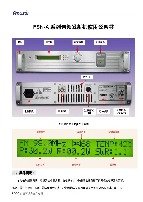
FSN-A系列发射机使用说明书
图四 图五为音量设定介面,音量设定范围为 0-13
图六为输出功率设节菜单
图五
图六 图七为功率设定介面,功率设定范围为 0-30W,步进 0.1W。80W 以上步进 1W。
图七 图八为立体声单声道设定菜单
图九为声道设置介面
图八
立体声
单声道 图十为温度设置菜单
图九
图十 图十一为温度设置介面,温度可设定范围为 60-70 摄氏度,步进 1 摄氏度。
二,功能设定
调节旋钮往左旋转向低端调节,往右旋转向高端调节,在主介面下按下进入 MENU,在 MENU 下按下为进 入设定介面,在设定介面按下为确认设定并返回 MENU。 图二为频率设定菜单
图二
图三为频率设定介面,频率设置范围为 87.5-108MHZ(特殊频率可以按客户要求定制,详请与本公司联系)
产品售出后,由我方负责免费保修 1 年(人为拆卸,烧毁除外)。售后保修日期以发货日期为准。 为所有客户提供全面的 24 小时技术支持。 为客户购买的产品架设和布局提供参考建议。
环境温度过高。 开机 LCD 无显示,LED 电源灯不亮。请检查发射机电源插座的保险丝是否熔断。 接收到的信号有电流声:请避免电源线、射频输出线和音频线相互交织在一起和远离周围电器干扰源。音频
信号输入线一定要用屏蔽线。
收音机接收时有破音现象:请将发射机音源输入音量调整适当。
五,售后保修服务
图二 进入主介面后,将实时显示工作Байду номын сангаас率,音量,FETMOS 工作温度,输出正向功率,输出反向功率和驻波比。 注意进入主介面后,输出功率将从 0 瓦开始向上调节,约 30 秒左右达到设定的最大功率。 在驻波或温度保护作用时,输出功率将自动降到 1 瓦以下并在屏幕显示报警信息,在故障排除后,输出功率将自 动调整到设定的输出功率。
超声波泥水界面仪使用说明书
前言非常感谢您选择本公司仪器!在使用本产品前,请详细阅读本说明书,请遵守本说明书操作规程及注意事项,并保存以供参考。
◆由于不遵守本说明书中规定的注意事项,所引起的任何故障和损失均不在厂家的保修范围内,厂家亦不承担任何相关责任。
请妥善保管好所有文件。
如有疑问,请联系我公司售后服务部门。
◆如果您需要电子版说明书,请登陆本公司网站下载,或拨打服务热线,联系我公司售后服务部门。
◆在收到仪器时,请小心打开包装,检查仪器及配件是否因运送而损坏,如有发现损坏,请联系我公司售后服务部门,并保留包装物,以便寄回处理。
◆当仪器发生故障,请勿自行修理,请联系我公司售后服务部门。
◆本说明书适用于V4.00系列的产品版本。
以下标识将会在本手册或者仪器上出现:注意保险丝接地端公司简介大连因斯特科技有限公司是专注于自动化领域的仪器仪表设计、制造、销售、安装、售后服务为一体的现代化高新技术企业,公司与国内外知名仪表企业精诚合作,采用进口原件研制生产具有国内领先、国际先进的自控仪表产品,开发“因斯特”品牌系列分析、流量、液位、压力等在线监测产品,长期与国外诸多知名仪表企业进行技术交流合作,产品不但性能品质过硬,还融入了符合中国思维模式的操作菜单界面。
产品不断更新换代,自投入市场以来,广泛应用于自来水、污水处理、石油、化工、电力、冶金、环保、制药等行业,得到了广大用户的一致好评。
公司拥有高级职称技术人员十余名,并长期与大连工业大学等高校合作,为企业不断输入技术、销售等多方面人才,确保满足不同客户的服务需求。
公司自主研发、生产、营销:PH计、ORP仪、化学膜溶解氧(DO)、荧光法溶解氧(DO)、浊度计(SS)、余氯检测仪、电导率、光电污泥浓度计(MLSS)、超声波污泥浓度计、超声波泥水界面仪、超声波液位计、超声波液位差计、超声波明渠流量计、电磁流量计(DN15-DN2000)、超声波流量计、COD 在线监测仪、氨氮在线监测仪、总磷(TP)在线监测仪、总氮(TN)在线监测仪、总磷总氮一体机、六价铬在线检测仪、总铜在线分析仪、总镍在线分析仪、总铬在线分析仪、总镉在线分析仪、总砷在线分析仪、总铅在线分析仪、总汞在线分析仪、总锰在线分析仪、挥发酚在线分析仪、氰化物在线分析仪、氟化物在线分析仪。
FSN超声波处理器说明书

目录1、概述 (2)2、工作原理 (2)3、整机结构名称 (2)4、电气原理 (3)5、产品特点.......................................................................................................3-46、使用条件 (4)7、安装操作指南 (5)8、维护保养 (5)9、配套清单 (5)9、变幅杆更换...................................................................................................6-7上海生析超声仪器有限公司1、概述上海生析超声仪器有限公司是一家集研究、设计、生产、销售以及维修超声波设备的专业产家。
生析公司有着集十多年生产超声波设备经验,设计及生产多种规格超声设备,产品种类有台式超声波清洗器、工业超声波清洗、超声波细胞破碎仪、超声波乳化仪、超声波处理器、超声波塑料焊接机、超声波换能器等等。
其产品广泛应用于全国高等院校的实验室、光学工业、珠宝首饰、航天工业、五金工业、汽车制造工业。
生析超声仪器有限公司以先进雄厚的技术力量,不断开发新产品,“高技术、高品质、优质的售后服务” 是生析公司的宗旨。
它将为广大客户提供全面的超声设备与服务。
2、工作原理超声换能器震动时产生的超声波作用于液体时,液体中每个气泡的破裂会产生能量极大地冲击波,相当于瞬间产生几百度的高温和高达上千个大气压,这种现象被称为“空化效应”。
超声空化是强超声在液体媒质中引起的一种特有的物理过程,它伴随有许多奇妙的现象和惊人的效应。
空化基本效应表现为高温效应,放电效应,发光效应,以及冲击、压力效应等等。
人们正是利用这些独特的效应而广泛应用于医药、生物、化学、物理等领域。
3、整机结构名称前面板后面板①连接线缆②套筒③换能器④变幅杆⑤液晶控制器⑥仪器机箱⑦输出插座(连接换能器)⑧仪器铭牌⑨保险丝座⑩总电源插座(11)总电源开关(12)输入插座(连接温度探头)(13)过流保护指示4、电气原理1.发生器本发生器的原理方框示意图:超声电路结构2.换能器所采用的压力换能器是夹心单螺钉结构,不同型号和功率的换能器配用不同规格和数量的电压陶瓷片。
超声波测速仪用户手册说明书

MechanicsAcousticsDebye-Sears EffectDETERMINE THE VELOCITY OF ULTRASONIC WAVES IN LIQUIDS.UE1070550 04/16 JSBASIC PRINCIPLESThe diffraction of light by ultrasonic waves in liquids was predicted by Brillouin in 1922, and the effect was con-firmed experimentally in 1932 by Debye and Sears and also by Lucas and Biquard . It is caused by the periodic variations in the refractive index of the liquid that are produced by ultrasonic waves. If a light beam is passed through the liquid perpendicular to the ultrasound direc-tion, the arrangement acts as a phase grating, which moves depending on the velocity of sound. Its grating constant corresponds to the wavelength of the ultra-sound, and thus depends on its frequency and the veloci-ty of sound in the medium. The movement of the phase grating can be neglected if the effect is observed on a screen at a large distance.In the experiment, a vertically orientated generator couples ultrasonic waves at frequencies between 1 MHz and 12 MHz into the test liquid. A monochromatic parallel light beam pass-es through the liquid in the horizontal direction and is diffract-ed by the phase grating (see fig. 1). The diffraction pattern contains several diffraction maxima spaced at regular dis-tances (see fig. 2).The k th-order maximum of the diffraction pattern is found at the diffraction angle αk , defined by(1) SLk tan λλ⋅=αk λL : ight wavelength, λS : ultrasound wavelength.Thus, the ultrasound wavelength λS can be determined from the separation between the diffraction maxima. Furthermore, according to the relationship(2) S λ⋅=f cit is possible to calculate the velocity of sound c in the liquid, since the frequency f of the ultrasonic waves is also known.Fig. 1: D iagram showing the diffraction of light by a phasegrating that is produced in a liquid by ultrasonic wavesFig. 2: D iffraction pattern caused by the diffraction of light at aphase grating produced in a liquid by ultrasonic wavesLIST OF APPARATUS1 Ultrasonic cw Generator with Probe 1002576 (U100061) 1 Test Vessel, Complete 1002578 (U10008)1 Laser Diode for D-S Effect, Red 1002577 (U10007)1 Laser Diode for D-S Effect, Green 1002579 (U10009)1 Pocket Measuring Tape,2 m 1002603 (U10073)1 Ultrasonic Coupling Gel 1008575 (XP999)SET-UP∙Fill the test vessel with distilled water and place it about3 m from the projection screen.∙Mount the multi-frequency probe vertically in the holder of the test vessel and connect it to the PROBE output of the ultrasonic generator (see Fig. 3).∙Mount the red laser diode in the laser holder of the test vessel and connect it to the LASER output of the ultra-sonic generator.Fig. 3: Experiment set-up for the diffraction of light at a phase grating produced in a liquid by ultrasonic waves EXPERIMENT PROCEDURE∙Measure the distance s between the multi-frequency probe and the screen.∙Switch on the ultrasonic cw generator.∙Switch on the laser and the multi-frequency probe.∙Set the frequency to 1 MHz.∙Adjust the amplitude of the transducer signal, and by means of the three adjusting screws of the transducer holder adjust the orientation of the multi-frequency probe so that standing waves are generated.∙On the screen measure the distance x2k between the –k th order and the +k th order diffraction maxima.∙Increase the frequency in steps of 1 MHz up to 12 MHz, and for each frequency measure the distance x2k and de-termine the diffraction order k.∙Replace the red laser diode with a green one and make a similar set of measurements. SAMPLE MEASUREMENTS AND EVALUA-TIONs =325 cmTable 1: Experiment data with light of wavelength λL = 652 nm (red laser)Table 2: Experiment data with light of wavelength λL = 532 nm (green laser)It is necessary to measure the distance s between the ultra-sound generator and the screen used to observe the diffrac-tion pattern, and the distance x2k between the -k th and the +k th diffraction maxima. From these two distances, it is possi-ble to calculate the diffraction angle αk for the k th-order maxi-mum, given by:sx⋅=α2tan2kk.This leads to the following equation for determining the ultra-sound wavelength λS:L2kS2λ⋅⋅⋅=λxsk.This is the equation by which the sound wavelengths in the right-hand column of both tables were calculated.3B Scientific GmbH, Rudorffweg 8, 21031 Hamburg, Germany, Figure 4 shows the calculated sound wavelength as a function of the frequency of the ultrasonic waves. The hyperbola curve was calculated according to Equation (2) as:fc =λS with s m 1482=cThe velocity of sound c determined by this curve-fitting proce-dure is in excellent agreement with the literature value.510f / M H zλ μ/ mFig. 4: S ound wavelength λS in water as a function of the fre-quency f。
超声机Aixplorer MACH 20产品说明书
AIXPLORER MACH 20 UltraFast TM IntelligenceIntelligence and Innovationin UltrasoundAixplorer MACH® 20 ultrasound systems leverage 10 years of clinical expertise to help you handle exams with ease and confi dence.Understanding your everyday challenges, the Aixplorer MACH® 20 performance meets innovation with leading edge UltraFast™ technology.The UltraFast™ technology allows for the acquisition up to 20,000 frames per second1, this technology off ers new possibilities for patient management. The next generation of the UltraFast™ technology — has 5x more computing power2.21 - Bercoff J, Ultrafast Ultrasound Imaging. Ultrasound Imaging - Medical Applications.2011 Aug; DOI: 10.5772/197292 - In comparison with Aixplorer® MultiWave™ systems AN ECO-DESIGNEDPRODUCTThe company is certifiISO 14001. This certificonfi rms that the companyhas voluntarily implementedan environmental management policy, demonstrating a strongcommitment to minimizeenvironmental impactthroughout theproduct’ life cycle.Image Quality for Improved Diagnostic Confi denceDesigned for optimal sensitivity and operator comfort, our multi-application transducer family can also be used on other Aixplorer MACH® 20 products.3High Quality B-Mode ImagingAixplorer MACH® 20 off ers excellent B-mode image quality with incredible defi nition in both fundamental and harmonic imaging modes.Designed to fi t the needs of radiologists, a portfolio of advanced features is availableto improve image quality, resolution and contrast.Innovative Imaging ModesShearWave™ PLUS (SWE TM PLUS) elastography is capable of visualizing, analyzing and quantifying the tissue stiffness in real-time 1,2,3. This non-invasive approach is clinically proven to be reliable and highly reproducible.Aixplorer MACH® 20 gives you the flexibility to benefit from proprietary ShearWave™ PLUSelastography on the transducers of your choice.4Key Attributes:• Increased SWE TM PLUS frame rates • Accelerated filling of the SWE TM PLUS box• Increased penetration to visualize deep lesions• Preserving the quality of theB-mode1 - Cosgrove D, Berg W, Doré J et al. Shear wave elastography for breast masses is highly reproducible. European Radiology. 2012 May; 22(5): 1023–1032.2 - Hudson J, Milot L, Parry C et al. Inter-and intra-operator reliability and repeatability of shear wave elastography in the liver: a study in healthy volunteers. Ultrasound Med Biol. 2013 Jun;39(6):950-53 - Garcovich M, Veraldi S, Di Stasio E et al. DLiver Stiffness in Pediatric Patients with Fatty Liver Disease: Diagnostic Accuracy and Reproducibility of Shear-Wave Elastography. Radiology . 2017 Jun; 283(3):820-827.4 - Peer Reviewed Articles ShearWave™ Elastography for Breast Imaging. MKG.EC.3355 - Peer Reviewed Articles ShearWave™ Elastography for Liver and Abdominal Imaging. MKG.EC.3376 - Peer Reviewed Articles ShearWave™ Elastography for Musculo-Skeletal System. MKG.EC.3377 - Correas J-M, Tissier A-M, Khairoune A et al. Prostate Cancer: Diagnostic Performance of Real-Time Shear-Wave Elastography. Radiology 2015 Apr;275(1):280-9.Angio PLanewaveUltraSensitive Imaging (Angio PL.U.S.)Angio PL.U.S. is a color mode that offers enhanced sensitivity and resolution of blood flow. Angio PL.U.S. provides the possibility to explore microvascular flow.TriVu™ ImagingTriVu is a real-time imagingmode that allows you to display morphology (B-mode), stiffness(SWE PLUS) and flow information (equivalent to Angio PL.U.S.), all inthe same image, simultaneously.TriVu is the answer to confident and timely diagnosis.Needle PLUS™Needle PLUS™ addresses the challenge of limited needle visibilityand the need to predict the needle trajectory. This real-time imagingmode allows you to perform biopsies with precision and confidencewithout loss of B-mode information.5General ImagingBreastWith over 200 publications in peer-reviewed medical journals, SWE has been proven to be a complementary tool for: breast lesion diagnosis and characterization 1, biopsy planning 2 and treatment; therapy monitoring 3 and prognostics. Allowing to performexams with ease and confidence for all breast morphologies.Muscles and T endonsShearWave PLUS with its unique ability to analyze tissue stiffness (up to 1,200 kPa or 20 m/s), and in real time, is an asset for tendinopathy assessment and muscle disorders quantification. By adding innovative imaging modes, such as Angio PL.U.S. and Needle PLUS™, ultrasound exams benefit from complementary diagnostic information.LiverAixplorer MACH® 20 offers a suite of diagnostic imaging tools for non-invasiveassessment and follow-up of liver diseases. The utility of SWE in the management of patients with chronic liver disease has been demonstrated in more than 160 clinical publications for evaluation 4 and diagnosis 5 of hepatic fibrosis and follow-up and monitoring of patients.Masculine HealthIn addition to conventional ultrasound modes, ShearWave™ PLUS in real time andAngio PL.U.S. make ultrasound a multi-parametric modality. Thus, it can be used for the detection and characterization of prostate and testicular lesions. Targeted biopsies can also be performed with confidence and precision.Ob GynThe advanced visualization capabilities of Aixplorer MACH® 20 let you clearly see fine morphological structural details of the ovaries, adnexae and endometrium, including difficult cases such as a fibroid uterus 6, 7. The system reveals the smallest of fetus structures and let you explore morphology and detect abnormalities in early stages of the pregnancy.61 - Berg WA, Cosgrove DO, Doré CJ, et al. Shear-wave elastography improves the specificity of breast US: the BE1 multinational study of 939 masses. Radiology. 2012 Feb;262(2):435-49.2 - Mullen R, Thompson JM, Moussa O et al.Shear-wave elastography contributes to accurate tumour size estimation when assessing small breast cancers. Clin Radiol. 2014;69(12):1259–1263.3 - Lee SH, Chang JM, Han W, et al. Shear-Wave Elastography for the Detection of Residual Breast Cancer After Neoadjuvant Chemotherapy. Ann Surg Oncol. 2015;22 Suppl 3:S376–S384.4 - Gao Y, Zheng J, Liang P, et al. Liver Fibrosis with Two-dimensional US Shear-Wave Elastography in Participants with Chronic Hepatitis B: A Prospective Multicenter Study. Radiology. 2018 Nov;289(2):407-415.5 - Garcovich M, Veraldi S, Di Stasio E, et al. Liver Stiffness in Pediatric Patients with Fatty Liver Disease: Diagnostic Accuracy and Reproducibility of Shear-Wave Elastography. Radiology. 2017;283(3):820–827.6 - Engineering Clinical Evaluation (Ece) V10 Endocavity Probes Evaluation in Gynecology Dr Shojai Aix En Provence; PM.TP/TR.0347 - V10 CMR Validation – Institut de Radiologie de Paris – Gynecology; PM.TP/TR.0361. Widescreen 21.5” Full HD monitorImage uniformity, deep blacks and refined detail2. Large 13.3” Full HD touch displayMore flexibility to define your workflow3. Intuitive control panelwith revolutionary SonicPad TM Improved user experience and workflow4. Low noise level, optimized cooling fan architecture Reduced noise for all environments5. Reduced footprint Suitable for any practiceDesigned to be the New Standard734512SuperSonic ImagineFor more information contact:+33 (0)4 42 99 24 24******************************M K G .E C .197 R e v A © S u p e r S o n i c I m a g i n e - A p r i l 2020Indications for Use: The SuperSonic Imagine Aixplorer MACH® range ultrasound diagnostic systems and transducers are intended for general purpose pulse echo ultrasound imaging, soft tissue viscoelasticity imaging and Doppler fluid flow analysis of the human body. The Aixplorer MACH® ultrasound diagnostic systems are indicated for use in the following applications, for imaging and measurement of anatomical structures: Abdominal, Small Organs, Musculoskeletal, Superficial Musculoskeletal, Vascular, Peripheral Vascular, Intraoperative, OB-GYN, Pelvic, Pediatric, Transrectal, Transvaginal, Urology, Neonatal/Adult Cephalic and Non-invasive Cardiac. In addition, the SuperSonic Imagine Aixplorer MACH® ultrasound diagnostic systems and associated transducers are intended for: measurements of abdominal anatomical structures; measurements of broadband shear wave speed, and tissue stiffness in internal structures of the liver and the spleen; measurements of brightness ratio between liver and kidney; visualization of abdominal vascularization, microvascularization and perfusion; quantification of abdominal vascularization and perfusion. The shearwave speed, beam attenuation, viscosity and stiffness measurements, the brightness ratio, the visualization of vascularization, microvascularization and perfusion, the quantification of vascularization and perfusion may be used as an aid to clinical management of adult and pediatric patients with liver disease. It is intended for use by licensed personnel qualified to direct the use of the medical ultrasound devices. CE certificate no. 26415, FDA cleared K180572.2020 Hologic Inc., All rights reserved. Hologic, SuperSonic, UltraFast, Aixplorer, Aixplorer MACH, Angio PL.U.S., Needle PL,US, NL SWE™, Planewave UltraSensitive, ShearWave, SonicPad, SWE and associated logos are trademarks and/or registered trademarks of Hologic, Inc., and/or its subsidiaries in the United States and other countries. This information is intended for medical professionals in the U.S. and other markets and is not intended as a product solicitation or promotion where such activities are prohibited. Because Hologic materials are distributed through websites, eBroadcasts and tradeshows, it is not always possible to control where such materials appear. For specific information on what products are available for sale in a particular country, please contact your local Hologic representative.。
超声波说明书内容
FUF-2000 基本型号
注: 开箱检查
FUF-2000P 系列便携式超声流 量表 FUF-2000-WJP1 外夹便携式 FUF-2000- WJP2 外夹笔记本便 携式 FUF-2000- WJP3 外夹手持式
FUF-2000F 系列在线式超声流量表
FUF-2000-FC 插入式 FUF-2000-FG 法兰式
OCT
OCT
RS232 接 口
模 拟
模 拟
一
继
继
电
电
电 23 5
源
+
输 出
输 出 一
集 电 级
发 射 级
器 输 出
器 输 出
+
模
模
模
模
上
上
下
下
拟
拟
拟
拟
游
游
游
游
电输
输
输
输
模
模
探
探
入
入
入
入
头
头
地
头
头
源地
3;
AI3
AI2
AI1
� 红
一 � 黑
+
� 红
一 � 黑
供电电源 通电运行
FUF-2000 型超声波流量表的供电电源可接入直流 8~36V 或交流 7~30V 范围内 的电源都能正常连续工作。 使用直流电源的用户订质时应注明
TIME DELTA
TIME DELTA
DSF 打印式
160×160×65 DSF 一体式
二 安装调试与测量操作
FUF-2000 联线
FUF-2000P 手 提 式 、FUF-2000P 手持式流量表在使用 AC220V 电源时联
西门子超声波转换器fus_sono
SOUND VELOCITY
SOUND VELOCITY MIN.
SOUND VELOCITY MAX.
声速的测量单位是 米/秒,通过输入最低和最高声速来设置测量范围。 . 如果声速超过所述的范围,便激励错误继电器动作或错误指示.
DKFD.PS.029.Q3.02
39
SITRANS F US SONOFLO 7. Starting up 方框图
PULSE UNIT
VOLUME PULSE XXXXXXXX UUUU
PULSE WIDTH XXXX UUUU
频率输出特性 characteristics
SOUND VELOCITY
PULSE UNIT: UNIT: kg UNIT: kg*10 UNIT: kg*100 UNIT: ton UNIT: lb UNIT: lb*10 UNIT: lb*100
在转换器设置菜单里, 前进键 此键为各菜单间的向前翻页键。 后退键 此键为各菜单间的向后翻页键。 改换键 此键用于改变设置或数值 选择键 此键用于选择数值的数位 锁定/开锁键 此键用于确定改变设置值或进入子菜单。
以复位方式使累加器均复位,进行调零并能使复位至工厂设置。 .
注意: 用户密码、校正因数、语言选择和自动零位均不能通过复位方式,返回到工厂设置 .
服务模态提供了把输出设置成固定值(强制输出)的模式。一旦脱离了服务模式, 原按服务模式进行设置的参数均被取消。
可以选择菜单语言。
在此菜单中能够选择操作员可访问信息。
声速范围定义是在质量流量子菜单中,选择校准质量流量,并输入声速1#和声速2#。当声 速超出音速1和音速2所规定的范围时,继电器将接通。 .
当已经选择了“故障”时,最低和最高声速均是错误信息部分。
超声波使用说明书
N&DN系列(LCD)超声波细胞粉碎机使用说明书宁波新芝生物科技股份有限公司地址:宁波市国家高新技术园区木槿路65号 315013电话:传真:网址:一. 概述随着生物产业的发展,应用超声波细胞粉碎机所做的实验要求也随之提高,如对样品温度的测定、控制,低温冷却样品及整机的智能化程度的提高等等,都提出了新的要求,为进一步完善此类仪器的各项性能,我公司在现有各种型号的超声波细胞粉碎机的基础上,吸收国外最新技术,结合微电脑控制、选频、测温、保护等软硬件技术而研制的超声波细胞粉碎机,它具有技术先进、性能可靠、操作简便、外型美观、显示清晰明亮、测温控温精确等优点。
超声波细胞粉碎机是一种利用强超声在液体中产生空化效应,对物质进行超声处理的多功能、多用途的仪器,能用于动植物组织、细胞、细菌、芽胞菌种的破碎,同时可用来乳化、分离、分散、匀化、提取、脱气、清洗及加速化学反应等等。
该机广泛应用于生物化学、微生物学、药物化学、表面化学、物理学、动物学、农学、医学、制药等领域教学、科研、生产。
Ф18 3/4”20-25KHz 200-950W 200 - 500mlФ20 3/4” -1000mlФ25 1” -1200ml本机由超声波发生器和超声波换能器组件两大部分组成。
超声波发生器(电源)是将220VAC、50Hz的单相电通过变频器件变为20-25kHz、约600V的交变电能、并以适当的阻抗与功率匹配来推动换能器,作纵向机械振动,振动波通过浸入在样品溶液中的钛合金变幅杆对被破碎的各类细胞产生空化效应,从而达到破碎细胞之目的。
其电原理由整流电源,开关电源、变频系统、功率放大器、锁相频率自动跟踪器、功率调节器、功率检测器、功率保护器及微电脑控制等组成。
换能器组件是由压电振子、变幅放大器所组成的产生机械能的聚能变幅装置。
仪器功能键说明(见附图):变幅杆选择开关航空插(超声)保险丝-8A保险丝-5A电源插座五. 参数设置:图2.面板和按键1.按安装图说明安装好本仪器,用专用的电源线连接发生器背面的电源插座,把换能器组件的信号输入接头与信号输出接口连接好。