
FITC免疫荧光检测试剂盒小鼠FITCImmunofluorescence
FITC 免疫荧光检测试剂盒(小鼠)
FITC Immunofluorescence Detection Kit (Mouse)
产品编号:E670005
包装规格:100 Tests/300 Tests 产品简介
免疫荧光检测试剂盒系列用于细胞或组织切片的免疫荧光染色。
在有适当的一抗检测特定的目标蛋白时,就可以使用免疫荧光检测试剂盒检测到红色、绿色或蓝色等荧光。
本试剂盒含有FITC 标记的驴抗小鼠IgG (H+L) 抗体,可以用于检测小鼠来源的相应一抗,在荧光显微镜或激光共聚焦显微镜下可以观察到鲜艳的绿色。
FITC 是一种常用的绿色荧光探针。
FITC 的吸收(激发)和发射峰分别为492 nm 和520 nm 。
产品特点
1. 本试剂盒含有抗荧光淬灭封片液,可以使荧光更加持久。
2. 操作简便,灵敏度高。
保存条件
驴抗小鼠FITC 标记二抗和抗荧光淬灭封片液-20°C 避光保存,其它试剂2-8°C 保存,保质期12个月。
试剂盒组成 组分
100 Tests 300 Tests 试剂 A 封闭液 (Blocking Solution)
10 mL 30 mL 试剂 B 驴抗小鼠FITC 标记二抗 (FITC Conjugated Donkey Anti-Mouse IgG)
100 μL 300 μL 试剂 C 免疫荧光染色二抗稀释液 (Secondary Antibody Dilution Buffer For IF Staining) 30 mL 90 mL 试剂 D 抗荧光淬灭封片液 (Anti-Fade Mounting Medium)
3 mL
10 mL 操作步骤
A. 免疫荧光染色的准备工作
荧光标记二抗的稀释:将荧光标记的二抗按照1:50-200的比例用本试剂盒提供的免疫荧光染色二抗稀释液进行稀释。
根据荧光的强弱,其稀释比例可以适当地提高或降低。
B. 贴壁细胞
1. 细胞爬片,PBST 洗3次,每次5 min 。
2. 加入1 mL 预冷的固定液,室温固定10 min 或更长时间。
3. PBST 洗3次,每次5 min 。
4. 0.1-0.5%Triton X-100室温通透10-15 min 。
5. PBST 洗3次,每次5 min 。
6. 封闭液室温封闭30-45 min 。
7. 一抗室温孵育60 min ,为增强与一抗的结合,建议4°C 孵育过夜,次日室温孵育60 min 。
8. PBST 洗4次,每次5 min 。
s a
n g
o n
b
i o t
e c
h
9. 加入50 μL 稀释的荧光标记的二抗,避光孵育45-60 min 。
10. PBST 洗3次,每次5 min ,期间适当注意避光操作。
11. 如有需要可进行DAPI 或Hoechst 复染细胞核。
12. 抗荧光淬灭封片液封片,指甲油密封盖玻片。
13. 荧光显微镜下可以观察到绿色的荧光。
C. 悬浮细胞
1. 离心收集细胞样品于1.5 mL 离心管中,去掉上清后轻轻弹散细胞。
2. 加入0.5 mL 固定液,混悬细胞,固定10 min 或更长时间。
3. 离心去除固定液,PBST 洗3次,每次5 min 。
4. 0.1-0.5%Triton X-100室温通透10-15 min 。
5. PBST 洗3次,每次5 min 。
6. 最后一次离心后吸除大部分液体并保留50 μL 液体,再缓缓悬起细胞,滴加至防脱载玻片上,尽量使细胞分布均匀。
7. 稍晾干,使细胞贴在载玻片上不易随液体流动。
8. 封闭液室温封闭30-45 min 。
9. 一抗室温孵育60 min ,为增强与一抗的结合,建议4°C 孵育过夜,次日室温孵育60 min 。
10. PBST 洗4次,每次5 min 。
11. 加入50 μL 稀释的荧光标记的二抗,避光孵育45-60 min 。
12. PBST 洗3次,每次5 min ,期间适当注意避光操作。
13. 如有需要可进行DAPI 或Hoechst 复染细胞核。
14. 抗荧光淬灭封片液封片,指甲油密封盖玻片。
15. 荧光显微镜下可以观察到绿色的荧光。
D. 冰冻切片
1. 用固定液固定10 min 或更长时间。
2. PBST 洗3次,每次5 min 。
3. 封闭液室温封闭30-45 min 。
4. 一抗室温孵育60 min ,为增强与一抗的结合,可以4°C 孵育过夜,次日室温孵育60 min 。
5. PBST 洗4次,每次5 min 。
6. 加入50 μL 稀释的荧光标记的二抗,避光孵育45-60 min 。
7. PBST 洗3次,每次5 min ,期间适当注意避光操作。
8. 如有需要可进行DAPI 或Hoechst 复染细胞核。
9. 抗荧光淬灭封片液封片,指甲油密封盖玻片。
10. 荧光显微镜下可以观察到绿色的荧光。
E. 石蜡切片
1. 切片常规脱蜡至水。
2. 根据每一种抗体的要求,对组织进行相应的抗原修复,也可选用本公司生产的抗原修复液。
3. PBST 洗3次,每次5 min 。
4. 封闭液室温封闭30-45 min 。
5. 一抗室温孵育60 min ,为增强与一抗的结合,可以4°C 孵育过夜,次日室温孵育60 min 。
6. PBST 洗4次,每次5 min 。
7. 加入50 μL 稀释的荧光标记的二抗,避光孵育45-60 min 。
8. PBST 洗3次,每次5 min ,期间适当注意避光操作。
9. 如有需要可进行DAPI 或Hoechst 复染细胞核。
10. 抗荧光淬灭封片液封片,指甲油密封盖玻片。
s a
n g
o n
b
i o t
e c
h
11. 荧光显微镜下可以观察到绿色的荧光。
注意事项
1. 荧光物质均易发生淬灭,染色后需尽快进行荧光显微镜下的观察。
如果不能及时观察可以4°C 避光保存,但随着存放时
间的延长可能会导致观察效果越来越差。
2. 如果观察时发现荧光过弱,可以适当提高一抗的浓度或适当提高荧光标记抗体的浓度。
3. 为了您的安全和健康,请穿实验服并戴一次性手套操作。
s a
n g
o n
b
i o t
e c
h。
小鼠cfos试剂盒使用方法
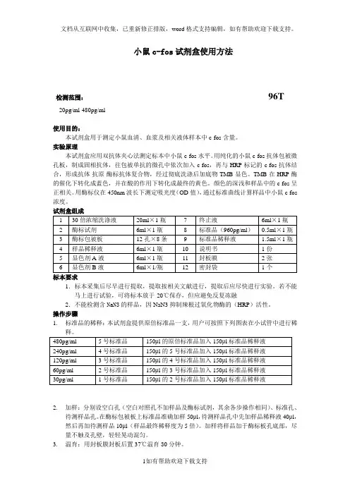
小鼠c-fos试剂盒使用方法检测范围:96T20pg/ml-480pg/ml使用目的:本试剂盒用于测定小鼠血清、血浆及相关液体样本中c-fos含量。
实验原理本试剂盒应用双抗体夹心法测定标本中小鼠c-fos水平。
用纯化的小鼠c-fos抗体包被微孔板,制成固相抗体,往包被单抗的微孔中依次加入c-fos,再与HRP标记的c-fos抗体结合,形成抗体-抗原-酶标抗体复合物,经过彻底洗涤后加底物TMB显色。
TMB在HRP酶的催化下转化成蓝色,并在酸的作用下转化成最终的黄色。
颜色的深浅和样品中的c-fos呈正相关。
用酶标仪在450nm波长下测定吸光度(OD值),通过标准曲线计算样品中小鼠c-fos 浓度。
试剂盒组成标本要求1.标本采集后尽早进行提取,提取按相关文献进行,提取后应尽快进行实验。
若不能马上进行试验,可将标本放于-20℃保存,但应避免反复冻融2.不能检测含NaN3的样品,因NaN3抑制辣根过氧化物酶的(HRP)活性。
操作步骤1.标准品的稀释:本试剂盒提供原倍标准品一支,用户可按照下列图表在小试管中进行稀2.加样:分别设空白孔(空白对照孔不加样品及酶标试剂,其余各步操作相同)、标准孔、待测样品孔。
在酶标包被板上标准品准确加样50μl,待测样品孔中先加样品稀释液40μl,然后再加待测样品10μl(样品最终稀释度为5倍)。
加样将样品加于酶标板孔底部,尽量不触及孔壁,轻轻晃动混匀。
3.温育:用封板膜封板后置37℃温育30分钟。
4.配液:将30倍浓缩洗涤液用蒸馏水30倍稀释后备用5.洗涤:小心揭掉封板膜,弃去液体,甩干,每孔加满洗涤液,静置30秒后弃去,如此重复5次,拍干。
6.加酶:每孔加入酶标试剂50μl,空白孔除外。
7.温育:操作同3。
8.洗涤:操作同5。
9.显色:每孔先加入显色剂A50μl,再加入显色剂B50μl,轻轻震荡混匀,37℃避光显色15分钟.10.终止:每孔加终止液50μl,终止反应(此时蓝色立转黄色)。
Annexin-V-FITC细胞凋亡检测试剂盒

2. Yang L, Zhou X, Yang J, Yin X, Han L, Zhao D. Aspirin inhibits cytotoxicity of prion peptide PrP106-126 to neuronal cells associated with microglia activation in vitro. J Neuroimmunol. 2008 Aug 13;199(1-2):10-7.
分钟,收集细胞,用50-100μl Annexin V-FITC结合液轻轻重悬细胞,涂片后,荧光显微镜下观察。 2. 对于贴壁细胞:
A. 把细胞培养液吸出至一合适离心管内,PBS洗涤贴壁细胞一次,加入适量胰酶细胞消化液(可含有EDTA)消化细胞。 室温孵育至轻轻吹打可以使贴壁细胞吹打下来时,吸除胰酶细胞消化液。需避免胰酶的过度消化。
使用说明:
1. 对于悬浮细胞: A. 在进行完细胞凋亡刺激后,1000g (约1000-2000rpm)离心5分钟,弃上清,收集细胞,用PBS轻轻重悬细胞并计数。 注意:PBS重悬不能省略,PBS重悬的过程同时也起到了洗涤细胞的作用,可以保证后续Annexin V-FITC的结合。
B. 取5-10万重悬的细胞,1000g离心5分钟,弃上清,加入195μl Annexin V-FITC结合液轻轻重悬细胞。 C. 加入5μl Annexin V-FITC,轻轻混匀。 D. 室温(20-25℃)避光孵育10分钟。可以使用铝箔进行避光。 E. 1000g离心5分钟,弃上清,加入190μl Annexin V-FITC结合液轻轻重悬细胞。 F. 加入10μl碘化丙啶染色液,轻轻混匀,冰浴避光放置。 G. 随即进行流式细胞仪检测,Annexin V-FITC为绿色荧光,PI为红色荧光。如果用于荧光显微镜下检测,1000g离心5
免疫荧光
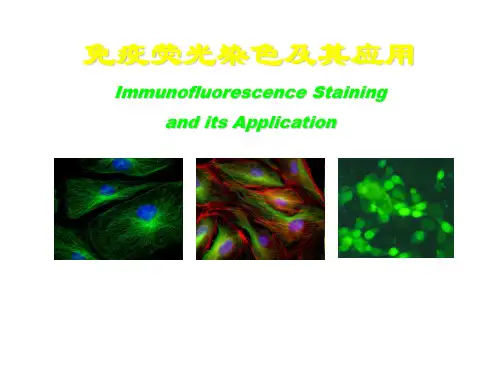
FITC-细胞桨;DAPI-细胞核
SYBR Green I 溶液染色: 病毒(小点)、细菌(大亮 点)、硅藻(细长的细胞) SYBR Green I 凝胶染色
染色体原位杂交检测
免疫荧光染色,多色叠加
实验内容
小鼠B细胞膜表面Ig(mIgG)测定——直接免疫荧光染色法
细胞膜或细胞内的抗原分子与相应的荧光素直 接标记的mAb结合后,形成带有荧光色素的抗原抗 体复合物。 经激发光激发后发出与荧光素相对应的特定波 长的荧光,其荧光强度与被测抗原分子表达密度成 正比例关系,由此可检测细胞与标记抗体对应抗原 的表达量和阳性细胞百分比。 活细胞表面保留有较完整的抗原或受体,根据 所测定的荧光强度和阳性细胞百分率即可知相应抗 原的密度和分布的比例。
3、用眼科剪将脾脏一端剪一小口后,将含5ml Hank’s液的注射器 从另一端刺入脾脏,缓缓注入Hank’s液,即可见脾细胞流入平皿, 在注射Hank’s液同时不断转动针头方向,直至脾脏变苍白为止。
4、将脾细胞悬液吸入试管,静置10分钟,以去除较大块未冲散 的脾组织块,然后将沉淀以上部分移入干净试管。
荧光
指一个分子或原子吸收了外界给予的能量后, 即刻引起发光;停止能量供给,发光亦瞬即停 止。
受到短波激发光照射,发射出波长较长的荧光。 若激发光停止照射,荧光立即熄灭。 荧光的衰减:取决于激发光强度和照射时间。
荧光发生机理
量子理论:光波短,光子能量强;光波长,光子能量小 某些物质接收紫外线和较短波照射------能量增高,处于 不稳定状态-----以光的形式向外释放多余的能量。
在碱性条件下,FITC的异硫氰酸基在水溶液中与 免疫球蛋白的自由氨基经碳酰胺化而形成硫碳氨基键, 成为标记荧光免疫球蛋白,即荧光抗体。 一个Ig分子上最多能标记15~20个FITC分子。
免疫荧光

LOGO
实验展示
不同的亚细胞定位
绿色:细胞骨架
红色:细胞核
LOGO
实验展示
GFP Anti-NOB1 DAPI
1
B
Anti-NOB1
DAPI
2
3
LOGO
注意事项
注意事项: •细胞密度要合适,状态较好; •PBS洗时动作要轻缓,避免冲下细胞; •操作时避免弄混盖玻片的正反面; •从孵育二抗开始要避光; •完成后最好及时镜检,照相;分别用不同波长激发荧光,在Photoshop软件 下合成一张图; •镜检时盖玻片应朝向物镜。
LOGO
免疫荧光
LOGO
1 2 3 4
免疫荧光原理 实验方法 实验展示
注意事项
LOGO
免疫荧光ห้องสมุดไป่ตู้理
免疫荧光技术(Immunofluorescence technique )又称荧光抗体技术,是标记免疫技 术中发展最早的一种。它是在免疫学、生物化学和显微镜技术的基础上建立起来的一 项技术。利用抗原抗体反应进行组织或细胞内抗原物质的定位。先将已知的抗原或抗 体标记上荧光素制成荧光标记物,再用这种荧光抗体(或抗原)作为分子探针检查细 胞或组织内的相应抗原(或抗体)。在细胞或组织中形成的抗原抗体复合物上含有荧 光素,利用荧光显微镜观察标本,荧光素受激发光的照射而发出明亮的荧光,可以看 见荧光所在的细胞或组织,从而确定抗原或抗体的性质、定位,以及利用定量技术测 定含量。
LOGO
免疫荧光原理
LOGO
免疫荧光原理
常见荧光素 1)FITC:黄色结晶粉末,吸收光:490~495nm,发射光:520~530nm,明亮 的黄绿色荧光。 2)RB200:橘红色粉末,吸收光570nm,发射光595~600nm,橘红色荧光。 3)TRITC:紫红色粉末,吸收550nm,发射光620nm,橙红色荧光。 4)镧系:Eu、Tb 5)PE:吸收光490~560nm,发射光595nm,红色荧光。 6)其它:酶作用后产生荧光物质。
免疫荧光

酶底物产物激发光发射光 Β-G MUG MU 360 450 AP MUP MU 360 450 HRP HPA二聚体 317 414 ⑷合适荧光素的选择 1)具有与蛋白质形成共价键的化学基团。 荧光素-N=C +NH-蛋白质荧光素-N-C-N-蛋白质 ‖ ︱ ︱‖ ︱ S H H SH 2)荧光效率高,标记后下降不明显。 3)荧光色泽与背景色泽对比鲜明。
实验步骤
直接法测抗 原
间接法测抗 体
⑴基本原理 将荧光素标记在相应的抗体上,直接与相应抗原反应。其优点是方法简便、特异性高,非特异性荧光染色少。 缺点是敏感性偏低;而且每检查一种抗原就需要制备一种荧光抗体。此法常用于细菌、病毒等微生物的快速检查 和肾炎活检、皮肤活检的免疫病理检查。 ⑵试剂与仪器 磷酸盐缓冲盐水(PBS):0.01mol/L,pH7.4 荧光标记的抗体溶液:以0.01mol/L,pH7.4的PBS进行稀释 缓冲甘油:分析纯无荧光的甘油9份+ pH9.2 0.2M碳酸盐缓冲液1份配制 搪瓷桶三只(内有0.01mol/L,pH7.4的PBS 1500ml) 有盖搪瓷盒一只(内铺一层浸湿的纱布垫) 荧光显微镜 玻片架
1)荧光色素
许多物质都可产生荧光现象,但并非都可用作荧光色素。只有那些能产生明显的荧光并能作为染料使用的有 机化合物才能称为免疫荧光色素或荧光染料。常用的荧光色素有:
⑴异硫氰酸荧光素(fluoresceinisothiocyanate,FITC)为黄色或橙黄色结晶粉末,易溶于水或酒精等溶 剂。分子量为389.4,最大吸收光波长为490--495nm,最大发射光波长520--530nm,呈现明亮的黄绿色荧光,结 构式如下:
标记的抗抗体是抗球蛋白抗体,同于血清球蛋白有种的特异性,如免疫抗鸡血清球蛋白只对鸡的球蛋白发生 反应,因此,制备标记抗体适用于任何抗原的诊断。
免疫荧光技术

葡聚糖凝胶G25或G50,用pH=7.4的0.01%PBS溶胀 后装柱,加入标记后的抗体溶液,然后用上述 PBS洗脱,取洗脱液加入20%三氯醋酸使蛋白沉淀 后,上清液中的荧光素应低于0.01ug/ml。
⑵ 除去标记不适当的抗体:
采用DEAE纤维素离子柱。将DEAE纤维素用pH=7.6的 0.01mol/L磷酸盐缓冲液平衡装柱,加入标记抗体溶液, 进行分步洗脱。 未结合荧光素的抗体,所帶负电少,最先洗出。 中间洗脱出来的是荧光标记合适的抗体部分。 过量结合荧光素的抗体,所帶负电多,最后洗出。 但经此法纯化,标记抗体损失达50%。
⑶ 除去非特异反应物质:
将荧光抗体用同一正常组织或同种动物其它组织 的组织粉预先吸收。 按每ml标记抗体加100mg组织粉比例混合, 4℃ 磁力搅拌1h,静置1h,1800r/min于4℃条件下离 心20min取上清液,再用每ml标记抗体加组织粉 比例重复一次。
(三)荧光抗体的鉴定
荧光素与蛋白结合率:
P:总蛋白质的量。F:荧光色素量。(物质的量)
*
*
固定标本染色以F/P=1.5左右为宜 活细胞染色以F/P=2.4左右为宜.
F/P值高则抗体分子结合的荧光素多。
F/P=
IgG (mg/ml)
荧光色素(μg/L ) 160 000x10 3 荧光色素 ( μg/L ) x 0.4 x 6
IgG (mg/ml) 390x10
病毒
无水乙醇,丙酮、CCl4
(3)固定后处理
抗原固定后必须立即以冷PH7.4 PBS冲洗,顺序经过三缸 浸泡,每缸3min,末次再以蒸馏水浸泡1min以脱盐。 标本固定干燥后最好立即进行荧光染色及镜检,如必须 保存则应保持干燥,置于4℃以下保存,一般细菌涂片或 组织切片经过固定后可保存一个月以上,但病毒和某些 组织 抗原标本则需-20℃以下保存。
人CD45分子检测试剂盒(流式细胞仪法-FITC)
当某一细胞通过流式细胞仪被检测到具有此 525nm 特异光信号,就可以确
定该细胞表面表达 CD45 分子。对某一细胞群体进行该项检测,可以确定
本试剂用于体外流式细胞仪半定量检测经 EDTA、枸橼酸钠或肝素钠 抗凝的骨髓细胞表面 CD45 分子的表达。
CD45 的荧光强度结合测向角可以用于流式细胞仪检测时设门,准确找 到白血病细胞群,然后进行细胞免疫分型表达分析。
用 CD45 与侧向角(side scatter,SSC),对数取样后可将骨髓细胞清晰地 分出淋巴细胞,单核细胞,成熟粒细胞,幼稚细胞和红细胞群,其理论依 据是 CD45 是所有白细胞的抗原。其表达量在淋巴细胞最高,单核细胞, 成熟粒细胞,早期造血细胞(blasts)依次减弱。红细胞(中,晚幼红细胞, 成熟红细胞)不表达 CD45[1-3]。SSC 反映细胞的颗粒性,成熟粒细胞 SSC 最高,依次为单核细胞,淋巴细胞,早期造血细胞,红细胞。以 CD45/SSC 双参数,对数取样时即可把各细胞群区分出来。若同时加上一个,二个甚 或三个荧光标记的单抗,则很容易识别非正常细胞群所表达的抗原。这样 可以排除正常细胞的干扰。在幼稚细胞百分率低的情况下或检测残存白血 病时尤为必要 。
异硫氰酸(FITC)标记小鼠抗人 CD45 单克隆抗体试剂是一种流式细胞 仪用试剂,主要用于白血病分型诊断时,应用流式细胞术 CD45/SSC 设门进 行急性白血病免疫分型[4]。
通过使用本品检测 CD45+细胞,只作为辅助判断白血病的免疫分型, 其百分率高低与白血病的具体类型、发病、疾病进程和预后并无直接关系, 需要临床医师根据病人的临床表现及其他检查结果综合做出判断。 【检验原理】
fitc的激发波长和发射波长
fitc的激发波长和发射波长
FITC指Fluorescein isothiocyanate,是一种有机化学物质,它具有荧光特性,可以用微量添加到物质中,从而使其具有调节特性。
FITC的激发波长是495 nm,发射波长是520 nm。
FITC的主要用途有:
一、用于染色细胞。
FITC可以充当FLUOR和CFAs的凝胶电泳染色剂,有助于更好地解释实验结果,而不会影响实验的灵敏度。
另外,FITC还可以通过免疫细胞染色法来识别特定的细胞,使其与研究相关的特定信号互相关联。
二、用于组织及细胞构建。
FITC可以把特定的核酸和蛋白质附着在特定的细胞表面,以便标记指定的细胞,例如脂肪细胞、神经元和血管细胞等。
三、用于真核生物中的遗传测定。
FITC可以运用在荧光原位杂交技术(FISH)中,将特定的核酸链结合在特定的序列上,以帮助实验者测定基因的表达和遗传变异情况。
四、用于EMT鉴定。
FITC可以配合其他技术,如荧光技术、传递电子显微镜和免疫细胞化学分析,用于鉴定多种疾病的机制,例如肝癌、肠癌和胰腺癌等。
FITC有着许多广泛的用途,激发波长495nm,发射波长为520nm,并且可以将特定的核酸和蛋白质附着在特定的细胞表面,以帮助实验者做出合理的判断,获得准确的实验结果。
免疫荧光法检测干细胞标志物
免疫荧光法检测干细胞标志物英文回答:Immunofluorescence is a widely used technique to detect and visualize specific proteins or molecules in biological samples. It utilizes the specific binding of antibodies to their target molecules, followed by the detection of these antibodies using fluorescent markers.In the context of stem cell research, immunofluorescence can be used to detect and identify specific markers that are characteristic of stem cells. Stem cells are undifferentiated cells with the ability to differentiate into various cell types. They express certain proteins or molecules that are unique to stem cells, and these markers can be detected using immunofluorescence.For example, one commonly used stem cell marker is Oct-4. Oct-4 is a transcription factor that plays a crucialrole in maintaining pluripotency in embryonic stem cells.To detect Oct-4 using immunofluorescence, researchers would first incubate the stem cells with a primary antibody that specifically binds to Oct-4. This primary antibody is typically derived from a different species than the sample (e.g., mouse anti-Oct-4 antibody for human stem cells). After washing away any unbound antibodies, a secondary antibody conjugated to a fluorescent dye (e.g., FITC or Alexa Fluor) is added. This secondary antibody recognizes and binds to the primary antibody, allowing for the visualization of Oct-4 under a fluorescence microscope. The presence of Oct-4 in the stem cells can be identified by the green fluorescence emitted by the FITC or Alexa Fluor dye.In addition to Oct-4, there are many other markers that can be detected using immunofluorescence to identify and characterize stem cells. These include Nanog, Sox2, SSEA-4, and CD133, among others. Each of these markers has its own specific antibody that can be used in immunofluorescence experiments.Immunofluorescence is a powerful tool in stem cellresearch as it allows for the visualization and identification of stem cells based on their unique markers. It provides valuable information about the presence, location, and abundance of stem cells in a sample. This information is crucial for understanding stem cell biology and for the development of stem cell-based therapies.中文回答:免疫荧光法是一种广泛应用于生物样本中特定蛋白质或分子检测和可视化的技术。
FITC
FluoroTag™FITC Conjugation KitProduct Number FITC1Storage Temperature 2−8 °CTECHNICAL BULLETINProduct DescriptionThe FluoroTag FITC Conjugation Kit is suitable for the conjugation of polyclonal and monoclonal antibodies with fluorescein isothiocyanate (FITC) for use in immunohistochemistry and immunofluorescence studies utilizing flow cytometry. It may also be used for conjugation of FITC to peptide hormones, cytokines, growth factors, and other proteins. The kit contains sufficient reagents for at least 5 conjugations. Fluorescein isothiocyanate (FITC), Isomer I is among the most widely used fluorescent labeling reagents due to the fluorophore’s high quantum efficiency and conjugate stability. FITC has an absorption maximum at 495 nm and emission maximum at 525 nm. FITC reacts with free amino groups of proteins to form stable conjugates (see Figure 1). FITC-protein conjugates, in particular FITC labeled antibodies, are used as specific probes in immunocytochemistry and flow cytometry applications.1,2Biologically active FITC-conjugates of peptide hormones and growth factors have been successfully prepared which identify receptors on target cells.3,4FITC has also been used as a site-specific probe for several other proteins.5,6,7The use of optimal labeling conditions is recommended. Overlabeling of proteins generally results in altered specificity, aggregation and/or precipitation of the protein. Fluorescent labeling of antibodies with high fluorophore to antibody ratios (molar F/P >6) usually results in increased non-specific binding (fluorescent background) and decreased quantum yield due to the fluorophore self-quenching effect.The FluoroTag Kit includes detailed procedures for both small and large scale conjugation of FITC to antibody. Small scale FITC conjugations are performed using three different molar ratios of FITC to antibody. Based on the molar ratio that gives the most satisfactory result, the large scale procedure can then be performed to optimally label the protein. The labeled protein is purified from the unconjugated fluorescein by a quick Sephadex®G-25M column. The F/P molar ratio of the purified protein is then determined by measuring the absorbance at 280 nm and at 495 nm.Reagents and Materials Provided• Fluorescein isothiocyanate, Isomer I (FITC), F7250.Five amber vials each containing 2.0 mg oflyophilized fluorescein isothiocyanate.• 0.1M Sodium Carbonate-Bicarbonate Buffer, pH9.0, C0688. Five capsules containing powder.• Phosphate Buffered Saline (PBS), P3813. Five packages containing powder.• Gel filtration columns. Two columns prepacked with Sephadex G-25M. These serve to separateunreacted FITC from the conjugate and for bufferexchange. The columns are preswollen in watercontaining 0.15% Kathon®CG/ICP II aspreservative. The gel filtration columns are supplied for two different reaction scales:Small Scale (Column A), B7533:One columnprepacked with Sephadex G-25M. The bed volume of the column is 3.5 ml and the bed height is2.6cm. The maximal sample volume is 0.3 ml.Large Scale (Column B), B4783:One columnprepacked with Sephadex G-25M. The bed volume of the column is 9.1 ml and the bed height is 5cm.The maximal sample volume is 1.5 ml.Reagents and Equipment Required but Not Provided• Standard glass vials (1.5−2 ml capacity) equipped with stirring bars.• Two dilution vials are needed for each conjugation.• Vortex mixer.• Standard glass tubes (12 X75 mm or 13 X100 mm) to collect fractions from Sephadex G-25M columns.• Aluminum foil -To protect reaction and FITC labeled protein from intense light.• Quartz cuvette -1 cm path length• UV/Visible spectrophotometer.2Precautions and DisclaimerThis product is for R&D use only, not for drug, household, or other uses. Please consult the Material Safety Data Sheet for information regarding hazards and safe handling practices.Preparation Instructions0.1 M Sodium Carbonate-Bicarbonate Buffer, pH 9.0,C0688-Add contents of one capsule to 50 ml of deionized water to make 0.1M sodium carbonate-bicarbonate buffer, pH9.0. This is used to dissolve FITC and to buffer the conjugation reaction. Phosphate Buffered Saline (PBS), P3813-Mix contents of one package with 800 ml of deionized water. Adjust volume to 1 liter to make 10 mM sodium phosphate buffer, 27 mM KCl, 138 mM NaCl, pH 7.4. This serves as an equilibration buffer for the Sephadex G-25 columns, for the elution of the labeled protein from the column, and for the final dilution of the labeled protein.Storage/StabilityStore at 2−8 °C.ProcedureThe protocol outlined describes the labeling of 1 mg of IgG at 5 mg/ml with FITC (Small Scale Conjugation Procedure). The procedure can be scaled up to 5 mg of IgG maintaining the same concentration and molar ratio of the reagents (Large Scale Conjugation Procedure). It is important to consider that the number and surface availability of amine groups (primarily -amine groups of lysine residues) vary greatly among proteins and even among different IgGs. This may result in a large variability of the level of labeling. Testing different FITC to antibody molar ratios to determine the optimal levels of labeling of the antibody is recommended.Small Scale Conjugation Procedure(1.0 mg IgG)This procedure describes the conjugation of FITC to1mg of IgG, using one of the following molar ratios in the reaction mixture: 5:1, 10:1 and 20:1 of FITC(MW389) to IgG (MW 150,000). The labeling is performed in a final reaction volume of 0.25 ml. In general, these reaction molar ratios result in fluorescein-antibody conjugates with F/P ratios of 1−2, 2−4 and 3−6, respectively. This procedure can be modified if a protein of a different molecular weight (or a different amount of IgG) is used in the labeling reaction. (See Table 2 reaction mixture ratios for whole IgG, IgM, or antibody fragments.)1.Dissolve the contents of one sodium carbonate-bicarbonate capsule (C0688) in 50 ml of deionized water. The pH of this buffer should be 9.0 ±0.1(See Application Notes 1&2).2.Prepare at least 0.25 ml of antibody solution at5.0mg/ml in 0.1 M carbonate-bicarbonate buffer,pH 9.0 (See Application Notes 3, 4 & 5). The A280of an IgG solution at 1.0 mg/ml is 1.4 (1.0 cm path length).3.Add 0.2 ml (1.0 mg) of the antibody solution intoeach reaction vial labeled “5:1”, “10:1”, or “20:1”. 4.Reconstitute one vial of FITC (F7250) in 2 ml of0.1M carbonate-bicarbonate buffer and vortex untilall FITC has dissolved. Label vial “20:1 FITC”. The solution should be freshly prepared before eachconjugation and used within 5 minutes (SeeApplication Note 6). The "20:1 FITC" solution isfurther used to prepare the "5:1" and "10:1"solutions.5.Prepare the required dilution of FITC in 0.1Mcarbonate-bicarbonate buffer as directed inTable1.Table 1.Dilution of FITC in 0.1M Carbonate-Bicarbonate Buffer6.Add 50 µl of the appropriate FITC dilution dropwisewhile stirring to the correspondingly labeledreaction vial.pletely cover the reaction vial with aluminumfoil to protect from light.8.Incubate all reaction vials for 2 hours at roomtemperature with gentle stirring.Isolation of labeled protein1.Empty contents of PBS package (P3813) into asuitable container. Add 800 ml of distilled ordeionized water and mix. Adjust to final volume of 1,000 ml.bel Sephadex G-25M, column A (B7533), “5:1”,“10:1”, or “20:1”. Support column over a suitable(100ml) beaker.3.Remove cap from the top of the column, cut openlower tip of column and let excess of liquid flowthrough. The column will not run dry.34.Equilibrate the column with 12 ml of PBS solution(6 X2 ml). If the column is not immediately used,close with top and bottom caps and store at 2−8 °C.5.Apply reaction mixture to top of column gel bed andcollect the flow through (Fraction 1)6.Elute the column with 2.5 ml of PBS, collecting0.25ml fractions (10 X0.25 ml). Monitor theabsorbance of each fraction at 280 nm. Two bands will be visible during elution. The conjugate ispresent in the first band (fractions 6−8)(See Application Note 7).7.Pool the main fractions. Do not collect fractions withA280<0.2.8.Wash the column with 35 ml (10 X column volumes)of PBS to remove unbound fluorophore. This issufficient to regenerate the column.9.For prolonged storage, wash the column with 10mlof PBS containing 0.05% sodium azide and storecapped at 2−8 °C, with 1 ml buffer above the gel. 10.Determine the fluorescein/protein ratio (F/P) of theconjugate using a spectrophotometer as described in Determination of Fluorescein/Protein Molar Ratio (F/P).11.For storage of the conjugate after determination ofthe F/P molar ratio, add 1% (w/v) BSA and 0.1%(w/v) sodium azide to the conjugate. Store at2−8°C, protected from light.Large Scale Conjugation Procedure(5.0 mg IgG)This procedure describes the scale-up of the conjugation reaction of FITC to 5 mg of IgG, using the molar ratio, which gives the most satisfactory results as obtained from the “Small Scale Conjugation Procedure.” The labeling is performed maintaining the same concentrations and ratios of reagents used previously. The reaction is performed in a final reaction volume of 1.25 ml.1.Add 1.0 ml (5.0 mg) of the antibody solution to areaction vial labeled “5:1”, “10:1”, or “20:1”.2.Reconstitute one vial of FITC (F7250) in 2 ml of0.1 M carbonate-bicarbonate buffer and vortex untilall FITC has dissolved.3.Prepare 10:1 or 5:1 dilution of FITC in 0.1Mcarbonate-bicarbonate buffer as directed inTable1, if necessary.4.Add 250 µl of the appropriate FITC dilutiondropwise while stirring to the reaction vial.pletely cover the reaction vial with aluminumfoil to protect from light.6.Incubate reaction vial for 2hours at roomtemperature with gentle stirring.Isolation of labeled proteinbel Sephadex G-25M, column B (B4783), “5:1”,“10:1”, or “20:1”. Support the column over asuitable (100 ml) beaker.2.Remove cap from the top of the column, cut openlower tip of column and let excess of liquid flowthrough. The column will not run dry.3.Equilibrate the column with 30 ml of PBS solution(6 X5 ml). If the column is not immediately used,close with top and bottom caps and store at 2−8 °C.4.Apply reaction mixture to top of the column gel bedand collect flow through (Fraction 1).5.Elute column with 10 ml of PBS, collecting 1.0 mlfractions (10 X1 ml). Monitor the absorbance ofeach fraction at 280 nm. Two bands will be visible during elution. The conjugate is present in the first band (fractions 3−5). (See Application Note 7.)6.Pool the main fractions. Do not collect fractionswith A280<0.4.7.Wash the column with 50 ml of PBS solution toremove unbound fluorophore. This is sufficient toregenerate the column.8.Preserve the column as instructed in step 9 of thesmall scale “Isolation of Labeled Protein” section. Determination of Fluorescein/Protein Molar Ratio (F/P) The F/P molar ratio is defined as the ratio of moles of FITC to moles of protein in the conjugate.To determine this ratio, it is necessary to first determine the absorbance of the conjugate sample at 280 nm and then at 495 nm.1.Place the conjugate sample in a quartz cuvette.For the large scale conjugation, dilute 0.1 ml of the FITC conjugate in 0.9 ml of PBS containing sodium azide. Read the absorbance of the conjugatesample at 280 nm and 495 nm. The absorbancereading of the conjugate sample should be between0.2 and 1.4 at 280 nm. If the absorbance reading isoutside this range, adjust the sample dilutionaccordingly.2.From the absorbance readings (A280and A495) ofthe conjugate sample, calculate the F/P of thefluorescein-IgG conjugate according to the84The protein concentration of the fluorescein-IgGFITC, use the general formula below, substituting the appropriate values for the particular protein:MW is the molecular weight of the protein.389 is the molecular weight of FITC.195 is the absorption E 0.1% of bound FITC at 490 nm at pH 13.0.(0.35 x A 495) is the correction factor due to the absorbance of 280(For E 0.1%and C values, see Table 2)FITC at 280 nm.8E 0.1%is the absorption at 280 nm of a protein at 1.0mg/ml.280280Application Notes1.Do not store sodium carbonate-bicarbonate buffermore than 1 week at 2−8 °C. The pH of the buffer may change upon storage. It is advised that fresh buffer be made just before use.2.The conjugation of FITC to proteins is affected bythe reaction conditions (concentration, temperature, pH).3.When conjugating antibodies with FITC, the startingmaterial should be free of contaminating serum proteins. Affinity isolated antibodies, IgG fractions, or Protein A purified immunoglobulin are generally acceptable.4.Protein solutions should not be prepared in bufferscontaining amines such as Tris, glycine or sodium azide since they inhibit the labeling reaction. If the buffer contains amines or sodium azide, dialyze protein solution (1 ml) against PBS, pH 7.4(1,000ml), overnight at 2−8 °C. Avoid dialysis at high pH values (>8.0−8.5) as this may be harmful to some proteins.5.If the antibody is in PBS pH 7.4 (without azide), add1 M carbonate-bicarbonate buffer (1 capsule (C0688) in 5 ml deionized water) to a final concentration of 0.1 M (e.g., 0.1 ml of 1 Mcarbonate-bicarbonate buffer to 0.9 ml IgG solution at 5.0 mg/ml).6.FITC is not stable in aqueous solutions. Do notstore FITC stock solutions.7.The second band containing unbound fluorophoreis retained on the column and will elute only with subsequent PBS washes.References1.Staines, W., et al., Three-colorimmunofluorescence histochemistry allowing triple labeling within a single section. J. Histochem. Cytochem., 36, 145 (1988).2.Carter, N., in Flow Cytometry: A PracticalApproach, M.G. Ormerod, Ed., IRL Press (1990).3.Carraway, K., et al., Location of the epidermalgrowth factor binding site on the EGF receptor. A resonance energy transfer study. Biochemistry , 29, 8741 (1990).4.Schwartz, J., and Vale, W., Fluorescent andcytotoxic analogs of corticotropin-releasing factor: probes for studying target cells in heterogeneous populations. Methods in Enzymol ., 168, 29 (1989).5.Adams, S., et al., Fluorescence ratio imaging ofcyclic AMP in single cells. Nature , 349, 694 (1991).6.Abbott, A., et al., Immunochemical andspectroscopic characterization of two fluorescein 5'-isothiocyanate labeling sites on Na+, K(+)-ATPase. Biochemistry , 30, 1692 (1991).7.Pavalko, F., and Burridge, K., Disruption of theactin cytoskeleton after microinjection of proteolytic fragments of alpha-actinin. J. Cell Biol.,114, 481 (1991).8.The, T., and Feltkamp, T., Conjugation offluorescein isothiocyanate to antibodies. I. Experiments on the conditions of conjugation. Immunol.,18, 865 (1970).Sephadex is a registered trademark of Pharmacia, Inc. Kathon is a registered trademark of the Rohm & Haas Company.LPG,KTA 12/05-15 Table 2.Reaction Mixture Ratios for Whole IgG, IgM or Antibody Fragments** This corresponds to volume in microliters of the appropriate FITC concentration (5:1, 10:1, or 20:1).***For IgM, a lower molar ratio is recommended as high levels of FITC may result in overlabeling of the antibody and self-quenching of the fluorophore.****For F(ab′) 2 use a 2-fold more concentrated FITC solution than used for IgG.*****For Fab′and Fc use a 4-fold more concentrated FITC solution than used for IgG.6Figure 1.The FITC Labeling ReactionFITC conjugation occurs through the free amino groups of proteins or peptides, forming a stable thiourea bond.Sigma brand products are sold through Sigma-Aldrich, Inc.Sigma-Aldrich, Inc. warrants that its products conform to the information contained in this and other Sigma-Aldrich publications.Purchaser must determine the suitability of the product(s) for their particular use. Additional terms and conditions may apply.Please see reverse side of the invoice or packing slip.。