蔡司CIRRUS HD-OCT
ZEISS CIRRUS 6000高性能OCT设备说明书
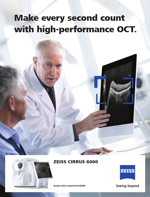
Make every second count with high-performance OCT.ZEISS CIRRUS 6000/cirrus6000High-Performance OCTAdvance your fast-paced practiceCIRRUS® 6000, the next-generation OCT from ZEISS, delivers high-speed image capture with HD imaging detail and a wider field of view, so you can make more informed decisions and spend more time with your patients.Performance OCT Faster imaging with greater detail, at 100,000 scans per second, for improved patient care.Proven analyticsComprehensive, clinically-validated tools to diagnoseand manage a range ofconditions.Patient-first designSeamless transfer of rawpatient data from previousgenerations of CIRRUS forcontinuity of patient care.2The power of 100,000 scans per secondFaster imaging:Reduce chair time and speed upyour practice.• 270% faster OCT scans and 43% faster OCTA scans.*• OCT cube scans in as little as 0.4 seconds.• High-speed imaging in combination with FastTrac™ eye tracking technology reduces the chance of motion artifacts such as those caused by blinks and saccades.Greater detail:View more in seconds and dig deeper with high-definition imaging.• 12×12 mm single-shot OCTA cube scan in addition to 8×8, 6×6 and 3×3 mm scans.• High-Definition AngioPlex scans (8×8 and 6×6 mm) for even greater microvascular detail without limiting the field of view.• 2.9 mm scan depth.*Compared to prior generations of CIRRUS. 4“The CIRRUS 6000 is all about its speed. With increased speed comes greatly improved resolution and detail on cube, raster and OCTA scans, and the new faster CIRRUS enables me to incorporate these more reliable scans into my daily workflow and make important treatment decisions for my patients.”Theodore Leng, MD, FACS,Byers Eye Institute at Stanford, United States512 mm HD 1 Line Raster 100x averaged. Image courtesy of Theodore Leng, MD, Byers Eye Institute, United States 12×12 mm single-shot OCTA of branch retinal vein occlusion (BRVO).Image courtesy of Jesse Jung, MD, East Bay Retina, United States612 mm HD 1 Line Raster 100x averaged. Image courtesy of Theodore Leng, MD, Byers Eye Institute, United States8×8 mm HD AngioPlex OCTA of BRVO.Image courtesy of Roger Goldberg, MD,Bay Area Retina Associates, United States diabetic retinopathy (NPDR). Image courtesy ofUnited Statesretinopathy (PDR). Image courtesy of Roger Goldberg,MD, Bay Area Retina Associates, United States7AngioPlex Metrix OCTA QuantificationAngioPlex ® Metrix ™ for Macula and ONHAngioPlex Metrix allows clinicians to objectivelyassess and track progressive eye diseases suchas diabetic retinopathy and glaucoma withquantification tools such as Vessel Density,Perfusion Density, and Foveal Avascular Zone (FAZ)for the macula, and Capillary Flux Index for theoptic nerve head.Proven analyticsCIRRUS-powered treatment decisionsAs the pioneering OCT technology, the CIRRUS platform offers clinicians extensive, clinically-validated applications for retina, glaucoma and anterior segment. The result: precise analysis, faster throughput and smarter decision-making across a wide spectrumof clinical conditions and patient types.RetinaMacular Change AnalysisThe CIRRUS data cube automatically stores and delivers each patient’s historical data to provide a variety of change assessments, including macular thickness change maps that help you understand your patient‘s response to treatment. Because every CIRRUS cube is tracked and registered to OCT scans from prior visits using CIRRUS’ FastTrac ™ Retinal Tracking Technology, you can confidently measure point-to-point changes in macular thickness. Visit 1Visit 28The CIRRUS suite of glaucoma analysis tools are designed to help you better visualize, detect, and manage all stages of glaucoma, from glaucoma suspects and mild glaucoma to severe glaucoma.GlaucomaAnterior SegmentPremier ModuleCIRRUS also enables comprehensiveimaging and quantification of theanterior segment for refractive surgeryplanning and follow-up, cornealevaluation and glaucoma assessment.CIRRUS RNFL thickness deviation mapshave been shown to be superior for detectinglocalized RNFL defects, compared to traditionalperipapillary RNFL thickness measurements.AutoCenter ™ – ZEISS’ patented algorithm automatically identifies theoptic nerve head using Bruch’s Membrane Opening (BMO) in 3-dimensionsfor more precise measurement of the neuro-retinal rim, accounting for tilteddiscs, disruptions to the RPE and other challenging pathology.Unique to ZEISS, Guided Progression Analysis ™ (GPA ™) provides both trend and event-based analyses that detect statistically significant change and quantify rate of change for key RNFL, ONH, andGCL/IPL parameters.Ganglion Cell Analysis helps identify macular glaucomatous damage, which can be missed with RNFL analysis bined GCL/IPL and RNFL thickness deviation maps provide a comprehensive widefield assessment.Anterior Segment9 mm epithelial thickness map of keratoconus highlights localizedepithelial thinning.9 mm high-definition cornea imaging with semi-automated measurement tools for flap thickness and residual stromal bed.9Combined GCA and RNFLDeviation Map RNFL ThicknessAnalysis RNFL Deviation Map Ganglion Cell AnalysisRNFL Thickness AnalysisPatient-first designUnique platform designed for the futureWith ZEISS CIRRUS 6000, your patient data is never left behind. The CIRRUS platform ensures seamless transfer of raw, dynamic patient data from previous generations of the device – enabling clinicians to maintain continuity of patient care, even as OCT technology evolves over time.10Technical specifications ZEISS CIRRUS 6000Analytical applicationsRetina:• M acular Thickness Analysis withReference Database (Diversified and Asian)• Macular Change Analysis• Advanced RPE Analysis• 3D Visualization• En Face Analysis• CIRRUS Wellness Exam Glaucoma:• Guided Progression Analysis• Ganglion Cell/IPL Thickness with Reference Database (Diversified and Asian)• RNFL Thickness with Reference Database (Diversified and Asian)• ONH Parameters with Reference Database (Diversified and Asian)• Average cup-to-disc ratio• Average, Superior and Inferior RNFL Thickness• CIRRUS Wellness ExamAnterior Segment:• 9 mm Epithelial Thickness andPachymetry Mapping• HD Cornea with Cornea Caliper Tool• C hamberView™ Full Anterior ChamberImaging for phakic IOL sizing and safetydistance measurements• A ngle imaging and measurement toolsfor Glaucoma (AOD, TISA, SSA)AngioPlex Metrix OCT Angiography Quantification: • Macular• Foveal Avascular Zone• Perfusion Density (ETDRS grid)• Vessel Density (ETDRS grid)• Optic Nerve Head• Capillary Perfusion Density• Capillary Flux Index• AngioPlex 2-visit comparisonKey ParametersMethodology:Spectral domain OCTOptical source:Superluminescent diode (SLD), 840 nm A-scan depth: 2.0 - 2.9 mm (in tissue)Scan speed:100,000 A-scans per secondMin. pupil diameter: 2.0 mmResolution:• Axial resolution• Transverse resolution 5 μm (in tissue), 1.95 μm (digital) 15 μm (in tissue)Refractive error adjustment:-20D to +20D (dopters)Fundus Imaging:• Methodology• Optical Source• Field of View (degrees)Line Scanning Laser Ophthalmoscope (LSO) SLD 750 nm36×30Posterior Segment scans:• OCT• OCTA Cube scan (Macula and Optic Disc)HD Raster (1, 5, 21-line, cross and radial); Raster scan length 3-12 mm; image averaging up to 100x3×3, 6×6, 8×8, 12×12 mm (Macula); 4.5×4.5 mm (Optic Nerve Head); 14×10 mm (Montage), 14×14 mm (Montage)Anterior Segment scans:Cube, HD Cornea, Pachymetry, HD Angle, Wide Angle-to-Angle,Anterior Chamber, 5-Line RasterInstrument SpecificationsWeight:35 kg (77 lbs) (without monitor)Dimensions (L × W × H):62.2 × 42.5 × 29.2 cm (24.4 × 16.7 × 11.4 in) (without monitor)Input Power:• Voltage and Mains Frequency• Electrical Class 230V, 100/120V, 50-60Hz IEC 60601-1 Class IComputer SpecificationsMonitor: 22” Widescreen HD Resolution: 1920×1080Internal storage: 2 TB with 128 GB SSD USB Ports: 8Input devices:Wireless keyboard, Wireless mouseProcessor:Intel® Core i7 (7th Gen)Operating system (Instrument):Windows® 10 EnterpriseSupported operating systems (Review Station):Windows® 10, Windows® 8.1, Windows® 7 (64 bit)11E N _31_010_0047I C Z X I /2019 I n t e r n a t i o n a l e d i t i o n : O n l y f o r s a l e i n s e l e c t e d c o u n t r i e s .T h e c o n t e n t s o f t h e b r o c h u r e m a y d i f f e r f r o m t h e c u r r e n t s t a t u s o f a p p r o v a l o f t h e p r o d u c t o r s e r v i c e o f f e r i n g i n y o u r c o u n t r y . P l e a s e c o n t a c t o u r r e g i o n a l r e p r e s e n t a t i v e s f o r m o r e i n f o r m a t i o n . S u b j e c t t o c h a n g e s i n d e s i g n a n d s c o p e o f d e l i v e r y a n d d u e t o o n g o i n g t e c h n i c a l d e v e l o p m e n t . T h e s t a t e m e n t s o f t h e h e a l t h c a r e p r o f e s s i o n a l g i v i n g t e s t i m o n i a l r e f l e c t o n l y t h e i r p e r s o n a l o p i n i o n s a n d e x p e r i e n c e s a n d d o n o t n e c e s s a r i l y r e f l e c t t h e o p i n i o n s w i t h w h o m t h e y a r e a f f i l i a t e d . T h e h e a l t h c a r e p r o f e s s i o n a l g i v i n g t h i s t e s t i m o n i a l m a y h a v e a c o n t r a c t u a l r e l a t i o n s h i p w i t h C a r l Z e i s s M e d i t e c , I n c ., a n d m a y h a v e r e c e i v e d f i n a n c i a l c o m p e n s a t i o n . A n g i o P l e x , A n g i o P l e x M e t r i x , A u t o C e n t e r , C I R R U S ,F a s t T r a c a n dG P A a r e e i t h e r t r a d e m a r k s o r r e g i s t e r e d t r a d e m a r k s o f C a r l Z e i s s M e d i t e c , I n c . o r o t h e r c o m p a n i e s o f t h e Z E I S S G r o u p i n G e r m a n y a n d / o r o t h e r c o u n t r i e s .© C a r l Z e i s s M e d i t e c , I n c ., 2019. A l l r i g h t s r e s e r v e d .Carl Zeiss Meditec AG Goeschwitzer Strasse 5207745 Jena Germany//med/contactsCarl Zeiss Meditec, Inc.5160 Hacienda Drive Dublin, CA 94568USA//med/contacts 0297CIRRUS 6000。
眼科主要设备及品牌
眼科主要设备及品牌眼科是医学领域中研究眼部疾病和疾病治疗的学科,它涉及到许多专业设备的使用。
本文将介绍眼科主要设备及其品牌,以匡助读者了解眼科领域的技术和产品。
1. 视力检查设备视力检查设备用于评估患者的视力状况,常用的设备包括:- 自动视力检查仪:例如Topcon公司的CV-5000S自动视力检查仪,它能够自动进行视力测试,并根据测试结果生成报告。
- 自动折射计:例如Nidek公司的AR-1自动折射计,它能够测量患者的屈光度和角膜曲率。
- 自动角膜地形图仪:例如Zeiss公司的Atlas 9000角膜地形图仪,它能够生成患者眼部的角膜地形图,用于评估角膜形状和曲率。
2. 眼底检查设备眼底检查设备用于评估患者的眼底情况,常用的设备包括:- 直接眼底镜:例如Heine公司的K180直接眼底镜,它能够通过放大镜观察患者的眼底结构。
- 间接眼底镜:例如Keeler公司的Vantage Plus间接眼底镜,它能够提供更广阔的视野,并通过光源照亮眼底。
- 荧光眼底摄影仪:例如Canon公司的CR-2 PLUS AF荧光眼底摄影仪,它能够拍摄患者眼底的彩色图象,并匡助医生进行诊断。
3. 眼压测量设备眼压测量设备用于测量患者眼内的压力,常用的设备包括:- 非接触式眼压计:例如Reichert公司的ORange眼压计,它能够通过无接触的方式测量眼内压力。
- 接触式眼压计:例如Keeler公司的Pulsair EasyEye眼压计,它能够通过与角膜接触的方式测量眼内压力。
4. 眼角膜手术设备眼角膜手术设备用于进行眼角膜手术,常用的设备包括:- 激光角膜磨镶机:例如Alcon公司的WaveLight EX500激光角膜磨镶机,它能够通过激光技术改变角膜形状,从而矫正视力。
- 角膜切割机:例如Moria公司的M2单通道角膜切割机,它能够用于进行角膜移植手术。
- 角膜接触镜:例如Menicon公司的Ortho-K角膜接触镜,它能够通过改变角膜形状来矫正近视。
ZEISS CIRRUS HD-OCT 扫描数据移动指南说明书

How do I move scan data if I scan under the wrong patient name?ZEISS Quick Help: CIRRUS ™ HD-OCTAn incorrectly stored scan can easily be moved to a different file using the MOVE SCAN feature on a CIRRUS HD-OCT instrument. Follow these steps:1. F rom the main screen of the CIRRUS software application, choose the patient scan you wish to relocate and select Analyze. With the patient scan highlighted, select the desired analysis as if generating a report/analysis of the scan (Figure 1). Confirm the Analysis you selected is displayed, then click Edit and select Move Scan to open the Move Scan dialog box (Figure 2).2. H ighlight and click the patient name to select the patient record into which you wish to move the scan data and then click Move (Figure 3). When prompted, click Yes to proceed (Figure 4) and return to the Analyze screen of the current patient. The relocated scan will no longer be listed inthe incorrect patient profile.Figure 1Figure 2Figure 4Figure 3Carl Zeiss Meditec AG Goeschwitzer Strasse 51–5207745 Jena Germany/med Carl Zeiss Meditec, Inc.5160 Hacienda Drive Dublin, CA 94568USA/medRefer to the CIRRUS HD-OCT User Manual Instructions for Usefor safe and effective operation of the instrument.S E R .9215 P r i n t e d i n t h e U n i t e d S t a t e s . C Z -V I I I /2017 U S A E d i t i o n .T h e c o n t e n t s o f t h e r e f e r e n c e g u i d e m a y d i ff e r f r o m t h e c u r r e n t s t a t u s o f a p p r o v a l o f t h e p r o d u c t o r s e r v i c e o ff e r i n g i n y o u r c o u n t r y . P l e a s e c o n t a c t o u r r e g i o n a l r e p r e s e n t a t i v e f o r m o r e i n f o r m a t i o n . S u b j e c t t o c h a n g e i n d e s i g n a n d s c o p e o f d e l i v e r y a n d a s a r e s u l t o f o n g o i n g t e c h n i c a l d e v e l o p m e n t . C I R R U S i s a e i t h e r t r a d e m a r k o r r e g i s t e r e d t r a d e m a r k o f C a r l Z e i s s M e d i t e c A G o r o t h e r c o m p a n i e s o f t h e Z E I S S G r o u p i n G e r m a n y a n d /o r o t h e r c o u n t r i e s . ©C a r l Z e i s s M e d i t e c , I n c ., 2017. A l l r i g h t s r e s e r v e d .。
蔡司CirrusHD-OCT参数

蔡司CirrusHD-OCT参数
Cirrus HD-OCT参数
莱卡M844手术显微镜技术参数
显微镜优点介绍:
灵活摆位
电磁锁
使用方便快捷简单
最大伸展距离1496mm
手术显微镜可选择将控制系统固定
最新专利的阻尼系统工作更加稳定
显微镜移动顺畅
包括摄像头控制器支架
内置附件电源
持续可调的线性平衡系统
可选择与手术灯整合
显微镜马达电动调节高度上下高度达500mm。
徕卡率先推出独创的APO OptiChromeTM(复消色差技术)。
QuadZoomTM(四光路光学系统),电动6:1变倍,2×2光路设计为每个人提供最佳视觉效果。
在始终同步的情况下为主刀及其助手提供100%的立体视效果和照明。
OttoFlexTM II提供更高对比度的明亮的全角度双光路眼底红光反射。
新型超低角度双目镜筒UltralowTM II,提供理想的“目镜-视野”距离。
具有二合一功能的控制器和视频监视屏。
StepCycleTM -半自动手术功能。
电动XY平移,可自动调节速度或手动重置。
HD-OCT测量黄斑区神经节细胞层-内丛状层厚度对开角型青光眼诊断的意义

HD-OCT测量黄斑区神经节细胞层-内丛状层厚度对开角型青光眼诊断的意义许小兰;郭竞敏;陆朵朵;李木;张虹;王军明【摘要】AIM: To evaluate the diagnostic accuracy of macular ganglion cell - inner plexiform layer ( GCIPL ) measurements using high- definition optical coherence tomography ( Cirrus HD - OCT ) ganglion cell analysis algorithm for detecting early and moderate to severe glaucoma. <br> METHODS:Twenty normal control persons, 26 patients with early glaucoma and 29 patients with moderate to severe glaucoma were enrolled in this study. Macular GCIPL, optic nerve head ( ONH ) parameters and peripapillary retinal nerve fiber layer ( RNFL ) thickness were measured in each subject. Then all measured results of each parameter were calculated using SPSS17. 0. Areas under the receiver operating characteristic curves ( AUC) of each parameter were calculated to compare the diagnostic accuracy for detecting early and moderate to severe glaucoma. <br> RESULTS: For detecting early glaucoma, AUC of average RNFL and seven clock value of RNFL were the biggest ( 0. 871 and 0. 896 respectively ), the AUC of parameters in GCIPL were also significant, among them, <br> the average GCIPL showed bigger AUC(0. 847) than the minimum GCIPL (0. 812). For diagnosing moderate to severe glaucoma, the AUC of rim area was 0. 992, which was bigger than that of average RNFL ( 0. 991 ). The minimum GCIPL showed bigger AUC ( 0. 983 ) than the average GCIPL (0. 967). For early glaucoma diagnosis, the sensitivity of average RNFL was thehighest (76. 9%), while the average GCIPL has the highest specificity (93. 5%). <br> CONCLUSION: AS a new diagnostic parameter for detecting glaucoma, GCIPL shows similar diagnostic potential compared with RNFL. For early glaucoma diagnosis, average RNFL is the most important parameter, while screening early glaucoma, average GCIPL should be paid more attention.%目的:评估利用高分辨率相干光断层扫描( Cirrus-HD OCT)测量黄斑区神经节细胞层-内丛状层( GCIPL)厚度参数对早期和中晚期青光眼的诊断意义。
ZEISS CLARUS 700 超宽角高清Retina成像系统说明书

HD Ultra-widefield Fundus Imaging with Fluorescein AngiographyUnsurpassed image quality with fluorescein angiography.CLARUS® 700 from ZEISS allows you to capture clear and accurate images from the macula to the far periphery,all with a single instrument that combines:• Ultra-wide field of view• True Color imaging from broad spectrum LED scans• Exceptional resolution• Fluorescein Angiography (FA)• Advanced imaging featuresZEISS CLARUS 700 is a truly comprehensive imaging system developed for eye care specialists,helping deliver state-of-the-art care to their patients.2Montage fluorescein angiography with non-proliferative diabetic retinopathy, presenting the finest details at the foveal avascular zone and offering an exceptional rendering of the smallest microaneurysms across the image–from the fovea to the periphery.3COLORCapture True Color to assist with differential diagnosis.CLARITYSee high-resolution details from the posterior pole to the periphery. COMPLETEComprehensive in every way to maximize workflow efficiency.A Comprehensive Imaging System Now you can manage all fundus imaging modalities without compromising on clarity—viewing high resolution in ultra-widefield.• Image from the superior and inferior retina with less peripheral distortion • Capture clear detail of vessel structure from early to late phase of fluorescein angiography• AutoBright control automatically optimizes the angiogram series preserving change in signalCombining ultra-widefield imaging with True Color, excellent clarity and a full suite of imaging modalities, ZEISS CLARUS 700 empowers you with features and capabilities that maximize workflow efficiency.• Quickly and easily compare images over time and between image capture modes• Provide a comfortable patient experience that ensures image integrity, with ergonomic chin and head rests to swivel motion and live IR previewTrue Color ImagingPowered by Broad Line Technology , the ZEISS CLARUS 700 captures images that closely resemble the coloration of the fundus as seen during clinical examination.Unlike CSLO (confocal scanning laser), Broad Line Technology enables the combination of ultra-wide fields of view and a full range of retinal imaging modes to generate images with high dynamic range, contrast, resolution and natural colors through sequential illumination of broad-spectrum red, green and blue light emitting diodes.1True Color with RGB Channel SeparationComplete suite of imaging modalitiesFluorescein Angiography of proliferative diabetic retinopathyRed channel: reveals the choroid in more detail. This may be helpful in visualizing choroidal lesions such as nevi or tumors.Green channel: provides excellent contrast of the retina, especially of vasculature and hemorrhages.Blue channel: increases visibility of anterior retinal layers, allowing easiervisualization of retinal nerve fiber layers defects and epiretinal membranes.4Red Green Blue1Data on file.Now, ultra-wide fundus imagingwith True Color and unmatched clarity.In one complete system.FAF-Green image of dry age-relatedmacular degenerationFAF-Blue image of geographic atrophy External EyeStereo image pairs can be captured for stereoscopic evaluation of the fundus.56Capillary nonperfusion Macular ischemia Intraretinal microvascularabnormalitiesDry AMDepithelium at the macula.78See the whole picture.Key to meeting current challenges in eye care is the ability to capture, integrate and transform high-quality data into meaningful analyses that enhance practice workflow and improve patient care.The ZEISS Integrated Diagnostic Imaging combines exam data from gold-standard devices like CLARUS ultra-widefield fundus imaging and CIRRUS™ HD-OCT from ZEISS and presents critical information from multiple sources into a single integrated point-of-view for more efficient and insightful treatment decisions.Retina Workplace9PrecisionFocusQuickly see the details in regions of interest by selecting where to optimize focus, without losing the macula focal point.AutoBrightSpend time analyzing images rather than adjusting them. ZEISS CLARUS 700 automatically optimizes the brightness of the image sequence throughout the angiogram, while still preserving the change in signal. And with the extremely large dynamic range, you’ll never be at risk of saturating the image.Advanced features to help you capture your best images.10OriginalAutoBrightGazePointFind the patient’s gaze angle quickly and accurately. CLARUS 700 uses AI to automatically find the optic nerve head and accurately derive the patient’s gazerather than relying on internal fixation.Technical Specifications CLARUS 700 from ZEISS11ParametersImaging Modes:• True Color (with Red, Green and Blue channel split)• Fluorescein Angiography • Autofluorescence-Green • Autofluorescence-Blue • External eye image (ocular surface)• StereoField of View (measured from the center of the eye):• Widefield (one image)• Ultra-widefield (two images)• Montage (up to six images)133˚200˚up to 267˚Resolution:• Optical7.3 µm Minimum Pupil Diameter: 2.5 mmWorking Distance:25 mm (patient’s eye to front lens)Compensation for ametropia:- 24 D to + 20 D continuous Light Sources:• Red LED • Green LED • Blue LED• Infrared laser diode 585 - 640 nm 500 - 585 nm 435 - 500 nm 785 nmAutomatic Operations:Aquisition Speed:• Auto-focus • Auto-gain Auto Montage Auto-laterality• Live IR Preview • Image Capture10 frames/second ≤ 0.2 secondsInstrument Specifications Instrument Weight:50 lbs (22.7 kg)Instrument Dimensions (W x D x H):15” (38.1 cm) x 18” (45.7 cm) x 27” (68.6 cm) Instrument Table:• Description• Table Dimensions • WeightWheelchair accessible, electronic lift 37” (94 cm) x 27.5” (70 cm) 81 lbs (37 kg)Instrument Input Power:• Voltage and Mains Frequency • Electrical Class 100-240VAC, 50/60 Hz IEC 60601-1 Class IAt-Instrument Computer Monitor: 22” Full HD MVA LCD with LED Backlight Touch Screen:Capacitive, Multi-Touch Resolution:1920 x 1080RAM:32GBProcessor:Intel ® 6th Generation Core i5-6500TEInput/Output:USB 3.0 x 3; RS-232 x 2; 1.5 kV Isolated Gigabit Ethernet Port x 2; HDMI; and DisplayPort Hard Drive:2 TB(minimum 200,000 images)Operating System:Windows 10Dimensions (W x D x H):21.5” (54.6 cm) x 2.5” (6.4 cm) x 13.75” (34.9 cm)Weight:17.2 lbs (7.8 kg)Mounting:VESA 75/100 mmDownload the ZEISS Image Library App directly from the App Store. Explore a wide selection of modalities such as ultra-widefield and OCTA.C A M .11445 C Z V I I /2019 U n i t e d S t a t e s e d i t i o n : O n l y f o r s a l e i n s e l e c t e d c o u n t r i e s . T h e c o n t e n t s o f t h e b r o c h u r e m a y d i f f e r f r o m t h e c u r r e n t s t a t u s o f a p p r o v a l o f t h e p r o d u c t o r s e r v i c e o f f e r i n g i n y o u r c o u n t r y . P l e a s e c o n t a c t o u r r e g i o n a l r e p r e s e n t a t i v e s f o r m o r e i n f o r m a t i o n . S u b j e c t t o c h a n g e s i n d e s i g n a n d s c o p e o f d e l i v e r y a n d d u e t o o n g o i n g t e c h n i c a l d e v e l o p m e n t . C L A R U S 700 a n d C I R R U S -HD D -O C T a r e e i t h e r t r a d e m a r k s o r r e g i s t e r e d t r a d e m a r k s o f C a r l Z e i s s M e d i t e c A G o r o t h e r c o m p a n i e s o f t h e ZE I S S G r o u p i n G e r m a n y a n d / o r o t h e r c o u n t r i e s . © C a r l Z e i s s M e d i t e c , I n c ., 2019. A l l r i g h t s r e s e r v e d.Carl Zeiss Meditec AG Goeschwitzer Strasse. 51-5207745 Jena GERMANY//med/contactsCarl Zeiss Meditec, Inc.5160 Hacienda Drive Dublin, CA 94568USA/us/clarus700 /us/med 0297CLARUS 700。
眼科主要设备及品牌
眼科主要设备及品牌引言概述:眼科是医学领域中专门研究眼部疾病和视觉问题的学科。
随着科技的不断进步,眼科设备的发展也取得了显著的发展。
本文将介绍眼科主要设备及其品牌,匡助读者更好地了解眼科医学领域的发展。
一、屈光手术设备1.1 激光角膜磨镶机激光角膜磨镶机是进行屈光手术的关键设备之一。
其主要功能是通过激光技术对角膜进行磨镶,以改变角膜的曲率,从而矫正屈光不正。
目前市场上主要的品牌有美国Alcon的Wavelight EX500、德国Schwind的Amaris 1050RS等。
1.2 激光近视手术设备激光近视手术设备是用于矫正近视的关键设备。
它通过激光技术对角膜进行切削,改变角膜的形状,从而改善近视问题。
目前市场上主要的品牌有美国Alcon的Wavelight EX500、德国Schwind的Amaris 1050RS等。
1.3 激光白内障手术设备激光白内障手术设备是用于治疗白内障的关键设备。
它利用激光技术对眼内晶状体进行切削和摧毁,以去除白内障。
目前市场上主要的品牌有美国Alcon的LenSx、德国Zeiss的VisuMax等。
二、眼底疾病检查设备2.1 荧光眼底血管造影仪荧光眼底血管造影仪是用于检查眼底血管病变的重要设备。
它通过注射荧光染料,观察和记录眼底血管的变化,以匡助医生诊断和治疗眼底疾病。
目前市场上主要的品牌有美国Optos的Daytona、德国Heidelberg的Spectralis等。
2.2 眼底OCT设备眼底OCT设备是用于眼底结构成像的重要设备。
它利用光学相干断层扫描技术,能够高清地观察和记录眼底各层结构,以匡助医生诊断和治疗眼底疾病。
目前市场上主要的品牌有美国Zeiss的Cirrus HD-OCT、德国Heidelberg的Spectralis OCT等。
2.3 视野检查设备视野检查设备是用于检测视野功能的重要设备。
它通过投射光点或者光线,观察患者的视野范围和变化,以匡助医生诊断和治疗眼底疾病。
OCT检查的原理及阅片
2. 黄斑
→ 前节,后节
3. 图像
→ 图像,数据,血管
➢ 扫描获得眼部断层结构(角膜、房角、视网膜、脉络膜、视盘) ➢ 血管成像
现代OCT的功能 ➢ 视网膜厚度分析
➢ 节细胞复合体厚度分析 ➢ RPE分析 ➢ 青光眼分析(视盘、节细胞复合体、视网膜厚度联合分析) ➢ 与正常数据库比较分析 ➢ 疾病随访(黄斑改变分析、指导性进展分析GPA)
视网膜中层: 显示视网膜内三层的水肿与积液 的情况(从内核层到外核层)
高清5线 方便快捷高 清,1s成像
高清十字扫描 简化操作
100x高清扫描 满足超高清图像 的需求
后节数据扫描模式
黄斑
视盘
常进行512×128次扫描,扫描范围: 6×6mm2 系统自动识别黄斑中心凹
进行200×200次扫描,扫描范围: 6×6mm2, 系统自动识别视乳头的中心,确定直径 3.46mm测量环的位置 从正方形格子中提取数据分析该环上RNFL 厚度
• 高清21线扫描
• HD cross
• 高清 十字线扫描
• HD radial
• 高清 放射线扫描
• HD 1 line(100x)
• 高清 单线百次扫描
• Angiography 3x3mm
• 血流3x3mm
• Angiography 6x6mm
• 血流6x6mm
• Angiography 8x8mm
黄斑视网膜厚度测量
cube扫描或放射状扫描, 可观察黄斑的全貌和测量黄斑区的视网膜厚度 黄斑区视网膜分区:3个环,9个区 每个区视网膜厚度的平均值 与正常数据库进行 统计比较,结果用色阶表示
17
视盘的测量和分析
采用视盘cube扫描程序对视盘的各项指标测量, 与正常值数据库进行比较和统计学分析
Cirrus HD-OCT
者6 2 例( 8 0 眼) 及视力正常人 3 O 例( 4 0 眼, 正常组) , P O A G患者分为早期 P O A G组( 2 8 例, 3 4眼) 、 进展期 P O A G组
( 2 2 例, 2 6 眼) 和晚期 P O A G组 ( 1 2 例, 2 0眼) , 采 用频域 C i r r u s H D — O C T和 S y n e m e d E P 一 9 3 0自 动 视野 计对 和 P O A G 患者 和 正常组 分别进 行 R N F L 厚 度 和视 野 检测 。频 域 C i r r u s H D — O C T检 测 R N F L厚度 采 用 o p t i c d i s c c u b e 2 0 0 x 2 0 0 模 式 。视野 检测采 用 S y n e m e d E P 一 9 3 0自动视 野计 3 0 。 一 2 程序 。将各 组患者 全周 平均 R N F L厚度及 视乳 头周
[ Ab s t r a c t ]Ob j e c t i v e T o e v a l u a t e t h e r e l a t i o n s h i p b e t w e e n d e f e c t o f v i s u a l i f e l d a n d p e r i p a p i l l a r y r e t i n a l n e r v e i f b e r 1 a y e r( RN F L ) t h i c k n e s s m e a s u r e d b y C i r r u s H D- O C T i n p r i ma r y o p e n a n g l e g l a u c o m a( P O AG ) . Me t h o d s F r o m Ma y 2 0 1 0 t o D e c e m b e r 2 0 1 3 , i n C N P C C e n t r a l H o s p i t a l , 6 2 p a t i e n t s w i t h P O AG( 8 o e y e s ) a n d 3 0 n o r ma l s u b j e c t s( 4 0 e y e s ) w e r e s e l e c t e d , a n d 6 2 p a t i e n t s w i t h P O AG w e r e d i v i d e d i n t o e a r l y s t a g e P O A G g r o u p ( 2 8 c a s e s , 3 4 e y e s ) , a d v a n c e d s t a g e P O A G g r o u p( 2 2 c a s e s , 2 6 e y e s ) a n d l a t e s t a g e P O AG g r o u p( 1 2 c a s e s , 2 0 e y e s ) . A l l h o i r z o n d e t e c t i o n w a s u s i n g
蔡司CIRRUS HD-OCT
蔡司CIRRUS HD-OCT三维高清光学相干断层扫描仪一、Cirrus HD-OCT特点:1、蔡司是OCT的鼻祖,Cirrus HD-OCT是蔡司公司的第四代OCT,20年的开发经验2、采用全球领先的蔡司光学,性能稳定,故障率低,图像清晰3、6700万个扫描数据,全球最高,保证数据的准确,无病变遗漏4、对视盘周围进行200×200三维断层扫描,获得6×6mm范围的RNFL绝对值,并双眼对比呈现,同时自动分析1.73mm半径环RNFL厚度结果,并有年龄相关中国人正常数据库比较。
5、黄斑扫描,有关不少于512×128线或200×200线,5线高分辨率(扫描线长度,部位和角度可调整)扫描,最长扫描线9mm,最小间隔10微米,固视点任意位置可调,扫描范围最大60°。
6、有唯一经过FDA文件认证的同时具备图像采集和定量分析(Analysis)的能力。
7、可同时完成眼底病和青光眼的分析。
8、自动对应任意扫描的相同部位视网膜厚度变化,同时分析最多8次RNFL结果,自动获得缺损进展的情况,保证青光眼进展期的分析。
9、瞳孔要求只需要2mm,满足小瞳孔病人的扫描,为业为最小。
10、3维PRE地形图,显示任意部位RPE形态。
11、3维内界膜(ILM)地形图,显示任意部位内界膜形态12、在任何扫描范围内,有坐标线定位在OCT图,眼底图,3D图,C-Scan层面图等上,且完全一致13、操作简单,全中文操作界面,只需要鼠标点击屏幕上的瞳孔中央,即可完成OCT和眼底扫描,无需操作者很强的眼科设备操作经验。
14、有中国人专用数据库,且唯一经过FDA及SFDA认证,保证结果准确15、报告数据量大,阅读简单,全中文16、扫描速度快,一次扫描只需5秒就可完成。
17、具有黄斑自动居中及视盘自动居中功能,保证扫描环总是以居中点进行测量。
简单快捷。
二、价格:110万元标配,含电动升降台三、营运方式及费用使用过程中,固定耗材只有打印纸/墨盒(硒鼓)/电费,无其它固定费用收费标准:四川多数医院收费为100元/眼/部位四、后期维护及维护年限一年保修,终身维修,由厂家中国分公司直接提供售后服务。
- 1、下载文档前请自行甄别文档内容的完整性,平台不提供额外的编辑、内容补充、找答案等附加服务。
- 2、"仅部分预览"的文档,不可在线预览部分如存在完整性等问题,可反馈申请退款(可完整预览的文档不适用该条件!)。
- 3、如文档侵犯您的权益,请联系客服反馈,我们会尽快为您处理(人工客服工作时间:9:00-18:30)。
蔡司CIRRUS HD-OCT三维高清光学相干断层扫描仪
一、Cirrus HD-OCT特点:
1、蔡司是OCT的鼻祖,Cirrus HD-OCT是蔡司公司的第四代OCT,20年的开发经验
2、采用全球领先的蔡司光学,性能稳定,故障率低,图像清晰
3、6700万个扫描数据,全球最高,保证数据的准确,无病变遗漏
4、对视盘周围进行200×200三维断层扫描,获得6×6mm范围的RNFL绝对值,
并双眼对比呈现,同时自动分析1.73mm半径环RNFL厚度结果,并有年龄相关
中国人正常数据库比较。
5、黄斑扫描,有关不少于512×128线或200×200线,5线高分辨率(扫描线长度,
部位和角度可调整)扫描,最长扫描线9mm,最小间隔10微米,固视点任意位
置可调,扫描范围最大60°。
6、有唯一经过FDA文件认证的同时具备图像采集和定量分析(Analysis)的能力。
7、可同时完成眼底病和青光眼的分析。
8、自动对应任意扫描的相同部位视网膜厚度变化,同时分析最多8次RNFL结果,
自动获得缺损进展的情况,保证青光眼进展期的分析。
9、瞳孔要求只需要2mm,满足小瞳孔病人的扫描,为业为最小。
10、3维PRE地形图,显示任意部位RPE形态。
11、3维内界膜(ILM)地形图,显示任意部位内界膜形态
12、在任何扫描范围内,有坐标线定位在OCT图,眼底图,3D图,C-Scan层面图
等上,且完全一致
13、操作简单,全中文操作界面,只需要鼠标点击屏幕上的瞳孔中央,即可完成
OCT和眼底扫描,无需操作者很强的眼科设备操作经验。
14、有中国人专用数据库,且唯一经过FDA及SFDA认证,保证结果准确
15、报告数据量大,阅读简单,全中文
16、扫描速度快,一次扫描只需5秒就可完成。
17、具有黄斑自动居中及视盘自动居中功能,保证扫描环总是以居中点进行测量。
简单快捷。
二、价格:110万元
标配,含电动升降台
三、营运方式及费用
使用过程中,固定耗材只有打印纸/墨盒(硒鼓)/电费,无其它固定费用
收费标准:四川多数医院收费为100元/眼/部位
四、后期维护及维护年限
一年保修,终身维修,由厂家中国分公司直接提供售后服务。