英文病例汇报模板
病历汇报英文演讲稿范文
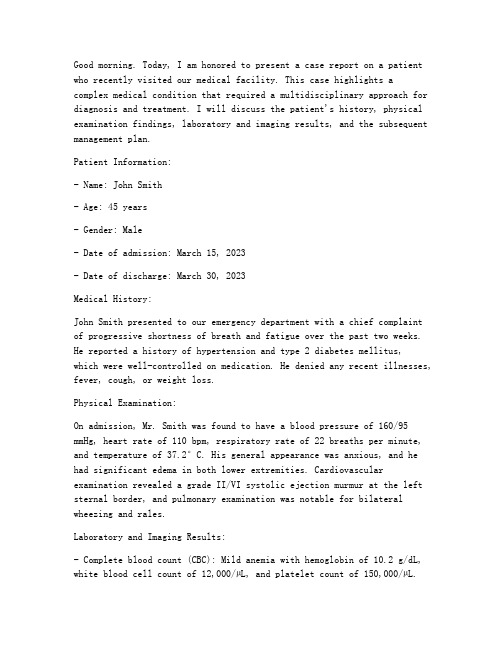
Good morning. Today, I am honored to present a case report on a patient who recently visited our medical facility. This case highlights a complex medical condition that required a multidisciplinary approach for diagnosis and treatment. I will discuss the patient's history, physical examination findings, laboratory and imaging results, and the subsequent management plan.Patient Information:- Name: John Smith- Age: 45 years- Gender: Male- Date of admission: March 15, 2023- Date of discharge: March 30, 2023Medical History:John Smith presented to our emergency department with a chief complaint of progressive shortness of breath and fatigue over the past two weeks. He reported a history of hypertension and type 2 diabetes mellitus,which were well-controlled on medication. He denied any recent illnesses, fever, cough, or weight loss.Physical Examination:On admission, Mr. Smith was found to have a blood pressure of 160/95 mmHg, heart rate of 110 bpm, respiratory rate of 22 breaths per minute, and tempera ture of 37.2°C. His general appearance was anxious, and he had significant edema in both lower extremities. Cardiovascular examination revealed a grade II/VI systolic ejection murmur at the left sternal border, and pulmonary examination was notable for bilateral wheezing and rales.Laboratory and Imaging Results:- Complete blood count (CBC): Mild anemia with hemoglobin of 10.2 g/dL, white blood cell count of 12,000/µL, and platelet count of 150,000/µL.- Electrolytes, renal function tests, and liver function tests were within normal limits.- Serologic tests for HIV, hepatitis B, and hepatitis C were negative.- Chest X-ray: Bilateral pulmonary edema and cardiomegaly.- Echocardiogram: Severe left ventricular dysfunction with an ejection fraction of 25%.- CT scan of the chest: Pulmonary embolism involving the left main pulmonary artery.Diagnosis:Based on the clinical presentation, laboratory findings, and imaging results, the patient was diagnosed with acute pulmonary embolism (PE) with secondary pulmonary hypertension and left ventricular dysfunction.Management Plan:- Anticoagulation therapy with heparin and apixaban was initiated to prevent further thromboembolic events.- Mechanical ventilation was required due to severe respiratory distress.- Inotropic support was provided to manage hypotension and improve cardiac output.- Treatment for secondary pulmonary hypertension included diuretics, nitrates, and inhaled bronchodilators.- Antibiotics were prescribed for a suspected lower respiratory tract infection.- The patient was also started on a low-sodium diet and received education on fluid management.Outcome:After a week of intensive care, Mr. Smith's clinical status improved significantly. His respiratory distress resolved, and he was able to beweaned off mechanical ventilation. His blood pressure stabilized, and the inotropic support was discontinued. By the time of discharge, his ejection fraction had improved to 30%, and he was discharged on apixaban and hydrochlorothiazide to manage his hypertension and diabetes.Conclusion:This case report illustrates the importance of early diagnosis and treatment of pulmonary embolism, which can be a life-threatening condition. The multidisciplinary approach, including emergency medicine, cardiology, pulmonology, and critical care, was crucial in managing this complex case. Mr. Smith's recovery demonstrates the potential for successful outcomes with appropriate medical intervention.Thank you for your attention, and I would be happy to answer any questions you may have.。
病例报告英文范文医护英语
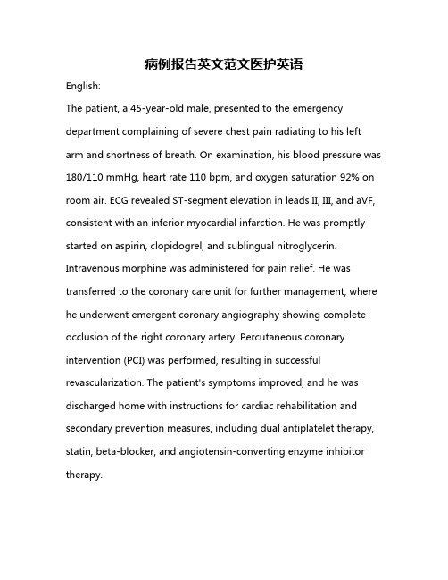
病例报告英文范文医护英语English:The patient, a 45-year-old male, presented to the emergency department complaining of severe chest pain radiating to his left arm and shortness of breath. On examination, his blood pressure was 180/110 mmHg, heart rate 110 bpm, and oxygen saturation 92% on room air. ECG revealed ST-segment elevation in leads II, III, and aVF, consistent with an inferior myocardial infarction. He was promptly started on aspirin, clopidogrel, and sublingual nitroglycerin. Intravenous morphine was administered for pain relief. He was transferred to the coronary care unit for further management, where he underwent emergent coronary angiography showing complete occlusion of the right coronary artery. Percutaneous coronary intervention (PCI) was performed, resulting in successful revascularization. The patient's symptoms improved, and he was discharged home with instructions for cardiac rehabilitation and secondary prevention measures, including dual antiplatelet therapy, statin, beta-blocker, and angiotensin-converting enzyme inhibitor therapy.Translated content:患者为45岁男性,就诊于急诊科,主诉严重胸痛放射至左臂,呼吸急促。
英语病历报告作文格式

英语病历报告作文格式Patient Medical Record Report.Patient Information:Full Name: John Doe.Gender: Male.Age: 45。
Address: 123 Main Street, City, State, Country.Contact Number: +1234567890。
Presenting Complaints:Mr. Doe presented with complaints of persistent chest pain, shortness of breath, and fatigue for the past two months. He reported a history of smoking for the past 20years and occasional alcohol consumption. There was no history of similar episodes in the past.Physical Examination:General: Mr. Doe appeared to be in moderate distress. His skin was pale, and there were no signs of jaundice or cyanosis.Cardiovascular: Heart rate was elevated at 100 beats per minute with irregular rhythm. Auscultation revealed a murmur in the mitral area.Respiratory: Breath sounds were diminished in the left lung base with evidence of crackles.Abdominal: Soft, non-tender abdomen with no organomegaly.Neurological: No focal neurological deficits were noted.Diagnostic Tests:Electrocardiogram (ECG): Showed irregular heartbeat with evidence of atrial fibrillation.Chest X-ray: Revealed enlarged heart with pulmonary congestion.Echocardiogram: Confirmed the presence of mitral valve regurgitation.Medical History:Mr. Doe had a history of hypertension for the past five years, which was well-controlled with medication. He had no known allergies to any medications. His family history was unremarkable for any cardiovascular diseases.Differential Diagnosis:Coronary Artery Disease (CAD)。
英语简要病历报告作文
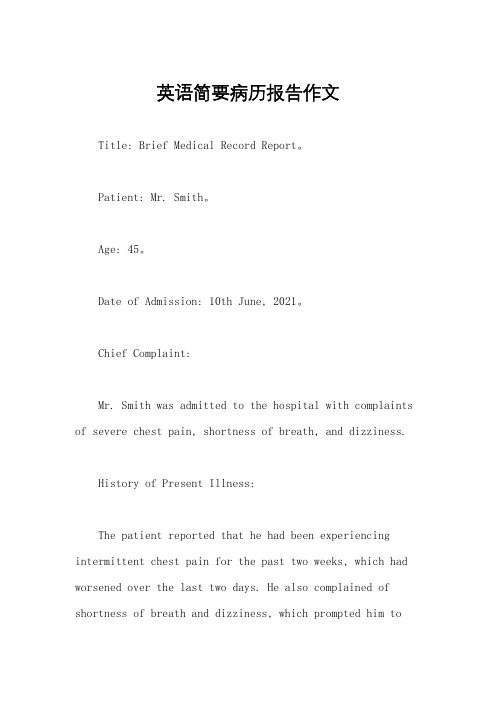
英语简要病历报告作文Title: Brief Medical Record Report。
Patient: Mr. Smith。
Age: 45。
Date of Admission: 10th June, 2021。
Chief Complaint:Mr. Smith was admitted to the hospital with complaints of severe chest pain, shortness of breath, and dizziness.History of Present Illness:The patient reported that he had been experiencing intermittent chest pain for the past two weeks, which had worsened over the last two days. He also complained of shortness of breath and dizziness, which prompted him toseek medical attention.Past Medical History:Mr. Smith has a past medical history of hypertension and hyperlipidemia. He is a smoker and has a family history of coronary artery disease.Physical Examination:On examination, the patient appeared pale and diaphoretic. His blood pressure was elevated at 160/100 mmHg, and his pulse was rapid at 110 beats per minute. Auscultation of the chest revealed diminished breath sounds and crackles in the lower lobes. The patient also had bilateral pedal edema.Diagnostic Tests:The patient underwent an electrocardiogram (ECG) which showed ST-segment elevation in the anterior leads, consistent with acute myocardial infarction. Laboratorytests revealed elevated cardiac enzymes, confirming the diagnosis of myocardial infarction.Treatment:Mr. Smith was immediately started on oxygen therapy, aspirin, and nitroglycerin to relieve his chest pain. He was also given a loading dose of clopidogrel and started on a heparin infusion. The patient was transferred to the coronary care unit for further management.Progress:The patient's symptoms improved with treatment, and his ECG showed resolution of the ST-segment elevation. He was monitored closely for any complications and was discharged after five days with instructions for cardiacrehabilitation and lifestyle modifications.Follow-up:Mr. Smith was advised to follow up with hiscardiologist for further evaluation and management of his coronary artery disease. He was also counseled on smoking cessation, dietary modifications, and regular exercise to reduce his risk of future cardiac events.Conclusion:Mr. Smith presented with acute myocardial infarction and was successfully treated with timely intervention. He was discharged in stable condition with a plan for long-term management to prevent further cardiovascular complications.This brief medical record report highlights the importance of prompt recognition and management of acute myocardial infarction to improve patient outcomes and reduce the risk of complications. It also emphasizes the need for comprehensive follow-up and lifestyle modifications to prevent future cardiac events.。
病例报告英语作文模板高中

病例报告英语作文模板高中Title: A Case Report: The Symptoms, Diagnosis, and Treatment of Influenza。
Introduction:Influenza, commonly known as the flu, is a contagious respiratory illness caused by influenza viruses. It can cause mild to severe illness and even lead to hospitalization or death, especially in high-risk groups. Here, we present a case report of a patient with influenza, detailing their symptoms, diagnosis, and treatment.Patient History:The patient, a 35-year-old male, presented to theclinic with complaints of fever, cough, sore throat, body aches, fatigue, and headache. The symptoms had started suddenly two days prior to the visit and had progressively worsened. The patient denied any recent travel history orcontact with sick individuals but reported exposure to crowded areas due to work.Clinical Examination:On examination, the patient appeared ill and fatigued. Vital signs revealed a temperature of 39.2°C (102.5°F), heart rate of 100 beats per minute, respiratory rate of 22 breaths per minute, and blood pressure within normal limits. Examination of the respiratory system revealed bilateral coarse crackles on auscultation.Diagnostic Evaluation:Given the patient's clinical presentation during the influenza season, a presumptive diagnosis of influenza was made. Nasopharyngeal swab specimens were collected for laboratory confirmation. Rapid influenza diagnostic tests (RIDTs) were performed, which yielded positive results for influenza A virus. Additionally, reverse transcription-polymerase chain reaction (RT-PCR) testing confirmed the presence of influenza A virus subtype H3N2.Treatment:Based on the diagnosis of influenza A, the patient was initiated on antiviral therapy with oseltamivir (Tamiflu). The treatment regimen included oral oseltamivir 75 mg twice daily for a duration of five days. In addition, supportive measures were implemented to alleviate symptoms and prevent complications. These measures included adequate hydration, rest, and over-the-counter analgesics for fever and body aches.Clinical Course:Following initiation of antiviral therapy and supportive measures, the patient's symptoms gradually improved over the course of the next week. Fever subsided within 48 hours of starting oseltamivir, and respiratory symptoms began to resolve. The patient was advised to complete the full course of antiviral therapy and to follow up if symptoms persisted or worsened.Discussion:Influenza is a common viral illness characterized by respiratory symptoms and systemic manifestations. It is typically diagnosed based on clinical presentation and confirmed by laboratory testing. Early initiation of antiviral therapy, such as oseltamivir, can reduce the severity and duration of symptoms, especially if started within 48 hours of symptom onset. Supportive measures play a crucial role in managing influenza, particularly in alleviating symptoms and preventing complications.Conclusion:This case report highlights the clinical presentation, diagnosis, and management of influenza in a young adult male. Prompt recognition of symptoms, timely diagnosis, and initiation of appropriate treatment are essential in managing influenza and preventing its spread in the community. Healthcare providers should remain vigilant during influenza season and advocate for vaccination as themost effective preventive measure against influenza infection.。
英文病例汇报实用句型
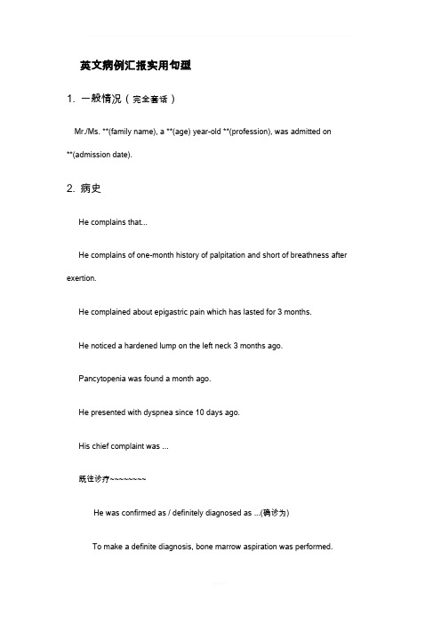
英文病例汇报实用句型1. 一般情况(完全套话)Mr./Ms. **(family name), a **(age) year-old **(profession), was admitted on**(admission date).2. 病史He complains that...He complains of one-month history of palpitation and short of breathness after exertion.He complained about epigastric pain which has lasted for 3 months.He noticed a hardened lump on the left neck 3 months ago.Pancytopenia was found a month ago.He presented with dyspnea since 10 days ago.His chief complaint was ...既往诊疗~~~~~~~~He was confirmed as / definitely diagnosed as ...(确诊为)To make a definite diagnosis, bone marrow aspiration was performed.He was suspected as...(疑似)The discomfort tended to worsening, which urged him to seek for medical care.He has been given 3 cycles of DA regimen for chemotherapy and complete remission was achieved only after the first cycle.He was given the thyroidectomy of the left lobe in local hospital.He was treated with antibiotics (details unknown), which didn't take effect as expected.The general condition is good at present.He was pain free now and hemodynamically stable.3. 查体Nothing noteworthy was found in the physical examination.There was nothing remarkable in the physical examination except for…The physical examination was otherwise normal except that…(上点小菜~~~血液科常见体征)皮肤粘膜generalized pallor,scattered petechiae,oral mucosal hematoma淋巴结enlarged lymph nodes头部yellow eyes (yellow-stained sclera)胸部tenderness in sternum,coarse breath sound, cardiac murmur, arrhythmia腹部enlargement of liver,splenomegaly4.辅助检查The laboratory findings suggested/indicated/demonstrated/showed that…Bone marrow film was performed, which confirmed the diagnosis of ALL.The results of blood routine showed that WBC count was 4,000 /cm3, while NEU count 2,500/cm3, hemoglobin 100 g/L, PLT count 100,000 /cm3.(/cm3 is pronounced as per cubic millimeter)Chest CT scan supported the diagnosis of NHL.Welcome To Download !!!欢迎您的下载,资料仅供参考!。
英文病历报告作文模板
英文病历报告作文模板Patient Information- Name: [Patient's Full Name]- Gender: [Male/Female]- Age: [Patient's age]- Date of Admission: [MM/DD/YYYY]Chief ComplaintThe patient presented with [specific symptoms/complaints] which started [duration].History of Present IllnessThe patient reported [detailed description ofsymptoms/complaints]. The symptoms worsened over the past [duration]. The patient experienced [associated symptoms] and tried [any self-medication or home remedies] but noticed no improvement. There was no history of trauma or injury.Past Medical HistoryThe patient has a history of [chronic/acute medical conditions, if any] which includes [specific conditions]. The patient has taken[previous medications/treatments] for these conditions.Social HistoryThe patient has a [specific occupation] and lives in [specific area]. The patient does [specific habits] such as smoking or drinking alcohol [frequency]. There is no significant family medical history.Physical Examination- Vital Signs:- Blood Pressure: [value] mmHg- Heart Rate: [value] bpm- Respiratory Rate: [value] bpm- Temperature: [value]C- General Appearance:The patient appears [general appearance of the patient].- Systemic Examination:- Cardiovascular: [specific findings]- Respiratory: [specific findings]- Gastrointestinal: [specific findings]- Neurological: [specific findings]- Musculoskeletal: [specific findings]Laboratory and Imaging Findings- Blood Test Results:- Complete Blood Count: [values]- Biochemical Profile: [values]- Others: [specific findings]- Imaging:- [Specific imaging tests performed]- Results: [specific findings]DiagnosisAfter evaluating the patient's medical history, physical examination, and laboratory/imaging findings, the following diagnosis was made:[Primary Diagnosis]Treatment and ManagementThe patient was started on [specific treatment plan] which includes [medications, therapies, or procedures]. The patient wasadvised to [specific instructions] and scheduled for [follow-up tests/appointments, if any].Follow-upThe patient will be followed up in [specific time frame] to assess the response to treatment and manage any complications that may arise. The patient was given contact information for any urgent concerns or changes in symptoms.Discussion and ConclusionThis case report highlights the presentation, evaluation, and management of a patient with [specific condition]. The patient's symptoms were appropriately addressed through a systematic approach involving history taking, physical examination, and laboratory/imaging investigations. The provided treatment plan aims to address the underlying cause and improve the patient's overall well-being. Continuous monitoring and follow-up will guide further management decisions.Note: This medical case report is fictional and serves as a template for educational purposes. Any resemblance to actualpatients is purely coincidental.。
英文版病例报告作文

英文版病例报告作文Patient: Mr. Smith。
Age: 45。
Gender: Male。
Complaint: Severe chest pain and shortness of breath。
History of Present Illness: Mr. Smith presented to the emergency room with complaints of severe chest pain and shortness of breath. He described the pain as crushing and radiating to his left arm. He also reported feeling lightheaded and dizzy. The symptoms started suddenly while he was at work and have been ongoing for the past 30 minutes.Past Medical History: Mr. Smith has a history of hypertension and hyperlipidemia. He is a smoker and admits to occasional alcohol consumption. He has no known drugallergies.Family History: His father had a history of myocardial infarction at the age of 50. His mother is alive and well with no significant medical history.Social History: Mr. Smith works as a sales manager and is under a lot of stress. He smokes a pack of cigarettes per day and drinks alcohol socially. He is currently single and lives alone.Physical Examination: On examination, Mr. Smith appeared diaphoretic and in distress. His blood pressure was 180/100 mmHg, heart rate was 110 beats per minute, and respiratory rate was 24 breaths per minute. His oxygen saturation was 92% on room air. Cardiac auscultation revealed muffled heart sounds and bilateral crackles on lung auscultation.Diagnostic Tests: ECG showed ST-segment elevation in leads II, III, and aVF, consistent with an inferior myocardial infarction. Cardiac enzymes were elevated,confirming the diagnosis of acute myocardial infarction.Treatment: Mr. Smith was immediately started on aspirin, clopidogrel, and heparin. He was also given nitroglycerinfor chest pain and oxygen supplementation to maintain oxygen saturation above 94%. He was then taken for emergent cardiac catheterization and percutaneous coronary intervention.Follow-up: Mr. Smith's symptoms improved after the intervention, and he was discharged home on dualantiplatelet therapy, statin, and beta-blocker. He was advised to quit smoking and reduce alcohol consumption. He was also referred to a cardiac rehabilitation program for further management.Outcome: Mr. Smith's condition improved significantly after the intervention, and he has been compliant with his medications and lifestyle modifications. He has not had any recurrent chest pain or shortness of breath since the hospitalization.。
- 1、下载文档前请自行甄别文档内容的完整性,平台不提供额外的编辑、内容补充、找答案等附加服务。
- 2、"仅部分预览"的文档,不可在线预览部分如存在完整性等问题,可反馈申请退款(可完整预览的文档不适用该条件!)。
- 3、如文档侵犯您的权益,请联系客服反馈,我们会尽快为您处理(人工客服工作时间:9:00-18:30)。
s s
23--35s 14--21s
FBG
3.72
g/L
1.7--4.5g/L
Liver function test
TP ALB ALP GGT ALT AST 67.5 34.1 51.8 23.2 6.9 12 g/L g/L U/L U/L U/L U/L
4/21
65--85g/L 40--55g/L 35--100U/L 7--45U/L 7--40U/L 13--35U/L
Discusion:
further treatment?
40--75%
4.82 4.8
4.74 4.49 4.36 X10~12/L 3.8--5.1
HB
110
114
112
106
103
g/L
115-150g/L 125-350X10~9/ L
PLT
398
373
389
413
370
X10~9/L
urine routine
LEU UWBC 3+ 44.3 /ul -
Imageological examination: 1、肝胆胰脾、双肾输尿管、膀胱、子宫 、附件无殊。 2、右下腹非均质性包块。
CT
Description:
右侧回盲部见斑片状软组织密度影及条索影 ,边缘毛糙,约29x43mm,增强明显不均匀强 化,内低密度坏死去无强化,右侧腹壁下见略 不规则环形强化影。阑尾区少量积气,见高密 度结石夹留置,呈术后改变,周围见多个稍大 淋巴结,增强明显强化。
Two months after appendicectomy;
Recurrent abdominal pain associated
with fever for one month
Present history 1
right ventral abdominal pain,2 months ago appendicectomy in Hongkong abdominal pain associated with fever for one month
Rebound tenderness(+);
Blood routine
21-Apr 29-Apr 4-May 8-May 10-May WBC
14.37 12.3 10.34 14.619.42 X10~9 /L 3.5--9.5
NEUT% 82.4 RBC
83.3 85.5 88.7 84.1 %
CT
1、阑尾术后改变并回盲部、阑尾周围脓肿, 建议治疗后复查; 2、阑尾周围多发淋巴结影; 3、肝右叶后上段不典型小血管瘤多考虑,随 访; 4、阴道穹窿部少量积液,请结合临床考虑。
Diagnosis
1. appendicitis after appendectomy
2. Abdominal Abscess
CASE REPORT
May 12th, 2016
Department of General Surger
Medical Records for Admission
Name: Sex: female Age: 31 Registration No.: Date of admission:
Chief compliant
Tumor marker
AFP
CEA CA125
4/7
11.2
0.48 22.1
ng/ml
ng/ml U/mL
0.89--8.78ng/ml
0--10ng/ml 0--35U/mL
CA153
CA199
5.5
7.5
U/mL
U/mL
0--28U/mL
0--37U/mL
LAB FINDINGS
ESR 24--23/ul
URBC EC
BXBGBSY HXBGBSY
8.4 4.8
8 1.5
/ul /ul
/HPF /HPF
0--18/ul 0--3/ul
0--5个/HP 0--3个/HPF
coagulation function
PT
INR APTT TT
15.3
1.33 37.9 18.2
s
10--14s
maximum body temperature up to 38.9℃
Family History
There are no similar diseases in his family
Physical Examination
right low abdominal pains tenderness(+)、