儿科英文病历模板
新生儿科英文病历
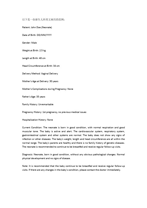
以下是一份新生儿科英文病历的范例:Patient: John Doe (Neonate)Date of Birth: DD/MM/YYYYGender: MaleWeight at Birth: 2.5 kgLength at Birth: 48 cmHead Circumference at Birth: 34 cmDelivery Method: Vaginal DeliveryMother's Age at Delivery: 30 yearsMother's Complications during Pregnancy: NoneFather's Age: 35 yearsFamily History: UnremarkablePregnancy History: 1st pregnancy, no previous medical issuesHospitalization History: NoneCurrent Condition: The neonate is born in good condition, with normal respiration and good muscular tone. The baby is active and alert. The cardiovascular system, respiratory system, gastrointestinal system and other systems are normal. The baby does not show any signs of infection or other diseases. The baby's weight, length and head circumference are all within the normal range. The baby's parents are healthy and there is no family history of genetic diseases. The neonate is recommended to continue to be breastfed and receive regular follow-up visits.Diagnosis: Neonate, born in good condition, without any obvious pathological changes. Normal physical development and no signs of disease.Note: It is recommended that the baby continue to be breastfed and receive regular follow-up visits. If there are any changes in the baby's condition, please contact the doctor immediately.。
儿科腺样体肥大门诊病历范文

儿科腺样体肥大门诊病历范文英文回答:Pediatric Adenotonsillar Hypertrophy Clinic Visit Note.Chief Complaint: Recurrent tonsillitis and nasal congestion.History of Present Illness:The patient is a 9-year-old male who presents with a 6-month history of recurrent tonsillitis. He has had 4 episodes of sore throat, fever, and swollen lymph nodes in the past 6 months. He has also had persistent nasal congestion and difficulty breathing through his nose. He has been treated with antibiotics for his tonsillitis, but the symptoms have recurred each time.Past Medical History:The patient has no significant past medical history. He is up-to-date on his vaccinations.Family History:The patient's mother has a history of recurrent tonsillitis.Social History:The patient lives at home with his parents and two siblings. He is in the 4th grade and doing well in school. He enjoys playing sports and spending time with his friends.Review of Systems:Constitutional: Fatigue, fever, night sweats.Head: Nasal congestion, difficulty breathing throughthe nose, sore throat.Eyes: No complaints.Ears: No complaints.Nose: Nasal congestion, difficulty breathing through the nose.Throat: Sore throat, swollen tonsils.Cardiovascular: No complaints.Respiratory: Difficulty breathing through the nose.Gastrointestinal: No complaints.Genitourinary: No complaints.Musculoskeletal: No complaints.Neurological: No complaints.Psychiatric: No complaints.Physical Examination:General: The patient is a well-nourished, well-developed 9-year-old male in no acute distress.Head: Normocephalic. No masses or tenderness.Eyes: Pupils equal and reactive to light. Extraocular movements intact. No conjunctival injection or discharge.Ears: Tympanic membranes are intact and mobile. No otorrhea.Nose: Nasal mucosa is inflamed and swollen. There is a deviated nasal septum to the right.Throat: Tonsils are enlarged and erythematous. There is a white exudate on the tonsils.Neck: No lymphadenopathy.Cardiovascular: Regular rate and rhythm. No murmurs,gallops, or rubs.Respiratory: Lungs are clear to auscultation bilaterally. No wheezes or rales.Gastrointestinal: Abdomen is soft and non-tender. No hepatosplenomegaly.Genitourinary: No abnormalities noted.Musculoskeletal: No abnormalities noted.Neurological: Cranial nerves are intact. Motor and sensory exams are normal.Assessment:Adenotonsillar hypertrophy.Recurrent tonsillitis.Nasal congestion.Plan:Adenotonsillectomy.Antibiotics for recurrent tonsillitis.Nasal saline irrigations.Follow-up:The patient will follow up in 2 weeks for a post-operative visit.中文回答:儿科腺样体肥大手术门诊病历。
湘雅二医院儿科英文病例模板
CHIEF COMPLAINT
Fever and cough for 10 days with leg pain.
Pharyngalgia and fever for 4 days. [færɪn'gældʒɪə] Cough and dyspnea for 2 days. [dɪs'pni:ə]
PHYSICAL EXAMINATION
Heart: The point of maximal impulse was in left 5th intercostal space on the mid clavicular line. The border of cardiac dullness was normal. Cardiac sounds were strong without splitting and heart rate was 140/min. Cardiac rhythm was regular. No pathological murmur was heard.
T 39.5℃, P 140/min, R 38/min, BP 80/50mmHg Temperature was thirty-nine point five centigrade, Heart rate was one hundred and forty beats per minute, Respiratory rate was thirty-eight times per minute, Blood pressure was eighty over fifty millimeter of mercury
supplementary foods [ˌsʌplɪˈmentri] vaccination [ˌvæksɪ'neɪʃn]
儿科腺样体肥大门诊病历范文

儿科腺样体肥大门诊病历范文英文回答:Pediatric Adenoid Hypertrophy Outpatient Medical Record.Patient Information:Name: John Smith.Age: 8 years old.Gender: Male.Date of Visit: January 15, 2022。
Chief Complaint: Difficulty breathing through the nose, snoring at night, recurrent ear infections.Present Illness:I have been experiencing difficulty breathing through my nose for the past few months. It feels like my nose is constantly blocked, and I have to breathe through my mouth most of the time. At night, I snore loudly, which disturbs my sleep. I also have been having recurrent ear infections, which are painful and affect my hearing. These symptoms have been bothering me and affecting my daily activities.Medical History:I have a history of recurrent upper respiratory tract infections, including colds and sore throats. I have also had a few episodes of tonsillitis in the past. My parents mentioned that I had similar symptoms when I was younger, but they improved as I grew older.Physical Examination:On examination, I was found to have nasal congestion with a nasal voice. My tonsils were not enlarged, but my adenoids were visibly enlarged, obstructing the nasal passage. My ears appeared normal externally, butexamination with an otoscope revealed fluid accumulation behind the eardrums.Diagnosis:Based on the history and physical examination findings, I have been diagnosed with pediatric adenoid hypertrophy. This condition refers to the enlargement of the adenoids, which are lymphoid tissues located at the back of the nasal cavity. Adenoid hypertrophy can cause nasal obstruction, snoring, and recurrent ear infections.Treatment Plan:To manage my condition, the following treatment plan has been recommended:1. Nasal Steroid Spray: I will be prescribed a nasal steroid spray to reduce the inflammation and shrink the adenoids, which will help improve nasal breathing.2. Antibiotics: Since I have recurrent ear infections,I will be prescribed a course of antibiotics to treat the current infection and prevent future episodes.3. Adenoidectomy: If my symptoms persist despite medical treatment or if there are complications such as recurrent sinusitis or middle ear infections, my doctor may recommend adenoidectomy. This surgical procedure involves the removal of the adenoids to alleviate the symptoms and prevent further complications.Follow-up:I have been advised to follow up with my doctor in two weeks to assess the response to treatment and discuss further management options if needed.中文回答:儿科腺样体肥大门诊病历范文。
儿科英文病历 case report
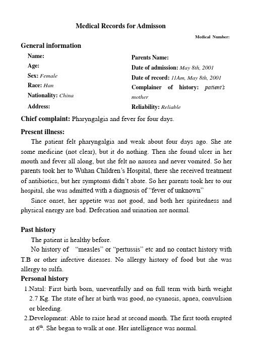
Medical Records for AdmissonMedical Number: General informationName:Age:Sex: Female Race:Han Nationality:China Address: Parents Name:Date of admission: May 8th, 2001 Date of record: 11Am, May 8th, 2001 Complainer of history: patient’s motherReliability: ReliableChief complaint: Pharyngalgia and fever for four days.Present illness:The patient felt pharyngalgia and weak about four days ago. She ate some medicine (not clear), but it do nothing. Then she found ulcer in her mouth and fever all along, but she felt no nausea and never vomited. So her parents took her to Wuhan Children’s Hospital, there she received treatment of antibiotics, but her symptom s didn’t abate. So her parents took her to our hospital, she was adm itted with a diagnosis of “fever of unknown”Since onset, her appetite was not good, and both her spiritedness and physical energy are bad. Defecation and urination are normal.Past historyThe patient is healthy before.No history of “measles” or “pertussis” etc and no contact history with T.B or other infective diseases. No allergy history of food but she was allergy to sulfa.Personal history1.Natal: First birth born, uneventfully and on full term with birth weight2.7 Kg. The state of her at birth was good, no cyanosis, apnea, convulsionor bleeding.2.Development: Able to raise head at second month. The first tooth eruptedat 6th. She began to walk at one. Her intelligence was normal.3.Nutrition: She was only feeded with breast milk before she was 6 monthsold. Then the additives were added. She was weaned from the breast at 14th month.4.Immunization: Inoculated on schedule after birth (such as B.C.G, D.P.Tand smallpox voccination).Physical examinationT 39.5℃, P 120/min, R 30/min, BP 110/90mmHg. She is well developed and moderately nourished. Active position. The skin was not stained yellow. No cyanosis. No pigmentation. No skin eruption. Spider angioma was not seen. No pitting edema. Superficial lymph nodes were found enlarged in her neck, but no flare and tenderness.HeadCranium: Hair was black and well distributed. No deformities. No scars. No masses. No tenderness.Ear: Bilateral auricles were symmetric and of no masses. No discharges were found in external auditory canals. No tenderness in mastoid area. Auditory acuity was normal.Nose:No abnormal discharges were found in vetibulum nasi. Septum nasi was in midline. No nares flaring. No tenderness in nasal sinuses.Eye:Bilateral eyelids were not swelling. No ptosis. No entropion. Conjunctiva was not congestive. Sclera was anicteric. Eyeballs were not projected or depressed. Movement was normal. Bilateral pupils were round and equal in size. Direct and indirect pupillary reactions to light were existent.Mouth: Oral mucous membrane was not smooth, and there were ulcer can be seen. Tongue was in midline. Pharynx was congestive. Tonsils were not enlarged.Neck: Symmetric and of no deformities. No masses. Thyroid was not enlarged. Trachea was in midline.ChestChestwall: Veins could not be seen easily. No subcutaneous emphysema.Intercostal space was neither narrowed nor widened. No tenderness. Thorax: Symmetric bilaterally. No deformities.Breast: Symmetric bilaterally.Lungs:Respiratory movement was bilaterally symmetric with the frequency of 30/min. thoracic expansion and tactile fremitus were symmetric bilaterally. No pleural friction fremitus. Resonance was heard during percussion. No abnormal breath sound was heard. No wheezes. No rales.Heart:No bulge and no abnormal impulse or thrills in precordial area. The point of maximum impulse was in 5th left intercostal space inside of the mid clavicular line and not diffuse. No pericardial friction sound. Border of the heart was normal. Heart sounds were strong and no splitting. Rate 120/min. Cardiac rhythm was regular. No pathological murmurs. Abdomen:Flat and soft. No bulge or depression. No abdominal wall varicosis. Gastralintestinal type or peristalses were not seen. There was not tenderness and rebound tenderness on abdomen or renal region. Liver was touched 1.5cm under the right costal margin. Spleen was 0.5 cm under the left. No masses. Fluidthrill negative. Shifting dullness negative. Borhorygmus 5/min. No vascular murmurs.Extremities: No articular swelling. Free movements of all limbs.Neural system:Physiological reflexes were existent without any pathological ones.Genitourinary system: Not examed.Rectum: not exanedInvestigationBlood-Rt: Hb 59g/L RBC 1.90T/L WBC 0.8G/L PLT 55G/LBlood cytology: A few immature lymphocytes could be seen.History summary1.Patient was female, 13 years old2.Pharyngalgia and fever for four days.3.No special past history.4.Physical examination: T 39.5℃, P 120/min, R 30/min, BP 110/90mmHg Superficial lymph nodes were found enlarged in her neck, but no flare and tenderness. Liver was touched 1.5cm under the right costal margin. Spleen was 0.5 cm under the left. No other positive signs.5.investigation information:Blood-Rt: Hb 59g/L RBC 1.90T/L WBC 0.8G/L PLT 55G/LBlood cytology: A few immature lymphocytes could be seen.Impression: Fever of UnkownAcute Lymphocyte leukaemia?Signature:。
儿童心肌炎病历模板范文

儿童心肌炎病历模板范文Medical Record Template for Childhood Myocarditis.Patient Information.Name:Date of Birth:Medical Record Number:History of Present Illness.Onset of symptoms (date and time):Symptoms: Describe the child's symptoms, including chest pain, shortness of breath, fatigue, fever, and rash.Duration of symptoms:Any recent illnesses or exposures:Family history of myocarditis or heart disease:Physical Examination.Vital signs (temperature, pulse, respiratory rate, blood pressure):General appearance: Describe the child's overall appearance, including any signs of distress, pallor, or cyanosis.Auscultation of the heart: Describe the presence of any murmurs, gallops, or extra heart sounds.Percussion of the heart: Outline the size and location of the heart.Respiratory examination: Describe the child's respiratory effort, breath sounds, and any evidence of wheezing or crackles.Abdominal examination: Assess for any hepatomegaly or splenomegaly.Investigations.Electrocardiogram (ECG): Describe any abnormalities, such as ST-segment elevation, T-wave inversion, or arrhythmias.Chest X-ray: Outline the size and shape of the heart, as well as any signs of pulmonary edema or pleural effusions.Echocardiogram: Evaluate the size and function of the heart, including ejection fraction, wall thickness, and any valvular abnormalities.Cardiac magnetic resonance imaging (MRI): Assess for myocardial inflammation and scarring.Blood tests: Obtain complete blood count, electrolytes,liver function tests, and cardiac enzymes (troponin, creatine kinase-MB).Diagnosis.Childhood myocarditis.Treatment.Supportive care: Provide oxygen, fluid resuscitation, and inotropic support as needed.Anti-inflammatory therapy: Administer corticosteroids (e.g., prednisone) to reduce inflammation.Immunosuppressive therapy: Consider immunosuppressive drugs (e.g., azathioprine, methotrexate) in severe cases.Antiviral therapy: If a viral etiology is suspected, administer antiviral medications (e.g., acyclovir, valacyclovir).Other: Additional therapies may include antibiotics (if infection is present), diuretics (for fluid retention), and antiarrhythmic drugs (for arrhythmias).Follow-Up.Schedule regular follow-up appointments to monitor the child's progress and adjust treatment as needed.Perform serial echocardiograms to assess cardiac function and identify any structural changes.Consider exercise testing to evaluate the child's tolerance for activity.Provide education and counseling to the child and family regarding the condition and its management.中文回答:儿童心肌炎病历模板范本。
早产儿门诊病历书写范文

早产儿门诊病历书写范文英文回答:Patient Name: XXX.Age: XXX.Date of Birth: XXX.Gender: XXX.Date of Admission: XXX.Chief Complaint: The patient was born prematurely at 32 weeks of gestation and is presenting with respiratory distress, feeding difficulties, and jaundice.History of Present Illness: The patient was born prematurely and has been experiencing respiratory distress since birth. The patient also has difficulty feeding andhas developed jaundice. The patient was admitted to the neonatal intensive care unit (NICU) for further evaluation and management.Past Medical History: The patient has a history of premature birth and is currently being monitored for potential complications associated with prematurity.Family History: No significant family history reported.Physical Examination: On examination, the patient appears jaundiced. The patient is tachypneic with increased work of breathing. The patient's abdomen is soft and non-tender. The patient's weight is below the 10th percentile for gestational age.Assessment and Plan: The patient is being evaluated for respiratory distress, feeding difficulties, and jaundice. The patient will undergo further diagnostic testing, including blood tests and imaging studies, to determine the underlying cause of the symptoms. The patient will also receive supportive care, including respiratory support andnutritional support, as needed.Disposition: The patient will be admitted to the NICU for further management and monitoring.中文回答:患者姓名,XXX.年龄,XXX.出生日期,XXX.性别,XXX.入院日期,XXX.主诉,患者早产,32周孕龄出生,出生后出现呼吸困难、进食困难和黄疸。
儿科入院病例书写范文
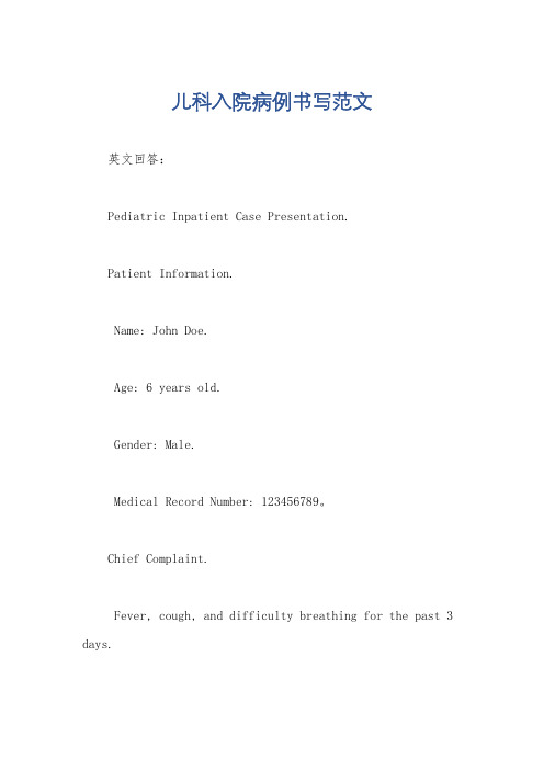
儿科入院病例书写范文英文回答:Pediatric Inpatient Case Presentation.Patient Information.Name: John Doe.Age: 6 years old.Gender: Male.Medical Record Number: 123456789。
Chief Complaint.Fever, cough, and difficulty breathing for the past 3 days.History of Present Illness.John Doe is a 6-year-old male who presents to the emergency department with a 3-day history of fever, cough, and difficulty breathing. The fever has been intermittent, reaching a maximum of 102.2°F. The cough is non-productive and has been getting worse over the past 3 days. Thedifficulty breathing has also been getting worse, and John Doe has been having difficulty sleeping at night.John Doe has no significant past medical history. He is up-to-date on all of his vaccinations. He lives with both parents and has no known allergies.Physical Examination.General: John Doe is a well-developed, well-nourished male in no acute distress.HEENT: John Doe's head is normocephalic and atraumatic. His ears are symmetrical and without discharge. His nose is midline and without discharge. His oropharynx is clearwithout erythema or exudates.Neck: John Doe's neck is supple without lymphadenopathy or thyromegaly.Chest: John Doe's chest is symmetrical with good expansion. Auscultation of the lungs reveals bilateral wheezing.Abdomen: John Doe's abdomen is soft, non-tender, and non-distended. No masses or hepatosplenomegaly are appreciated.Extremities: John Doe's extremities are warm and well-perfused. No clubbing, cyanosis, or edema is noted.Laboratory Studies.Complete blood count: White blood cell count12,000/mm3, hemoglobin 12.5 g/dL, platelets 300,000/mm3。
- 1、下载文档前请自行甄别文档内容的完整性,平台不提供额外的编辑、内容补充、找答案等附加服务。
- 2、"仅部分预览"的文档,不可在线预览部分如存在完整性等问题,可反馈申请退款(可完整预览的文档不适用该条件!)。
- 3、如文档侵犯您的权益,请联系客服反馈,我们会尽快为您处理(人工客服工作时间:9:00-18:30)。
Nanjing children’s hospitalMedical Records for AdmissonWard:321 Bed Number:32178 Medical Number: 696235 General informationName:Son of ***Sex: MaleAge: 3 hBirthplace: *** county,Anhui provinceRace:HanAddress:***town,***county,Anhu i province Date of admission:3:31pm Oct 16th,2015Date of record: 3:31pm Oct 16th,2015Parents Name: father *** Mother ***Complainer of history: patient’s fatherReliability: ReliableChief complaint: Shortness of breath and moaning for 3h Present illness:The afflicted baby was delivered 3h ago and had instaneous shortness of breath along with obtuse response and moaning.No aspnea or seizure or scream were observed. In local Hospital he received treatment of “naloxone、mezlocillin and Vit K1”, but his symptoms didn’t abate. So the parents took him to our hospital, he was admitted with a diagnosis of “acute respiratory dyspnea syndrome” .Breast feed has not been initiated.He has not vomitted,defecated or urinated since he was born,.Past historyNo history of any kind of disease.No history of infective diseases. No history of picking front tooth.No history of wiping oral cavity.Personal history1.Natal: second birth born,Gestational age:28 weeks;Multiplets:No;In Vitro fertilization:NoCause of premature birth:unknown;Time of birth:2015-10-16 11:00Mode of delivery:spontaneous delivery,born in local hospital Deliverer:practioner;Apgar scale:1min 7’,5min unknownResuscitation:No;Birth weight:1350gThe characteristics and volume of amniotic fluid remains unknown,as well as whether there was fetal aspiration of amniotic fluid or meconium.we also have no idea if there was Premature rupture of the amniotic fluid.Umbilical cord is normal;Jaundice:unknown2.Nutrition: breast feed has not been initiated.He had no signof defecation or vomit.3.Immunization: B.C.G and hepatitis B vaccine have not beeninoculated after birth;Other vaccination remains unknown. Family historyFather:Name:***;Age:29ys;Vocation:worker;Healthcondition:good;Blood type:ABMother:Name:***;Age:26ys;Vocaton:peasant;Healthcondition:good;Blood type:unknownMother’s history of pregnancies and births:2 pregnancies,no abortion;This pregnancy was normal.Existing siblings: a healthy sister of 2 and half years oldNo history of drug allergy or physical contact with pets;No history of drug abuse or venereal disease.Physical examinationT 34.1℃, P 126/min, R 70/min, BP 54/26mmHg.Weight 1380g. General descriptionHis consciousness is clear;He has Premature appearance,slight cyanosis on the face.He had shortness of breath along with obtuse response and moaning.Skin and mucosaThere is slight cyanosis of the skin.Elasticity is good. No edema.No sign of dehydration.No jaundice.No dermatoma.No bleeding or rash.HeadNo deformities of the Cranium.Anterior fontanelle 2cm*2cm,skin tensity is flat and soft..Hair is black.No hematoma or edema.Seams at the width of 2mm have not been sealed.Eye:Bilateral pupils were round and equal in size about 2mm*2mm. Movement was normal.Direct and indirect pupillary reactions to light were sharp.Conjunctiva was not congestive.No discharges were found around the eyes.Ear:No discharges were found in external auditory canals. No deformity in external auditory canals.Bilateral auricles werewell developed.Nose:No nares flaring. No deformity.Mouth: Oral mucous membrane was intact. No cleft palate. Neck: No resistence during examination. No masses at bilateral sternocleidomastoid muscles.ChestThorax: Symmetric bilaterally.Lungs:Respiratory movement was hyperactive with three concave sign.Breath sound was rough. Moan and goan can be heard as well.Heart:Heart rate 126/min.Heart sounds were strong and no splitting. Cardiac rhythm was regular. No pathological murmurs.Abdomen:Flat and soft. Skin was normal.Gastralintestinal type or peristalses were not seen. No discharges or flare were found in the umbilical area.No umbilical hernnia. Abdominal muscles were soft.Liver was soft.Liver was touched 2cm under the right costal margin but unreachable in subxiphoid regions. Spleen was unreachable under the left costal margin. No masses. Shifting dullness negative. Borborygmus was existent. Spine and Extremities: No deformity. Muscle tensity of all limbs were normal.Extremitis were warm.Capillary filling time was 2s. Neural system: Physiological reflexes were existent without any pathological ones.Anus and External genitalia: External genitalia appeared to be male’s.Anus was existent.No hypospadias.No testes were touched in bilateral scrotum.Specialized InvestigationLaboratory and instrument inspectionBlood sugar(2015-10-16,our hopital):10.8mmol/LClinical Impression:1. ARDS(Acute respiratory dyspnea syndrome) of prematurity2.pneumonia of prematurity3.very low birth weight infantSignature:Diagnosis for admission:1. ARDS(Acute respiratory dyspnea syndrome)of prematurity2.pneumonia of prematurity3.very low birth weight infantSignature:。