英文病历样本
新生儿科英文病历
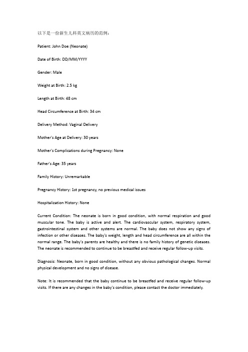
以下是一份新生儿科英文病历的范例:Patient: John Doe (Neonate)Date of Birth: DD/MM/YYYYGender: MaleWeight at Birth: 2.5 kgLength at Birth: 48 cmHead Circumference at Birth: 34 cmDelivery Method: Vaginal DeliveryMother's Age at Delivery: 30 yearsMother's Complications during Pregnancy: NoneFather's Age: 35 yearsFamily History: UnremarkablePregnancy History: 1st pregnancy, no previous medical issuesHospitalization History: NoneCurrent Condition: The neonate is born in good condition, with normal respiration and good muscular tone. The baby is active and alert. The cardiovascular system, respiratory system, gastrointestinal system and other systems are normal. The baby does not show any signs of infection or other diseases. The baby's weight, length and head circumference are all within the normal range. The baby's parents are healthy and there is no family history of genetic diseases. The neonate is recommended to continue to be breastfed and receive regular follow-up visits.Diagnosis: Neonate, born in good condition, without any obvious pathological changes. Normal physical development and no signs of disease.Note: It is recommended that the baby continue to be breastfed and receive regular follow-up visits. If there are any changes in the baby's condition, please contact the doctor immediately.。
门诊病历英文模板
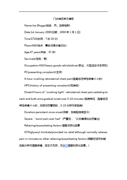
门诊病历英文模板Name:Joe Bloggs(姓名:乔。
伯劳格斯)Date:1st January 2000(日期:2000年1月1日)Time:0720(时间:7时20分)Place:A&E(地点:事故与急诊登记处)Age:47 years(年龄:47岁)Sex:male(性别:男)Occupation:HGV(heavy goods vehicle)driver(职业:大型货运卡车司机) PC(presenting complaint)(主诉)4-hour crushing retrosternal chest pain(胸骨后压榨性疼痛4小时)HPC(history of presenting complaint)(现病史)Onset:4 hours of“crushing tight”retrosternal chest pain,radiating to neck and both arms,gradual onset over 5-10 minutes.(起病特征:胸骨后压榨性疼痛4小时,向颈与双臂放射,5-10分钟内渐起病)Duration:persistent since onset(间期:发病起持续至今)Severe:“worst pain ever had”(严重性:“从未痛得如此厉害过)Relieving/exacerbating factors缓解与恶化因素GTN(glyceryl trinitrate)provided no relief although normally relieves pain in minutes,no other relieving/exacerbating factors.(硝酸甘油平时能)。
英文病历模版
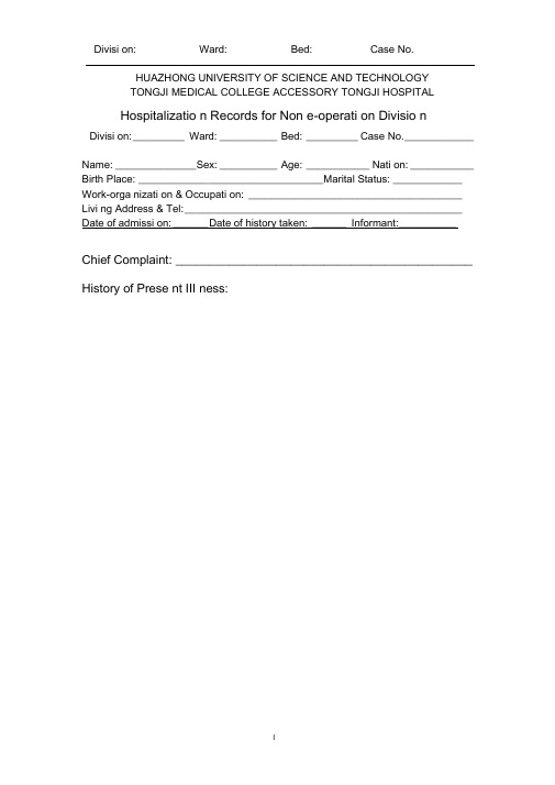
HUAZHONG UNIVERSITY OF SCIENCE AND TECHNOLOGYTONGJI MEDICAL COLLEGE ACCESSORY TONGJI HOSPITALHospitalizatio n Records for Non e-operati on Divisio nDivisi on: _________ Ward: __________ Bed: _________ Case No. ____________Name: ______________Sex: __________ Age: ___________ Nati on: ___________ Birth Place: ________________________________Marital Status: ____________ Work-orga nizati on & Occupati on: _____________________________________ Livi ng Address & Tel: ________________________________________________ Date of admissi on: ______ Date of history taken: ______ Informant:__________Chief Complaint: ____________________________________________History of Prese nt III ness:Past History:Gen eral Health Status: l.good 2.moderate 3.poorDisease history: (if any, please write dow n the date of on set, brief diag no sticand therapeutic course, and the results.)Respiratory system:1. None2.Repeated pharyngeal pain3.chronic cough4.expectoration:5. Hemoptysis6.asthma7.dyspnea8.chest painCirculatory system:1.No ne2.Palpitatio n3.exerti onal dysp nea4..cya no sis5.hemoptysis6.Edema of lower extremities7.chest pain8.s yn cope9.hypertensionDigestive system:1.None2.Anorexia3.dysphagia4.sour regurgitation5.eructation6.nausea7.Emesis8.melena9.abdominal pain 10.diarrhea11.hematemesis 12.Hematochezia 13.ja un diceUrinary system:1.N one2.Lumbar pain3.uri nary freque ncy4.uri nary urge ncy5.dysuria6.oliguria7.polyuria8.retention of urine9.ineontinence of urine10.hematuria ll.Pyuria 12.n octuria 13.puffy faceHematopoietic system:1.N one2.Fatigue3.dizz in ess4.gi ngival hemorrhage5.epistaxis6.subcuta neous hemorrhageMetabolic and en docri ne system:1.None2.Bulimia3.anorexia4.hot intoleranee5.cold intoleranee6.hyperhidrosis7.Polydipsia8.amenorrhea9.tremor of hands 10.character cha nge II.Marked obesity12.marked emaciati on 13.hirsutism 14.alopecia15.Hyperpigme ntatio n 16.sexual fun cti on cha ngeNeurological system:1.N one2.Dizz in ess3.headache4.paresthesia5.hypo mn esis6. Visual disturbanee7.lnsomnia8.somnolence9.s yn cope 10.c onv ulsi on II.Disturba nee of con scious ness12.paralysis 13. vertigoReproductive system:1.No ne2.othersMusculoskeletal system:1.None2.Migrating arthralgia3.arthralgia4.artrcocele5.arthremia6.Dysarthrosis7.myalgia8.muscular atrophyIn fectious Disease:1.N one2.Typhoid fever3.Dyse ntery4.Malaria 4.Schistosomiasis4. Leptospirosis 7.Tuberculosis 8.Epidemic hemorrhagic fever9.othersVacci ne ino culati on:1.No ne2.Yes3.Not clearVacci ne detail _________________________________________ Trauma an d/or operatio n history: Operati ons:1.No ne2.YesOperati on details: ______________________________________ Traumas:1.No ne2.YesTrauma details: _________________________________________ Blood tran sfusi on history:1.None2.Yes ( 1.Whole blood 2.Plasma3.Ingredient transfusion)Blood type: ___________ Tran sfusi on time: _________Tran sfusi on react ion1.None2.YesCli nic manifestation: ____________________________ Allergic history:1.No ne2.Yes3.Not clearallergen: _______________________________________________cli nical manifestation: ____________________________________Pers onal history:Custom livi ng address: ____________________________________________ Reside nt history in en demic disease area:Smoki ng: 1.No 2.YesAverage pieces per day; about yearsGivi ng-up 1.No 2.Yes (Time: )Drinking: 1.No 2.YesAverage grams per day; about yearsGivi ng-up 1.No 2.Yes(Time: ) Drug abuse: 1.No 2.YesDrug names: ______________________________________ Marital and obstetrical history:Married age: _________ years old Pregnancy ___________ t imesLabor ______________ times(1.Natural labor: ______ times 2.0perative labor: __________ times3. __________________ Natural abortion: _______________ times4.Artificial abortion: ____ times5. _______________________ Premature labor: _________ times6.stillbirth _________________ times)Health status of the Mate:1.Well2.Not fineDetails: _______________________________________________ Men strual history:Menarchal age: _______ Duration ________ d ay Interval ____ daysLast menstrual period: ____________ Menopausal age: ______ y ears oldAmount of flow: 1.small 2. moderate 3. largeDysme no rrheal: 1. prese nee 2.abse ncMe nstrual irregularity 1. No 2.Yes Family history: (especially pay atte ntio n to the in fectious and hereditary diseaserelated to the present illness)Father: l.healthy 2.ill: _________ 3.deceased cause: ____________________ Mother :1.healthy 2.ill: ________ 3.deceased cause: ____________________ Others: ________________________________________________________The an terior stateme nt was agreed by the in forma nt.Sig nature of in forma nt: Datetime:Physical Exam inationVital sig ns:Temperature: ______ C Blood pressure: ______ / ______ mmHg Pulse: ____ bpm (l.regular 2.irregular ) Respiration: __ bpm (l.regular 2.irregular )Gen eral con diti ons:Development: I.Normal 2.Hypoplasia 3.HyperplasiaNutrition : l.good 2.moderate 3.poor 4.cachexiaFacial expressi on: 1.no rmal 2.acute 3.chr onic other ___________________ Habitus: l.asthenic type 2.sthenic type 3.ortho-thenic typePositi on: l.active 2.positive pulsive 4.other _______________________ Consciousness: l.clear 2.somnolence 3.confusion 4.stupor 5.slight coma6. mediate coma7.deep coma8.deliriumCooperation: 1Yes 2.No Gait: 1.normal 2.abnormal _____Skin and mucosa:Color: 1.normal 2.pale 3.red ness 4.cya no sis 5.ja un dice 6.pigme ntatio nSk in erupti on: 1.No 2.Yes( type: _________ d istribution: _________________ )Subcuta neous bleed ing: 1.no 2.yes (type:______ distribution: ____________ ) Edema:1. no 2.yes ( locati on and degree ________________________________ ) Hair: 1.no rmal 2.abnormal(details ____________________________________ ) Temperature and moisture: normal cold warm dry moist dehydration Liver palmar :1.no2.yes Spider angioma (location: ________________________________ )Others: _________________________________________________________Lymph no des: enl argeme nt of superficial lymph no de:1. no2.yesDescripti on: ________________________________________________Head:Skull size:〔.normal 2.abnormal (description: __________________________ ) Skull shape: 1.no rmal 2.abnormal(description: _________________________ ) Hair distributio n :1. no rmal 2.abnormal(description: ____________________ ) Others: __________________________________________________________ Eye: exophthalmos: _________ eyelid: ___________ conjunctiva: _________ sclera: _______________ Cornea: _____________________Pupil: l.equally round and in size 2.un equal (R _____ mm L _______ m m)Pupil reflex:〔.normal 2.delayed (R __ s L _ s ) 3.abse nt (R _ L ___ )others: ______________________________________________________ Ear: Auricle〔.normal 2.desformation (description: ______________________ ) Discharge of exter nal auditory can al:1. no 2.yes (l.left 2.right quality: ___ )Mastoid tendern ess 1.no 2.yes (l.left 2.right quality: _________________ )Disturba nee of auditory acuity:1. no 2.yes(1」eft 2.right description: ___ ) Nose: Flaring of alae nasi :1.no 2.yes Stuffy discharge 1.no 2.yes(quality ____ ) Tendern ess over para nasal sinu ses:1. no 2.yes (location: ____________ ) Mouth: Lip ______________ Mucosa ____________ Tongue ________________ Teeth:1.normal 2.Agomphiasis 3. Eurodontia 4.others: _____________________Gum :1. normal 2.ab no rmal (Descripti on_________________________ )Tonsil: __________________________ P harynx: _____________________Sound: 1.no rmal 2.hoarse ness 3.others: ___________________________Neck:Neck rigidity 1. no 2.yes ( tra nsvers fin gers)Carotid artery: 1.normal pulsation 2.increased pulsation 3.marked distentionTrachea location: l.middle 2.deviation (〔.leftward ________ 2.rightward _____ ) Hepatojugular vein reflux: 1. n egative 2.positiveThyroid: 1.normal 2.enlarged ________ 3.bruit (1.no 2.yes ________________ ) Chest:Chest wall: 1.no rmal 2.barrel chest 3.pro minence or retractio n:(left _______ right ________ Precordial prominence _________ )Percussi on pai n over sternum 1.No 2.Yes Breast:〔.Normal 2.ab no rmal __ Lung: Inspection: respiratory movement〔.normal 2.abnormal ____________ Palpati on: vocal tactile fremitus:1. no rmal 2.ab no rmal ____________ pleural rubb ing sen sati on :1. no 2.yes __________________Subcuta neous crepitus sen sati on :1. no 2.yes ____________ Percussion:1. resonance 2. Hyperresonance &location _____________3 Flatness&location ________________________________4. dulln ess & location: _____________________________5. tympa ny &location: ______________________________lower border of lung: (detailed percussi on in respiratory disease) midclavicular line : R: ____ in tercostae L: ____ in tercostaemidaxillary line: R: ____ in tercostae L: ____ in tercostaescapular li ne: R: _______ in tercostae L: ____ in tercostaemoveme nt of lower borders:R: _____ cmL: _________ cm Auscultation: Breathing sound : 1.normal 2.abnormal _______________Rales:1. no 2.yes ________________________________ Heart: Inspection: Apical pulsation: 1.normal 2.unseen 3.increase 4.diffuseSubxiphoid pulsation: 1.no 2.yesLocati on of apex beat: 1. no rmal 2.shift ( ____ in tercosta,dista nee away from left MCL ____ cm) Palpati on:Apical pulsation:1. normal 2.lifting apex impulse 3.negative pulsationThrill:1. no 2.yes(location: __________ phase: ________________ )Percussi on: relative dull ness border: 1.no rmal 2.ab no rmal(Dista nee betwee n An terior Medli ne and left MCL _____ cm) Auscultation: Heart rate: __ bpm Rhythm:1.regular 2.irregular ______ Heart sound: 1.no rmal 2.abnormal ______________________Extra sound: 1.no 2.S3 3.9 4. opening snapP2 _____________ A _________ Pericardial frictio n soun d:1. no 2.yesMurmur: 1.no 2.yes (location __________ phase _____________quality _____ intensity ________ t ran smissio n _________effects of position ________________________________effects of respiration _____________________________ Peripheral vascular sig ns :1.N one2.paradoxical pulse3.pulsus alter nans4. Water hammer pulse5.capillary pulsati on6.pulse deficit7.Pistol shot sound8.Duroziezsig nAbdome n:Inspection: Shape: 1.normal 2.protuberanee 3.scaphoid 4.frog-belly Gastric patter n 1. no 2.yes In test inal pattern 1. no 2.yesAbdo minal vein varicosis 1.no 2.yes(direction: ______________ )Operatio n scarl. no 2.yes _______________________________ Palpation: l.soft 2. tensive (location: ______________________________ )Tendern ess: 1.no 2.yes(location: ______________________ )Rebo und tendern ess:1. no 2.yes(location: _____________ ) Fluctuatio n: l.prese nt 2.absce ntSuccussi on splash: 1.n egative 2.positiveLiver: ______________________________________________Percussion: Liver dullness border: 1.normal 2.decreased 3.absentUpper hepatic border:Right Midclavicular Line _______ In tercostaShift dullness:1.negative 2.positive Ascites: _____________ degreePai n on percussi on in costovertebral area: 1.n egative 2.positve __ Auscultation: Bowel sounds : 1.normal 2.hyperperistalsis 3.hypoperistalsis4.abse nee Gurgli ng soun d:1. no 2.yesVascular bruit 1.no 2.yes (location ____________________ ) Gen ital orga n: 1.un exam ined 2.no rmal 3.ab no rmalAnus and rectum: 1.un exam ined 2.no rmal 3.ab no rmalSpine and extremities:Spi ne: 1.no rmal 2.deformity (l.kyphosis 2.lo rdosis 3.scoliosis)3.Tenderness(location ____________________________ )Extremities: 1.no rmal 2.arthremia & arthrocele (location __________________ )3.A nkylosis (location _________ )4.Aropachy: 1.no 2.yes5.Muscular atrophy (location ______________________ )Neurological system: 1.no rmal 2.abnormal _____________________________ Importa nt exam in ati on results before hospitalizedSummary of the history: ______________________________________ Initial diagnosis: ____________________________________________Recorder:Corrector:。
英文病历样本
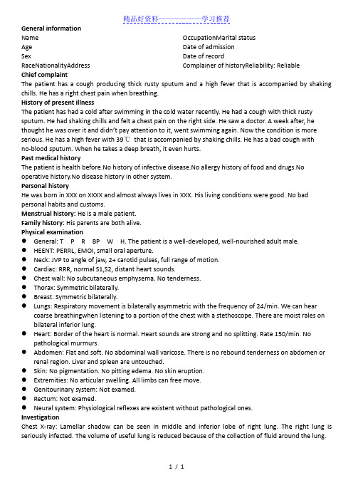
精品好资料——————学习推荐1 / 1 General informationName Age Sex RaceNationalityAddress OccupationMarital statusDate of admissionDate of recordComplainer of historyReliability: Reliable Chief complaintThe patient has a cough producing thick rusty sputum and a high fever that is accompanied by shaking chills. He has a right chest pain when breathing.History of present illnessThe patient has had a cold after swimming in the cold water recently. He had a cough with thick rusty sputum. He had shaking chills and felt a chest pain on the right side. He saw a doctor. A week after, he thought he was over it and didn’t pay attention to it, went swimming again. Now the condition is more serious. He has a high fever with 39℃ that is accompanied by shaking chills. He has a bad cough with no -blood sputum. When he takes a deep breath, it even hurts.Past medical historyThe patient is health before.No history of infective disease.No allergy history of food and drugs.No operative history.No disease history in other system.Personal historyHe was born in XXX on XXXX and almost always lives in XXX. His living conditions were good. No bad personal habits and customs.Menstrual history: He is a male patient.Family history: His parents are both alive.Physical examination● General: T P R BP W H. The patient is a well -developed, well -nourished adult male. ● HEENT: PERRL, EMOI, small oral aperture.● Neck: JVP to angle of jaw, 2+ carotid pulses, full range of motion.● Cardiac: RRR, normal S1,S2, distant heart sounds.● Chest wall: No subcutaneous emphysema. No tenderness.● Thorax: Symmetric bilaterally.● Breast: Symmetric bilaterally.● Lungs: Respiratory movement is bilaterally asymmetric with the frequency of 24/min. We can hearcoarse breathingwhen listening to a portion of the chest with a stethoscope. There are moist rales on bilateral inferior lung.● Heart: Border of the heart is normal. Heart sounds are strong and no splitting. Rate 150/min. Nopathological murmurs.● Abdomen: Flat and soft. No abdominal wall varicose. There is no rebound tenderness on abdomen orrenal region. Liver and spleen are untouched.● Skin: No pigmentation. No pitting edema. No skin eruption.● Extremities: No articular swelling. All limbs can free move.● Genitourinary system: Not examed.● Rectum: Not examed.● Neural system: Physiological reflexes are existent without pathological ones.InvestigationChest X -ray: Lamellar shadow can be seen in middle and inferior lobe of right lung. The right lung is seriously infected. The volume of useful lung is reduced because of the collection of fluid around the lung.。
英文病历书写范例

英文病历书写范例(内科)Medical Records for AdmissonMedical Number: 701721General informationName: Liu SideAge: EightySex: MaleRace: HanNationality: ChinaAddress: NO.35, Dandong Road, Jiefang Rvenue, Hankou, Hubei.Tel: 857307523Occupation: RetiredMarital status: MarriedDate of admission: Aug 6th, 2001Date of record: 11Am, Aug 6th, 2001Complainer of history:patient’s son and wifeReliability: ReliableChief complaint: Upper bellyache ten days, haematemesis, hemafecia and unconsciousness for fo ur hours.Present illness:The patient felt upper bellyache about ten days ago. He didn’t pay attention to it and thought heha date something wrong. At 6 o’cloc k this morning he fainted and rejected lots of blood and gore. T hen hemafecia began. His family sent him to our hospital and received emergent treatment. So the patient was accepted because of “upper gastrointestine hemorrhage and exsanguine shock”. Since the disease coming on, the patient didn’t urinate. Past historyThe patient is healthy before.No history of infective diseases. No allergy history of food and drugs.Past history Operative history: Never undergoing any operation. Infectious history: No history of s evere infectious disease. Allergic history: He was not allergic to penicillin or sulfamide. Respirator y system: No history of respiratory disease. Circulatory system: No history of precordial pain. Ali mentary system: No history of regurgitation.Genitourinary system: No history of genitourinary disease.Hematopoietic system: No history of anemia and mucocutaneous bleeding. Endocrine system: No acromegaly. No excessive sweats. Kinetic system: No history of confinement of limbs. Neural sys tem: No history of headache or dizziness. Personal historyHe was born in Wuhan on Nov 19th, 1921 and almost always lived in Wuhan. His living condition s were good. No bad personal habits and customs.Menstrual history: He is a male patient. Obstetrical history: NoContraceptive history: Not clear.Family history: His parents have both deads. Physical examinationT 36.5℃, P 130/min, R 23/min, BP 100/60mmHg. He is well developed and moderately nourished.Active position. His consciousness was not clear. His face was cadaverous and the skin was not sta ined yellow. No cyanosis. No pigmentation. No skin eruption. Spider angioma was not seen. No pi tting edema. Superficial lymph nodes were not found enlarged. HeadCranium: Hair was black and white, well distributed. No deformities. No scars. No masses. No ten derness.Ear: Bilateral auricles were symmetric and of no masses. No discharges were found in external au ditory canals. No tenderness in mastoid area. Auditory acuity was normal.Nose: No abnormal discharges were found in vetibulum nasi. Septum nasi was in midline. No nare s flaring. No tenderness in nasal sinuses. Eye: Bilateral eyelids were not swelling. No ptosis. No e ntropion. Conjunctiva was not congestive. Sclera was anicteric. Eyeballs were not projected or dep ressed. Movement was normal. Bilateral pupils were round and equal in size. Direct and indirect p upillary reactions to light were existent.Mouth: Oral mucous membrane was not smooth, and there were ulcer can be seen. Tongue was in midline. Pharynx was congestive. Tonsils were not enlarged.Neck: Symmetric and of no deformities. No masses. Thyroid was not enlarged. Trachea was in mi dline. ChestChestwall: Veins could not be seen easily. No subcutaneous emphysema. Intercostal space was nei ther narrowed nor widened. No tenderness.Thorax: Symmetric bilaterally. No deformities. Breast: Symmetric bilaterally.Lungs: Respiratory movement was bilaterally symmetric with the frequency of 23/min. thoracic e xpansion and tactile fremitus were symmetric bilaterally. No pleural friction fremitus. Resonance was heard during percussion. No abnormal breath sound was heard. No wheezes. No rales. Heart: No bulge and no abnormal impulse or thrills in precordial area. The point of maximum imp ulse was in 5th left intercostal space inside of the mid clavicular line and not diffuse. No pericardi al friction sound. Border of the heart was normal. Heart sounds were strong and no splitting. Rate 150/min. Cardiac rhythm was not regular. No pathological murmurs.Abdomen: Flat and soft. No bulge or depression. No abdominal wall varicosis. Gastralintestinal ty pe or peristalses were not seen. Tenderness was obvious around the navel and in upper abdoman. T here was not rebound tenderness on abdomen or renal region. Liver and spleen was untouched. No masses. Fluidthrill negative. Shifting dullness negative. Borhorygmus not heard. No vascular mur murs. Extremities: No articular swelling. Free movements of all limbs.Neural system: Physiological reflexes were existent without any pathological ones. Genitourinary system: Not examed. Rectum: not exanedInvestigationBlood-Rt: Hb 69g/L RBC 2.70T/L WBC 1. 1G/L PLT 120G/L History summary1. Patient was male, 80 years old2. Upper bellyache ten days, haematemesis, hemafecia and unconsciousness for four hours.3. No special past history.4. Physical examination: T 37.5℃, P 130/min, R 23/min, BP 100/60mmHg Superficial lymph node s were not found enlarged. No abdominal wall varicosis. Gastralintestinal type or peristalses were not seen. Tenderness was obvious around the navel and in upper abdoman. There was not rebound tenderness on abdomen or renal region. Liver and spleen was untouched. No masses. Fluidthrill ne gative. Shifting dullness negative. Borhorygmus not heard. No vascular murmurs. No other positive signs. 5. investigation information:Blood-Rt: Hb 69g/L RBC 2.80T/L WBC 1.1G/L PLT 120G/LImpression: upper gastrointestine hemorrhage Exsanguine shock出院小结(DISCHARGE SUMMARY), ===============Department of GastroenterologyChanghai Hospital,No.174 Changhai Road Shanghai, China Phone: 86-21-25074725-803 DISCHARGE SUMMARYDA TE OF ADMISSION: October 7th, 2005 DA TE OF DISCHARGE: October 12th, 2005 ATTE NDING PHYSICIAN: Yu Bai, MD PA TIENT AGE: 18ADMITTING DIAGNOSIS:V omiting for unknown reason: acute gastroenteritis?BRIEF HISTORYA 18-year-old female with a complaint of nausea and vomiting for nearly one month who was see n at Department of Gastroenterology in Changhai Hospital, found to have acute gastroenteritis and non-atrophic gastritis. The patient was subsequently recovered and discharged soon after medicati on.REVIEW OF SYSTEMShe has had no headache, fever, chills, diarrhea, chest pain, palpitations, dyspnea, cough, hemopty sis, dysuria, hematuria or ankle edema.PAST MEDICAL HISTORYShe has had no previous surgery, accidents or childhood illness.SOCIAL HISTORY: She has no history of excessive alcohol or tobacco use.FAMIL Y HISTORYShe has no family history of cardiovascular, respiratary and gastrointestinal diseases. PHYSICAL EXAMINA TIONTemperature is 37, pulse 80, respirations 16, blood pressure 112/70. General: Plump girl in no app arent distress. HEENT: She has no scalp lesions. Her pupils are equally round and reactive to light and accommodation. Extraocular movements are intact. Sclerae are anicteric. Oropharynx is clear. There is no thyromegaly. There is no cervical or supraclvicular lymphadenopathy. Cardiovascular: Regular rate andrhythm, normal S1, S2. Chest: Clear to auscultation bilateral. Abdomen: Bowel sounds present, no hepatosplenomagaly. Extremities: There is no cyanosis, clubbing or edema. Neurologic: Cranial n erves II-XII are intact. Motor examination is 5/5 in the bilateral upper and lower extremities. Sens ory, cerebellar and gait are normal.LABORATORY DATAWhite blood cells count 5.9, hemoglobin 111g/L, hematocrit 35.4. Sodium 142, potassium 4.3, chl oride 106, CO2 25, BUN 2.6mmol/L, creatinine 57μmol/L, glucose 4.1mmol/L, Albumin 36g/L. Endoscopic ExamChronic non-atrophic gastritisHOSPITAL COURSEThe patient was admitted and placed on fluid rehydration and mineral supplement. The patient im proved, showing gradual resolution of nausea and vomiting. The patient was discharged in stable c ondition.DISCHARGE DIAGNOSIS Acute gastroenteritisChronic non-atrophic gastritisPROGNOSISGood. No medications needed after discharge. But if this patient can not get used to Chinese food, she had better return to UK as soon as possible to prevent the relapse of acute gastroenteritis. The patient is to follow up with Dr. Bai in one week. ___________________________ Yu Bai, MD D: 12/10/2005。
英文病例范文
英文病例范文(中英文版)English Sample Medical Case:John Doe, a 35-year-old male, presented to the emergency department with a chief complaint of severe abdominal pain. The pain started suddenly two hours prior to his arrival and was described as sharp and radiating to his back. Upon examination, his vital signs were stable, but he appeared pale and diaphoretic. The abdomen was tender to palpation, particularly in the upper right quadrant. Laboratory investigations revealed an elevated white blood cell count and increased liver function tests. A diagnosis of acute cholecystitis was suspected, and an ultrasound was ordered to confirm the presence of gallstones.张三,35岁男性,因剧烈腹痛到急诊科就诊。
疼痛在就诊前两小时突然开始,表现为尖锐并向背部放射。
检查时,他的生命体征稳定,但面色苍白且出汗。
腹部触诊时,尤其右上象限区域明显疼痛。
实验室检查显示白细胞计数升高和肝功能测试异常。
英文病历报告作文模板
英文病历报告作文模板Patient Information- Name: [Patient's Full Name]- Gender: [Male/Female]- Age: [Patient's age]- Date of Admission: [MM/DD/YYYY]Chief ComplaintThe patient presented with [specific symptoms/complaints] which started [duration].History of Present IllnessThe patient reported [detailed description ofsymptoms/complaints]. The symptoms worsened over the past [duration]. The patient experienced [associated symptoms] and tried [any self-medication or home remedies] but noticed no improvement. There was no history of trauma or injury.Past Medical HistoryThe patient has a history of [chronic/acute medical conditions, if any] which includes [specific conditions]. The patient has taken[previous medications/treatments] for these conditions.Social HistoryThe patient has a [specific occupation] and lives in [specific area]. The patient does [specific habits] such as smoking or drinking alcohol [frequency]. There is no significant family medical history.Physical Examination- Vital Signs:- Blood Pressure: [value] mmHg- Heart Rate: [value] bpm- Respiratory Rate: [value] bpm- Temperature: [value]C- General Appearance:The patient appears [general appearance of the patient].- Systemic Examination:- Cardiovascular: [specific findings]- Respiratory: [specific findings]- Gastrointestinal: [specific findings]- Neurological: [specific findings]- Musculoskeletal: [specific findings]Laboratory and Imaging Findings- Blood Test Results:- Complete Blood Count: [values]- Biochemical Profile: [values]- Others: [specific findings]- Imaging:- [Specific imaging tests performed]- Results: [specific findings]DiagnosisAfter evaluating the patient's medical history, physical examination, and laboratory/imaging findings, the following diagnosis was made:[Primary Diagnosis]Treatment and ManagementThe patient was started on [specific treatment plan] which includes [medications, therapies, or procedures]. The patient wasadvised to [specific instructions] and scheduled for [follow-up tests/appointments, if any].Follow-upThe patient will be followed up in [specific time frame] to assess the response to treatment and manage any complications that may arise. The patient was given contact information for any urgent concerns or changes in symptoms.Discussion and ConclusionThis case report highlights the presentation, evaluation, and management of a patient with [specific condition]. The patient's symptoms were appropriately addressed through a systematic approach involving history taking, physical examination, and laboratory/imaging investigations. The provided treatment plan aims to address the underlying cause and improve the patient's overall well-being. Continuous monitoring and follow-up will guide further management decisions.Note: This medical case report is fictional and serves as a template for educational purposes. Any resemblance to actualpatients is purely coincidental.。
英文完全病历模板-详细版
Admission RecordName:* Nativity: * district, * citySex:male Race: HanAge:55 Date of admission:2020-09-07 14:30 Marital status: be married Date of record:2020-09-07 15:23 Occupation:teacher Complainer:patient himself Medical record Number: * Reliability: reliablePresent address: NO*, building*, * village,* district, *city, *provinceChief complaint: cough and sputum for more than 6 years, worsening for 2 weeksHistory of present illness: The patient complained of having paroxysmal cough and sputum 6 years ago. At that time, he was diagnosed as “COPD” in another hospital and no regular treatment was applied. Cough and sputum worsened and were accompanied by tachypnea 2 weeks ago with no inducing factors. Small amounts of white and mucous sputum were hard to cough up. Compared to daytime, tachypnea worsened in the night or when sputum can’t be cough up. The patient can’t lie flat at the night because of prominent tachypnea and prefer a high pillow. He had no fever, no chest pain, no dizziness, no diarrhea, no abdominal pain, no obvious decrease of activity tolerance. On 20*-0*-*, the patient went to *Hospital for medical consultation. CT lung imaging indicated: lesion accompanied by calcification in the superior segment, the inferior lobe of the right lung, the possibility of obsolete tuberculosis; emphysema, bullae formation and sporadic inflammation of bilateral lung; calcified lesion in the inferior lobe of the left lung; arteriosclerosis of coronary artery.Pulmonary function tests indicated:d obstructive ventilation dysfunction; bronchial dilation test was negative2.moderate decrease of diffusion function, lung volume, residual volume and the ratio of lungvolume; residual volume were normalThe patient was diagnosed as “AECOPD” and prescribed cefoxitin to anti-infection for a week, Budesonide and Formoterol to relieve bronchial muscular spasm and asthma,amb roxol to dilute sputum, and traditional Chinese medicine (specific doses were unknown).The patient was discharged from the hospital after symptoms of cough and sputum slightly relieved with a prescription of using Moxifloxacin outside the hospital for 1 week. Cough and sputum were still existing, thus the patient came to our hospital for further treatment and the outpatient department admitted him in the hospital with “COPD”. His mental status, appetite, sleep, voiding, and stool were normal. No obvious decrease or increase of weight.Past history: The patient was diagnosed as type 2 diabetes 1 years ago and take Saxagliptin (5mg po qd) without regularly monitoring the levels of blood sugar. The patient denies hepatitis, tuberculosis, malaria, hypertension, mental illness, and cardiovascular diseases. Denies surgical procedures, trauma, transfusion, food allergy and drug allergy. The history of preventive inoculation is not quite clear.Personal history: The patient was born in *district, * city and have lived in * since birth. He denies water contact in the schistosome epidemic area. Smoking 10 cigarettes a day for 20 years and have stopped for half a month. Denies excessive drinking and contact with toxics.Marital history: Married at age of 27 and have two daughters. Both the mate and daughters are healthy.Family history: Denies familial hereditary diseases.Physical ExaminationT: 36.5℃ P:77bpm R: 21 breaths/min BP:148/85mmHgGeneral condition:normally developed, well-nourished, normal facies, alert, active position, cooperation is goodSkin and mucosa: no jaundiceSuperficial lymph nodes: no enlargementHead organs: normal shape of headEyes:no edema of eyelids; no exophthalmos; eyeballs move freely; no bleeding spots of conjunctiva; no sclera jaundice; cornea clear; pupils round, symmetrical in size and acutely reactive to light.Ears: no deformity of auricle; no purulent secretion of the external canals; no tenderness over mastoidsNose: normal shape; good ventilation;no nasal ale flap; no tenderness over nasal sinus; Mouth: no cyanosis of lips; no bleeding spots of mouth mucosa; no tremor of tongue; glossy tongue in midline; no pharynx hyperemia; no enlarged tonsils seen and no suppurative excretions; Neck: supple without rigidity, symmetrical; no cervical venous distension; Hepatojugular reflux is negative; no vascular murmur; trachea in midline; no enlargement of thyroid glandChest: symmetrical; no deformity of thoraxLung:Inspection:equal breathing movement on two sidesPalpation: no difference of vocal fremitus over two sides;Percussion: resonance over both lungs;Auscultation: decreased breath sounds over both lungs; no dry or moist rales audible; no pleural friction rubsHeart:Inspection: no pericardial protuberance; Apex beat seen 0.5cm within left mid-clavicular at fifth intercostal space;Palpation: no thrill felt;Percussion: normal dullness of heart bordersAuscultation: heart rate 78bpm; rhythm regular; normal intensity of heart sounds; no murmurs or pericardial friction sound audiblePeripheral vascular sign: no water-hammer pulse; no pistol shot sound; no Duroziez’s murmur; no capillary pulsation sign; no visible pulsation of carotid arteryAbdomen:Inspection: no dilated veins; no abnormal intestinal and peristaltic waves seenPalpation: no tenderness or rebounding tenderness; abdominal wall flat and soft; liver and spleen not palpable; Murphy's sign is negativePercussion: no shifting dullness; no percussion tenderness over the liver and kidney regionAuscultation: normal bowel sounds.External genitalia: uncheckedSpine: normal spinal curvature without deformities; normal movementsExtremities: no clubbed fingers(toes); no redness and swelling of joints; no edema over both legs; no pigmentation of skins of legsNeurological system: normal muscle tone and myodynamia; normal abdominal and bicipital muscular reflex; normal patellar and heel-tap reflex; Babinski sign(-);Kerning sign(-) ; Brudzinski sign(-)Laboratory DataKey Laboratory results including CT imaging and pulmonary function test have been detailed in the part of history of present illness.Abstract*, male, 55 years old. Admitted to our hospital with the chief complaint of cough and sputum for more than 6 years, worsening for 2 weeks. Cough and sputum worsened and were accompanied by tachypnea 2 weeks ago. The patient can’t lie flat in the night because of prominent tachypnea and prefer a high pillow.Physical Examination: T: 36.5℃,P: 77bpm, R: 21 breaths per minute, BP:148/85mmHg. Decreased breath sounds over both lungs; no dry or moist rales audible.Laboratory data: CT lung imaging indicates: lesion accompanied by calcification in superior segment, inferior lobe of right lung, possibility of obsolete tuberculosis; emphysema, bullae formation and sporadic inflammation of bilateral lung; calcified lesion in inferior lobe of left lung. Pulmonary function tests indicate: mild obstructive ventilation dysfunction, bronchial dilation test was negative moderate decrease of diffusion function.Primary Diagnosis:1.AECOPD2.Type 2 Diabetes3.Primary Hypertension Doctor’s Signature:。
英文病历标准模版
英文病历标准模版Patient ProfileName: Si RuihuaDepartment: ___ Power ___Sex: FemalePresent Address: Electric Power Bureau Age: 80 yearsDate of n: May 17.2003nality: Chinese XinjiangDate of Record: May 17.2003Marital Status: MarriedReliability: Reliablen: Family ___History of Allergy: None reportedChief Complaints___。
breathlessness。
and precordial pain for the last hour。
There were no precipitating factors。
and the fort could not be relieved by rest。
As a result。
she came to the hospital for help。
She did not experience syncope。
cough。
headache。
diarrhea。
or vomiting during the course of the illness。
Her appetite。
sleep。
voiding。
and stool were normal.Medical History___.______。
___ distress。
She had a heart rate of 120 beats per minute and a blood pressure of 160/90 mmHg。
Her respiratory rate was 28 breaths per minute。
and her oxygen n was 90% on room air。
soap英文病历
soap英文病历Patient Information:Name: John SmithAge: 45 yearsGender: MaleDate of Admission: June 5, 2021Date of Discharge: June 10, 2021Chief Complaint:The patient presented with a persistent cough and difficulty breathing for the past week.History of Present Illness:Mr. Smith reports that he developed a cough one week ago, which has progressively worsened. He also complains of shortness of breath, especially during physical activities. He denies any chest pain, fever, or weight loss. The cough is non-productive and is not associated with any sputum or blood. He does not have a history of allergies or recent exposure to respiratory irritants.Past Medical History:The patient has a history of asthma, which is well-controlled with daily use of an inhaler. He had a similar episode of respiratory distress three years ago and was treated with corticosteroids and bronchodilators at that time. He denies any recent hospitalizations or surgeries.Family History:There is no significant family history of respiratory diseases.Social History:Mr. Smith is a non-smoker and does not consume alcohol regularly. He works as an office manager and is not exposed to any occupational hazards. He lives with his wife and two teenage children. He denies any recent travel or contact with sick individuals.Physical Examination:Upon examination, the patient appears in no acute distress. Vital signs are stable with a temperature of 98.6°F (37°C), blood pressure of 120/80 mmHg, heart rate of 80 beats per minute, and respiratory rate of 16 breaths per minute. Auscultation of the lungs reveals bilateral wheezing and decreased breath sounds in the lower lung fields. There is no evidence of cyanosis or clubbing. The cardiovascular and abdominal examinations are within normal limits.Diagnostic Tests:A chest X-ray was ordered to evaluate the patient's respiratory symptoms. The X-ray showed bilateral diffuse patchy infiltrates, consistent with bronchial asthma. Pulmonary function tests were performed, revealing a decreased forced expiratory volume in one second (FEV1) and forced vital capacity (FVC), indicating obstructive lung disease.Assessment and Plan:The patient's symptoms, physical examination findings, and diagnostic test results are consistent with a diagnosis of exacerbation of bronchial asthma. The patient was started on a short course of oral corticosteroids, a short-acting bronchodilator,and an inhaled corticosteroid. He was also provided education regarding trigger avoidance and proper inhaler technique. Close follow-up was scheduled to monitor his response to treatment and adjust the management plan if necessary.Follow-Up:The patient will be seen for a follow-up visit in two weeks to evaluate his response to treatment and adjust his medication regimen if needed. He was instructed to monitor his lung function at home using a peak flow meter and seek medical attention if there is a significant decrease in his peak flow readings or if his symptoms worsen. The importance of regular follow-up visits and adherence to the prescribed medication regimen was emphasized. Summary:Mr. Smith, a 45-year-old male with a history of asthma, presented with a persistent cough and difficulty breathing. A diagnosis of exacerbation of bronchial asthma was made based on his symptoms, examination findings, and diagnostic tests. The patient was started on appropriate treatment and provided with education regarding trigger avoidance and inhaler technique. Close follow-up was arranged to monitor his response to treatment and ensure optimal management.。
- 1、下载文档前请自行甄别文档内容的完整性,平台不提供额外的编辑、内容补充、找答案等附加服务。
- 2、"仅部分预览"的文档,不可在线预览部分如存在完整性等问题,可反馈申请退款(可完整预览的文档不适用该条件!)。
- 3、如文档侵犯您的权益,请联系客服反馈,我们会尽快为您处理(人工客服工作时间:9:00-18:30)。
General information
Name Age
Sex
Race Nationality Address Occupation
Marital status
Date of admission Date of record Complainer of history Reliability: Reliable
Chief complaint
The patient has a cough producing thick rusty sputum and a high fever that is accompanied by shaking chills. He has a right chest pain when breathing.
History of present illness
The patient has had a cold after swimming in the cold water recently. He had a cough with thick rusty sputum. He had shaking chills and felt a chest pain on the right side. He saw a doctor. A week after, he thought he was over it and didn’t pay attention to it, w ent swimming again. Now the condition is more serious. He has a high fever with 39℃that is accompanied by shaking chills. He has a bad cough with
no-blood sputum. When he takes a deep breath, it even hurts.
Past medical history
The patient is health before. No history of infective disease. No allergy history of food and drugs.
No operative history. No disease history in other system.
Personal history
He was born in XXX on XXXX and almost always lives in XXX. His living conditions were good. No bad personal habits and customs.
Menstrual history: He is a male patient.
Family history: His parents are both alive.
Physical examination
General: T P R BP W H. The patient is a well-developed, well-nourished adult male.
HEENT: PERRL, EMOI, small oral aperture.
Neck: JVP to angle of jaw, 2+ carotid pulses, full range of motion.
Cardiac: RRR, normal S1,S2, distant heart sounds.
Chest wall: No subcutaneous emphysema. No tenderness.
Thorax: Symmetric bilaterally.
Breast: Symmetric bilaterally.
Lungs: Respiratory movement is bilaterally asymmetric with the frequency of 24/min. We can hear coarse breathing when listening to a portion of the chest with a stethoscope.
There are moist rales on bilateral inferior lung.
Heart: Border of the heart is normal. Heart sounds are strong and no splitting. Rate 150/min. No pathological murmurs.
Abdomen: Flat and soft. No abdominal wall varicose. There is no rebound tenderness on abdomen or renal region. Liver and spleen are untouched.
Skin: No pigmentation. No pitting edema. No skin eruption.
Extremities: No articular swelling. All limbs can free move.
Genitourinary system: Not examed.
Rectum: Not examed.
Neural system: Physiological reflexes are existent without pathological ones. Investigation
Chest X-ray: Lamellar shadow can be seen in middle and inferior lobe of right lung. The right lung is seriously infected. The volume of useful lung is reduced because of the collection of fluid around the lung.。