奥林巴斯BX51显微镜使用手册
BX51使用手册

- . -使用说明书BX51/BX52系统显微镜- . -本手册使用于BX51/BX52系统金相显微镜。
为了确保平安、获得最优性能并使您完全熟悉这种显微镜的使用,我们建议您在操作显微镜前全面、仔细地看完这本手册。
为了供您进一步参考,应该把本手册放在靠近工作台并容易拿到的位置。
A X 9 8 5 5- . -BX51/52 为了让显微镜发挥最正确性能,正确安装和调节及其重要。
如果您将自行安装显微镜,请仔细阅读第7节,“安装〞〔第27页到29页〕。
重要要平安使用显微镜,必须阅读本节1 各局部名称1-34-52 反射光明场观察步骤6-73 使用调解装置8-203-1 镜座..........................................................................................................................................................................................................................8-10电压指示光强预置按钮使用使用滤光片3-2 聚焦装置 (11)卸下微调焦旋钮调整粗调焦旋钮力环粗调焦旋钮限位杆3-3 载物台......................................................................................................................................................................................................... 12-14放置样品调整X轴Y轴旋钮旋物台调整载台高度3-4 观察镜筒.................................................................................................................................................................... 15-17调整瞳距调整屈光度使用眼罩使用目镜测微尺选择三眼目镜筒的光路调整倾角3-5 聚光镜....................................................................................................................................................................18-19聚光镜定中心物镜与聚光镜的相容3-6 油浸物镜 (20)使用油浸物镜3-7物镜工作修正 (20)4 故障检修指导5 规格6 光学特性7安装更换灯泡请阅读本节- . -- . -BX51/BX52本显微镜使用 UIS 〔万能无限远〕光学系统,只能与适用于BX2系列的UIS 目镜、物镜和聚光镜一起使用。
奥林巴斯相机说明书

奥林巴斯相机说明书篇一:Olympus 荧光显微镜操作手册奥林巴斯(Olympus)荧光显微镜操作手册Olympus BX51物镜: 4 ×0.16(无DIC)10×0.4020×0.7540×1.00 oil100×1.40oil荧光滤色块转盘: 1. WU蓝2. WIB 绿(长通)3. WIBA 绿(带通)4. WIG 红5. CFP 青6. YFP 黄操作步骤:(注意:样品须在低倍镜下放置和取下)DIC观察:1. 打开明场电源开关(“︱”为开,“○”为关)2. 将样品置于载物台上,用样品夹夹好3. 将起偏器、检偏器、DIC棱镜推入光路,荧光滤块转盘拨到“1”位置,DIC棱镜应与相应的物镜倍数相匹配4. 先选用低倍物镜(“10×”)5. 调节透射光的强度,调节焦距,找到视野6. 换到高倍镜头,观察样品7. DIC观察时,光路选择拉杆拉到中间位置,既可观察,也可拍照荧光观察:1. 打开明场电源开关2. 打开汞灯电源开关3. 将样品置于载物台上,用样品夹夹好4. 检偏器、DIC棱镜在光路外5. 将荧光光路shutter打开(“○”为开,“●”为关),需保护样品时关闭shutter6. 光路选择拉杆推至最里边7. 根据样品的标记情况将荧光滤块转盘转到相应的位置8. 通过两组减光滤片调节激发光强度9. 从低倍镜开始观察,调焦,找到预观察视野,10. 依次换到高倍镜头,观察样品11. 拍照时光路选择拉杆完全拉出普通明场观察:1. 打开明场电源开关(“︱”为开,“○”为关)2. 将样品置于载物台上,用样品夹夹好3. 起偏器、检偏器、DIC棱镜在光路外,荧光滤块转盘拨到“1”位置,DIC棱镜拨到明场(BF)位置4. 先选用低倍物镜(“4×”)5. 调节透射光的强度,调节焦距,找到视野6. 依次换到高倍镜头,观察样品7. 光路选择拉杆拉到中间位置既可观察,也可拍照关机:1. 关闭汞灯电源(注意:汞灯需使用半小时以上方可关闭,关闭半小时以后方可再次开启)2. 将透射光调到最小,关闭明场电源开关3. 将镜头转到低倍镜,取出样品,若使用过油镜用干净的擦镜纸擦拭镜头4. 确认数据已经保存,关闭软件5. 使用光盘拷贝数据(禁止使用移动储存设备拷贝数据)6. 关闭电脑,登记使用时间、荧光数字等使用情况Olympus IX71物镜:物镜倍数相差环4 ×0.13 PHL10×0.30 PH120×0.45 PH140×0.60 PH260×1.35 oil (无相差)荧光滤色块转盘: 1. WU蓝2. WIB 绿(长通)3. WIBA 绿(带通)4. WIGA 红5. CFP6. YFP操作步骤:相差观察:1. 打开明场电源开关(“︱”为开,“○”为关)2. 将样品置于载物台上3. 将光路选择旋钮调至观察位置4. 从低倍镜开始观察,调节到与镜头相匹配的相差环,荧光滤块转盘拨到“1”的位置5. 调节透射光光强,调节焦距,找到预观察视野6. 依次换到高倍镜,(注意调节相差环)观察样品7. 拍照时将光路选择旋钮调至相机位置荧光观察:1. 打开明场电源开关2. 打开汞灯开关3. 将样品置于载物台上4. 将荧光光路shutter打开(“○”为开,“●”为关),需保护样品时关闭shutter5. 将光路选择旋钮调至观察位置6. 根据样品的标记情况将荧光滤块转盘转到相应的位置7. 通过两组减光滤片调节激发光强度8. 从低倍镜开始观察,调焦,找到预观察视野,9. 依次换到高倍镜头,观察样品10. 拍照时将光路选择旋钮调至相机位置普通明场观察:1. 打开明场电源开关(“︱”为开,“○”为关)2. 将样品置于载物台上3. 将光路选择旋钮调至观察位置4. 从低倍镜开始观察,相差环拨到明场(BF)位置,荧光滤色块转盘拨到“1”的位置5. 调节透射光光强,调焦,找到预观察视野6. 依次换到高倍镜,观察样品7. 拍照时将光路选择旋钮调至相机位置关机:1. 关闭汞灯电源(注意:汞灯需使用半小时以上方可关闭,关闭半小时以后方可再次开启)2. 将透射光强调到最小,透射光选择按钮按出3. 关闭明场电源开关4. 将镜头转到低倍镜,取出样品,若使用过油镜用干净的擦镜纸擦拭镜头5. 确认数据已经保存,关闭软件6. 使用光盘拷贝数据(禁止使用移动储存设备拷贝数据)7. 关闭电脑,登记使用时间、荧光数字等使用情况Olympus SZX16物镜:1×变倍:0.7~11.5荧光滤色块转盘:UV 蓝GFPHQ绿RFP2 红操作步骤:明场观察:将样品置于载物台上推入光路选择拉杆,荧光滤块转盘拨到空位选择合适的光源冷光源观察使用不透明的底板(黑或白),打开环形光电源开关(“︱”为开,“○”为关),调节光强度,找到预观察视野,调节变倍比,观察样品⑵底光源观察使用透明玻璃底板,打开底座电源开关(“︱”为开,“○”为关),将LBD滤片推入光路,调节反光镜方向、对比度和光强度,找到预观察视野,调节变倍比,观察样品4. 拍照时将光路选择拉杆拉出荧光观察:1. 将样品置于载物台上2. 一般选择黑色不透明底板3. 打开汞灯电源开关4. 荧光光路挡板推出 1. 2. 3. ⑴篇二:OLYMPUS在各国外使用总结经验及说明奥林巴斯 XZ-1 tipsXZ-1from Jonathon Donahue====================Here's a grab bag of XZ-1 information... from my posts, and others, on Dpreview... and from other places. 这里是一个大杂烩的XZ-1信息... 从我的帖子,和其他人,DPREVIEW ... 和其他地方。
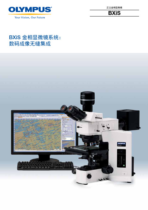
奥林巴斯BXiS金相显微镜系统: 数码成像无缝集成 正立金相显微镜BXiS 1 使用说明书

BXiS 金相显微镜系统:数码成像无缝集成正立金相显微镜BXiS今日,各种各样的检查应用,都要求光学检查系统能够以多种方式完成高效的图像处理工作。
无论您是需要使用白光成像进行基本的测量,还是需要使用具有色彩高保真度的偏振光进行严格的材料鉴定,奥林巴斯都能以其灵活的方案满足您的需求。
BXiS ——您的个性风格。
在奥林巴斯产品方案中,高级显微镜产品可搭载特定的数码显微照相装置,保证了图像的高分辨率和精确的色彩还原。
奥林巴斯全面解决方案还包括高级图像软件。
该软件融合了各种操作功能,包括基本的图像拍摄、图像处理、报表生成、数据导出以及数据、图像和报表的全球网络连接共享。
BXiS ——您的个性系统。
奥林巴斯允许用户自由创建工作方案以满足不同环境的工作流程和其它需要。
用户在专心工作的同时,奥林巴斯还给用户提供了各种便捷、省时的工具,让日常的例行工作也变得轻松容易。
BXiS ——您的个性方案。
BXiS 系统——不论是现在还是未来,都能满足您个性风格的任何操作应用拥有 BX iS , iS 代表您的个性风格(i ndividual S tyle )。
3多用途系统,满足您的个性风格奥林巴斯致力于创建可支持各种级别工作的显微镜系统方案5让工作流程精简高效OLYMPUS Stream 软件,满足您的每个需求BXiS 简化图像的拍摄流程BXiS 操作,不易疲劳9创建个人专属的奥林巴斯系统BXiS 按您的样本和方案设计完美的系统BXiS 拥有各种物镜各种奥林巴斯数码照相装置BXiS 拍摄您需要的图像从简单测量到复杂的图像分析16轻松扩展至未来的应用程序BXiS 的扩展功能可适应未来的需要奥林巴斯系统支持升级功能17系统图、规格BX51 / BX51M / BXFM 系统图BX61 系统图BX41M-LED 系统图BX51 IR 系统图规格外形尺寸让工作流程精简高效时间和工作环境同样重要,这正是BXiS的图像和控制软件可以个性化设置工作流程的原因。
Olympus BX51BX52系统显微镜操作规程(修正版补充)

反射荧光观察
1、准备工作:按观察方法,安装与其匹配的荧光组件和物镜,对准高压汞灯。
2、打开电源总开关到“I”(开)等弧光到稳定状态。
3、把样品放在载物台上。
4、根据样品的荧光指示剂选择与之匹配的荧光组件。
5、把物镜放在光路中聚焦样品。
6、按需要选择ND滤光片,调整整个视野亮度均匀一致并亮度最大。
7、调节视场光阑和孔径光阑,开始观察。
荧光镜的同时观察
1)反射光荧光和相衬的同时观察
1、把空的荧光滤色镜组件,或空位,转到光路中。
2、转动相衬转盘,显示出与物镜上Ph号相同的号码。
3、对准,在环位置和相板之间调节光轴。
4、选用与激发光对应的荧光组件,并将“Shutter”置于“O”位置,接通光路。
5、调节透射光强度,使相衬观察和荧光观察时图像的亮度协调,然后开始观察。
2)反射光荧光和透射光诺马斯基微分干涉衬(DIC)的同时观察
1、把空的荧光滤色镜组件,或空位,转到光路中。
2、调节万能滤光镜中的起偏镜,得到正交尼柯尔位置。
3、把微分干涉衬棱镜插入物镜转换器上的相应的位置。
4、转动万能滤光镜上的转盘,选择与用来观察的物镜相匹配的诺马斯基棱镜。
5、转动物镜转换器,将所需物镜转入光路中。
6、将样品放在载物台上,并聚焦。
7、调节透射光照明装置上的视场光阑和万能聚光镜上的孔径光阑。
8、旋转微分干涉衬棱镜旋钮,选用最理想的图像对比度。
9、选用与激发光相对应的荧光组件并把“Shutter”置于“O”位置。
10、调节透射光强度,使图像亮度适合同时进行荧光和DIC观察。
奥林匹斯BX-51微观镜 - 用户指南说明书

Olympus BX-51 Microscope ---User’s GuideVersion 1.0Edited by Dr. Hartmut G. Hedderich6/23/2021The following guide describes the use of the Olympus BX-51 Microscope. The guide is intended to assist instrument users after the initial training session.Olympus BX-51 Microscope –User’s Guide, Ver.1.0edited by Dr. Hartmut G. HedderichThe Olympus BX-51 Microscope can perform the following measurements: •Bright Field Microscopy in transmission and epi modes•Dark Field Microscopy in epi mode•Fluorescence Microscopy in epi mode•Polarization Microscopy in epi and transmission modes1. HardwareThe microscope has a total of 3 light sources: two tungsten lamps for transmission and epi microscopy and a mercury lamp for fluorescence microscopy. WARNING: never use the mercury lamp with the dark field cube or dark field objectives! The intense light will destroy the cube! The intensity of the tungsten lamps can be set with the illumination adjustment knob at the front right edge of the microscope.The specimen plate has a holder for microscope slides. When using slides full x-y-z control is available. The slide holder can be removed for large size substrates (i.e. well plates). When using substrates that do not fit the slide holder the x control is lost! Always re-install slide holder!In the standard setup the Olympus is equipped with the following objectives: x2, x10, x40 and x100 for bright field (BF) and x10 and x100 for dark field (DF) microscopy. The DF objectives are easily recognized by their size – they are much bigger than the BF objectives. Only BF objectives are used for fluorescence microscopy!The filter cube changer has a total of 6 positions. 5 of those are permanently occupied by the following filter cubes (see data sheets in addendum):•position 1 – BF•position 2 – U/B (ultraviolet excitation/blue emission) – model U-MWU2•position 3 – B/G (blue excitation/green emission) – model U-MWB2•position 4 – G//R (green excitation/red emission) – model U-MWG2•position 5 – DF•position 6 – open, can be used for user specific filters (user needs to provide excitation, emission and dichroic filters)There is a shutter at the bottom right corner of the filter cube changer. It needs to be in the open position in order to get light to the eye piece or the camera.The Olympus has an extra module to connect an external detector or spectrograph. Those need to be provided by the user.The analyzer for epi polarization microscopy is permanently installed below the eye piece.A high-end DP-71 camera is used to take pictures, time-lapse pictures or movies as well as standard movies.All levers have pictograms pointing out how a component performs under different lever positions. Hint: the extra module camera position (lever in) points the mirror to the back! Pull the lever all the way out to have the light go to the DP-71 camera!There is a neutral density (ND) filter at the arm of the microscope. Use the filter for BF microscopy. Remove the ND filter once a fluorescence cube is chosen or you perform DF or polarization microscopy!2. StartupCovid-19 Regulations: please watch the following YouTube movie (https://youtu.be/-VqncPchM1I) to see how the microscope is cleaned prior to use and after finishing your experiments!Turn on tungsten (white light) source. The on/off switch is at the right side of the microscope. There is also a switch to change between top (epi) and bottom (transmission) illumination. Set the intensity with the illumination adjustment knob.The mercury lamp does have its own power supply. Please follow the instructions on top of the power supply. Specifically, leave the lamp on for a minimum of 15 minutes before switching it off. Leave the lamp off for at least 15 minutes, so it has time to cool off! The life time of the mercury lamp is limited to 200-300 hours. Not following those rules will dramatically diminish the lifetime of the lamp!Place your sample on the specimen table. You may focus your sample through the eye piece.3. SoftwareStart the software by clicking the DP Controller icon on the desktop. This will start the camera software. Push the Start button on the left side of the Capture tab to turn the camera on. The monitor will show a preview screen with a live image.There are a total of 7 tabs beneath the image screen. Those are from left to right: Capture, Color, Level, Scale, Timelapse/Movie, Microscope and User Setting.The Capture tab contains the most important information and will help you to get good images. The following parameters can be set here:•Image Size– it will start up with 4080x3072 resolution. Please change it to 1360x1024 for images or 680x512 for movies (frame rate limited).•Exposure Mode– it starts up in Auto mode. I strongly recommend to switch to Manual mode. This will give you an image exactly how you want it.•Exposure Time– move the slider to set the exposure time while watching the life image on the screen (range: 1/44,000s to 60s)•ISO Sensitivity– for DSL cameras use the lowest possible ISO setting to avoid the emergence of white noise.•Objective– if you want a scale on your image you need to have the correct objective being displayed here. Since two 10x and 100x objectives (one each for BF and DF) are installed it helps to go to the Microscope tab to select the correct objective. Each objective was calibrated individually!•Accumulation– leave at Average modeThe Color tab contains two options: white and black balance. They cannot be used together! White balance provides the possibility to adjust the RGB (red-green-blue) components of the image. This can be helpful to make certain particles stand out more. Black balance darkens the image and sharpens the edges of blurry particles.The Level tab gives the possibility to move the spectral range of a BF image.The Scale tab gives the user many options to put a scale on the image. There are options for color changes of line and numbers, length of scale line, positioning, etc. Choose what will work best for you.Timelapse/Movie tab becomes interesting if you have a reaction going on or you have something moving on your substrate. The options are timelapse which will take images in a user-defined time interval and total time. This will produce a stack of picture. It is also possible to combine those pictures to a movie by using timelapse movie. The last option is a straight up movie. Please make sure to set the image size to 680x512 pixels!Microscope tab is used to tell the program which objectives are installed and which one is active. It is important to choose the correct objective in order to have a correct scale imprinted. Each objective was calibrated individually.UserSetting tab gives the possibility to save a certain setup. Since the microscope is setup for each new slide, it usually is not an option that is needed.Use the x-y-z stage to get your specimen in focus and to the correct position. Use the exposure time setting to get optimum light characteristics. Hit the camera button to take an image. The image will show up in a second window. There are several options to manipulate your image post capture. The most important one is image composition which allows to combine several images to a new picture. This is an important option to see for example which parts of your specimen fluoresce and which ones do not.4. Known Problems/MaintenanceSometimes it seems impossible to get the sample in focus. This is most often seen with the x40 and x100 objectives. The reason is that the objective lens got dirty. Usually, this happens when a user runs the objective into the specimen. Please inform the person-in-charge of the microscope right away. He will clean the objectives with solvent and special tissue paper. Please do not attempt to clean microscope objectives yourself!When the mercury lamp gets close to the end of its lifetime, it will be harder to get the burner started or it won’t start at all. Please inform us about this observation. We will change the mercury lamp and re-align the microscope correctly.5. Turn Off/Shut Down MicroscopeSave your images and copy them to a flash drive if necessary. Close the program. Logoff the computer.Turn off ALL light sources. This is especially important for the mercury lamp which has a limited lifetime. Remove all your samples. Please do not leave any samples with the microscope. Samples and substrates that are left will be discarded!Please return the microscope to the following positions:•Move the specimen table to the bottom•Switch to the x2 objective•Close shutter•Return filter changer to position 1 (BF)•Put ND filter back in its position•Push lamp lever to the inward position (tungsten lamp)Finally, clean the microscope as demonstrated in the cleaning movie!6. Addendum• Position 2: Data Sheet for Filter Cube U-MWU2:Transmittance/WavelengthThe values of transmittance were measured using Lambda900 (PerkinElmer Life & Analytical Sciences).•Position 3: Data Sheet for Filter Cube U-MWB2:Excitation filter EmissionfilterDichromaticfilter460-490 520IF 500Transmittance/WavelengthThe values of transmittance were measured usingLambda900 (PerkinElmer Life & Analytical Sciences).•Position 4: Data Sheet for Filter Cube U-MWG2:Excitation filter EmissionfilterDichromaticfilter510-550 590 570Transmittance/WavelengthThe values of transmittance were measured usingLambda900 (PerkinElmer Life & Analytical Sciences).。
奥林巴斯 BX51WI BX61WI 固定台显微镜使用手册说明书

Olympus is about life. About photographic innovations that capture precious moments of life. About advanced medical technology that saves lives. About information- and industry-related products that make possible a better living. About adding to the richness and quality of life for everyone. Olympus. Quality products with a FIXED STAGE UPRIGHT MICROSCOPE BX51WIFIXED STAGE UPRIGHT MICROSCOPE WITH MOTORIZED FOCUSING BX61WIUNIVERSALINFINITY SYSTEMBX51WI with Luigs & Neumann Accessories.Combined with WI-DPMC.BX51WI with Burleigh AccessoriesA dual commitment:Preventing vibration andprotecting living cell specimensOne design theme was central to the development ofthe new fixed stage microscopes from Olympus — achieve an evenhigher standard of stability and reliability in electro-physiological applications.The result is a wide range of advanced new features to avoid and prevent vibration. These innovations include the introduction of a new observation method along withdetailed analysis of operability and further refinements in image clarity. These improvements work together to make patch clamp operations smoother and more efficient than ever before. Combined with the traditional excellence of UIS optics, the new Olympus fixed stage microscopes define new levels of quality in both performance and ease of use.Fluorescence macro objectives for membrane potential observation Full-system physiological confocal microscope,BX61WI with Z-axis motor is LSM ready7×Intermediate magnification 0.35 x 80×Intermediate magnification4 xN .A .0.95X L UM P L F L20x W Interchanging low and high magnifications without changing objectives.A new concept in vibration-free design.A major concern for researchers conducting electro-physiology experiments is the vibration which occurswhen switching objectives and the resulting interference this can cause to the specimens and adjacent equipment. To solve this problem, Olympus introduces a new concept —the provision of an intermediate magnification changer in combination withthe new High N.A. long working distance 20x objective that allows the user to switch between low and high magnifications without the need to switch objectives.New 20x objective (XLUMPLFL20XW) N.A. 0.95; W.D.: 2.0mm The new 20x water immersion objective makes high-resolutionobservation possible with a wide range of intermediate magnification lenses. Since exchanges between low and high magnification are performed through the intermediate magnification changer, vibration is reduced to a minimum and the usual concern about collisions between objectives and patch clamp electrodes is eliminated.CondenserFilter turretIR-DIC port IR-DICXLUMPLFL20xW N.A. 0.95Video port FL/DICLight path exchange leverIntermediate magnification change lever IR filter(not to use with visible light DIC observations)Observation port FL/DICDual port WI-DPMCMirror unit exchange turret 0.35x(0.25x) lensTube lensMirror unitDIC PrismAnalyzerC mountDichroic mirror 4x lensPolarizer1/4 wave plateDIC elementExcitation light Fluorescence light IR-DICTransmitted lightSimultaneous fluorescence and IR-DIC observations With the included 690nm dichromatic mirror in the WI-DPMC,fluorescence light is sent to the front port, and IR-DIC light is sent to the back port allowing two cameras to image simultaneously with no vibration introduced by light path selection.Variable magnification dual port (WI-DPMC)The WI-DPMC rear camera port includes a 2 position intermediate magnification selector. A high magnification 4x intermediate lens is included and a (0.25x, or 0.35x) low magnification lens is optional.High or low magnification selection is via a single lever with no click-stops or detents allowing a specimen to be scanned and measured with minimal disturbance from vibration. 775nm and 900nm IR-DIC compatible.*Available for 0.5x, 1x and 2x intermediate magnification lenses by special order.Variable Click-stopsAll click-stops, as when selecting between camera and observation modes, can be adjusted to the point of no click and thus no vibration.q Vibration-free shutterThe fluorescence shutter slides horizontally with no detents and no vibration.y A waterproofing sheetA waterproofing sheet, attached by the supplied magnets, provides protection against liquid overflow and spills. The sheet is large enough to protect the frame, condenser and focusing mechanisms .e Ample space around the condenserFrame designed for ample space around the condenser, making it easy to adjust Nomarski DIC contrast, exchange filters,adjust the condenser's aperture stop and to easily switch between visible light,Nomarski DIC or IR-DIC.w Mirror unit turret with adjustable click releaseThe click-stop on the 6 position turret can be released with a precision screwdriver.r Front focus knobs close to the operator's handFine focus control is located at the front on both sides of the microscope body. The knob on the right integrates both coarse and fine focus control.t Coarse focus lock leverWhen engaged at the desired position, the objective can be raised with the coarse focus knob and then returned precisely to its original position.Front operation with no shock and no noise.A new concept in experimental operation.rewqThe new front operation system prevents interference in patch clamping work. The design concept is simple and allows frequently performed operations like focusing or filter exchange to be done easily at the front of the unit.Ample space is provided on both sides of the microscope frame and condenser, so the necessary manipulation equipment can be positioned close to themicroscope.tu Remote power supply and hand switchThe remote TH4 power supply for transmitted light is designed with no cooling fan to minimize electrical noise.Features on/off and intensity controls. Can also be used with the optional TH4-HS hand switch providing light intensity and on/off control a maximal distance away from the Faraday cage.uyRaising the objective and lowering the stage to enable small animal experimentsThe arm height raising kit (WI-ARMAD) provides an additional 40mm of clearance and is mounted between the microscope frame and the reflected light illuminator. Small animal experiments usually do not require transmitted light thus allowing the removal of the substage condenser assembly. After removal, the stage may be lowered an additional 50mm, providing a total clearance increase of 90mm.A variety of convenient units toadd light sources and control the lightLamphouse adapter U-LHADThis adapter allows the mounting of the dual port (U-DP) between the microscope frame and lamp housing.Rectangular field stop BX-RFSSDesigned for use with CCD cameras, prevents photobleaching of the specimen outside of the imaging area.Pinhole unit BX-RFSPOTLighting the cell via a pinhole allows experimentation on reaction to light. Optional MELLES GRIOT's ø16pinhole is used.New functionality and solutions to meet a wide variety of needs.A powerful new concept.(photo shown is BX-RFA)Experimenting with small animalsPhotoactivationNormal configuration40mm more clearance via WI-ARMAD18mmW.D.18mm 40mmW.D.BX-RFSSBX Stage and adapter for injection experimentsThe stage adapter WI-STAD is designed to allow the attachment ofa traditional microscope right or left hand stage to the WI frame.The compact design of the BX2 stage (U-SVRB-4, or U-SVLB-4)reduces the distance between the specimen and the manipulatorand creates a stable platform for injections.MicroinjectionConfocal Microscope SystemBX61WI — Built in Z-axis focus motorThe BX61WI frame incorporates a precise Z-axis focus motor with 0.01µm step size. Designed to incorporate the Olympus Fluoview scan unit and software, the BX61WI is ready for confocal z-stacks. Microscope frame includes programmable buttons for a wide variety of applications.Convenient, optional focusing hand switch U-FH forremote operationThe remote hand switch allows the user to control the microscope remotely via a 2 meter connection cable. Hand switch allows the selection of coarse and fine focus movement, and nosepiece escape/ return. Hand switch buttons can also be custom programmed for individual needs.Optional Olympus Fluoview Confocal System FV500/FV300 With the scanning unit set at the back of the microscope body, compact layout in the cage is possible.Moving the microscope and scanning unit together(mover available by special order)Allows X and Y movement of both the microscope and scan unit together while the stage and specimen are fixed.Additional lasers and accessoriesAn assortment of lasers can easily be attached to satisfy a wide variety of applications.Setting example: BX61WI+FV300 U-FHUltimate image clarity for electro-physiological experim A new concept in live cell observation.Senarmont compensation for Nomarski DIC observation When using a Senarmont equipped condenser, all contrast adjustments are performed with the 1/4 wave plate below the condenser, thus eliminating the risk of bumping the stage, specimen, manipulators or nosepiece.IR-DIC/ Nomarski DIC observationOblique illumination observationOblique observation optimizes contrast by changing the direction of the specimen shadowOlympus has developed an oblique condenser (WI-OBCD) whose long working distance enables the angles of shadow to be altered through 360 degrees without moving the specimen. Requiring no additional accessories, oblique illumination is easy to set up and control. Plastic dishes (normally unsuitable for all types of DIC) are easy to image with oblique illumination. The oblique illumination slit aperture is variable in size and on a slider allowing quick changeover.IR-DIC Optimized Optics:Designed for observations at 775nm to 900nmThanks to the precisely aberration-compensated IR-DIC optics covering from visible to near infrared light of 775nm/900nmwavelength, the clarity of images observed under near infrared light has been improved still further, allowing clear observation of even deep sections of brain slice.• Visible light DICAllows operator high-resolution observation of the tissue surface.• 775nm IR-DICIn combination with an IR camera allows observation within the tissue slice. Optics are corrected for visible and IR wavelengths allowing fast switching between wavelengths with minimal refocusing.• 900nm Nomarski DICAllows observation deeper into the tissue (requires special polarizer and analyzer optimized for 900nm).Analyzer DIC prismObjectiveDIC prism 1/4 wave platePolarizerIR filter(not to use with visiblelight DIC observation)Nucleus of solitary tract from slice of rat medulla oblongata (thickness: 400µm )Prof. Fusao KatoSchool of Medicine Physiology Dept.,Jikei UniversityKato & Shigetomi, J. Physiol.(2001), 530: 469-486Universal condenser with DIC for improved contrastSuitable for use in visible and 775nm/900nm near-infrared light,the U-UCD8 universal condenser is a high N.A., short working distance condenser offering improved contrast in nerve cell observations, for example.AdjustableU-UCD8WI-TP137WI-DICTWI-OBCDRotatablements.Transverse cryostat section through the hippocampus of a mouseat postnatal day 10 was stained with a mouse monoclonal anti-neurofilament-L (Chemicon, MAB1615) .An FITC-conjugated anti-mouse antibody was used for detection of NF-L.Objective: XLFLUOR4x/340Masaharu Ogawa,Ph.DLaboratory for Cell Culture Development,Brain Science Institute, RikenObserving changes in membrane potentialFluorescence macro observationMeasuring changes inmembrane electric potential by using the XLUMPLFL20xW objective with N.A. 0.95The XLUMPLFL20xW objective, with its high N.A., and 2.0mm of working distance allows the measurement of cell membrane electric potential (as seen right). Also, the 4x macro objective (XLFLUOR4x/340) can be used to measure membrane potential at the tissue level. A water immersion cap (XL-CAP) can be attached to the macro 2x or 4x objectives to eliminate disturbances caused by water ripples.2x and 4x Macro lenses with high numerical apertures provide fluorescence imagesDesigned for GFP imaging of large cells such as neurons 2x and 4x low magnification fluorescence objectives and a special GFP observation mirror unit are available. The objectives have a long working distance for maximum flexibility. An optional waterimmersion cap (XL-CAP)is also available toremove imageaberrations caused by ripples on water surfaceof immersed specimens.Imaging of neuronal activity with voltage sensitive dyeSpread of neural activity in area CA1 of acute rat hippocampal slice (400µm thick) in response to a single stimulation applied to Schaffer collateral pathway imaged (at frame rate of 0.7 ms/frame) with a fluorescent voltage sensitive dye (VSD; Di-4-ANEPPS). The fluorescent image (90x60 pixels) captured by a digital high-speed CCD camera (MiCAM01, Brain Vision Inc.; with 20x objective and 0.5x adapter) is superimposed on the illustration of a hippocampal slice (upper left panel). The image is enlarged and shown on the illustration of pyramidal cells (solid line) (lower left panel). Each laminar of CA1 is shown as follows: SO-A, Stratum oriens-alveus; SP, Stratum pyramidal; SR, Stradum radiatum. The individual somas of cells were visible (indicated by dotted circle on the image) and were found along the stratum pyramidal. The changes in the fluorescence of VSD (optical signal) in accordance with the membrane potential change upon a stimulation (Stim) onto Schaffer collateral (Sch) were pseudo-color encoded and shown as consecutive images (upper right panel; number in each image shows time from the stimulation (ms)). The depolarizing signal (red) spread along Schaffer collateral,which was followed by a hyperpolarizing signal (blue) originated in stratum pyramidal. The time courses of optical signals in representative pixels are shown in lower right traces.Takashi Tominaga Ph.D, Brain-Operative Device Lab., Brainway Group, Brain Science Institute, RikenU-SLRE XLFLUOR2x/340XLFLUOR4x/340U-MF/XL U-MGFPA/XL U-MGFP/XLIntermediate magnification changer U-ECA, U-CAThe U-ECA, which includes a 2x intermediate magnification position,allows quick magnification changes to a camera or observer without the need to change objectives. The U-CA includes a 4 position turret that allows rapid switching between a 1x, 1.25x, 1.6x and 2x positions. Both changers accept standard Olympus adapters for attaching a wide range of cameras.* U-ECA and U-CA are notrecommended for IR observation with the U-TR30 trinocular observation head.AccessoriesMulti double port tube U-DPTSThe U-DPTS accepts an optional dichroic mirror allowing theincoming to be split between visible and infrared and be observed simultaneously using two cameras.* A fluorescence mirror unit is required.UIS ObjectivesU-CMDPTSU-PMDPTSU-DPTSU-ECAU-CAU-TVCACBX51WI/BX61WI specificationsSan-Ei building, 22-2, Nishi Shinjuku 1-chome, Shinjuku-ku, Tokyo, JapanPostfach 10 49 08, 20034, Hamburg, Germany2 Corporate Center Drive, Melville, NY 11747-3157, U.S.A.491B River Valley Road, #12-01/04 Valley Point Office Tower, Singapore 2483732-8 Honduras Street, London EC1Y OTX, United Kingdom.104 Ferntree Gully Road, Oakleigh, Victoria, 3166, AustraliaSpecifications are subject to change without any obligation on the part of the manufacturer.ISO9001Certificate No.69372CertificationDesign and productionadheres to ISO9001international quality standard.ISO 9001ISO14001Design and production at the OlympusOptical Co. Ltd. Ina Plant conforms withISO14001 specifications forenvironmental management systems.CertificationCertificate No. 70933ISO 14001U K A S*All brands are trademarks or registered trademarks of their respective owners.Web site address: BX51+WI-DPMC dimensions (unit: mm)。
奥林巴斯金相显微镜安全操作及保养规程

奥林巴斯金相显微镜安全操作及保养规程前言奥林巴斯金相显微镜是一款广泛应用于金相显微研究领域的高级显微镜。
在日常使用过程中,为了确保实验结果的准确性和仪器设备的安全性,必须对其进行正确的操作和保养。
本文档将介绍奥林巴斯金相显微镜的安全操作及保养规程,供用户参考。
安全操作1.操作前的准备在进行任何操作之前,必须确定目标样品和相关仪器已全部准备好。
•检查样品,确保其没有气泡、残留广告胶、油污等污染物•检查显微镜,确保其所有部件在正确的位置并牢固•准备好必要的工具和附件2.操作步骤•打开设备前,必须检查电源线和设备是否漏电。
•打开灯源,调节光强至适当亮度,调节对比度。
•调节双眼管与衍射光源的高度一致,同时开启防眩光门。
•放置样品,在样品平台上旋转调节至所需要的视角。
•镜头调焦,对焦样品,检查样品是否清晰。
•调整目镜度盘,合适的视野大小。
•更换镜头、调整目镜等操作,使用前必须关闭灯源,并关闭电源。
•完成操作后,关闭灯源,关闭电源,清理并保管好样品及相关器材。
3.注意事项•操作过程中注意使用手套,避免手指直接接触样品。
•操作过程中严禁把手指伸进仪器内部。
•操作结束后,及时清理仪器表面和样品平台等。
保养规程1.日常保养•每次使用后,必须按照规定流程进行清洁,清除镜头和样品平台等器材表面的残留物。
•定期使用专业清洁剂对镜头、目镜、光源器材进行清洁,并用干净软布擦干。
•定期检查各部件连接是否牢固,是否有断裂、变形等情况。
2.镜头保养•镜头表面不可被手指触碰,且不可用硬物擦拭,可以使用专业软布进行轻柔擦拭。
•镜头存储时,应该收起来,并使用专业防潮防尘套包裹。
3.电器元件保养•定期清除灰尘,避免电路发生故障。
•定期检查电源线和电器元件连接是否正常。
总结在使用奥林巴斯金相显微镜进行实验时,必须遵守正确的操作程序以及仔细遵守使用说明书中的安全操作技巧。
同样,用户还需定期对仪器进行日常保养,状况检查和设备保养。
当我们使用这些仪器的时候,必须始终牢记仪器设备的安全可靠性和正确性,只有这样,我们才能保证实验结果的准确性并延长仪器使用寿命。
奥林巴斯显微镜安装套件用户手册说明书

USER MANUAL#23 Olympus Type Microscope Mount Set14-0010Version 2.0020131.800.828.6972For information regarding applicable intellectual property, please visit /patents.Information in this publication supersedes that in all previously published material. Due to our ongoing development program, Vincent Associates reserves the right to discontinue or change specifications or de-signs, at any time, without incurring any obligation.Version 2.002013Vincent Associates, a Division of VA, Inc.803 Linden Ave.Rochester, NY 14625Tel: 585-385-5930Fax: 585-385-6004UNIBLITZ®, N-CAS®and VINCENT ASSOCIATES® are registered trademarks of VA, Inc.Printed in the U.S.A.WarrantyLIMITED PRODUCT WARRANTY: All Products manufactured by VINCENT ASSOCIATES® (MANUFACTURER) are warranted to meet published specifications and to be free of defects in materials and workmanship as defined in the specifications for 365 days - one year - (WARRANTY PERIOD) from the date of original shipment of the product. DSS series shutters are additionally warranted to achieve two million cycles within the WARRANTY PERIOD (as defined in the CYCLE WARRANTY CRITERION). MANUFACTURER will, at its own option within the WARRANTY PERIOD, repair or replace without charge any listed item discovered to be defective excepting transportation charges. Burned out or otherwise damaged actuator coils are not covered under this warranty. Any defective product returned to the MANU-FACTURER must follow the RETURN MATERIAL AUTHORIZATION PROCEDURE as defined below. This warranty does not extend to cover damage resulting from alteration, misuse, negligence, abuse, normal wear and tear, or accident. The MANUFACTURER will consider the return of unused equipment if returned within 30 days from the original date of shipment, subject to a 20% restocking charge. This offer does not apply to used or damaged equipment. This warranty extends only to the original purchase and is not availa-ble to any third party, including any purchaser assemblies or other Products of which the goods may become component equipment.CYCLE WARRANTY CRITERION: One "cycle" is considered one open and one closure of the shutter. DSS Shutter must be operated with the ED12DSS driver or equivalent H-Bridge type shutter driver circuit at +10.7VDC across the actuator coil for the specified duration. DSS Shutter must be operated within the de-fined environmental, electrical and mechanical specifications as listed on the device's data sheet. After one year (WARRANTY PERIOD), the cycle warranty is null and void. If returned, the device must be accompa-nied by a written statement indicating the approximate number of cycles contained on the device, include all parameters to which the shutter was operated and follow the RETURN MATERIAL AUTHORIZATION PROCEDURE as defined below.RETURN MATERIAL AUTHORIZATION PROCEDURE: MANUFACTURER will only accept returned Products from customers that have obtained an RMA (Return Material Authorization) number from the MANUFACTURER. The customer must also include an itemized statement of defect(s). The Product will then be evaluated per the MANUFACTURER'S standard repair guidelines. Any Product which has been re-turned to the MANUFACTURER but which is found to meet the applicable specifications and not defective in materials and workmanship shall be subject to the MANUFACTURER's standard evaluation charge. The MANUFACTURER assumes no liability for customer returned material.LIMIT OF LIABILITY: The buyer's exclusive remedy and the limit of MANUFACTURER'S liability for any loss whatsoever shall not exceed the purchase price paid by the buyer for the goods to which a claim is made. MANUFACTURER does not give any implied warranties of merchantability, fitness for a particular purpose, or of any other nature in connection with the sale of any Products.IntroductionThe #23 mount set adapts specific shutter housing types to an Olympus type microscope. No modifi-cation to the microscope should be necessary. A filter area is also provided to allow the user to install an op-tical filter between the shutter housing and the microscope illuminator. The housed versions of the LS, VS, and XRS series shutters, as well as the CS25 and CS35, can be configured with this mount set type. A clear-ance cutoff (1.38in/35.1mm from aperture center) is provided with the VS series only.Assembly ProcedureMount AssemblyUnpack the Mount SetAs you remove the packing material from the box, check for small items that may be attached tothe packing material. Make sure the following parts were included in the box:(see parts list table and Figure 1)Step Two:Screw the rear mount sub -assembly to the back of the shutter housing, tighten securely. (see Figure 3)Step One:Using (3) 2-56 flat head screws attach #23 front mount to the front of the shutter housing. (see Figure 2)INSTALLATION STEPS: Step Three:Figure 4SpecificationsDue to our ongoing product development program, Vincent Associates reserves the right to discontinue or change specifications at any time, without incurring and obligations. Teflon is a registered trade mark of E.I. Dupont U.S. Pat No.3.427.293.6,652,165. Drawings shown for illustrative purposed only. Updated 9/2013ALL DIMENSIONS MAX[mm] INCH。
- 1、下载文档前请自行甄别文档内容的完整性,平台不提供额外的编辑、内容补充、找答案等附加服务。
- 2、"仅部分预览"的文档,不可在线预览部分如存在完整性等问题,可反馈申请退款(可完整预览的文档不适用该条件!)。
- 3、如文档侵犯您的权益,请联系客服反馈,我们会尽快为您处理(人工客服工作时间:9:00-18:30)。
使用说明书BX51/BX52系统显微镜本手册使用于BX51/BX52系统金相显微镜。
为了确保安全、获得最优性能并使您完全熟悉这种显微镜的使用,我们建议您在操作显微镜前全面、仔细地看完这本手册。
为了供您进一步参考,应该把本手册放在靠近工作台并容易拿到的位置。
A X 9 8 5 5为了让显微镜发挥最佳性能,正确安装和调节及其重要。
如果您将自行安装显微镜,请仔细阅读第7节,“安装”(第27页到29页)。
重要要安全使用显微镜,必须阅读本节1 各部分名称1-3 4-52 反射光明场观察步骤6-73 使用调解装置8-203-1 镜座..........................................................................................................................................................................................................................8-10电压指示光强预置按钮使用使用滤光片3-2 聚焦装置 (11)卸下微调焦旋钮调整粗调焦旋钮张力环粗调焦旋钮限位杆3-3 载物台......................................................................................................................................................................................................... 12-14放置样品调整X轴Y轴旋钮旋转载物台调整载台高度3-4 观察镜筒.................................................................................................................................................................... 15-17调整瞳距调整屈光度使用眼罩使用目镜测微尺选择三眼目镜筒的光路调整倾角3-5 聚光镜....................................................................................................................................................................18-19聚光镜定中心物镜与聚光镜的相容3-6 油浸物镜 (20)使用油浸物镜3-7物镜工作修正 (20)4 故障检修指导5 规格6 光学特性7 安装更换灯泡请阅读本节本显微镜使用UIS (万能无限远)光学系统,只能与适用于BX2系列的UIS目镜、物镜和聚光镜一起使用。
(一些为BX系列设计的配件也可以使用,详情请洽奥林巴斯公司或参见产品目录。
)如果使用不适合的附件,效果会很不好安全注意事项①图1(图1)1. 把显微镜安装在坚固、平坦的桌面或者工作台上,不要堵塞镜基下面的通风口。
不要把显微镜放在易变形的表面上,因为这样会影响空气流通变热甚至起火。
2. 灯室的表面和附近在使用中会变得非常热,安装显微镜时,一定要在灯座周围特别是上方,保留足够的空间(10厘米或以上)。
3. 在装显微镜时,使电源线远离灯屋。
当电源线插拔除到灯屋时,电源线表面有可能会融化并导致漏电。
4. 更换灯泡时,要把开关①拨到“ ”(关)的位置,然后从墙上插座拔出电源线,以免触电或者着火。
如果显微镜使用中或者刚刚使用后,要等到灯屋和灯泡冷却后才能更换灯泡。
(图1)指定卤素灯泡12V100WHAL (PHILIPS 7724)12V50WHAL-L (LIFE JC)#显微镜还内置了一个保险丝(更换保险丝请与制造商或者指定代理商联系)5. 始终使用奥林巴斯公司提供的电源线。
如果不能使用指定电源线,就无法保证产品的安全和性能。
6. 始终要将显微镜的接地端与墙上插座的接地端牢固连接。
如果设备没有接地,奥林巴斯就不能保证电气安全和设备性能。
7. 不要把金属物放到显微镜的通风口里,会导致漏电受到电击并且设备损坏。
安全标志下面的标志是标在显微镜上的。
须清楚这些符号的含义,始终按照最安全的方法使用显微镜。
警告警告牌/标志贴在操作和使用显微镜时需要特别注意的地方。
始终注意警告信息。
21准备图21. 显微镜是精密仪器,操作时要小心,并避免突然和剧烈的震动2. 不要再有阳光直射、高温或多尘、以及容易受到震动的地方使用显微镜。
(操作环境条件参考第5节,“规格”。
) 3. 移动显微镜时,谨慎地抓在镜基和镜臂附近, 如图2所示 (重量:大约16KG ) # 如果安装了照相模块,显微镜系统会很重。
小心握住中间部分移动。
# 如果握住载物台,粗调焦钮,观察筒的双目部分或灯屋,将会损伤显微镜。
4. BX51/52显微镜系列可以和两个中间附件共同使用, (例如U-CA 变倍器, U-EPA2调节器,等等)当用两个中间附件时,请先阅读中间附件的操作说明。
BX51/BX522维护和保养1. 清洁各种玻璃部件时,要用试镜布轻轻擦拭。
除掉指纹油渍时,要用少量的乙醚(70%)和酒精(30%)混合溶液能的电火花来源,如进行开关操作的电子设。
还要记住,只能在通风良好的房间中使用这些化学物品。
2.不要使用有机溶剂擦拭玻璃不见以外的显微镜去他部位。
如果要清洁这些部件,请使用一块无毛柔软的布蘸少量中性清洁剂擦拭。
3. 不要拆开显微镜的任何部位。
4. 不使用显微镜时,等到灯座完全冷却后,把它用所提供的防尘罩盖上3警告如果不按照本手册指定方式操作显微镜,可能会危害用户的安全。
另外,也可能损坏显微镜。
应始终按照本手册操作显微镜。
◎如果还没有组装起来显微镜,请阅读第7节,“组装”(第27页到第29页)。
透射光模型说明BX51TF或BX52TF 显微镜体光路选择按钮(第16页)瞳间距调节刻度(第15页)聚光器高度调节钮(第18页)内六角起子(存放位置)屈光度调节环(第15页)标本夹(第12页)Aperture iris diaphragm ring(Page 19)细微调旋钮(第11页)(可分离)Pre-focusing Lever(第11页)Filter mount(第9页)视场光圈调节环(第18页)光强度调节旋钮(第8页)(灯电压调节旋钮)粗调焦旋钮张力调节环(第11页) 4主开关(第1页)LED电压指示灯光强预置按钮(第8页)光强预置调节螺丝(第8页)Y轴旋钮(第13页)X轴旋钮(第13页)粗调焦旋钮(第11页)可选择滤光按钮(第9页)滤光按钮(第9页)ND6, ND25和LBD 从前到后BX51/BX52透射光/反射光模型说明BX51TRF和BX52TRF显微镜操作对那些透射光模型说明是一样的除透射光/反射光开关和反射光系统之外(垂直照明器,反射光灯室)。
注意反射光照明器* 反射光灯屋·普通照明器· 100 W水银灯屋(BX-URA2) (U-LH100HG)·荧光照明器· 100 W APO水银灯屋(BX-RFA) (U-LH100HGAPO)· 75 W氙气灯屋(U-LH75XEAPO)· 100 W 卤素灯屋(U-LH100-3)·系统灯屋(U-ULH, 卤素灯规格)落射反射光选择钮(只有一个卤素光源的除外)* 对于反射光照明器和反射光灯屋,请参考使用手册。
(相关操作装置)(页码)6⑩⑦⑥⑧⑿⑧⒀⑨②⑧⑾④⑤BX51/BX52⑾①③⒁◎请将“观察步骤”一页另行复印,贴在显微镜旁边3-1 镜座1 电压指示器(图3)8①图3②图4②①1. 顺时针转动光强调节钮①提高电压,使照明更亮2. LED 指示灯数字②显示出正确电压2 设置光强预置按钮(图4)◎光强预置按钮①能够把光强预置与预先选择的水平,无论此光强调节钮 的设置在什么位置。
在LBD 滤光片使用时光强预置按钮事先调整到最适合摄像的光强 (大约标记在9V )。
1. 把光强预置按钮①按倒ON 位置上。
(按钮处于ON 位置时,按钮发亮)。
2. 使用小的平头改锥,转动预置调节螺丝②以获得所需要的光强。
顺时针转动 旋钮增加光强。
3. 当把光强预置按钮按倒OFF 时,亮度就恢复到由光强调节钮设置的位置。
# 光强预置按钮位于ON 时,转动光强调节钮对亮度不起作用。
3 使用滤色片(图5-10)◎用以下方法其中之一可以把滤光片放到光路中 · 按下内置滤光片按钮使之进入光路(第9页)· 在显微镜滤光片衬垫里放入滤光槽并使之进入光路(第9页)· 在U-FC 滤光盒里插入滤光片, 贴在滤光片上并通过滑动滤光片控制杆使之进入光路 (第9页)使用内置滤光片 (图5) BX51/BX52①②③④ 图5⑤图6 图7⑥从①到④每一个按钮按下都可以使之进入光路, 再按一次就从光路中移出。
* 设定可以由当地奥林巴斯代理商设置。
安装单独滤光片 (图6) 直径45 mm 的滤光片⑤安装在镜基上的滤光片插槽里。
如果需要多重滤光片,请购买U-FC 滤光片盒。
# 即使一个滤光片盒用了,不超过3mm 的滤光片仍可以安装在滤光片插槽里。
使用滤光片盒 (图7 – 10)把滤光片装进滤光盒◎滤光片盒可容纳的滤光片直径45 mm 厚度2.7 mm 或更小。
◎滤光片盒又两个槽,一个在左边另一个在右边。
1. 把所有的滤光片控制杆都移到OUT 的位置除非有的插槽里 插上了滤光片。
2. 滑动控制杆⑥到IN 的位置, 确保发出喀嚓声安全到位。
(图7)3.固定控制杆到图7所示的位置上, 按照箭头方向插入滤光片到盒子中。
4. 用同样的方法插入另两个滤光片。
10⑨⑦图9 图10⒁⑧⑩安装滤光片盒1. 完全松开滤光片盒的夹紧螺丝⒁. (图8)2. 排列滤光片⑦在滤光片盒子的底面与安置的槽孔⑧在过滤器衬垫上,然后攫取滤光片盒对位。
3. 转动滤光片盒把边与底部对齐(图9)。
4. 排列制动螺丝⒁与定位孔⑨ 在滤光衬垫上, 然后拧紧螺丝紧固滤光片盒。
# 当安装滤光片盒时,载物台下降时可能会碰到, 因此,小心使用当降低安装了滤光片盒的载物台时。
使用滤光片盒 (图10)上述的三种滤光片可以装入滤光片盒中。
移动左右两侧控制杆⑩在IN 的位置把对应的滤光片放入光路中。
3-2 调焦1卸下细微调旋钮BX51/BX52 (图11)③②图11①①◎微调焦旋钮可以卸下,以免操作X轴旋钮和Y轴旋钮时碍手。
