Vasculogenic mimicry and its clinical significance in medulloblastoma
肝细胞癌组织中肿瘤相关成纤维细胞与血管生成拟态的相关性及临床意义

肝细胞癌组织中肿瘤相关成纤维细胞与血管生成拟态的相关性及临床意义陶明珠,胡书娅,郭庆喜,刘祖平,杨成万Relationship and clinical significance between a-SMA+CAFs in hepatocellular carcinoma tissues and vasculogenic mimicryTao Mingzhu,Hu Shuya,Guo Qingxi,Liu Zuping,Yang ChengwanThe Affiliated Hospital of Southwest Medical University,Sichuan Luzhou646000,China.[Abstract]Objective:To investigate the relationship between a-SMA*carcinoma-associated fibroblasts(CAFs)and vasculogenic mimicry(VM)in hepatocellular carcinoma(HCC)and its clinical significance.Methods:Tumor biopsies of60patients pathologically diagnosed as hepatocellular carcinoma were collected and the expres-sional levels of Vimentin and a一SMA in cancer stroma.The Vimentin+stromal cells and a一SMA+CAFs werecounted pathologically as well.The presence of VM was detected via double staining of CD31and PAS.Relationshipbetween the expressional levels of CAFs and VM,and association of the expressional levels of CAFs and VM with clin・ic pathological characteristics were analyzed statistically.Results:The high expression of a-SMA+CAFs in Vimentin+stromal cells and the positive expression rates of VM were61.7%(37/60)and35.0%(21/60), respectively.Data showed that both the high expression of a-SMA*CAFs and the positive expression rates of VM were positivelycorrelated with tumor diameter,Edmondson classification,clinical stage and liver cirrhosis(P<0.05).The high expression of a-SMA+CAFs was positively correlated with the positive expression rates of VM in HCC.Conclusion:Both CAFs and VM play a critical role in the development of HCC.The a-SMA+CAFs may be involved in and promote the formation of VM.[Key words]liver neoplasms,hepatocellular carcinoma,carcinoma-associated fibroblasts, vasculogenic mimicry,im・munohistochemistryModem Oncology2019,27(23):4234-4237【摘要】目的:探讨肝细胞癌(hepatocellular carcinoma,HCC)中a-SMA+肿瘤相关成纤维细胞(carcinoma-associated fibroblasts,CAFs)与血管生成拟态(vasculogenic mimicry,VM)的相关性及其临床意义。
上皮-间充质转换与血管生成拟态形成在肝细胞癌中的表达及临床意义

上皮-间充质转换与血管生成拟态形成在肝细胞癌中的表达及临床意义李珍杰;李建生;张金平;保洁;李东颖【摘要】目的研究上皮-间充质转换(epithelial-mesenchymal transition,EMT)相关蛋白及血管生成拟态(vasculogenic mimicry,VM)在肝细胞癌(hepatocellular carcinoma,HCC)中的表达及其与复发、转移的关系,探讨EMT在HCC VM形成中的可能作用机制.方法应用免疫组化法检测200例临床资料和随访资料完整的手术切除HCC病例中E-cadllerin的表达,CD31/PAS双重染色观察HCC中VM的分布特征.比较其在HCC中的临床病理意义和预后的关系.结果 200例HCC组织中38例存在VM,阳性率为19.0%;VM的阳性表达与病理分级、TNM分期及预后有关(P<0.05).200例HCC组织E-cadllerin表达97例,阳性率为48.5%;E-cadllerin的表达与病理分级、TNM分期、预后有关(P<0.05).且VM阴性组E-cadllerin表达率均高于VM阳性组(P<0.05).结论 HCC组织中存在VM;VM、E-cadllerin与HCC的侵袭、转移及预后密切相关.HCC可能通过EMT过程促进VM形成.【期刊名称】《胃肠病学和肝病学杂志》【年(卷),期】2015(024)005【总页数】3页(P535-537)【关键词】上皮-间充质转换;血管生成拟态;肝细胞癌【作者】李珍杰;李建生;张金平;保洁;李东颖【作者单位】郑州大学第一附属医院消化科,河南郑州450052;郑州大学第一附属医院消化科,河南郑州450052;郑州大学第一附属医院消化科,河南郑州450052;郑州大学第一附属医院消化科,河南郑州450052;郑州大学第一附属医院消化科,河南郑州450052【正文语种】中文【中图分类】R735.7肝细胞癌(hepatocellular carcinoma,HCC)是我国常见的恶性肿瘤,其恶性程度高,总体预后较差[1]。
医学英语写作与翻译
第三部分医学英语的写作任务一标题的写作(Title)标题的结构1. 名词+介词Blindness(视觉缺失)after Treatment for Malignant Hypertension 2. 名词+分词Unilateral Neurogenic Pruritus Following Stroke中风后单侧神经性瘙痒3. 名词+不定式Suggestion to Abolish Icterus Index Determination(黄疸指数测定)where Quantitative Bilirubin Assay(胆红素定量)is Available建议能做胆红素定量的化验室不再做黄疸指数测定4. 名词+同位语Gentamicine, a Selelctive Agent for the isolation of Betahemolytic Streptocc ociβ-溶血性链球菌庆大霉素是分离β-溶血性链球菌的选择性药物5. 名词+从句Evidence that the V-sis Gene Product Transforms by Interaction with the Receptor for Platelet-derived Growth Factor血小板源性生长因子.V-sis 基因产物由血小板生成因子受体相互作用而转化的依据6. 动名词短语Preventing Stroke in patients with Atrial Fibrillation心房纤维性颤动心旁纤颤患者中风预防Detecting Acute Myocardial Infarction(急性心肌梗死)byRadio-immunoassay for Creative Kinase(酐激酶)用放射免疫法测定酐激酶诊断急性心肌梗死7. 介词短语On Controlling Rectal Cancer8. 陈述句Dietary Cholesterol is Co-carcinogenic协同致癌因素for Human Colon Cancer9. 疑问句Home or Hospital BirthsIs Treatment of Borderline Hypertension Good or Bad?注意副标题的作用1.数目:Endoluminal Stent-graft 带支架腔内搭桥for Aortic Aneurysms动脉瘤: A report of 6 cases带支架腔内搭桥治疗动脉瘤的六例报告2.重点:Aorto-arteritis 大动脉炎Chest X-ray Appearance and Its Clinical Significance大动脉炎胸部X线表现及临床意义3.方法:Gallstone Ileus(胆结石梗阻): A Retrospective Study 4.作用:Carcinoembryonic Antigen in Breast-cancer Tissue: A useful prognostic indictor乳腺癌组织中癌胚抗原——一种有用的预后指示5.疑问:Unresolved—Do drinkers have less coronary heart disease? 6.连载顺序:Physical and Chemical Studies of Human Blood Serum: II. A study of miscellaneous Disease conditions人类血清的理论研究:II. 多种病例的研究7.时间:A Collaborative 综合Study of Burn Nursing in China: 1995-1999常见标题句式举例1. 讨论型:Discussion of/ on; An approach to; A probe into; Investigation of; Evaluation of / on汉语中的“初步体会”、“试论”、“浅析”之类的谦辞可以不译。
乙醛脱氢酶1和人表皮生长因子受体2在胃癌中的表达及临床意义

乙醛脱氢酶1和人表皮生长因子受体2在胃癌中的表达及临床意义石庆芳; 李玲玲; 杨俊娥; 杜秋越; 王大庆; 周风举【期刊名称】《《解放军医学杂志》》【年(卷),期】2019(044)011【总页数】5页(P948-952)【关键词】乙醛脱氢酶1; 人表皮生长因子受体2; 胃癌; 肿瘤干细胞【作者】石庆芳; 李玲玲; 杨俊娥; 杜秋越; 王大庆; 周风举【作者单位】053000 河北衡水河北医科大学附属衡水市人民医院(哈励逊国际和平医院)肿瘤内科; 300100 天津天津市中心妇产科医院病理科【正文语种】中文【中图分类】R735.2我国胃癌发病率居消化道恶性肿瘤首位,半数患者就诊时已经是晚期,且术后复发转移率高,5年生存率<40%,预后极差。
2018年,全球癌症统计数据显示,胃癌发病率居恶性肿瘤第5位,死亡率居第3位[1]。
近年来,肿瘤干细胞与恶性肿瘤发生发展及其耐药的关系成为研究热点[2]。
2007年,Ginestier等[3]发现,乙醛脱氢酶1(aldehyde dehydrogenase 1,ALDH1)作为乳腺癌干细胞标志物,其高表达提示乳腺癌预后不良。
有研究表明,ALDH1在乳腺癌、骨肉瘤、头颈部肿瘤等多种实体肿瘤中高表达,与恶性肿瘤的发生发展明显相关[4-8]。
近年来有研究报道了与胃癌发生及预后有关的肿瘤干细胞标志物[9-10]。
本研究检测胃癌组织和癌旁组织ALDH1、人表皮生长因子受体2(human epidermal growth factor receptor,HER-2)蛋白的表达,探讨其与胃癌发生发展及临床病理因素的关系,以期为胃癌治疗提供新靶点。
1 资料与方法1.1 一般资料收集2015年7月-2018年3月就诊于河北医科大学附属衡水市人民医院的162例胃癌患者的手术标本、胃镜活检标本及临床病理资料。
其中男93例,女69例,年龄25~80岁,中位年龄67岁。
收集的标本均未接受任何抗肿瘤治疗,临床病理证实为腺癌。
TWIST1在人脐静脉内皮细胞增殖、迁移及血管形成中的作用
生物技术进展2022年第12卷第1期142~148Current BiotechnologyISSN 2095‑2341研究论文ArticlesTWIST1在人脐静脉内皮细胞增殖、迁移及血管形成中的作用谢月樵,郭丹,王楠,尤娜,黄琬玲,马小彤*中国医学科学院血液病医院(中国医学科学院血液学研究所),北京协和医学院,实验血液学国家重点实验室,国家血液系统疾病临床医学研究中心,细胞生态海河实验室,天津300020摘要:探讨TWIST1在原代人脐静脉内皮细胞(human umbilical vein endothelial cells ,HUVECs )增殖、迁移及体外血管生成中的作用。
用有靶向人TWIST1基因shRNA (pLL3.7-shTwist1-GFP )的慢病毒液感染试验组细胞,同时以携带Scramble shRNA 的慢病毒液(pLL3.7-shCtrl -GFP )感染对照组细胞,用流式细胞术测定细胞感染效率,实时荧光定量PCR(real -time fluorescent quantitative PCR ,qRT -PCR )检测shRNA 的基因沉默效率。
通过制作细胞生长曲线、Annexin V/7AAD 染色流式细胞术、细胞划痕实验、小管形成实验、qRT -PCR 检测TWIST1表达降低对HUVECs 的增殖、凋亡、迁移、血管形成能力以及血管生长因子受体2(vascular endothelial growth factor receptor 2,VEGFR2)基因表达的影响。
试验组TWIST1基因表达下降为对照组的30%,表明shTWIST1能有效降低TWIST1基因的表达。
与对照组相比,敲降TWIST1能明显抑制HUVECs 的增殖(P <0.01),诱导细胞凋亡(P <0.05)。
试验组HUVECs 划痕愈合率、体外生成的血管样结构数目和总小管分支长度均显著低于对照组(P <0.01);与对照组相比,试验组HUVECs 中VEGFR2的表达显著降低(P <0.01)。
211097962_雷公藤红素抗肿瘤作用及机制研究进展
生物技术进展 2023 年 第 13 卷 第 1 期 77 ~ 82Current Biotechnology ISSN 2095‑2341进展评述Reviews雷公藤红素抗肿瘤作用及机制研究进展董立强 , 王斌 , 苏适 , 刘东琦绥化学院,黑龙江 绥化 152061摘 要:雷公藤红素是我国传统中药雷公藤中的天然活性成分,具有抗类风湿、抗炎、抗肿瘤等多种生物学活性。
近年来,雷公藤红素由于低毒、多靶点、广谱性等优势,在抗肿瘤治疗中备受关注。
雷公藤红素可以通过调控PI3K/AKT 、NF -κB 、MAPK 和STAT3等多种信号通路抑制肿瘤增殖、侵袭和转移,诱导肿瘤细胞凋亡。
综述了雷公藤红素的抗肿瘤作用及机制,以期促进雷公藤红素的深入研究与应用。
关键词:雷公藤红素;细胞凋亡;信号通路;抗肿瘤DOI :10.19586/j.2095⁃2341.2022.0167 中图分类号:R284.1 文献标志码:AProgress on Celastrol in Anti -tumor Effects and MechanismDONG Liqiang , WANG Bin , SU Shi , LIU DongqiSuihua University , Heilongjiang Suihua 152061, ChinaAbstract :Celastrol is one of the natural active ingredient in traditional Chinese medicine Tripterygium wilfordii , which has many biological activities such as anti -rheumatoid , anti -inflammatory , anti -tumor and so on. In recent years , celastrol has attracted much attention in anti -tumor therapy due to its advantages of low toxicity , multiple targets and broad spectrum. Celastrol can in‐hibit tumor proliferation , invasion and metastasis , and induce tumor cell apoptosis via regulating various signaling pathwayssuch as PI3K/AKT , NF -κB , MAPK and STAT3 pathway. This paper summarized the anti -tumor effects and mechanism of celas‐trol , in order to provide reference for the future research.Key words :celastrol ; apoptosis ; signaling pathway ; anti -tumor雷公藤红素(celastrol )属于五环三萜类化合物,分离自传统中药雷公藤或南蛇藤的根皮、茎和叶,分子量为450.61,易溶于乙醇、三氯甲烷、二甲基亚砜等有机溶剂[1]。
贝伐单抗治疗脑胶质瘤的临床应用进展
贝伐单抗治疗脑胶质瘤的临床应用进展刘秋华(综述);胡锡琮(审校)【期刊名称】《实用癌症杂志》【年(卷),期】2014(000)008【总页数】3页(P1035-1036,1040)【关键词】贝伐单抗;脑胶质瘤【作者】刘秋华(综述);胡锡琮(审校)【作者单位】530011 广西中医药大学附属瑞康医院射波刀中心;530011 广西中医药大学附属瑞康医院射波刀中心【正文语种】中文【中图分类】R739.4原发性颅脑肿瘤中40%~50%为脑胶质瘤。
而胶质母细胞瘤(glioblastoma,GBM)患者只有9.8%的生存时间能超过5年[1]。
影响脑胶质瘤生存的重要因素是肿瘤的复发与转移。
而肿瘤的复发和转移原因之一就是肿瘤血管生成(angiogenesis)[2],抗血管生成治疗近年来成为实体肿瘤治疗方案中的重要组成部分,其抗肿瘤血管生成治疗的代表药物是贝伐单抗(bevazicumab,BEV)[3]。
BEV是由罗氏公司研发的1种单克隆抗体,是目前世界上第一个被批准用于抑制血管生长的单克隆抗体药物,于2004年2月获得了美国食品和药品管理局(FDA)批准上市,并于2009年3月被FDA批准可单药治疗复发性GBM[4]。
现就BEV的作用机制及其在脑胶质瘤中的临床应用作一综述。
1 贝伐单抗的概述1.1 贝伐单抗的作用机制原发肿瘤往往存在数年无血管状态,称肿瘤的无血管期,此时肿瘤病灶靠邻近组织血管获取营养和氧气,肿瘤处于休眠状态,肿瘤组织很少超过2~3 mm3;一旦肿瘤组织内某亚群细胞表型受某些因素激发,从而导致肿瘤血管生成,肿瘤组织获得更多营养和氧气,肿瘤组织快速增长,此时称为肿瘤的有血管期,此时期也促进肿瘤播散和转移[5]。
近30多年来,分子机制研究发现,肿瘤血管生成相关的因子有30余种,如表皮生长因子 (epidermal growth factor,EGF)、促血小板生长因子(platelet-derived growth factor,PDGF)、胰岛素样生长因子(insulin-like growth factor,IGF)、血管内皮生长因子(vascular endothelial growth factor,VEGF)等[6]。
血管生成拟态和血管生成及其意义
国外医学肿瘤学分册2003年6月第30卷第3期血管生成拟态和血管生成及其意义张诗武1,高欣1综述孙保存2审校(1天津武警医学院病理教研室,天津3001622.天津市肿瘤医院病理科,天津300060)摘要:血管生成拟态(vascu[ogenk:mimicry)是近雨年来提出的一种全新的肿瘤内血管生成模式。
其特点为:肿瘤细胞通过自身变形和基质重塑产生血管样通道,通道内无内皮细胞衬覆,通道外基底膜PAS染色为阳性。
该血管样通道生成机制、管壁结构等都与传统的肿瘤内血管生成不一样,因此通过血管生成拟态构建的血管样通道有可能成为某些肿瘤新的治畹靶§.jj关键词:血管生成拟态;血管生成;葡萄膜黑色素瘤中圈分类号:R73023文献标识码:A文章编号:1000—8225(2003】030180031999年美国Iowa7s大学的Maniotis等”。
研究人的眼葡萄膜黑色素瘤微循环而发现了一种与经舆的肿瘤血管生成(angiogenesis)途径完全不同的、不依赖内皮细胞的全新的肿瘤血管生成模式。
Maniotis等认为在葡萄膜黑色素瘤中,黑色素瘤细胞通过自身变形并与细胞外基质相互作用模仿血管壁结构形成可输送血液的管道系统,从而重建肿瘤的微循环,并在某个环节与宿主血管相连使肿瘤获得血液供应,并将这个过程命名为血管生成拟态(vasculogenicmimicry)。
Mani—otis和Folberg等|“认为可以通过…些无创伤的检蠢和诊断于段对眼葡萄膜黑色素瘤进行确诊,以避免对肿瘤组织的刺激而造成肿瘤的扩散。
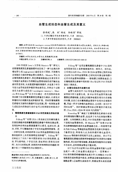
同时因葡萄膜黑色素瘤有其独特的微循环发生机制,用一些抑制血管生成拟态的药物进行治疗也可以收到较好的临床疗效.,1葡萄膜黑色素瘤微循环PAS阳性图案及其临床意义FHberg等”1采用UEA一1荧光标记及共聚焦激光扫描显微镜来检测葡萄膜黑色素瘤中特殊的微血管,发现各种各样的血管图案都可以看见,包括长的、直的、环形、平行聚集的和十字交叉的血管样图案等。
- 1、下载文档前请自行甄别文档内容的完整性,平台不提供额外的编辑、内容补充、找答案等附加服务。
- 2、"仅部分预览"的文档,不可在线预览部分如存在完整性等问题,可反馈申请退款(可完整预览的文档不适用该条件!)。
- 3、如文档侵犯您的权益,请联系客服反馈,我们会尽快为您处理(人工客服工作时间:9:00-18:30)。
©2011 L a n d e s B i o s c i e n c e .D o n o t d i s t r i b u t e .Cancer Biology & Therapy 13:5, 1-8; March 1, 2012; © 2012 Landes BioscienceresearCh paper*Correspondence to: Yi-quan Ke; Email: yiquanke@ Submitted: 10/15/11; Revised: 12/18/11; Accepted: 12/18/11/cbt.13.5.19108IntroductionMedulloblastoma is the most common primary malignantbrain tumor in children and young adults. Over the past three decades, many medulloblastoma patients have successfully been cured by multimodal treatments, including surgery, postsurgi-cal radiotherapy and chemotherapy. However, these treatments also resulted in chronic suffering for patients. Cognitive dysfunc-tion and neurological problems were two of the most common complications. Moreover, evidence indicates that the prognosis for medulloblastoma is still poor in a significant proportion of patients.1 Thus, to develop more effective and harmless treat-ments, we need to explore medulloblastoma in detail.Angiogenesis has been considered to be the most impor-tant way for tumors to sustain growth. Without angiogenesis, tumors cannot grow beyond 2–3 mm 3.2 Current research on vasculogenic mimicry (VM) has reinforced this viewpoint. In 1999, Maniotis et al. reported a new blood supply system inde-pendent of endothelial vessels in malignant melanoma, named VM. VM is defined as a fluid-conducting channels formed by highly invasive and genetically dysregulated melanoma cells. Endothelial cells are not present in VM channels as detected by light microscopy, immunohistochemistry and transmission Vasculogenic mimicry (VM), a process involving the formation of a tubular structure by highly invasive and genetically dysregulated tumor cells, can supplement the function of blood vessels to transport nutrients and oxygen to maintain the growth of tumor cells in many malignant tumors. We aimed to explore the existence of VM and its clinical significance in medulloblastoma in this study. VM was identified in 9 out of 41 (22%) medulloblastoma tissues. Immunohistochemical studies revealed that the presence of VM was associated with the expression of MMp-2, MMp-14, epha2 and laminin 5γ2. Tumor tissues with VM were associated with lower microvessel density (MVD), which was indirect evidence of the blood supply function of VM. survival analysis and log-rank tests showed that patients with VM had shorter overall survival time than those without VM. Multivariate analysis and the Cox proportional hazards model identified VM as independent prognostic factor for overall survival. Our results confirmed the existence of VM for the first time and revealed that VM is a strong independent prognostic factor for survival in patients with medulloblastoma.Vasculogenic mimicry and its clinical significancein medulloblastomashi-yong Wang,1,2 Li Yu,3 Geng-qiang Ling,1,2 sha Xiao,3 Xin-lin sun,1,2 Zhen-hua song,1,2 Yi-jing Liu,1,2 Xiao-dan Jiang,1,2Ying-qian Cai 1,2 and Yi-quan Ke 1,2,*1Department of Neurosurgery; and 3pathology; Zhujiang hospital; 2Institute of Neurosurgery; Key Laboratory on Brain Function repair and regeneration of Guangdong;southern Medical University; Guangzhou, ChinaKey words: medulloblastoma, vasculogenic mimicry, microvessel density, prognosis, angiogenesisAbbreviations: VM, vasculogenic mimicry; MVD, microvessel density; KPS, karnofsky performance scale; D/N, desmoplastic/nodular; MBEN, medulloblastoma with extensive nodularity; LCA, large cell/anaplastic; SI, staining index; PI3K,phosphatidylinositol-3-kinaseT h i s m a n u s c r i p t h a s b e e n p u b l i s h e d o n l i n e , p r i o r t o p r i n t i n g . O n c e t h e i s s u e i s c o m p l e t e a n d p a g e n u m b e r s h a v e b e e n a s s i g n e d , t h e c i t a t i o n w i l l c h a n g e a c c o r d i n g l y .electron microscopy.3 Studies revealed that VM exists in many malignant tumors,4-8 and VM is a prognostic factor for poorer clinical outcome.9-13 Research on angiogenesis inhibitors such as Anginex, TNP-470 and endostatin revealed that they could abrogate new vessels formed by human vascular endothelial cells in vitro. Interestingly, these inhibitors did not affect tumor cell VM formation under the same conditions and even induced the formation of VM.14 Medulloblastoma is abundant in vessels, but results from anti-angiogenic treatments are far from satisfac-tory.15 These data suggest that VM functions as a supplementary blood supply system for the growth of tumors and contributes to the failure of anti-angiogenic treatments aimed at depriving tumors of blood supply.This is the first study demonstrating the presence of VM and its clinical significance in medulloblastoma. These findings will provide additional information that will hopefully lead to improvement in targeted therapies for medulloblastoma.Results Clinical characteristics of the patients. Tables 1 and 2 sum-marize the clinical and pathological characteristics of 41 medul-loblastoma patients.©2011 L a n d e s B i o s c i e n c e .D o n o t d i s t r i b u t e .cases. However some channels had characteristics clearly differ-ent from blood vessels in 9 out of 41 cases. The CD34-negative PAS-positive channels were found in 5 out of 25 classic medul-loblastomas, 2 out of 5 desmoplastic medulloblastomas and 2 out of 4 large cell/anaplastic (LCA) medulloblastomas. The PAS-positive patterns, also described as a fluid-conducting ECM meshwork lined with tumor cells, coexisted with endo-thelial cell-lined blood vessels and had direct connections with them. The PAS-positive patterns extended directly from CD34-positive vessels, branched out into several smaller PAS-positive patterns which interconnected into a cluster of back-to-back looping patterns. At high magnification, translucent red blood cells were found spreading along the PAS-positive pat-terns (Fig. 1A–D ). In the adjacent tissue sections, we stained with eosin after PAS-CD34 dual staining and red blood cells were highlighted. We confirmed red blood cells spreading along PAS-positive patterns. In some regions, the patterns were splayed open, showing hollow channels containing red blood cells (Fig. 1E ). These characteristics strongly supported the belief that the PAS-positive patterns were a part of the medul-loblastoma microcirculation.VM is associated with the expression of MMP-2, MMP-14, EphA2 and laminin 5γ2 in medulloblastoma. The results of immunohistochemical studies for VEG F, MMP-2, MMP-14, EphA2 and laminin 5γ2 were summarized in Figure 2 and Table 3. The expression of MMP-2 (p = 0.001), MMP-14 (p = 0.039), laminin 5γ2 (p = 0.005) and EphA2 (0.036) were signifi-cantly higher in the VM-positive group than in the VM-negative group, whereas the expression of VEGF (p = 0.518) showed no difference between these two groups.No association was found between VM and clinical and pathological data. The presence of VM in medulloblastoma was not associated with age (p = 0.187), gender (p = 0.819), Karnofsky Performance Scale (KPS) score (0.224), hydrocephalus (0.580), histological classification (0.286), tumor location (p = 0.724), tumor size (p = 0.530), tumor metastasis (p = 0.479), and the extent of tumor resection (p = 0.819) (Table 2).VM-positive medulloblastoma has lower microvessel den-sity. We compared the MVD counts between VM positive and VM negative medulloblastoma. The results demonstrated that VM positive medulloblastomas had lower levels of MVD (38.12 ± 5.64) when compared with VM negative medulloblastomas (58.53 ± 11.53), (p = 0.000) (Table 4).VM is a prog nostic factor for shorter survival time in medulloblastoma. We used Kaplan-Meier survival analysis to compare the survival times between VM-positive (total 9, censored 2) and VM-negative patients (total 32, censored 18) or between below-median MVD (total 21, censored 9) and above-median MVD (total 20, censored 11). The results dem-onstrated that the VM-positive group had a shorter survival time (median 30.47 mo, 95% CI, 24.00–36.94 mo) when com-pared with the VM-negative group (median 56.43 mo, 95% CI 46.98–65.89 mo) (p < 0.0001) (Fig. 3). There was no sig-nificant correlation between MVD and overall survival time of the medulloblastoma patients (p = 0.386) (Fig. 4). Univariate analysis showed that survival time was significantly correlatedVM exists in medulloblastoma. We used the CD34 anti-gen as a hallmark to identify endothelial cells, and we usedperiodic acid-Schiff (PAS) staining to highlight the basement membrane of tumor blood vessels and the PAS-positive pat-terns in medulloblastoma tissue sections. Tumor blood vessels positive for CD34 showed brown staining on the luminal sur-face and a PAS-positive reaction in the basement membrane. CD34-positive blood vessels played a dominant role in mostTable 1. Clinical data of medulloblastoma patientsDataMedian age (years)9 (1.5–37)Median MVD counts 51.5Median Kps score 80 (50–90)Median tumor size (cm 3)21.0Median survival time (months)51.2Table 2. The association of clinicopothological data and VM in medulloblastoma patientsnVM +VM -χ2*pGender Male 266200.0530.819Female 15312Age (years)age ≤31239 3.3530.1873< age ≤1520614age >15909KPS score ≤70206141.4760.224≥8021318HydrocephalusNo154110.3070.580Yes26521Histology Classic 30525 2.5050.286Desmoplastic725LCa422Tumor locationmidline 307230.1250.724lateral 1129Tumor size ≤median 224180.3940.530>median 19514Metastasis positive 10370.5000.479Negative 31625Resection Total 266200.0530.819partial15312*pearson χ2 test (asymptotic significance, two-sided).©2011L a n d e s B i o s c i e n c e.D o n o t d i s t r i b u t e.Figure 1. representative microphotographs of endothelial cell-lined blood vessels and VM in medulloblastoma. (a) pas-positive patterns were indirect communication with CD34 positive endothelial cell-lined blood vessels (brown stainings). The pas-positive patterns extended directly fromCD34-positive vessels and branched out into smaller pas-positive patterns that interconnected with a cluster of back-to-back looping patterns (Boxed area). (B) In high magnification field, translucent red blood cells were found spreading along the pas-positive patterns (red arrow). (C) Tumor cells delimited by cross-linking pas-positive patterns. Boxed area was illustrated in oil immersion objective in (D). (D) In some regions, pas-positive patterns showed hollow channels filled with translucent red blood cells (red arrow). (e) patterns in medulloblastoma were stained with CD34 and pas dual staining in combination with eosin staining to highlight red blood cells. pas-positive patterns splayed open and filled with red blood cells, withouthints of endothelial cells in certain areas (red arrow). Tissue stains: CD34 and pas dual staining with hematoxylin counterstaining (a–D) and in combi-nation with eosin (e). Original magnifications, x200 (a), x400 (B), x400 (C), x1,000 (D), x400 (e).©2011 L a n d e s B i o s c i e n c e .D o n o t d i s t r i b u t e .DiscussionThe vascularization of tumors is heterogeneous. Angiogenesis (the tumor vasculature arising from sprouting of endothelial cells from local vessels) and vasculogenesis (vasculature formed by colonization of circulating endothelial progenitor cells from the bone marrow) were once two well-accepted paradigms for thewith VM (p = 0.0001), metastasis (p = 0.044) and KPS score (p = 0.01). Patient age, gender, hydrocephalus, tumor histo-logical classification, tumor size, tumor location and extent of resection were not significant variables. Multivariate analysis and the Cox proportional hazards model identified VM and metastasis as independent prognostic factors for overall sur-vival time.Figure 2. Immunohistochemical studies of the expression of VeGF, MMp-2, MMp-14, epha2 and laminin 5γ2 in medulloblastoma. (a, C, e, G and I) arerepresentative micrographs from VM-positive tissue sections. (B, D, F, h and J) are representative micrographs from VM-negative tissue sections.(a and B) expression of VeGF in medulloblastoma. (C and D) expression of MMp-2 in medulloblastoma. (e and F) expression of MMp-14 in medulloblas-toma. (G and h) expression of epha2 in medulloblastoma. (h and I) expression of laminin 5γ2 in medulloblastoma. Original magnifications, x400 (a–J).©2011 L a n d e s B i o s c i e n c e .D o n o t d i s t r i b u t e .expression of MMP-2, MMP-14, EphA2 and laminin 5γ2 in VM-positive tissues compared with VM-negative tissues, espe-cially in those VM lesions. These components play significant roles in the formation of VM. Studies in melanoma indicated that cooperative interactions between laminin 5γ2-chain and MMP-2 and MMP-14 are required for the formation of VM. EphA2 can activate phosphatidylinositol-3-kinase (PI3K), which regulates the activity of MMP-14. Activated MMP-14 leads to the activation of pro-MMP-2 to an active MMP2 proteinase. Active MMP-2 can cleave laminin 5γ2-chain into promigratory γ2' and γ2x fragments, which can lead to the formation of VM.26,27 Our formation of tumor vascularization. The blood vessels formed by angiogenesis and vasculogenesis are both initiated and regulated by angiogenic factors secreted by tumor cells.16 However, theseobservations have been challenged over the last decade by thediscovery of VM in a series of malignant tumors. Until now, the underlying molecular mechanisms for VM were not completely understood. Previous research on melanoma, breast cancer, ovarian cancer suggest that the plasticity of cancer cells enablesthem to mimic the phenotypes and functions of endothelial cells, thus leading to the formation of patterned matrix type of VM-patterned networks of interconnected loops of PAS-positiveextracellular matrix that may be solid or hollow, with no involve-ment of endothelial cells.17-19 The plasticity of highly malignanttumor cells has some similarities to the stemness of cancer stemcells. Recently, reports reveal that glioblastoma stem-like cells (CSC) can differentiate into functional endothelial cells, which harbor the same genomic alterations as cancer cells.20,21 Andthere is evidence suggesting that glioblastoma stem-like cells are capable of forming blood vessels de novo-tubular type of VM.5 Together, these observations strongly suggest that the plasticity of highly malignant tumor cells and cancer stem-like cell may mimic the role of normal stem cells and facilitate the formation of vascular network to sustain the blood supply of fast-growing tumor.22Morphologically, VM channels are patterned networks of solid or hollow interconnected loops of PAS-positive extracel-lular matrix, lined externally by tumor cells. Functionally, they interconnect with endothelial blood vessels and conduct plasma and red blood cells. Previous studies in mela-noma provided considerable evidence that the looping patterned matrix conducts fluid in vitro and in vivo. Melanoma cells cultured in three-dimensional matrix gel formed looping patterns which can transmit fluid after direct microinjection and by passive absorption.23 In a xenograft mouse model of uveal melanoma, intravenous tracer material can perfuse to a looping patterned matrix.24 Further studies in patients with uveal melanoma using laser scanning confocal microscopy revealed similar results.25 Based on three morphological mani-festations, we believe that the PAS-positive patterns in medulloblastoma are a part of the tumor microcirculation, but not blood leaks or hemorrhage. First, the patterned matrix was in direct communication with blood ves-sels. Second, in certain regions, these patterns branched out into smaller patterns with hol-low channels filled with red blood cells. Third, using the CD34 and PAS dual staining in combination with eosin staining to highlight the red blood cells in tissue sections, we found that red blood cells spread along the PAS-positive patterns.In this article, immunohistochemical studies revealed significant increases in the Table 3. expression of VeGF, MMp-2, MMp-14, epha2 and laminin 5γ2 in medulloblastoma tissuesVM +VM -Z p value VeGF 65.20 ± 36.2071.94 ± 54.66-0.6460.518MMp-2112.56 ± 56.7760.48 ± 60.07-3.1970.001MMp-14115.16 ± 85.5573.47 ± 53.64-2.0040.039epha252.07 ± 18.3933.78 ± 20.88-2.0950.036Laminin 5γ2129.91 ± 95.8459.03 ± 63.11-2.8030.005Table 4. Correlation of MVD with VM in medulloblastoma N MVD (mean ± SD)Z p VM +938.12 ± 5.64VM -3258.53 ± 11.53-4.220.000Figure 3. The presence of VM in medulloblastoma was associated with shorter survival time by Kaplan-Meier survival analysis and log-rank test (VM-positive 9, censored 2 vs. VM-neget-ive 32, censored 18).©2011 L a n d e s B i s c i e n c e .D o n o t d i s t r i b u t e .metastasis,22 there was no association between VM and metastasis in our study. Our obser-vation needs to be further studied in larger amount of samples. The MVD counts in the VM-positive group were significantly less than in the VM-negative group, which is indirect evidence of the blood supply function of VM. A previous study revealed that MVD has no prognostic significance in medulloblastoma.29 Our observations in medulloblastoma lead us to the same conclusion.Observations in a series of malignant tumors reveal that VM might serve as an unfavorable prognostic factor. However, there has been no research on the prognostic signifi-cance of VM in medulloblastoma prior to our study. Although VM had no association with patients’ clinicopathological characteristics in this study, Kaplan-Meier survival analy-ses and log-rank tests revealed that medullo-blastomas with VM have a poorer clinical outcome compared with those without VM. Univariate analysis and multivariate analysis showed that VM is an independent prognos-tic factor for overall survival time in patientswith medulloblastoma. Our observation in medulloblastoma is consistent with previous studies. It is noteworthy that while VM cor-relates with the MVD, MVD does not cor-relate with overall survival, yet VM does. It is not surprising that, although we found no correlation between VM and pathological classifications or between VM and metastasis which was consid-ered as inconclusive results in this study, VM is indeed associated with more malignant biological behaviors such as invasiveness and metastasis that directly affect patient clinical outcome.3 Moreover, currently widespread used anti-angiogenic drugs may have no effect on VM,14 even inducing VM formation when blood vessels are destroyed leaving a hypoxic environment.30 Therefore,newly developed drugs based on anti-angiogenic strategies must take both anti-angiogenic and anti-VM treatment into serious consideration.In conclusion, this pilot study provided direct evidence that VM, a supplementary microcirculation for angiogenesis, exists in medulloblastoma and is an adverse predictor of clinical outcome.Materials and Methods Tissue samples. A total of 41 paraffin-embedded medullo-blastoma tissues between 1999 and 2010 were obtained from the Department of Pathology, Zhujiang Hospital, Southern Medical University. All tissue samples were collected from patients who did not undergo therapy before the surgical opera-tion to remove the tumor. Tumor sections were reviewed by two neuropathologists to verify the diagnosis of medulloblas-toma in accordance with the 2007 World Health Organizationresults provide evidence that VM may be associated with the deg-radation and remodeling of the extracellular matrix in medul-loblastoma. Research on VEGF reveals that it is a stimulator of endothelial cell proliferation, and VEGF can induce the forma-tion of VM through a VEGFR-1dependent manner, increasing the expression of VM associated proteins in melanoma.28 In our research, the expression of VEGF had no significant correlation with VM in medulloblastoma. It is possible that VM in medul-loblastoma might be regulated through a pathway distinct from VEGF.We have confirmed the existence of VM in medulloblas-toma. Moreover, we explored whether the presence of VM has any association with clinicopathological data and its prognosticsignificance in medulloblastoma. In this retrospective study of 41 patients with medulloblastoma, we found that VM had no association with clinicopathological characteristics. Notably VM had no association with histological classification, which wasinconsistent with previous studies in glioma, renal cell carcinomaand mesothelial sarcoma.9,10,12Among all the subtypes of medul-loblastoma, the LCA variant is notable for its high aggressive-ness and relatively poorer clinical outcome.1However, it might be unconvincing to draw the above conclusion as there are only four examples of the LCA subtype in this study. The true asso-ciation has yet to be conclusively determined. Metastases in this study were found in 10 patients: 8 with spinal cord metas-tases and 2 with extracranial metastases. Although the genera-tion of VM by highly aggressive tumor cells facilitates tumor Figure 4. Comparison of Kaplan-Meier survival curves (log-rank test) in patients with medulloblastoma according to patients with MVD values below the group median (total 21,censored 9) vs. those with values above the group median (total 20, censored 11).©2011 L a n d e s B i o s c i e n c e .D o n o t d i s t r i b u t e .percentage of positively stained cells in 10 visual fields of eachsection was converted into a score as follows: 0 for <10% posi-tive cells, 1 for <25% positive cells, 2 for <50% positive cells, 3 for >50% positive cells. The score of intensity of each section was graded as follows: Strong intensity staining was scored as 3, moderate as 2, weak as 1 and negative as 0. The staining index was determined by multiplying the average percentage of positive cells and the score of intensity. The assessment of immunohisto-chemistry results was completed by two independent investiga-tors blinded to the clinicopathological data. Discrepancies were resolved by consensus.9Microvessel density. Microvessel density counts were com-pleted according to the method described by Foote et al. This portion of the study was completed by two additional inde-pendent observers unaware of the clinical outcome of patients. Tumor sections were examined under 200x magnification, and the average number of microvessels in 10 randomly selected visual fields represented the MVD of the tumor. Any endothe-lial cell or endothelial cell cluster positive for CD34 and clearly separate from the adjacent cluster was considered to be a single countable microvessel. The average MVD for the 41 specimens was calculated and patients were grouped according to whether their MVD was lower (below-median MVD) or higher (above-median MVD) than median. The overall survival times for these two groups were compared, and the association between VM and MVD was determined.Statistical analysis. Statistical analyses were performed using SPSS 13.0 for Windows (SPSS Inc.). A p value of less than 0.05 was defined as statistically significant. The differential expres-sion levels of VEGF, MMP-2, MMP-14, laminin 5γ2, EphA2 in the VM-positive and VM-negative groups were determined by the Mann-Whitney test. MVD counts in the VM-positive and VM-negative groups were also compared by the Mann-Whitney test. Associations between VM and clinical and pathological data were determined by the Pearson’s χ2 test. The survival analysis was calculated by the Kaplan-Meier method and the differ-ences between groups were compared using the log-rank test. Univariate analysis was performed to identify prognostic vari-ables for overall survival. Multivariate analysis was performed to identify the independent prognostic factors for survival using the Cox regression hazard model. Data on surviving patients and patients without follow-up information were considered censored data in the analysis.Disclosure of Potential Conflicts of InterestNo potential conflicts of interest were disclosed.AcknowledgementsWe express our gratitude to Professor Shengli An (Department of Biostatistics, Southern Medical University) for the analysis of statistical data and Mr. Xuan Lu (Department of Pathology, Zhujiang Hospital, Southern Medical University) for the sec-tioning of tissues. This project was supported by grants from the National Science Foundation of China (No. 30901174), and from the Science and Technology Planning Project of Guangdong (No. 2008B030301152).(WHO) classification of central nervous system (CNS) tumors. According to the 2007 WHO classification of CNS tumors, medulloblastomas are separated into the classic tumor and four main histological subtypes:desmoplastic/nodular (D/N), medulloblastoma with extensive nodularity (MBEN), ana-plastic medulloblastoma and large cell medulloblastoma. D/N medulloblastoma and MBEN share fundamental clinical and pathological features as do anaplastic medulloblastoma and large cell medulloblastoma.1,31 We divided the 41 patients into three groups: classic medulloblastoma, desmoplastic medullo-blastoma that contained D/N and MBEN medulloblastomas and LCA medulloblastoma that contained anaplastic medullo-blastoma and large cell medulloblastoma. Detailed clinical and pathological data were collected for all samples, including age, gender, KPS score, hydrocephalus, histological classification, size, location, metastasis, total or partial resection. Research Ethics Committee approval has been obtained for the use of all human tissues in this study.CD34-PAS dual staining. For CD34-PAS dual staining par-affin sections were cut at 5 μm. Slides were deparaffinized twice with xylene and subsequently hydrated against a concentration gradient in ethanol solutions. Antigen retrieval was performed using citrate buffer (pH 6.0) at 96°C for 20 min. For demon-stration of endothelial cells in medulloblastoma, the slides were incubated with mouse monoclonal anti-human CD34 antibody (Santa Cruz Biotechnology, catalog: sc-52312, dilution 1:100) at 4°C over night. After washing with PBS, the slides were treated with PowerVision Two-Step Histostaining Reagent (Zhongshan G oldenbridge Biotechnology, catalog: PV-6002) for 30 min at 37°C. The chromogen used to highlight a positive reaction was 3,3'-diaminebenzidine tetrahydrochloride (DAB), resulting in a brown product. To highlight the matrix-associated vascular channels of medulloblastoma, slides were stained following the PAS staining procedures before counterstaining with Mayer’s hematoxylin. Finally, the slides were covered with a permanent mounting medium. To highlight the red blood cells in PAS posi-tive patterns, the adjacent sections were stained with eosin after the above procedures.Immunohistochemistry. To determine the expression of VEGR, MMP-2, MMP-14, EphA2 and laminin 5γ2 in medul-loblastoma by immunohistochemistry, tissue sections were pre-pared as previously described. Slides were then incubated over night at 4°C with a monoclonal antibody against VEGF (Santa Cruz Biotechnology, catalog: sc-80437, dilution 1:50); EphA2 (Millipore, catalog: 05-480, dilution 1:100), MMP-2 (Abcam, catalog: ab2462, dilution 1:100), MMP-14 (Abcam, catalog: ab53712, dilution 1:100), or a polyclonal antibody against lam-inin 5γ2 (Abcam, catalog: ab96327, dilution 1:100). Appropriate positive and negative controls for each antibody were included. The staining index (SI) was used to evaluate the expression level of each marker based on the varying degrees of staining inten-sity and percentage of positively stained cells in the specimens. Therefore, a combined intensity and percentage positive scoring method was used. Ten visual fields of each tissue section were selected randomly under the microscope at 400x magnification, and 100 cells in each visual field were counted. The average。